Filters
▼Clonality
▼Type
▼Reactivity
▼Gene Name
▼Isotype
▼Host
▼Application
▼Clone
▼Active Proteins
AAA Biotech also known as AAA Bio or AAABio provides a variety of high-quality recombinant and natural/native proteins that are proven to work in a wide range of experiments. Explore our products to find the active protein that best fits your needs or experimental model.
Viewing 1350-1400 of 2567 product results
PHOSPHORYLASE B, Active Protein (Cat# AAA50505)
Wnt1, Active Protein (Cat# AAA75438)
WISP1, Active Protein (Cat# AAA75458)
Factor X, Active Protein (Cat# AAA71837)
GST Yb2, Active Protein (Cat# AAA71846)
sDLL4, Active Protein (Cat# AAA75582)
Pepsinogen I, Active Protein (Cat# AAA75592)
IL21, Active Protein (Cat# AAA75599)
IL-13 alpha 2 receptor (IL13RA2), Active Protein (Cat# AAA76103)
CD200 (MOX1), Active Protein (Cat# AAA76111)
 Bioactivity
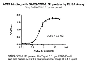
(The activity was determined by immobilized Spike Protein S1 binding with human ACE2 in a functional ELISA assay, the ED50 was determined to be 3.8nM.2019-ncov S1-his tagged (coating at 0.5ug-well) binding with Human ACE2-Fc (cat. The linear range was found to be 0.1-5 ug/ml)
Bioactivity
(The activity was determined by immobilized Spike Protein S1 binding with human ACE2 in a functional ELISA assay, the ED50 was determined to be 3.8nM.2019-ncov S1-his tagged (coating at 0.5ug-well) binding with Human ACE2-Fc (cat. The linear range was found to be 0.1-5 ug/ml)
COVID 19 Spike S1 Coronavirus, Active Protein (Cat# AAA76120)
PAI2, Active Protein (Cat# AAA75570)
PDGF CC, Active Protein (Cat# AAA75572)
sFRP1, Active Protein (Cat# AAA75573)
Transforming Growth Factor-Beta 3, Active Protein (Cat# AAA76535)
(a) Analysis by RP-HPLC.
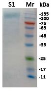
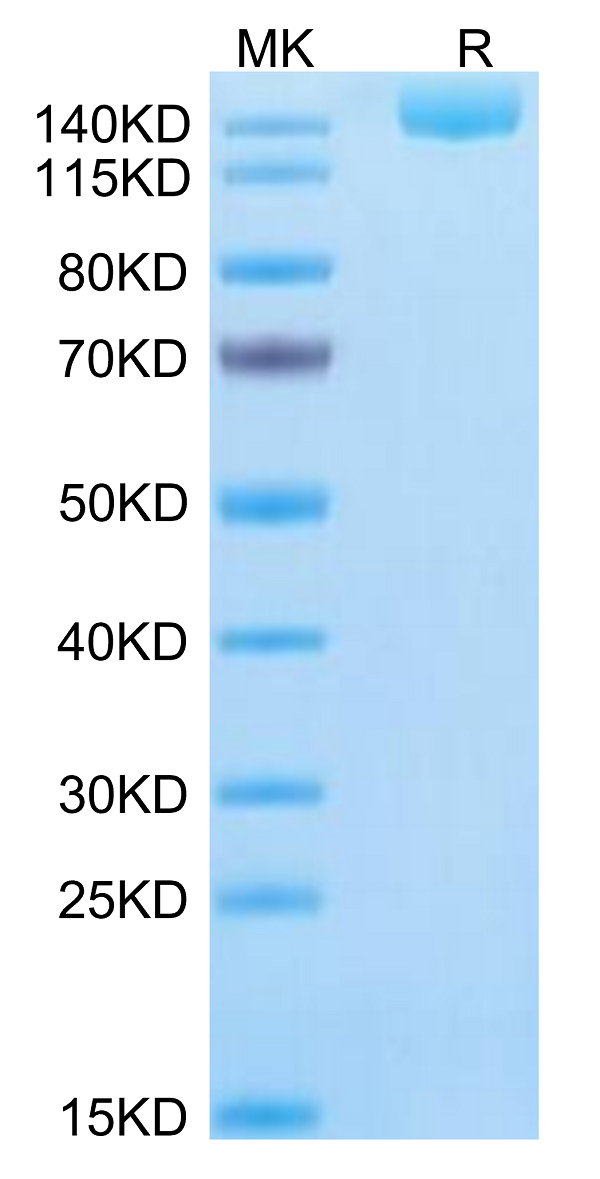
(b) Analysis by SDS-PAGE.
 ELISA
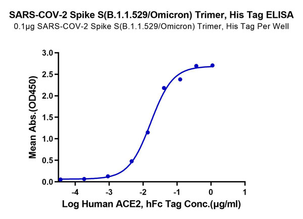
(SARS COV2 S B.1.1.529 trimer ACE functional binding ELISA assay2019-ncov S B.1.1.529 trimer(coating at 0.1ug-well) binding with Human ACE2-Fc.The linear range was found to be 23 ng/ml)
ELISA
(SARS COV2 S B.1.1.529 trimer ACE functional binding ELISA assay2019-ncov S B.1.1.529 trimer(coating at 0.1ug-well) binding with Human ACE2-Fc.The linear range was found to be 23 ng/ml)
SARS COV2 Spike protein S B.1.1.529 (omicron) Trimer, Active Protein (Cat# AAA76382)
 Application Data
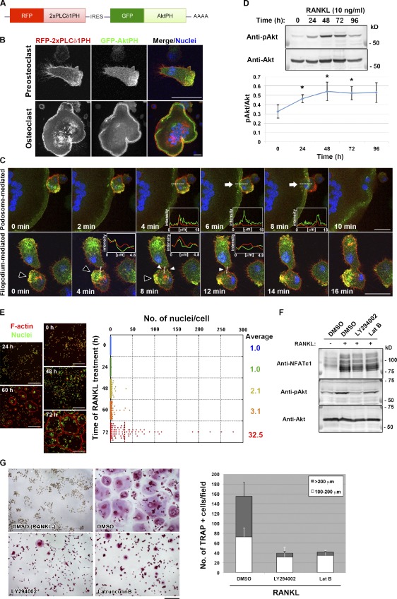
(Polarized membrane extensions mediate osteoclast fusion.(D, top) Immunoblot analysis of lysates of RAW264.7 macrophages stimulated with RANKL for the indicated times with antibodies to Ser473-phosphorylated (p) or total forms of Akt.)
Application Data
(Polarized membrane extensions mediate osteoclast fusion.(D, top) Immunoblot analysis of lysates of RAW264.7 macrophages stimulated with RANKL for the indicated times with antibodies to Ser473-phosphorylated (p) or total forms of Akt.)
Soluble RANK ligand, Active Protein (Cat# AAA76383)
 Application Data
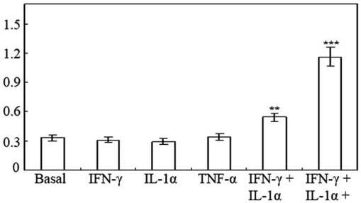
((A) Nitrite production by HT-29 cells following 48 h of treatment with cytokines.Effect of 0–50 ng/ml TNF-? on nitrite production induced by 100 U/ml IFN-? and 10 ng/ml IL-1? in HT-29 cells following 48 h of treatment.)
Application Data
((A) Nitrite production by HT-29 cells following 48 h of treatment with cytokines.Effect of 0–50 ng/ml TNF-? on nitrite production induced by 100 U/ml IFN-? and 10 ng/ml IL-1? in HT-29 cells following 48 h of treatment.)
Interferon-beta 1b, Active Protein (Cat# AAA76387)
 Application Data
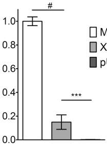
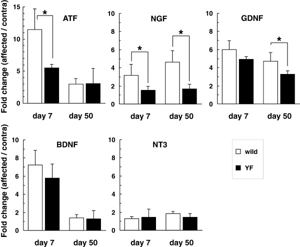
(Effects of ACM from Olig2PC-Astros and NPC-Astros on neurons.(h and i) RT-PCR analysis of the expression of the antioxidant defense-related genes, GCLC and NFE2L2 (n=3), and the neurotrophic growth factor genes, BDNF, GDNF and NT-3 (n=3).)
Application Data
(Effects of ACM from Olig2PC-Astros and NPC-Astros on neurons.(h and i) RT-PCR analysis of the expression of the antioxidant defense-related genes, GCLC and NFE2L2 (n=3), and the neurotrophic growth factor genes, BDNF, GDNF and NT-3 (n=3).)
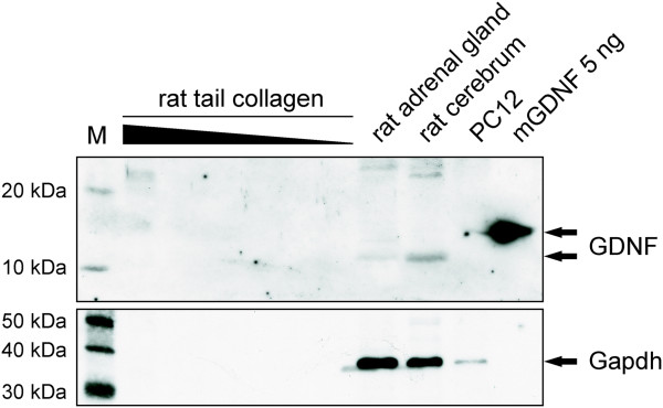
Glial-Derived Neurotrophic Factor, Active Protein (Cat# AAA76397)
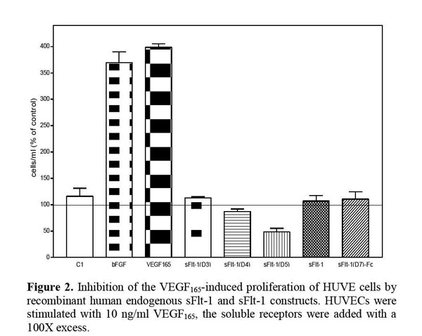
 Application Data
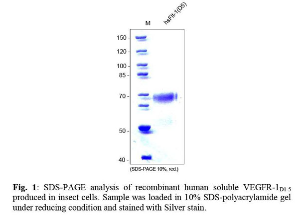
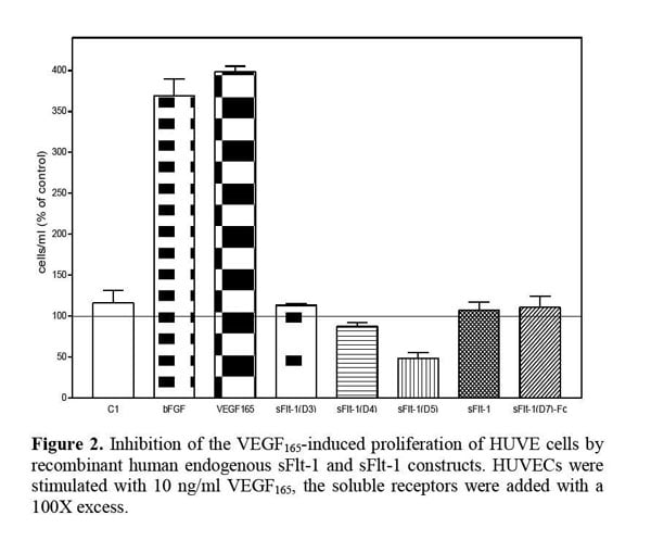
VEGFR-1/Flt-1 (D5), soluble, Active Protein (Cat# AAA79174)
 Application Data
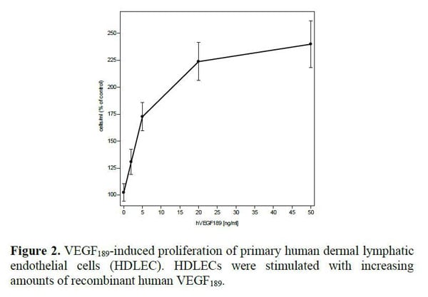
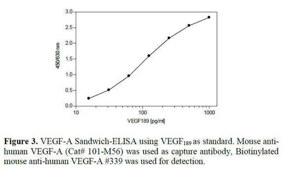
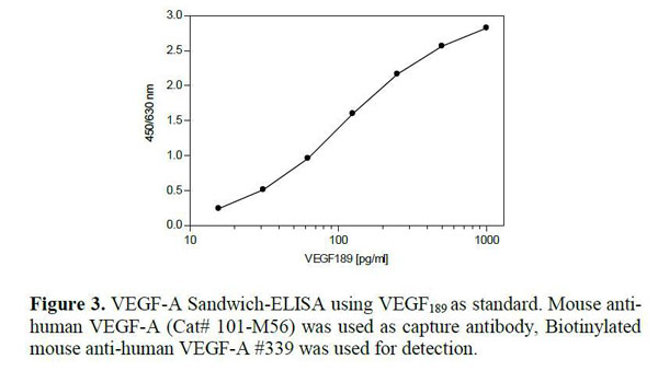
VEGF189, Active Protein (Cat# AAA79179)
 SDS-PAGE
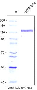
(SDS-PAGE analysis of recombinant human soluble TIE-2/Fc produced from insect cells. Sample was loaded in 10% SDS-polyacrylamide gel under reducing condition and stained with Coomassie blue.)
SDS-PAGE
(SDS-PAGE analysis of recombinant human soluble TIE-2/Fc produced from insect cells. Sample was loaded in 10% SDS-polyacrylamide gel under reducing condition and stained with Coomassie blue.)
TIE-2/Fc Chimera, soluble, Active Protein (Cat# AAA79181)
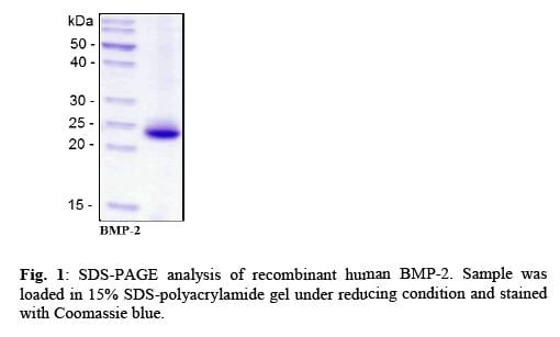
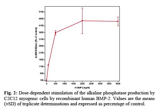 Application Data
BMP-2, Active Protein (Cat# AAA79187)
GDF-11, Active Protein (Cat# AAA79190)
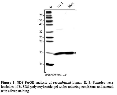
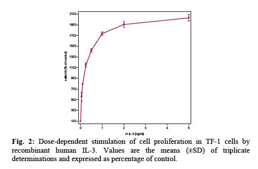 Application Data
IL-3, Active Protein (Cat# AAA79193)
 Application Data
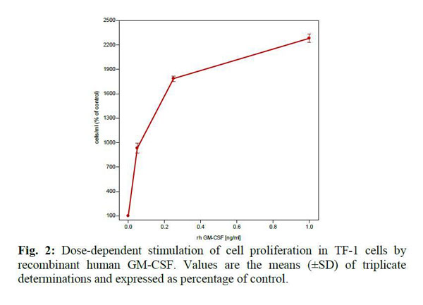
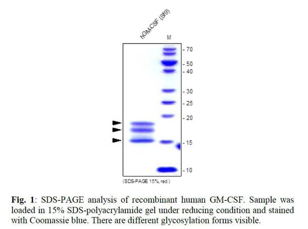
GM-CSF, Active Protein (Cat# AAA79214)
 Application Data
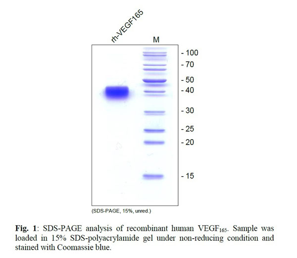
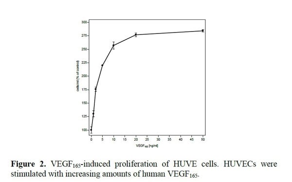
VEGF165, Active Protein (Cat# AAA79220)
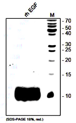 SDS-PAGE
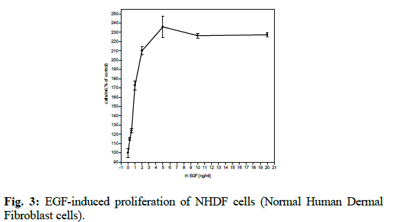
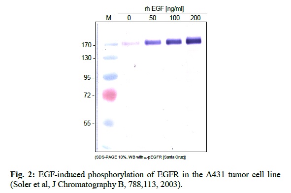
EGF, Active Protein (Cat# AAA79221)
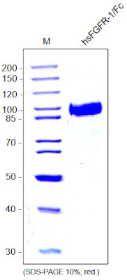
FGFR-1/Fc Chimera, soluble, Active Protein (Cat# AAA79237)
 Application Data
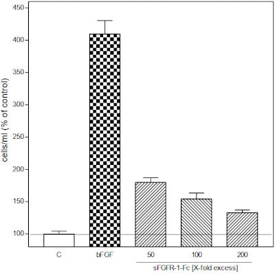
(Inhibition of the basic FGF-induced proliferation of HUVE cells by recombinant human sFGFR-1-Fc. HUVECs were stimulated with 10 ng/ml bFGF, the soluble receptor was added with a 50 - 200X excess)
Application Data
(Inhibition of the basic FGF-induced proliferation of HUVE cells by recombinant human sFGFR-1-Fc. HUVECs were stimulated with 10 ng/ml bFGF, the soluble receptor was added with a 50 - 200X excess)
FGFR-1/Fc Chimera, soluble, Active Protein (Cat# AAA79238)
 Application Data
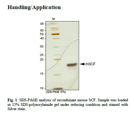
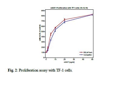
SCF, Active Protein (Cat# AAA79243)
GDNF, Active Protein (Cat# AAA79245)
 Application Data
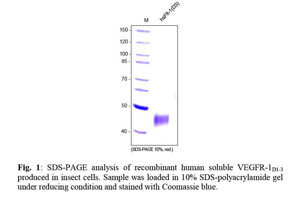
VEGFR-1/Flt-1 (D3), soluble, Active Protein (Cat# AAA79257)
TGF-beta1, Active Protein (Cat# AAA79258)
IGF-BP2, Active Protein (Cat# AAA79281)
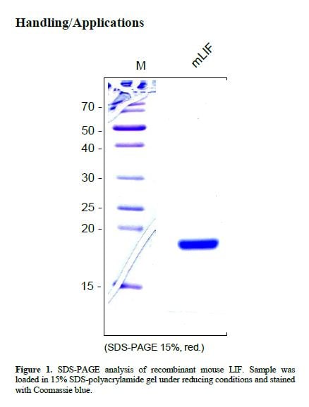
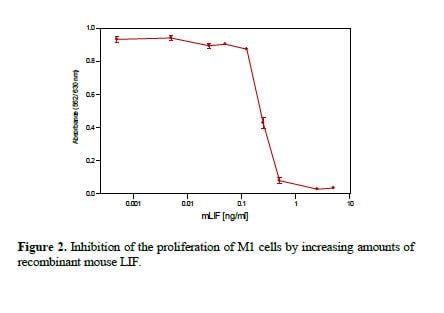 Application Data
LIF, Active Protein (Cat# AAA79285)
Enterokinase, Active Protein (Cat# AAA79292)
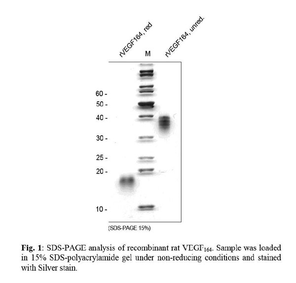
 Application Data
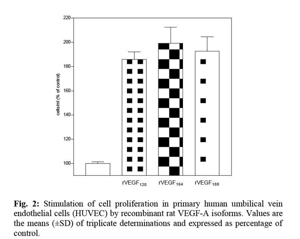
VEGF164, Active Protein (Cat# AAA79293)
 Application Data
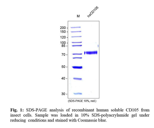
CD105/Endoglin, soluble, Active Protein (Cat# AAA79306)
IGF-1 LR3, Active Protein (Cat# AAA79323)
 Application Data
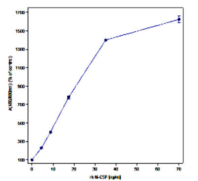
(Fig. 2: Dose-dependent stimulation of cell proliferation in NFS60 cells by recombinant human M-CSF. Values are the means (±SD) of triplicate determinations and expressed as percentage of control.)
Application Data
(Fig. 2: Dose-dependent stimulation of cell proliferation in NFS60 cells by recombinant human M-CSF. Values are the means (±SD) of triplicate determinations and expressed as percentage of control.)
M-CSF, Active Protein (Cat# AAA79123)
 ELISA
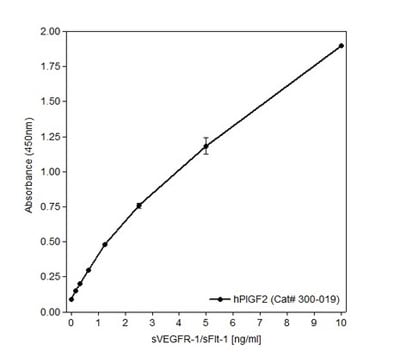
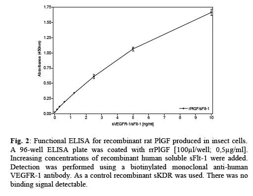
(Figure 2.Functional ELISA: Recombinant human PlGF-2 was coated with 0.5ug/ml and increasing amounts of recombinant human sFlt-1(5) was added as standard . The monoclonal mouse anti-human VEGFR1/Flt-1 antibody in combination with a goat anti-mouse Biotin antibody was used for detection.)
ELISA
(Figure 2.Functional ELISA: Recombinant human PlGF-2 was coated with 0.5ug/ml and increasing amounts of recombinant human sFlt-1(5) was added as standard . The monoclonal mouse anti-human VEGFR1/Flt-1 antibody in combination with a goat anti-mouse Biotin antibody was used for detection.)
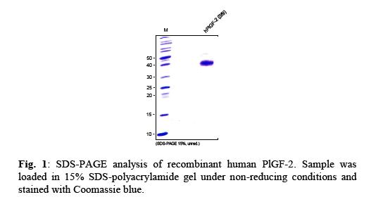
PlGF-2, Active Protein (Cat# AAA79127)
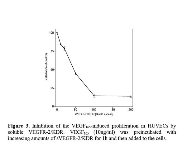 Application Data
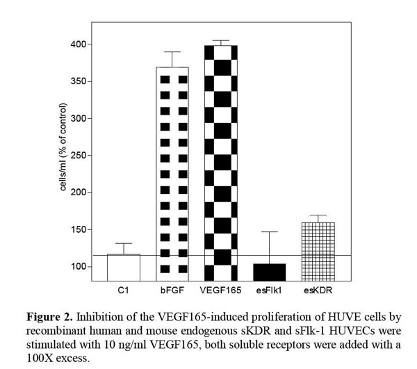
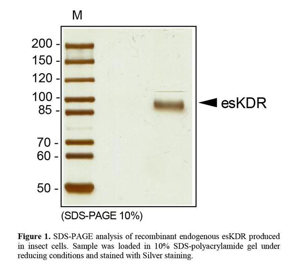
VEGFR-2/KDR (native), soluble, Active Protein (Cat# AAA79129)
Glu-C, Active Protein (Cat# AAA79131)
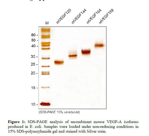
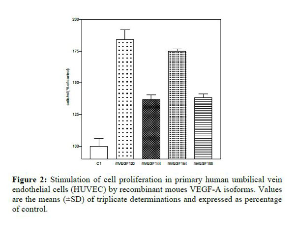 Application Data
VEGF188, Active Protein (Cat# AAA79133)
 BioLISA
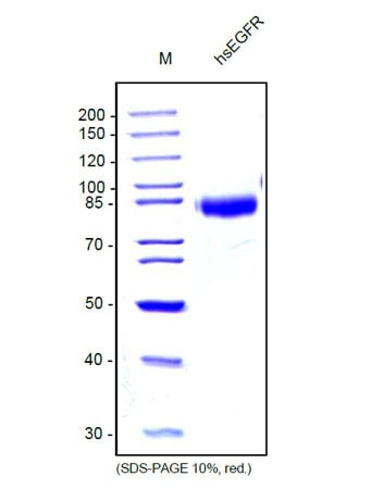
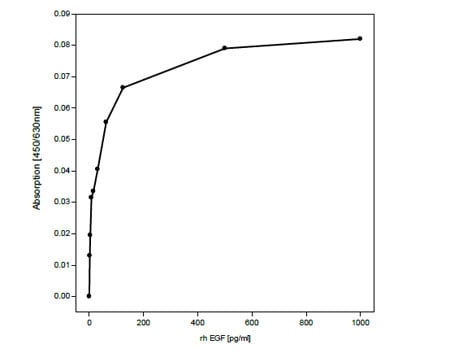
(Fig 2: Binding of sEGFRto recombinant human EGF in a functional ELISA. EGF [AAA79134] was coated with 200ng/well to a 96-well plate and sEGFR was added with increasing concentrations up to 4000ng/ml. Detection was performed using a polyclonal rabbit anti-human EGFR [] (1 ug/ml, 100 ul/well) and a goat anti-rabbit Biotin conjugated secondary antibody.)
BioLISA
(Fig 2: Binding of sEGFRto recombinant human EGF in a functional ELISA. EGF [AAA79134] was coated with 200ng/well to a 96-well plate and sEGFR was added with increasing concentrations up to 4000ng/ml. Detection was performed using a polyclonal rabbit anti-human EGFR [] (1 ug/ml, 100 ul/well) and a goat anti-rabbit Biotin conjugated secondary antibody.)
EGFR, soluble, Active Protein (Cat# AAA79134)
 Application Data
PlGF, Active Protein (Cat# AAA79139)
CYR61, Active Protein (Cat# AAA79141)
Maltase Dehydrogenase, Active Protein (Cat# AAA78969)
What Are Active Proteins?
Proteins are large molecules made up of long chains of amino acids.
They will typically fold into a very particular 3-dimensional shape/conformation, that is sometimes referred to as their “native” form, which allows them to work properly in the body. For the purposes of product categorization, AAA Biotech will typically refer to proteins purified from their original animal host as being “native” proteins (this is to signify their difference compared to their “recombinant” or “synthetic” protein counterparts).
If a protein successfully folds into the correct shape, it is will typically display high fidelity characteristics to its original protein in its original animal host, and be classified as an active protein, as it will be able to function “normally” in most enzymatic or binding capacities. If it loses this shape, due to factors such as heat or strong chemicals (such as detergents), it becomes inactive and is no longer able to perform its basic functions. All of the proteins in this category are made under strict quality control, and they are active, pure, low in contaminants, and stable.
Most are stored as freeze-dried powders and come without extra tags, so they’re very close to the actual natural/native form.
Key Applications of Active Proteins
1. Scientific Research
- Aid in the study of how proteins function in the body
- Aid in understanding various disease processes
2. Drug Development
- Powerful tools to investigate how potential drugs interact with specific proteins
- Ideal for identifying drug targets
3. Cell Culture
- Are routinely utilized to support cell growth and function (e.g., using exogenous growth factors)
- Can be used to promote cellular development into specific types (differentiation)
4. Diagnostics
- Regularly utilized in tests to detect diseases or infections (e.g., COVID-19, cancer)
- Note: All products are strictly for research-use only (RUO).
5. Therapeutics
- Some active proteins are used directly as treatments (e.g., insulin, enzymes)
- Note: All products are strictly for research-use only (RUO).
6. Vaccine Development
- Used to create or test vaccines by mimicking parts of viruses or bacteria
7. Biochemical Assays
- They can facilitate the characterization of enzyme activity, binding strength, or protein interactions in lab tests
Why Buy Active Proteins from AAA Biotech?
- High biological activity – Verified to perform as expected or indicated on datasheet
- Strict quality control – We are confident in our active proteins’ reliability and consistency
- High purity & low endotoxin – Ideal for applications involving sensitive or precious samples/components
- Freeze-dried for stability – Long shelf life and straightforward storage
- Mostly tag-free – Closer to natural/native protein form
FAQ
1. What are active proteins used for in research?
Active proteins are used primarily in the study of how proteins function, in characterizing/discovering drug interactions, supporting cell growth, running biochemical assays, and in development of diagnostics or therapeutics.
2. How are AAA Biotech's active proteins validated?
AAA Biotech’s active proteins are validated through strict quality control and functional assays to ensure they are properly folded and active. “Active”, though, can be an ambiguous term, so if a specific “activity” or “binding” capability of a protein is of crucial interest to you, please inquire with us prior to purchase, and we will provide further details on how the “Active” modifier was determined to be applicable.
3. Are these proteins tested for biological activity?
Yes, all active proteins from AAA Biotech are tested to confirm they have the expected biological activity before being offered for use. Though, said “biological activity” can be either “enzymatic”, “binding”, or both.