Filters
▼Clonality
▼Type
▼Reactivity
▼Gene Name
▼Isotype
▼Host
▼Application
▼Clone
▼Active Proteins
AAA Biotech also known as AAA Bio or AAABio provides a variety of high-quality recombinant and natural/native proteins that are proven to work in a wide range of experiments. Explore our products to find the active protein that best fits your needs or experimental model.
Viewing 1150-1200 of 2567 product results
 Application Data
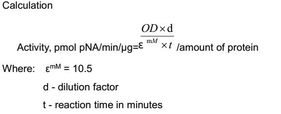
(Caspase 3 is a member of the cysteine-aspartic acid protease (caspase) family. Sequential activation of caspases plays a central role in the execution-phase of cell apoptosis. Caspases exist as inactive proenzymes that undergo proteolytic processing at conserved aspartic residues to produce two subunits, large and small, that dimerize to form the active enzyme. This protein cleaves and activates caspases 6 and 7; and the protein itself is processed and activated by caspases 8, 9, and 10. Caspase 3 can hydrolyze the peptide substrate acetyl-Asp-Glu-Val-Asp-p-nitroanilide (Ac-DEVD-pNA) resulting in the release of the p-nitroaniline (pNA) moiety. p-Nitroaniline has a high absorbance at 405nm. Thus the activity of recombinant human caspase 3 can be measured by calculate the concentration of the pNA released from the substrate. The reaction was performed in adding 50L 2×buffer (50mM HEPES, 100mM NaCl, 10mM DTT, 2mM EDTA, 10% glycerol) to 96 well plates, then add 50L various concentration of caspe 3 (diluted by 1×buffer, 25mM HEPES, 50mM NaCl, 5mM DTT, 1mM EDTA, 5% glycerol) to each well, finally, add 5L 4mmol Ac-DEVD-pNA to each well. Cover the 96 well plates and incubate at 37 for 2h. p-Nitroaniline (pNA) standard curve prepare by double dilute 200M pNA with 1×buffer and record the OD value at 405nm. Calculate the caspase 3 activity in pmol of pNA released per min per g recombinat human caspase 3.The specific activity of recombinant human caspase 3 is 2196pmol/min/g.)
Application Data
(Caspase 3 is a member of the cysteine-aspartic acid protease (caspase) family. Sequential activation of caspases plays a central role in the execution-phase of cell apoptosis. Caspases exist as inactive proenzymes that undergo proteolytic processing at conserved aspartic residues to produce two subunits, large and small, that dimerize to form the active enzyme. This protein cleaves and activates caspases 6 and 7; and the protein itself is processed and activated by caspases 8, 9, and 10. Caspase 3 can hydrolyze the peptide substrate acetyl-Asp-Glu-Val-Asp-p-nitroanilide (Ac-DEVD-pNA) resulting in the release of the p-nitroaniline (pNA) moiety. p-Nitroaniline has a high absorbance at 405nm. Thus the activity of recombinant human caspase 3 can be measured by calculate the concentration of the pNA released from the substrate. The reaction was performed in adding 50L 2×buffer (50mM HEPES, 100mM NaCl, 10mM DTT, 2mM EDTA, 10% glycerol) to 96 well plates, then add 50L various concentration of caspe 3 (diluted by 1×buffer, 25mM HEPES, 50mM NaCl, 5mM DTT, 1mM EDTA, 5% glycerol) to each well, finally, add 5L 4mmol Ac-DEVD-pNA to each well. Cover the 96 well plates and incubate at 37 for 2h. p-Nitroaniline (pNA) standard curve prepare by double dilute 200M pNA with 1×buffer and record the OD value at 405nm. Calculate the caspase 3 activity in pmol of pNA released per min per g recombinat human caspase 3.The specific activity of recombinant human caspase 3 is 2196pmol/min/g.)
Caspase 3, Active Protein (Cat# AAA150098)
 Application Data
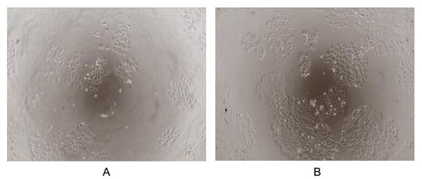
(Tumor protein p53, also known as p53, cellular tumor antigen p53 (UniProt name), phosphoprotein p53, tumor suppressor p53, antigen NY-CO-13, or transformationrelated protein 53 (TRP53), is any isoform of a protein encoded by homologous genes in various organisms, such as TP53 (humans) and Trp53 (mice). TP53 involved in cell cycle regulation as a trans-activator that acts to negatively regulate cell division by controlling a set of genes required for this process. One of the activated genes is an inhibitor of cyclin-dependent kinases. To test the effect of TP53 on cell apoptosis, Jurkat cells were seeded into triplicate wells of 96-well plates at a density of 5,000 cells/well with 1% serum standard 1640 including various concentrations of recombinant human TP53. After incubated for 72h, cells were observed by inverted microscope and cell proliferation was measured by Cell Counting Kit-8 (CCK-8). Briefly, 10uL of CCK-8 solution was added to each well of the plate, then the absorbance at 450nm was measured using a microplate reader after incubating the plate for 1-4 hours at 37?. Proliferation of Jurkat cells after incubation with TP53 for 72h observed by inverted microscope was shown in Figure 1. Cell viability was assessed by CCK-8 (Cell Counting Kit-8) assay after incubation with recombinant TP53 for 72h. The result was shown in Figure 2. It was obvious that TP53 significantly inhibit cell viability of Jurkat cells.)
Application Data
(Tumor protein p53, also known as p53, cellular tumor antigen p53 (UniProt name), phosphoprotein p53, tumor suppressor p53, antigen NY-CO-13, or transformationrelated protein 53 (TRP53), is any isoform of a protein encoded by homologous genes in various organisms, such as TP53 (humans) and Trp53 (mice). TP53 involved in cell cycle regulation as a trans-activator that acts to negatively regulate cell division by controlling a set of genes required for this process. One of the activated genes is an inhibitor of cyclin-dependent kinases. To test the effect of TP53 on cell apoptosis, Jurkat cells were seeded into triplicate wells of 96-well plates at a density of 5,000 cells/well with 1% serum standard 1640 including various concentrations of recombinant human TP53. After incubated for 72h, cells were observed by inverted microscope and cell proliferation was measured by Cell Counting Kit-8 (CCK-8). Briefly, 10uL of CCK-8 solution was added to each well of the plate, then the absorbance at 450nm was measured using a microplate reader after incubating the plate for 1-4 hours at 37?. Proliferation of Jurkat cells after incubation with TP53 for 72h observed by inverted microscope was shown in Figure 1. Cell viability was assessed by CCK-8 (Cell Counting Kit-8) assay after incubation with recombinant TP53 for 72h. The result was shown in Figure 2. It was obvious that TP53 significantly inhibit cell viability of Jurkat cells.)
Tumor Protein p53, Active Protein (Cat# AAA150104)
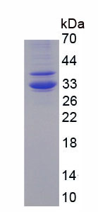
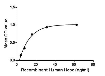 Bioactivity
(Figure. The binding activity of Hepc with FPN.Hepcidin (Hepc) is a regulator of iron metabolism. Hepcidin inhibits iron transport by binding to the iron export channel ferroportin which is located on the basolateral surface of gut enterocytes and the plasma membrane of reticuloendothelial cells (macrophages). Hepcidin ultimately breaks down the transporter protein in the lysosome. Inhibiting ferroportin prevents iron from being exported and the iron is sequestered in the cells. Besides, Ferroportin (FPN) has been identified as an interactor of Hepc, thus a binding ELISA assay was conducted to detect the interaction of recombinant human Hepc and recombinant human FPN. Briefly, Hepc were diluted serially in PBS, with 0.01% BSA (pH 7.4). Duplicate samples of 100uL were then transferred to FPN-coated microtiter wells and incubated for 2h at 37. Wells were washed with PBST and incubated for 1h with anti-Hepc pAb, then aspirated and washed 3 times. After incubation with HRP labelled secondary antibody, wells were aspirated and washed 3 times. With the addition of substrate solution, wells were incubated 15-25 minutes at 37. Finally, add 50uL stop solution to the wells and read at 450nm immediately. The binding activity of Hepc and FPN was shown in Figure 1, and this effect was in a dose dependent manner.)
Bioactivity
(Figure. The binding activity of Hepc with FPN.Hepcidin (Hepc) is a regulator of iron metabolism. Hepcidin inhibits iron transport by binding to the iron export channel ferroportin which is located on the basolateral surface of gut enterocytes and the plasma membrane of reticuloendothelial cells (macrophages). Hepcidin ultimately breaks down the transporter protein in the lysosome. Inhibiting ferroportin prevents iron from being exported and the iron is sequestered in the cells. Besides, Ferroportin (FPN) has been identified as an interactor of Hepc, thus a binding ELISA assay was conducted to detect the interaction of recombinant human Hepc and recombinant human FPN. Briefly, Hepc were diluted serially in PBS, with 0.01% BSA (pH 7.4). Duplicate samples of 100uL were then transferred to FPN-coated microtiter wells and incubated for 2h at 37. Wells were washed with PBST and incubated for 1h with anti-Hepc pAb, then aspirated and washed 3 times. After incubation with HRP labelled secondary antibody, wells were aspirated and washed 3 times. With the addition of substrate solution, wells were incubated 15-25 minutes at 37. Finally, add 50uL stop solution to the wells and read at 450nm immediately. The binding activity of Hepc and FPN was shown in Figure 1, and this effect was in a dose dependent manner.)
Hepcidin, Active Protein (Cat# AAA150123)

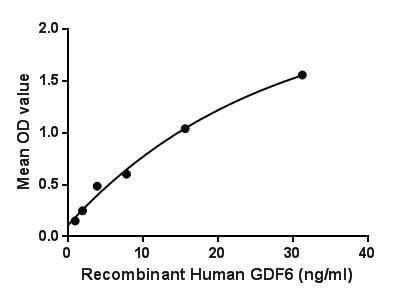 Bioactivity
(Figure. The binding activity of GDF6 with BMPR.)
Bioactivity
(Figure. The binding activity of GDF6 with BMPR.)
Growth Differentiation Factor 6, Active Protein (Cat# AAA150125)
 Bioactivity
(Figure. Cell suppresstion of HepG2 cells after stimulated with SFRP1.)
Bioactivity
(Figure. Cell suppresstion of HepG2 cells after stimulated with SFRP1.)
Secreted Frizzled Related Protein 1, Active Protein (Cat# AAA150141)
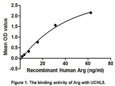
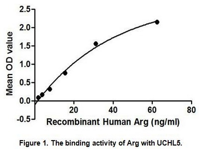
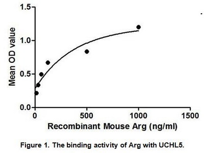
 Application Data
Arginase (ARG), Active Protein (Cat# AAA148185)
 Application Data
Arginase (ARG), Active Protein (Cat# AAA148186)
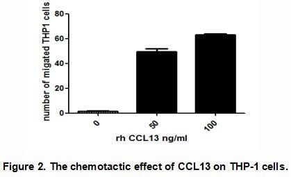
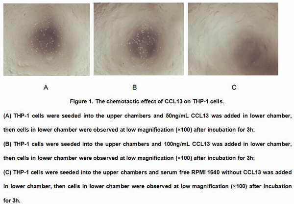
 Application Data
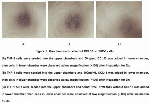
Monocyte Chemotactic Protein 4 (MCP4), Active Protein (Cat# AAA146618)
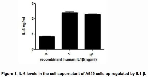
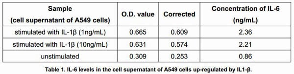
 Application Data
Interleukin 1 Beta (IL1b), Active Protein (Cat# AAA146620)
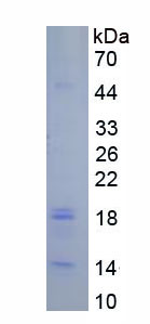
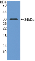
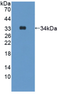
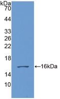
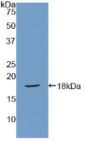
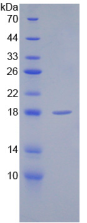
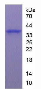
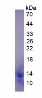
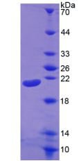
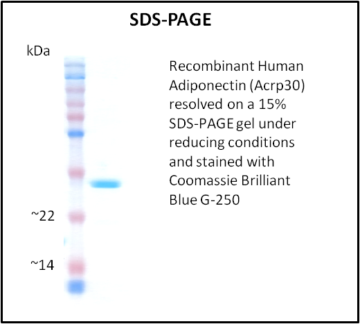
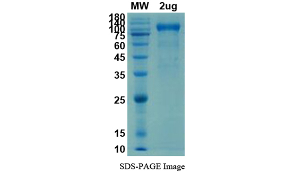
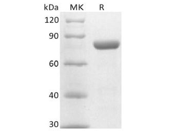
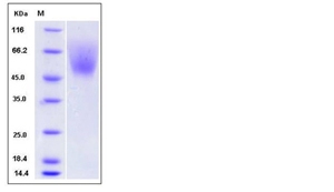
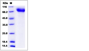
 SDS_PAGE
(Sample: Active recombinant PAM, Human)
SDS_PAGE
(Sample: Active recombinant PAM, Human)
Peptidylglycine Alpha Amidating Monooxygenase (PAM), Active Protein (Cat# AAA150334)
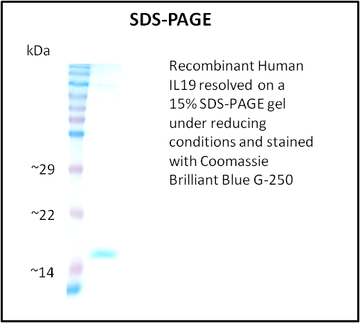
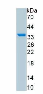
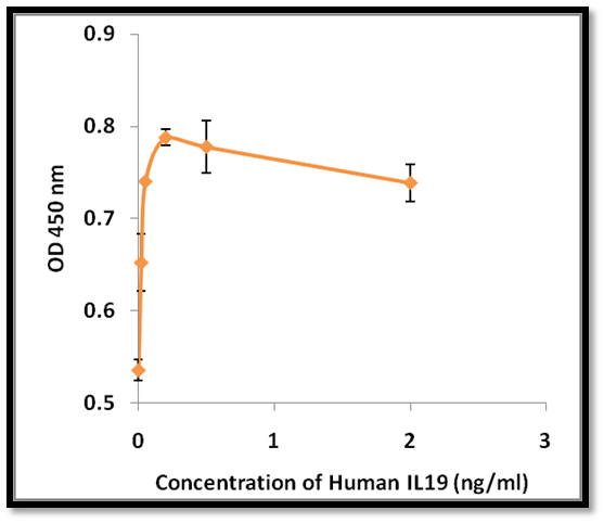 Application Data
IL19, Active Protein (Cat# AAA214302)
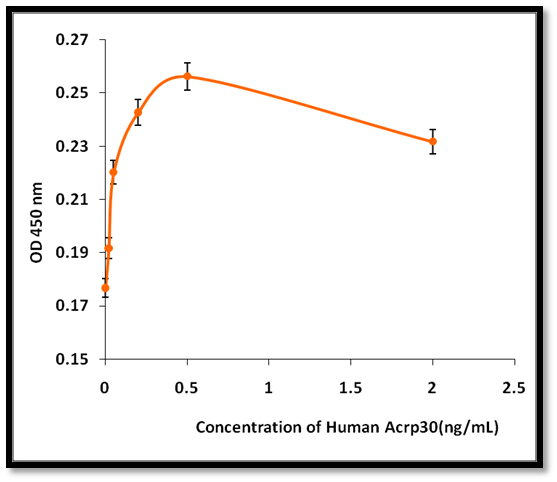 Application Data
Adiponectin (Acrp30), Active Protein (Cat# AAA214306)
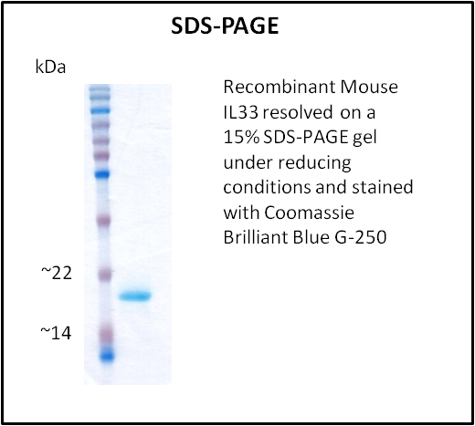
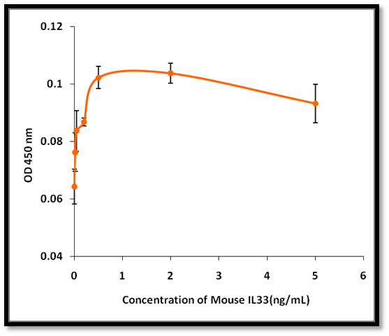 Application Data
IL33, Active Protein (Cat# AAA214317)
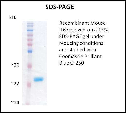
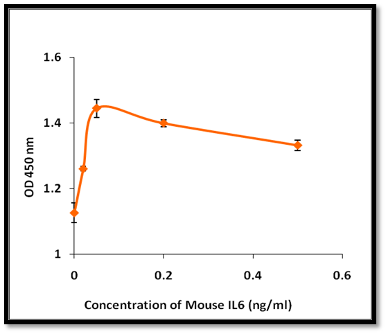 Application Data
IL6, Active Protein (Cat# AAA214318)
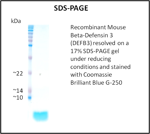
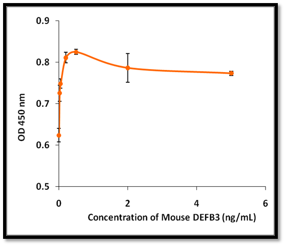 Application Data
Beta-Defensin 3 (DEFB3), Active Protein (Cat# AAA214325)
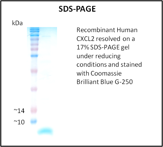
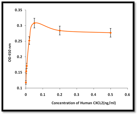 Application Data
CXCL2, Active Protein (Cat# AAA214224)
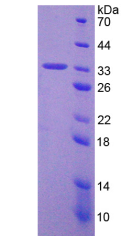
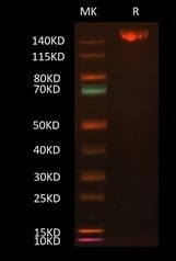
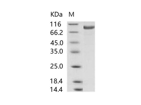
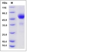
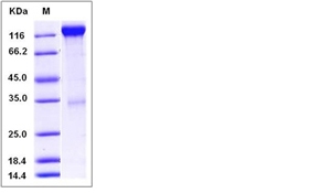
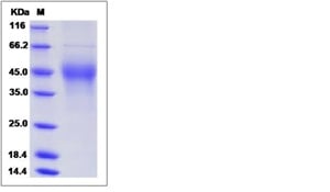
 SDS-PAGE
AGO2 / Argonaute 2 / EIF2C2, Active Protein (Cat# AAA173551)
CD40, Active Protein (Cat# AAA75431)
CD80 (B7-1), Active Protein (Cat# AAA76106)
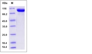
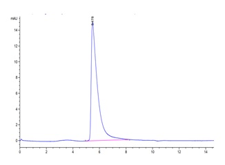 Application Data
(The purity of 2019-nCoV Spike S is greater than 95% as determined by SEC-HPLC.)
Application Data
(The purity of 2019-nCoV Spike S is greater than 95% as determined by SEC-HPLC.)
COVID 19 Spike S Trimer Coronavirus, Active Protein (Cat# AAA76122)
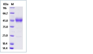
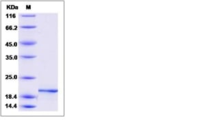
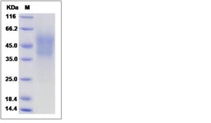
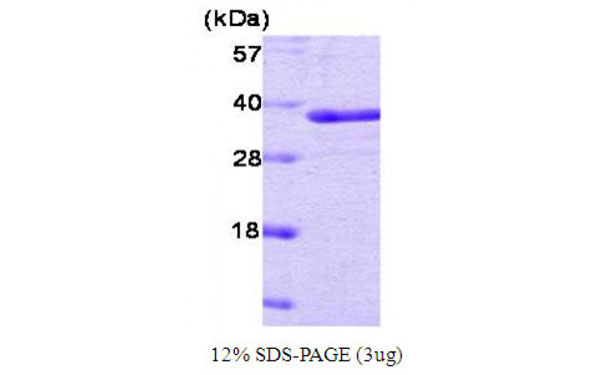 SDS-PAGE
(Figure annotation denotes ug of protein loaded and % gel used.)
SDS-PAGE
(Figure annotation denotes ug of protein loaded and % gel used.)
PTP1B, Active Protein (Cat# AAA107757)
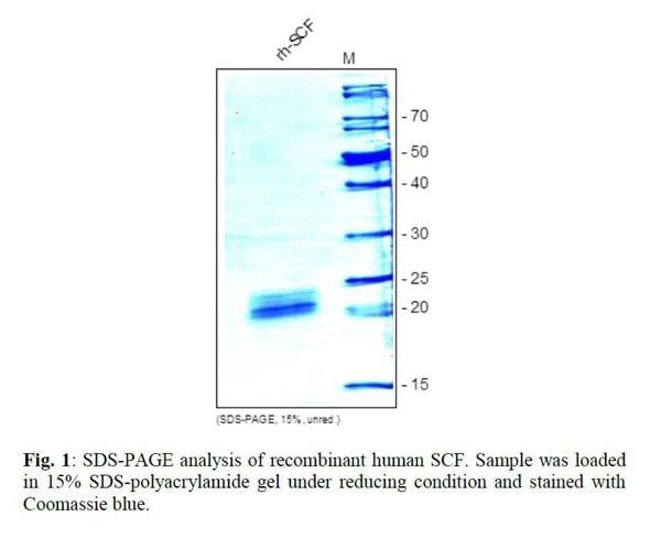
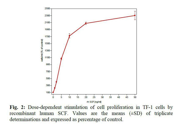 Application Data
SCF, Active Protein (Cat# AAA79310)
GPR15L, Active Protein (Cat# AAA79328)
TNF-alpha, Active Protein (Cat# AAA79194)
Chemerin, Active Protein (Cat# AAA79234)
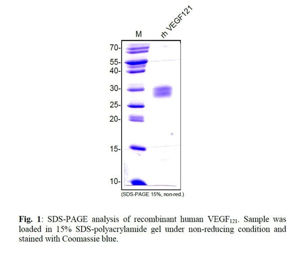
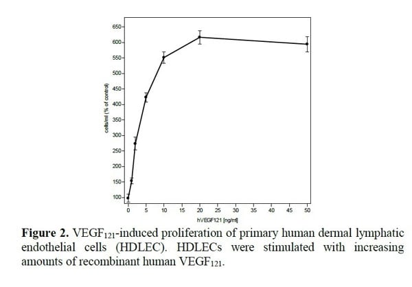 Application Data
VEGF121, Active Protein (Cat# AAA79142)
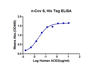
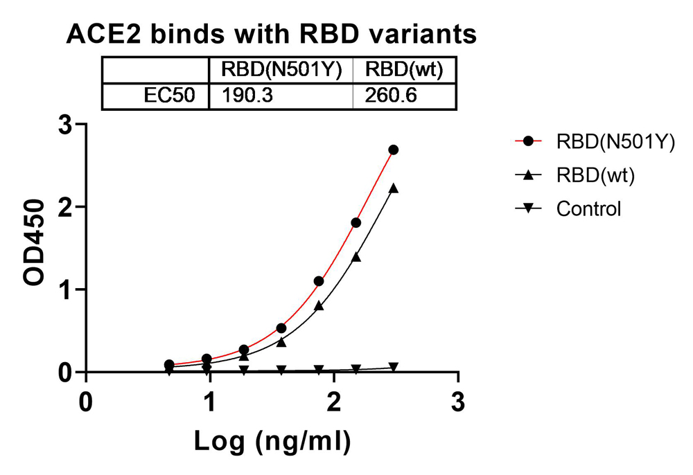 Bioactivity
ACE2, Active Protein (Cat# AAA119897)
 Bioactivity
MERS-CoV Spike/S1 Protein, Active Protein (Cat# AAA177990)
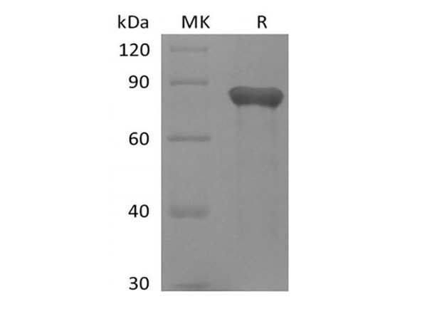
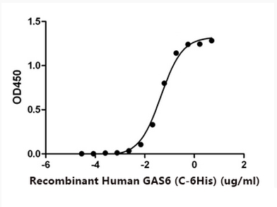 Bioactivity
GAS6, Active Protein (Cat# AAA177950)
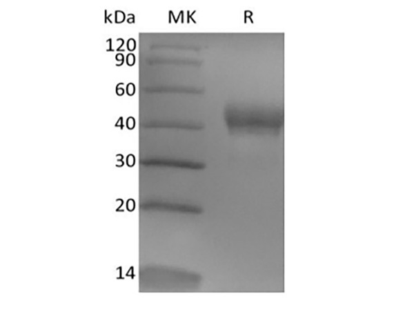
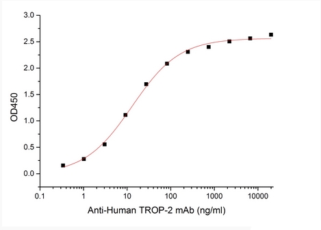 Bioactivity
Tumor-associated Calcium Signal Transducer 2/TROP-2, Active Protein (Cat# AAA177960)
 Bioactivity
Nectin-4, Active Protein (Cat# AAA177966)
Activin receptor type-2B (ACVR2B), Active Protein (Cat# AAA116934)
IL6R alpha, Active Protein (Cat# AAA75439)
Factor X, Active Protein (Cat# AAA75328)
Activin Receptor Type-2B (ACVR2B), Active Protein (Cat# AAA235620)
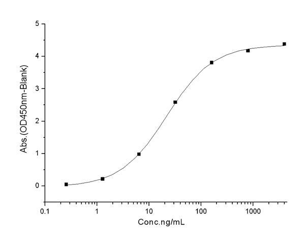
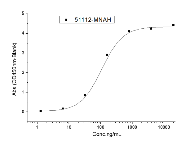
 Application Data
(Immobilized mouse PVR-His at 10 ug/ml (100 ul/well) can bind mouse TIGIT-Fc, The EC50 of mouse TIGIT-Fc is 0.25-0.55 ug/ml.)
Application Data
(Immobilized mouse PVR-His at 10 ug/ml (100 ul/well) can bind mouse TIGIT-Fc, The EC50 of mouse TIGIT-Fc is 0.25-0.55 ug/ml.)
TIGIT, Active Protein (Cat# AAA258211)
 Application Data
TIGIT, Active Protein (Cat# AAA258213)
 Application Data
(Measured by its binding ability in a functional ELISA. Immobilized mouse SIRPA-His at 10 ug/ml (100 ul/well) can bind human CD47-Fc, The EC50 of human CD47-Fc is 0.05-0.13 ug/ml.)
Application Data
(Measured by its binding ability in a functional ELISA. Immobilized mouse SIRPA-His at 10 ug/ml (100 ul/well) can bind human CD47-Fc, The EC50 of human CD47-Fc is 0.05-0.13 ug/ml.)
SIRP alpha, Active Protein (Cat# AAA258215)
 Application Data
(Immobilized mouse VEGF164 at 10 ug/ml (100 ul/well) can bind mouse KDR-Fc, The EC50 of mouse KDR-Fc is 0.11-0.27 ug/ml.)
Application Data
(Immobilized mouse VEGF164 at 10 ug/ml (100 ul/well) can bind mouse KDR-Fc, The EC50 of mouse KDR-Fc is 0.11-0.27 ug/ml.)
VEGFR2/KDR, Active Protein (Cat# AAA258218)
 Application Data
(Measured by its binding ability in a functional ELISA. Immobilized mouse PTN at 10 ug/ml (100 ul/well) can bind rat SDC1-Fc, The EC50 of rat SDC1-Fc is 0.4-1.1 ug/ml.)
Application Data
(Measured by its binding ability in a functional ELISA. Immobilized mouse PTN at 10 ug/ml (100 ul/well) can bind rat SDC1-Fc, The EC50 of rat SDC1-Fc is 0.4-1.1 ug/ml.)
Pleiotrophin/PTN, Active Protein (Cat# AAA258219)
 Application Data
(Measured by its binding ability in a functional ELISA. Immobilized mouse TEK-His at 10 ug/ml (100 ul/well) can bind human Ang2-Fc with a linear range of 6.25-200 ng/ml.)
Application Data
(Measured by its binding ability in a functional ELISA. Immobilized mouse TEK-His at 10 ug/ml (100 ul/well) can bind human Ang2-Fc with a linear range of 6.25-200 ng/ml.)
TIE2, Active Protein (Cat# AAA258224)
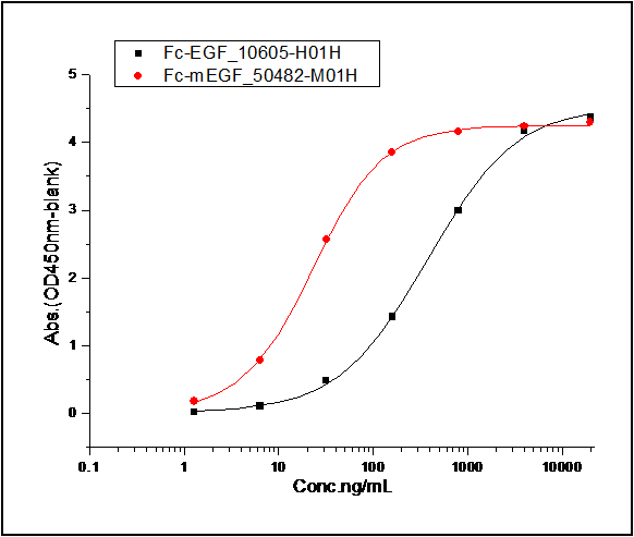 Application Data
(Measured by its binding ability in a functional ELISA. 1. Immobilized mouse EGFR-his at 10 ug/mL (100 ul/well) can bind human EGF-Fc, The EC50 of human EGF-Fc is 60-90 ng/mL. 2. Immobilized mouse EGFR-his at 10 ug/mL (100 ul/well) can bind mouse EGF-Fc, The EC50 of mouse EGF-Fc is 70-100 ng/mL.)
Application Data
(Measured by its binding ability in a functional ELISA. 1. Immobilized mouse EGFR-his at 10 ug/mL (100 ul/well) can bind human EGF-Fc, The EC50 of human EGF-Fc is 60-90 ng/mL. 2. Immobilized mouse EGFR-his at 10 ug/mL (100 ul/well) can bind mouse EGF-Fc, The EC50 of mouse EGF-Fc is 70-100 ng/mL.)
EGFR, Active Protein (Cat# AAA258226)
 Application Data
(Measured in a cell proliferation assay using TF-1 human erythroleukemic cells. The ED50 for this effect is typically 0.5-3 ng/ml.)
Application Data
(Measured in a cell proliferation assay using TF-1 human erythroleukemic cells. The ED50 for this effect is typically 0.5-3 ng/ml.)
Erythropoietin, Active Protein (Cat# AAA258227)
 Application Data
(Measured in a cell proliferation assay using TF-1 human erythroleukemic cells. The ED50 for this effect is typically 2-10 ng/mL.)
Application Data
(Measured in a cell proliferation assay using TF-1 human erythroleukemic cells. The ED50 for this effect is typically 2-10 ng/mL.)
Erythropoietin, Active Protein (Cat# AAA258228)
 Application Data
(Measured by its ability to inhibit NGF-induced proliferation of TF-1 human erythroleukemic cells. The ED50 for this effect is typically 0.2-1 ug/mL in the presence of 10 ng/mL of recombinant mouse NGF.)
Application Data
(Measured by its ability to inhibit NGF-induced proliferation of TF-1 human erythroleukemic cells. The ED50 for this effect is typically 0.2-1 ug/mL in the presence of 10 ng/mL of recombinant mouse NGF.)
TrkA, Active Protein (Cat# AAA258230)
 Application Data
(Measured in a cell proliferation assay using M-NFS-60 mouse myelogenous leukemia lymphoblast cells. The ED50 for this effect is typically 2-10 ng/ml.)
Application Data
(Measured in a cell proliferation assay using M-NFS-60 mouse myelogenous leukemia lymphoblast cells. The ED50 for this effect is typically 2-10 ng/ml.)
M-CSF, Active Protein (Cat# AAA258231)
 Application Data
(Immobilized mouse CSF1 at 2 ug/ml (100 ul/well) can bind mouse CSF1R-Fch, The EC50 of mouse CSF1R-Fch is 60-220 ng/mL.)
Application Data
(Immobilized mouse CSF1 at 2 ug/ml (100 ul/well) can bind mouse CSF1R-Fch, The EC50 of mouse CSF1R-Fch is 60-220 ng/mL.)
M-CSF, Active Protein (Cat# AAA258232)
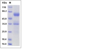
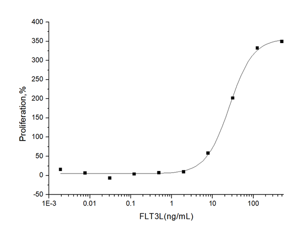 Application Data
(Measured in a cell proliferation assay using BaF3 mouse pro-B cells transfected with mouse Flt-3. The ED50 for this effect is typically 7-30 ng/mL.)
Application Data
(Measured in a cell proliferation assay using BaF3 mouse pro-B cells transfected with mouse Flt-3. The ED50 for this effect is typically 7-30 ng/mL.)
Flt3 Ligand, Active Protein (Cat# AAA258233)
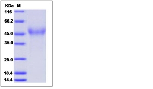
 Application Data
(Measured by its binding ability in a functional ELISA. Immobilized mouse CD320-His at 10 ug/ml (100 ul/well) can bind biotinylated mouse TCN2-His, The EC50 of biotinylated mouse TCN2-His is 0.10-0.24 ug/mL.)
Application Data
(Measured by its binding ability in a functional ELISA. Immobilized mouse CD320-His at 10 ug/ml (100 ul/well) can bind biotinylated mouse TCN2-His, The EC50 of biotinylated mouse TCN2-His is 0.10-0.24 ug/mL.)
CD320, Active Protein (Cat# AAA258237)
What Are Active Proteins?
Proteins are large molecules made up of long chains of amino acids.
They will typically fold into a very particular 3-dimensional shape/conformation, that is sometimes referred to as their “native” form, which allows them to work properly in the body. For the purposes of product categorization, AAA Biotech will typically refer to proteins purified from their original animal host as being “native” proteins (this is to signify their difference compared to their “recombinant” or “synthetic” protein counterparts).
If a protein successfully folds into the correct shape, it is will typically display high fidelity characteristics to its original protein in its original animal host, and be classified as an active protein, as it will be able to function “normally” in most enzymatic or binding capacities. If it loses this shape, due to factors such as heat or strong chemicals (such as detergents), it becomes inactive and is no longer able to perform its basic functions. All of the proteins in this category are made under strict quality control, and they are active, pure, low in contaminants, and stable.
Most are stored as freeze-dried powders and come without extra tags, so they’re very close to the actual natural/native form.
Key Applications of Active Proteins
1. Scientific Research
- Aid in the study of how proteins function in the body
- Aid in understanding various disease processes
2. Drug Development
- Powerful tools to investigate how potential drugs interact with specific proteins
- Ideal for identifying drug targets
3. Cell Culture
- Are routinely utilized to support cell growth and function (e.g., using exogenous growth factors)
- Can be used to promote cellular development into specific types (differentiation)
4. Diagnostics
- Regularly utilized in tests to detect diseases or infections (e.g., COVID-19, cancer)
- Note: All products are strictly for research-use only (RUO).
5. Therapeutics
- Some active proteins are used directly as treatments (e.g., insulin, enzymes)
- Note: All products are strictly for research-use only (RUO).
6. Vaccine Development
- Used to create or test vaccines by mimicking parts of viruses or bacteria
7. Biochemical Assays
- They can facilitate the characterization of enzyme activity, binding strength, or protein interactions in lab tests
Why Buy Active Proteins from AAA Biotech?
- High biological activity – Verified to perform as expected or indicated on datasheet
- Strict quality control – We are confident in our active proteins’ reliability and consistency
- High purity & low endotoxin – Ideal for applications involving sensitive or precious samples/components
- Freeze-dried for stability – Long shelf life and straightforward storage
- Mostly tag-free – Closer to natural/native protein form
FAQ
1. What are active proteins used for in research?
Active proteins are used primarily in the study of how proteins function, in characterizing/discovering drug interactions, supporting cell growth, running biochemical assays, and in development of diagnostics or therapeutics.
2. How are AAA Biotech's active proteins validated?
AAA Biotech’s active proteins are validated through strict quality control and functional assays to ensure they are properly folded and active. “Active”, though, can be an ambiguous term, so if a specific “activity” or “binding” capability of a protein is of crucial interest to you, please inquire with us prior to purchase, and we will provide further details on how the “Active” modifier was determined to be applicable.
3. Are these proteins tested for biological activity?
Yes, all active proteins from AAA Biotech are tested to confirm they have the expected biological activity before being offered for use. Though, said “biological activity” can be either “enzymatic”, “binding”, or both.