Filters
▼Clonality
▼Type
▼Reactivity
▼Gene Name
▼Isotype
▼Host
▼Application
▼Clone
▼Active Proteins
AAA Biotech also known as AAA Bio or AAABio provides a variety of high-quality recombinant and natural/native proteins that are proven to work in a wide range of experiments. Explore our products to find the active protein that best fits your needs or experimental model.
Viewing 1050-1100 of 2567 product results
 Application Data
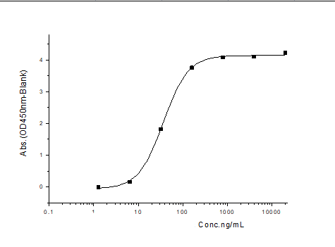
(Measured by its binding ability in a functional ELISA. Immobilized human CADM1 at 2 ug/ml (100 ul/well) can bind biotinylated human CRTAM with a linear range of 12.5-400 ng/ml.)
Application Data
(Measured by its binding ability in a functional ELISA. Immobilized human CADM1 at 2 ug/ml (100 ul/well) can bind biotinylated human CRTAM with a linear range of 12.5-400 ng/ml.)
CRTAM, Active Protein (Cat# AAA258000)
 Application Data
(Measured in a cell proliferation assay using BaF3 mouse pro-B cells transfected with mouse Flt-3. The ED50 for this effect is typically 2-11 ng/mL.)
Application Data
(Measured in a cell proliferation assay using BaF3 mouse pro-B cells transfected with mouse Flt-3. The ED50 for this effect is typically 2-11 ng/mL.)
Flt3 Ligand, Active Protein (Cat# AAA257867)
 Application Data
(Measured by its binding ability in a functional ELISA. Immobilized human GFRA1-Fc at 10 ug/mL (100 uL/well) can bind GDNF/Biotin, the EC50 of human GDNF/Biotin is 20-50 ng/mL.)
Application Data
(Measured by its binding ability in a functional ELISA. Immobilized human GFRA1-Fc at 10 ug/mL (100 uL/well) can bind GDNF/Biotin, the EC50 of human GDNF/Biotin is 20-50 ng/mL.)
GFR Alpha-1/GFRA1, Active Protein (Cat# AAA257871)

 Application Data
(Measured by its ability to inhibit rhIFN-gamma mediated protection of WISH cells infected with vesicular stomatitis virus(VSV). The ED50 for this effect is typically 2-8 ug/mL.)
Application Data
(Measured by its ability to inhibit rhIFN-gamma mediated protection of WISH cells infected with vesicular stomatitis virus(VSV). The ED50 for this effect is typically 2-8 ug/mL.)
IFNGR1, Active Protein (Cat# AAA257873)
 Application Data
(Measured by its binding ability in a functional ELISA. Immobilized human human B7-H2 at 1 ug/ml (100 ul/well) can bind human ICOS with a linear range of 1.6-200 ng/ml.)
Application Data
(Measured by its binding ability in a functional ELISA. Immobilized human human B7-H2 at 1 ug/ml (100 ul/well) can bind human ICOS with a linear range of 1.6-200 ng/ml.)
ICOS, Active Protein (Cat# AAA257874)
 Application Data
(Measured by its binding ability in a functional ELISA. Immobilized TGFBR2h (1-166Q) at 10 ug/mL (100 uL/well) can bind TGFB1-His/Biotin, the EC50 of human TGFB1-His/Biotin is 130-300 ng/mL.)
Application Data
(Measured by its binding ability in a functional ELISA. Immobilized TGFBR2h (1-166Q) at 10 ug/mL (100 uL/well) can bind TGFB1-His/Biotin, the EC50 of human TGFB1-His/Biotin is 130-300 ng/mL.)
TGFBR2, Active Protein (Cat# AAA257875)
 Application Data
(Measured by its binding ability in a functional ELISA. Immobilized PPICZa-IFNA2 at 10 ug/mL (100 uL/well) can bind IFNaR2-Fc, the EC50 of IFNaR2-Fc is 60-200 ng/mL.)
Application Data
(Measured by its binding ability in a functional ELISA. Immobilized PPICZa-IFNA2 at 10 ug/mL (100 uL/well) can bind IFNaR2-Fc, the EC50 of IFNaR2-Fc is 60-200 ng/mL.)
IFNAR2, Active Protein (Cat# AAA257877)
 Application Data
(Measured by its ability to induce alkaline phosphatase production by C3H10T1/2 mouse embryonic fibroblast cells. Nakamura, T. et al. (1997) Biochem. Biophys. Res. Commun.237:465. The ED50 for this effect is typically 2-10 ug/mL.)
Application Data
(Measured by its ability to induce alkaline phosphatase production by C3H10T1/2 mouse embryonic fibroblast cells. Nakamura, T. et al. (1997) Biochem. Biophys. Res. Commun.237:465. The ED50 for this effect is typically 2-10 ug/mL.)
Sonic Hedgehog, Active Protein (Cat# AAA257878)
 Application Data
(Measured by its ability to inhibit TNF-alpha mediated cytotoxicity in L-929 mouse fibrosarcoma cells in the presence of the metabolic inhibitor actinomycin D. The ED50 for this effect is typically 5-40 ng/mL in the presence of 1 ng/mL recombinant human TNF-alpha.)
Application Data
(Measured by its ability to inhibit TNF-alpha mediated cytotoxicity in L-929 mouse fibrosarcoma cells in the presence of the metabolic inhibitor actinomycin D. The ED50 for this effect is typically 5-40 ng/mL in the presence of 1 ng/mL recombinant human TNF-alpha.)
TNFR2/TNFRSF1B, Active Protein (Cat# AAA257881)
 Application Data
(Measured in a cell proliferation assay using TF-1 human erythroleukemic cells. The ED50 for this effect is typically 0.5-2.5 ng/ml.)
Application Data
(Measured in a cell proliferation assay using TF-1 human erythroleukemic cells. The ED50 for this effect is typically 0.5-2.5 ng/ml.)
Oncostatin M/OSM, Active Protein (Cat# AAA257885)
 Application Data
(Measured by its binding ability in a functional ELISA. Immobilized PDGFRbeta-His at 10 ug/ml (100 ul/well) can bind Cynomolgus PDGFB, The EC50 of Cynomolgus PDGFB is 3-8 ng/ml.)
Application Data
(Measured by its binding ability in a functional ELISA. Immobilized PDGFRbeta-His at 10 ug/ml (100 ul/well) can bind Cynomolgus PDGFB, The EC50 of Cynomolgus PDGFB is 3-8 ng/ml.)
PDGFRB, Active Protein (Cat# AAA257890)
 Application Data
(Measured by its binding ability in a functional ELISA. Immobilized human BMPR-II-Fc at 10 ug/mL (100 ul/well) can bind biotinylated human BMP2-Fc, The EC50 of biotinylated human BMP2-Fc is 80-110 ng/mL.)
Application Data
(Measured by its binding ability in a functional ELISA. Immobilized human BMPR-II-Fc at 10 ug/mL (100 ul/well) can bind biotinylated human BMP2-Fc, The EC50 of biotinylated human BMP2-Fc is 80-110 ng/mL.)
BMPR2, Active Protein (Cat# AAA257894)
 Application Data
(Measured in a cell proliferation assay using BaF3 mouse pro-B cells transfected with human FGFR2b. The EC50 for this effect is typically 2-14 ng/mL.)
Application Data
(Measured in a cell proliferation assay using BaF3 mouse pro-B cells transfected with human FGFR2b. The EC50 for this effect is typically 2-14 ng/mL.)
FGF10, Active Protein (Cat# AAA257898)
 Application Data
(Measured by its binding ability in a functional ELISA. Immobilized Human IGF1 at 2 ug/ml (100 ul/well) can bind Human IGFBP4 His, the EC50 of Human IGFBP4 His is 10-60 ng/mL.)
Application Data
(Measured by its binding ability in a functional ELISA. Immobilized Human IGF1 at 2 ug/ml (100 ul/well) can bind Human IGFBP4 His, the EC50 of Human IGFBP4 His is 10-60 ng/mL.)
IGF1, Active Protein (Cat# AAA257900)
 Application Data
(Measured in a cytotoxicity assay using L929 mouse fibrosarcoma cells in the presence of the metabolic inhibitor actinomycin D. The ED50 for this effect is typically 1-5 ng/mL.)
Application Data
(Measured in a cytotoxicity assay using L929 mouse fibrosarcoma cells in the presence of the metabolic inhibitor actinomycin D. The ED50 for this effect is typically 1-5 ng/mL.)
TNF-alpha, Active Protein (Cat# AAA257901)
 Application Data
(Measured by its ability to bind human LIF-Fc in a functional ELISA.)
Application Data
(Measured by its ability to bind human LIF-Fc in a functional ELISA.)
LIFR, Active Protein (Cat# AAA257904)
 Application Data
(Measured by its ability to neutralize Activin-mediated inhibition on MPC11 cell proliferation. The ED50 for this effect is typically 0.5-3 ug/mL in the presence of 10 ng/ml Recombinant Human ctivin A.)
Application Data
(Measured by its ability to neutralize Activin-mediated inhibition on MPC11 cell proliferation. The ED50 for this effect is typically 0.5-3 ug/mL in the presence of 10 ng/ml Recombinant Human ctivin A.)
Follistatin, Active Protein (Cat# AAA257910)
 Application Data
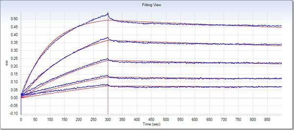
(Measured by its binding ability in a functional ELISA. Immobilized recombinant human Angiopoietin-2 at 10 ug/ml (100 ul/well) can bind Human Tie2 / Fc chimera with a range of 0.2-20 ug/ml.)
Application Data
(Measured by its binding ability in a functional ELISA. Immobilized recombinant human Angiopoietin-2 at 10 ug/ml (100 ul/well) can bind Human Tie2 / Fc chimera with a range of 0.2-20 ug/ml.)
TIE2, Active Protein (Cat# AAA257917)
 Application Data
(Measured by its binding ability in a functional ELISA. Immobilized EPOR (hFc Tag) at 2 ug/mL (100uL/well) can bind Cynomolgus Erythropoietin-His, the EC50 of Cynomolgus Erythropoietin-His is 4-20 ng/mL.)
Application Data
(Measured by its binding ability in a functional ELISA. Immobilized EPOR (hFc Tag) at 2 ug/mL (100uL/well) can bind Cynomolgus Erythropoietin-His, the EC50 of Cynomolgus Erythropoietin-His is 4-20 ng/mL.)
EPOR, Active Protein (Cat# AAA257918)
 Application Data
(Measured by its ability to bind biotinylated human DMP1 in a functional ELISA.)
Application Data
(Measured by its ability to bind biotinylated human DMP1 in a functional ELISA.)
Factor H, Active Protein (Cat# AAA257920)
 Application Data
(Measured by its ability to bind recombinant human EphrinB2 in a functional ELISA.)
Application Data
(Measured by its ability to bind recombinant human EphrinB2 in a functional ELISA.)
EphB2, Active Protein (Cat# AAA257925)
 Application Data
(Measured by its binding ability in a functional ELISA. Immobilized human CD40L-His at 10 ug/mL (100 uL/well) can bind Human CD40-Fc. The EC50 of Human CD40-Fc is 10-30 ng/mL.)
Application Data
(Measured by its binding ability in a functional ELISA. Immobilized human CD40L-His at 10 ug/mL (100 uL/well) can bind Human CD40-Fc. The EC50 of Human CD40-Fc is 10-30 ng/mL.)
CD40, Active Protein (Cat# AAA257926)

 Application Data
(Measured by its binding ability in a functional ELISA. Immobilized human CD70 at 10 ug/mL (100 ul/well) can bind human CD27 with a linear range of 40-60 ng/ml.)
Application Data
(Measured by its binding ability in a functional ELISA. Immobilized human CD70 at 10 ug/mL (100 ul/well) can bind human CD27 with a linear range of 40-60 ng/ml.)
CD70, Active Protein (Cat# AAA257927)
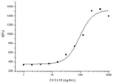 Application Data
(Measured by its ability to chemoattract BaF3 mouse pro-B cells transfected with mouse CXCR6. The ED50 for this effect is 50-250 ng/mL.)
Application Data
(Measured by its ability to chemoattract BaF3 mouse pro-B cells transfected with mouse CXCR6. The ED50 for this effect is 50-250 ng/mL.)
CXCL16, Active Protein (Cat# AAA257935)
 Application Data
(Measured by its binding ability in a functional ELISA. Immobilized PVRL1/NECTIN1 Protein, Human, Recombinant (His Tag) at 2 ug/ml (100 ul/well) can bind Nectin 3 Protein, Human, Recombinant (ECD, hFc Tag), the EC50 of Nectin 3 Protein, Human, Recombinant (ECD, hFc Tag)is 30-140 ng/mL.)
Application Data
(Measured by its binding ability in a functional ELISA. Immobilized PVRL1/NECTIN1 Protein, Human, Recombinant (His Tag) at 2 ug/ml (100 ul/well) can bind Nectin 3 Protein, Human, Recombinant (ECD, hFc Tag), the EC50 of Nectin 3 Protein, Human, Recombinant (ECD, hFc Tag)is 30-140 ng/mL.)
Nectin 3, Active Protein (Cat# AAA257936)
 Application Data
(Measured by its binding ability in a functional ELISA. 1. Immobilized recombinant human PVRL3 at 1 ug/ml (100 ul/well) can bind biotinylated Nectin-1 with a linear range of 6.4-800 ng/ml. 2. Immobilized recombinant human PVRL3 at 1 ug/ml (100 ul/well) can bind Nectin-1/ Fc chimera with a linear range of 0.156-5 ng/ml.)
Application Data
(Measured by its binding ability in a functional ELISA. 1. Immobilized recombinant human PVRL3 at 1 ug/ml (100 ul/well) can bind biotinylated Nectin-1 with a linear range of 6.4-800 ng/ml. 2. Immobilized recombinant human PVRL3 at 1 ug/ml (100 ul/well) can bind Nectin-1/ Fc chimera with a linear range of 0.156-5 ng/ml.)
Nectin 3, Active Protein (Cat# AAA257937)
 Application Data
(Measured by its binding ability in a functional ELISA. Immobilized human EphB4-his at 2 ug/ml (100 ul/well) can bind human EphrinB2 Fc chimera with a linear range of 1-25 ng/ml.)
Application Data
(Measured by its binding ability in a functional ELISA. Immobilized human EphB4-his at 2 ug/ml (100 ul/well) can bind human EphrinB2 Fc chimera with a linear range of 1-25 ng/ml.)
Ephrin B2, Active Protein (Cat# AAA257941)
 Application Data
(Measured by its binding ability in a functional ELISA. Immobilized human EFNB2 at 10ug/mL (100uL/well) can bind biotinylate human EphB4-Fc, the EC50 of biotinylate human EphB4-Fc is 5-60 ng/mL.)
Application Data
(Measured by its binding ability in a functional ELISA. Immobilized human EFNB2 at 10ug/mL (100uL/well) can bind biotinylate human EphB4-Fc, the EC50 of biotinylate human EphB4-Fc is 5-60 ng/mL.)
Ephrin B2, Active Protein (Cat# AAA257943)

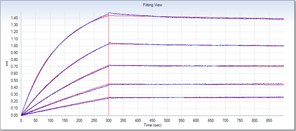
 Application Data
(Loaded Anti-CD112 Antibody on AMC Biosensor, can bind Nectin-2 Protein, Human, Recombinant (His Tag) with an affinity constant of 34.4 nM as determined by Octet RED System.)
Application Data
(Loaded Anti-CD112 Antibody on AMC Biosensor, can bind Nectin-2 Protein, Human, Recombinant (His Tag) with an affinity constant of 34.4 nM as determined by Octet RED System.)
Nectin-2, Active Protein (Cat# AAA257812)
 Application Data
(Measured in a cell proliferation assay using Balb/c 3T3 mouse embryonic fibroblasts. The ED50 for this effect is typically 0.15-0.75 ng/mL.)
Application Data
(Measured in a cell proliferation assay using Balb/c 3T3 mouse embryonic fibroblasts. The ED50 for this effect is typically 0.15-0.75 ng/mL.)
FGF1, Active Protein (Cat# AAA257816)
 Application Data
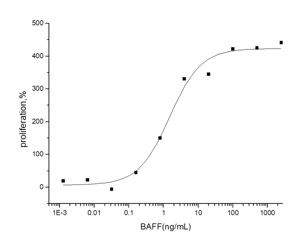
(Measured in a cell proliferation assay in mouse splenocytes. The ED50 for this effect is typically 0.6-3.2 ng/mL.)
Application Data
(Measured in a cell proliferation assay in mouse splenocytes. The ED50 for this effect is typically 0.6-3.2 ng/mL.)
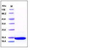
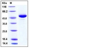
BAFF/BLyS, Active Protein (Cat# AAA257823)
 Application Data
(Measured by its binding ability in a functional ELISA. Immobilized human GPC5 at 5 ug/ml (100 ul/well) can bind human bFGF with a linear ranger of 0.156-2.5 ng/ml.)
Application Data
(Measured by its binding ability in a functional ELISA. Immobilized human GPC5 at 5 ug/ml (100 ul/well) can bind human bFGF with a linear ranger of 0.156-2.5 ng/ml.)
Glypican 5, Active Protein (Cat# AAA257827)
 Application Data
(Measured by its ability to inhibit Wnt3a-induced alkaline phosphatase production by C3H10T1/2 cells. The ED50 for this effect is approximately 0.1-0.4 ug/ml in the presence of 10 ng/mL of mouse Wnt3a.)
Application Data
(Measured by its ability to inhibit Wnt3a-induced alkaline phosphatase production by C3H10T1/2 cells. The ED50 for this effect is approximately 0.1-0.4 ug/ml in the presence of 10 ng/mL of mouse Wnt3a.)
DKK1, Active Protein (Cat# AAA257837)
 Application Data
(Measured by its ability to compete with human EphrinA3 / Fc for binding to immobilized mouse EphA6-his in a functional ELISA assay.)
Application Data
(Measured by its ability to compete with human EphrinA3 / Fc for binding to immobilized mouse EphA6-his in a functional ELISA assay.)
Ephrin A3, Active Protein (Cat# AAA257841)
 Application Data
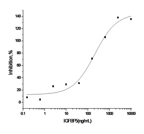
(Measured by its ability to inhibit the biological activity of IGF-I or IGF-II on MCF-7 human breast cancer cells. The ED50 for this effect is 0.1-0.5 ug/mL in the presence of 14 ng/mL rhIGF-II.)
Application Data
(Measured by its ability to inhibit the biological activity of IGF-I or IGF-II on MCF-7 human breast cancer cells. The ED50 for this effect is 0.1-0.5 ug/mL in the presence of 14 ng/mL rhIGF-II.)
IGFBP5, Active Protein (Cat# AAA257845)
 Application Data
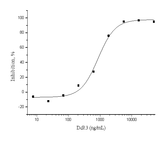
(Measured by its ability to inhibit Fas Ligand induced apoptosis of Jurkat human acute T cell leukemia cells. The ED50 for this effect is typically 0.5-3 ug/mL in the presence of 200 ng/mL recombinant human Fas ligand.)
Application Data
(Measured by its ability to inhibit Fas Ligand induced apoptosis of Jurkat human acute T cell leukemia cells. The ED50 for this effect is typically 0.5-3 ug/mL in the presence of 200 ng/mL recombinant human Fas ligand.)
DcR3, Active Protein (Cat# AAA257848)
 Application Data
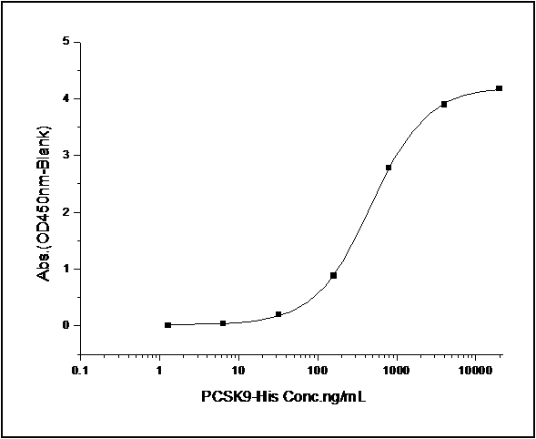
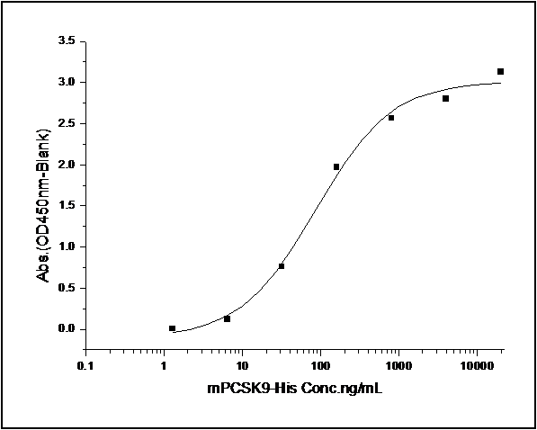
(Measure by its ability to bind with human PCSK9 in a functional ELISA. Immobilized mouse PCSK9 at 10 ug/ml (100 ul/well) can bind biotinylated recombinant human LDLR. The EC50 of biotinylated human LDLR is 0.12 ug/ml.)
Application Data
(Measure by its ability to bind with human PCSK9 in a functional ELISA. Immobilized mouse PCSK9 at 10 ug/ml (100 ul/well) can bind biotinylated recombinant human LDLR. The EC50 of biotinylated human LDLR is 0.12 ug/ml.)
LDLR/LDL Receptor, Active Protein (Cat# AAA257851)
 Application Data
(Measured by its binding ability in a functional ELISA. Immobilized human EphB4 at 2 ug/ml (100 ul/well) can bind human EphrinB2 with a linear range of 1-125 ng/ml.)
Application Data
(Measured by its binding ability in a functional ELISA. Immobilized human EphB4 at 2 ug/ml (100 ul/well) can bind human EphrinB2 with a linear range of 1-125 ng/ml.)
EphB4, Active Protein (Cat# AAA257853)
 Application Data
(Measured by its binding ability in a functional ELISA. Immobilized human MDK at 10 ug/ml (100 ul/well) can bind mouse SDC4-Fc with a linear range of 0.16-1.25 ug/ml.)
Application Data
(Measured by its binding ability in a functional ELISA. Immobilized human MDK at 10 ug/ml (100 ul/well) can bind mouse SDC4-Fc with a linear range of 0.16-1.25 ug/ml.)
Midkine, Active Protein (Cat# AAA257854)
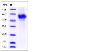
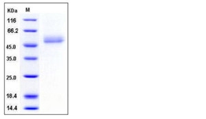
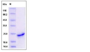
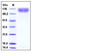
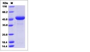
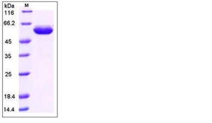
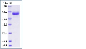
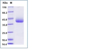
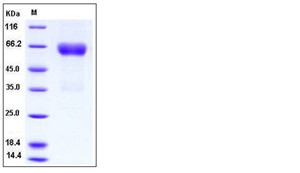
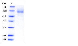
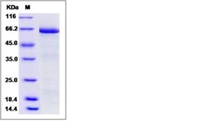
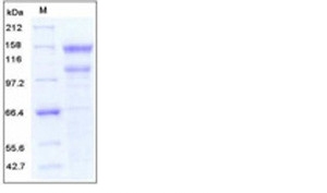
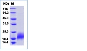
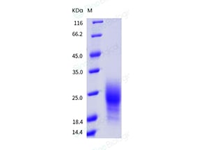
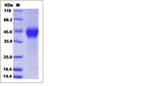
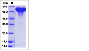
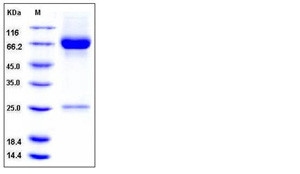 SDS-PAGE
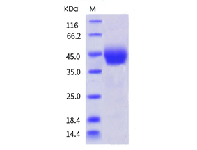
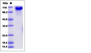
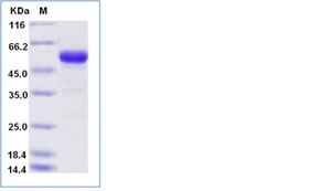
MMP-8, Active Protein (Cat# AAA257855)
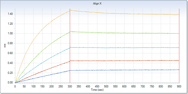
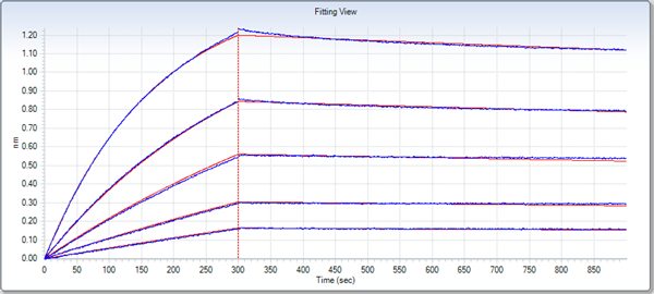

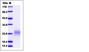
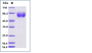
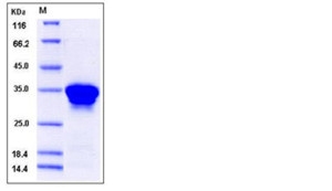
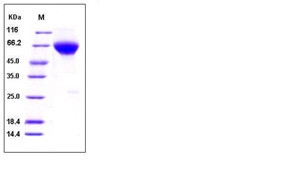
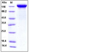
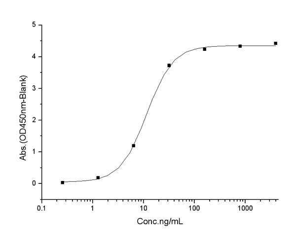
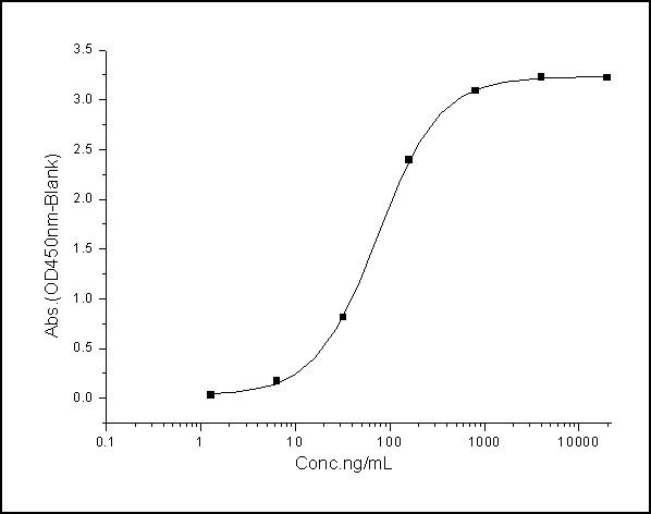
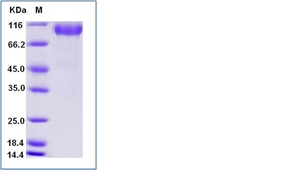
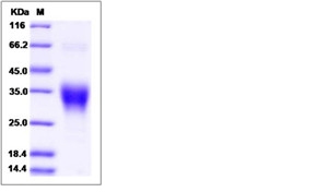
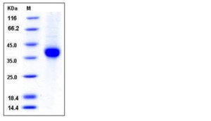
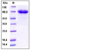
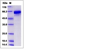
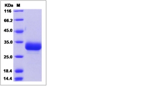
 Application Data
CD32B/Fcgr2b, Active Protein (Cat# AAA257858)
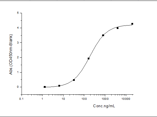
 Application Data
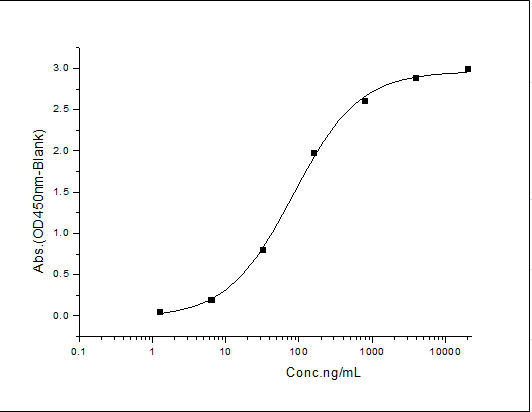
(Measured by its binding ability in a functional ELISA. Immobilized Human PDGFRB at 2 ug/ml (100 ul/well) can bind Mouse PDGF-B His, the EC50 of Mouse PDGF-B His is 4-24 ng/mL.)
Application Data
(Measured by its binding ability in a functional ELISA. Immobilized Human PDGFRB at 2 ug/ml (100 ul/well) can bind Mouse PDGF-B His, the EC50 of Mouse PDGF-B His is 4-24 ng/mL.)
PDGF-B, Active Protein (Cat# AAA258203)
 Application Data
(Measured in a serum-free cell proliferation assay using MCF-7 human breast cancer cells. Karey, K.P. et al. (1988) Cancer Research 48:4083. The ED50 for this effect is typically 5-40 ng/mL.)
Application Data
(Measured in a serum-free cell proliferation assay using MCF-7 human breast cancer cells. Karey, K.P. et al. (1988) Cancer Research 48:4083. The ED50 for this effect is typically 5-40 ng/mL.)
Transferrin, Active Protein (Cat# AAA258207)
 Application Data
(Measured in a serum-free cell proliferation assay using MCF-7 human breast cancer cells. Karey, K.P. et al. (1988) Cancer Research 48:4083. The ED50 for this effect is typically 0.05-0.2 ug/mL.)
Application Data
(Measured in a serum-free cell proliferation assay using MCF-7 human breast cancer cells. Karey, K.P. et al. (1988) Cancer Research 48:4083. The ED50 for this effect is typically 0.05-0.2 ug/mL.)
Transferrin, Active Protein (Cat# AAA258208)
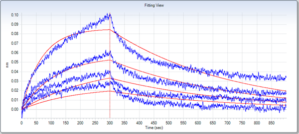
 Application Data
(Immobilized mouse TIGIT-His at 10 ug/ml (100 ul/well) can bind mouse PVR-Fch, The EC50 of mouse PVR-Fch is 0.31-0.73 ug/ml.)
Application Data
(Immobilized mouse TIGIT-His at 10 ug/ml (100 ul/well) can bind mouse PVR-Fch, The EC50 of mouse PVR-Fch is 0.31-0.73 ug/ml.)
TIGIT, Active Protein (Cat# AAA258212)
 Application Data
(Measured by its binding ability in a functional ELISA. Immobilized mouse CD27-His at 10 ug/mL (100 uL/well) can bind mouse CD70, the EC50of mouse CD70 is 10-50 ng/mL.)
Application Data
(Measured by its binding ability in a functional ELISA. Immobilized mouse CD27-His at 10 ug/mL (100 uL/well) can bind mouse CD70, the EC50of mouse CD70 is 10-50 ng/mL.)
CD70, Active Protein (Cat# AAA258236)
 Application Data
(Measured by its binding ability in a functional ELISA. Immobilized mouse EFNB3-His at 10 ug/mL (100 uL/well) can bind biotinylated mouse EPHB3-His, the EC50 of biotinylated mouse EPHB3-His 0.02-0.4 ug/mL.)
Application Data
(Measured by its binding ability in a functional ELISA. Immobilized mouse EFNB3-His at 10 ug/mL (100 uL/well) can bind biotinylated mouse EPHB3-His, the EC50 of biotinylated mouse EPHB3-His 0.02-0.4 ug/mL.)
Ephrin B3, Active Protein (Cat# AAA258239)

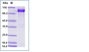
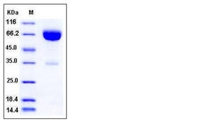
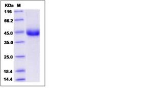
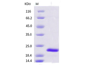
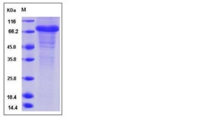
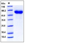
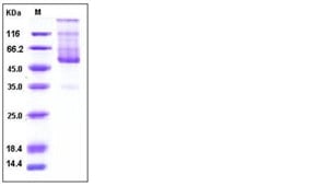
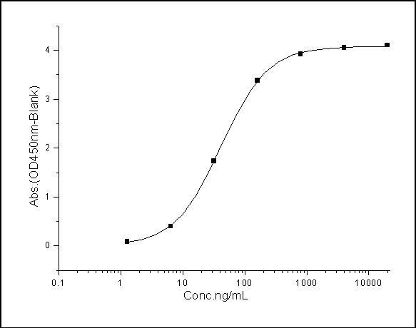
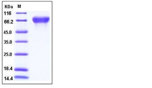
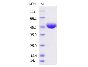
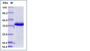
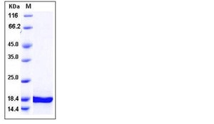
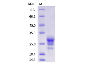
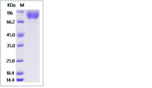 Application Data
PDGFRA, Active Protein (Cat# AAA258242)
 Application Data
(Measured by its ability to inhibit recombinant rmWnt3a induced alkaline phosphatase production by C3H10T 1/2 cells. The ED50 for this effect is 0.05-0.3 ug/mL.)
Application Data
(Measured by its ability to inhibit recombinant rmWnt3a induced alkaline phosphatase production by C3H10T 1/2 cells. The ED50 for this effect is 0.05-0.3 ug/mL.)
DKK1, Active Protein (Cat# AAA258244)
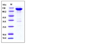
 Application Data
(Measured by its ability to bind rat Fc-VEGFC in functional ELISA.)
Application Data
(Measured by its ability to bind rat Fc-VEGFC in functional ELISA.)
Neuropilin-2, Active Protein (Cat# AAA258245)
What Are Active Proteins?
Proteins are large molecules made up of long chains of amino acids.
They will typically fold into a very particular 3-dimensional shape/conformation, that is sometimes referred to as their “native” form, which allows them to work properly in the body. For the purposes of product categorization, AAA Biotech will typically refer to proteins purified from their original animal host as being “native” proteins (this is to signify their difference compared to their “recombinant” or “synthetic” protein counterparts).
If a protein successfully folds into the correct shape, it is will typically display high fidelity characteristics to its original protein in its original animal host, and be classified as an active protein, as it will be able to function “normally” in most enzymatic or binding capacities. If it loses this shape, due to factors such as heat or strong chemicals (such as detergents), it becomes inactive and is no longer able to perform its basic functions. All of the proteins in this category are made under strict quality control, and they are active, pure, low in contaminants, and stable.
Most are stored as freeze-dried powders and come without extra tags, so they’re very close to the actual natural/native form.
Key Applications of Active Proteins
1. Scientific Research
- Aid in the study of how proteins function in the body
- Aid in understanding various disease processes
2. Drug Development
- Powerful tools to investigate how potential drugs interact with specific proteins
- Ideal for identifying drug targets
3. Cell Culture
- Are routinely utilized to support cell growth and function (e.g., using exogenous growth factors)
- Can be used to promote cellular development into specific types (differentiation)
4. Diagnostics
- Regularly utilized in tests to detect diseases or infections (e.g., COVID-19, cancer)
- Note: All products are strictly for research-use only (RUO).
5. Therapeutics
- Some active proteins are used directly as treatments (e.g., insulin, enzymes)
- Note: All products are strictly for research-use only (RUO).
6. Vaccine Development
- Used to create or test vaccines by mimicking parts of viruses or bacteria
7. Biochemical Assays
- They can facilitate the characterization of enzyme activity, binding strength, or protein interactions in lab tests
Why Buy Active Proteins from AAA Biotech?
- High biological activity – Verified to perform as expected or indicated on datasheet
- Strict quality control – We are confident in our active proteins’ reliability and consistency
- High purity & low endotoxin – Ideal for applications involving sensitive or precious samples/components
- Freeze-dried for stability – Long shelf life and straightforward storage
- Mostly tag-free – Closer to natural/native protein form
FAQ
1. What are active proteins used for in research?
Active proteins are used primarily in the study of how proteins function, in characterizing/discovering drug interactions, supporting cell growth, running biochemical assays, and in development of diagnostics or therapeutics.
2. How are AAA Biotech's active proteins validated?
AAA Biotech’s active proteins are validated through strict quality control and functional assays to ensure they are properly folded and active. “Active”, though, can be an ambiguous term, so if a specific “activity” or “binding” capability of a protein is of crucial interest to you, please inquire with us prior to purchase, and we will provide further details on how the “Active” modifier was determined to be applicable.
3. Are these proteins tested for biological activity?
Yes, all active proteins from AAA Biotech are tested to confirm they have the expected biological activity before being offered for use. Though, said “biological activity” can be either “enzymatic”, “binding”, or both.