Filters
▼Clonality
▼Type
▼Reactivity
▼Gene Name
▼Isotype
▼Host
▼Application
▼Clone
▼Active Proteins
AAA Biotech also known as AAA Bio or AAABio provides a variety of high-quality recombinant and natural/native proteins that are proven to work in a wide range of experiments. Explore our products to find the active protein that best fits your needs or experimental model.
Viewing 800-850 of 2567 product results
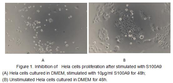
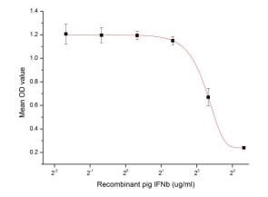 Bioactivity
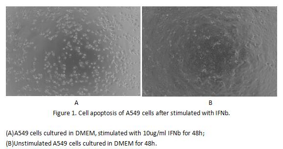
(Figure 2. Cell apoptosis of A549 cells after stimulated with IFNb.)
Bioactivity
(Figure 2. Cell apoptosis of A549 cells after stimulated with IFNb.)
Interferon Beta (IFNb), Active Protein (Cat# AAA153005)

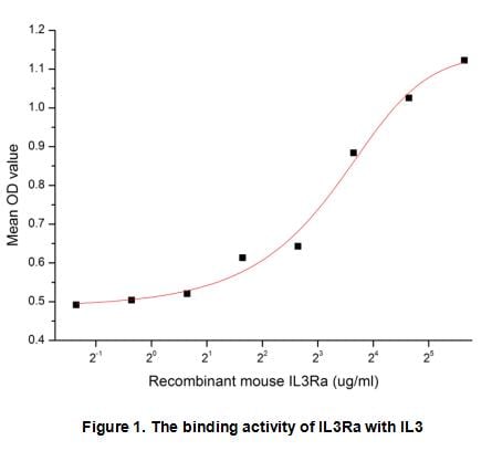 Bioactivity
(Interleukin 3 receptor alpha (IL3Ra), also known as CD123 (Cluster of Differentiation 123), is a subunit of the functional high-affinity mouse IL-3 receptor which is a heterodimer. The alpha subunit alone binds IL-3 with low affinity. The beta subunit doe)
Bioactivity
(Interleukin 3 receptor alpha (IL3Ra), also known as CD123 (Cluster of Differentiation 123), is a subunit of the functional high-affinity mouse IL-3 receptor which is a heterodimer. The alpha subunit alone binds IL-3 with low affinity. The beta subunit doe)
Interleukin 3 Receptor Alpha (IL3Ra), Active Protein (Cat# AAA153091)
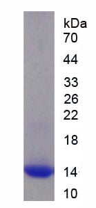

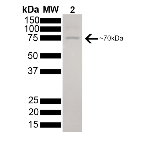
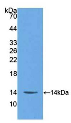 WB (Western Blot)
(Sample: Recombinant S100A9, Human;Antibody: Rabbit Anti- Human S100A9 Ab)
WB (Western Blot)
(Sample: Recombinant S100A9, Human;Antibody: Rabbit Anti- Human S100A9 Ab)
S100 Calcium Binding Protein A9 (S100A9), Active Protein (Cat# AAA153103)
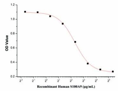
 Bioactivity
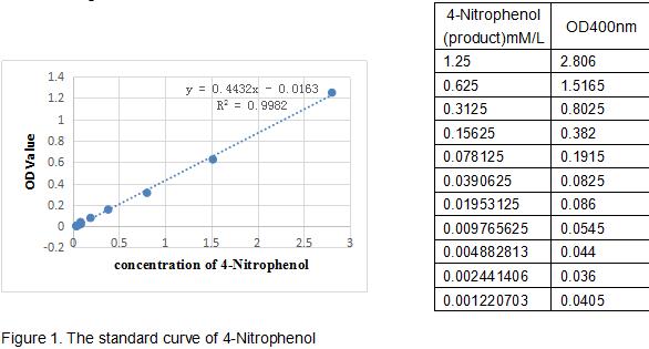
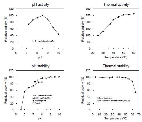
(One unit of enzyme activity is defined as the 1ug of enzyme required to convert 1pmol of 4-Nitrophenyl acetate to 4-Nitrophenol in 1min at 37 degree C. The specific activity of recombinant mouse CES1 is 158 pmol/min/ug.)
Bioactivity
(One unit of enzyme activity is defined as the 1ug of enzyme required to convert 1pmol of 4-Nitrophenyl acetate to 4-Nitrophenol in 1min at 37 degree C. The specific activity of recombinant mouse CES1 is 158 pmol/min/ug.)
Carboxylesterase 1 (CES1), Active Protein (Cat# AAA153113)
 Bioactivity
(carboxylesterase 1(CES1) also known as Liver carboxylesterase 1 is a serine esterase and member of a large multigene carboxylesterase family. The protein Involved in the detoxification of xenobiotics and in the activation of ester and amide prodrugs. Hydr)
Bioactivity
(carboxylesterase 1(CES1) also known as Liver carboxylesterase 1 is a serine esterase and member of a large multigene carboxylesterase family. The protein Involved in the detoxification of xenobiotics and in the activation of ester and amide prodrugs. Hydr)
Carboxylesterase 1 (CES1), Active Protein (Cat# AAA153114)
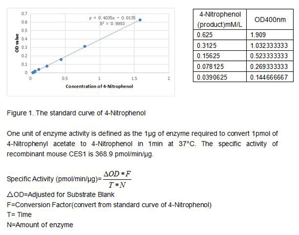
 Bioactivity
(Catalase (CAT) is an antioxidant enzyme present in all aerobic organisms. It is known to catalyze H2O2 into water and oxygen in an energy-efficient manner in the cells exposed to environmental stress. H2O2 will have specific absorbance at 240 nm . when we)
Bioactivity
(Catalase (CAT) is an antioxidant enzyme present in all aerobic organisms. It is known to catalyze H2O2 into water and oxygen in an energy-efficient manner in the cells exposed to environmental stress. H2O2 will have specific absorbance at 240 nm . when we)
Catalase (CAT), Active Protein (Cat# AAA153117)
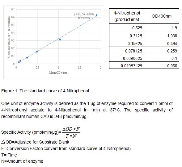 Bioactivity
(Carbonic Anhydrase IX (CA9) also known as membrane antigen MN and renal cell carcinoma (RCC) associated antigen G250, is a transmembrane enzyme expressed primarily in carcinoma cells. It is one of the best markers for hypoxia and for RCC. rhCA9 correspond)
Bioactivity
(Carbonic Anhydrase IX (CA9) also known as membrane antigen MN and renal cell carcinoma (RCC) associated antigen G250, is a transmembrane enzyme expressed primarily in carcinoma cells. It is one of the best markers for hypoxia and for RCC. rhCA9 correspond)
Carbonic Anhydrase IX (CA9), Active Protein (Cat# AAA153124)
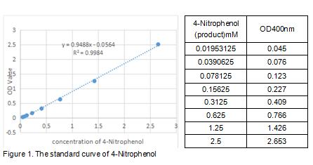
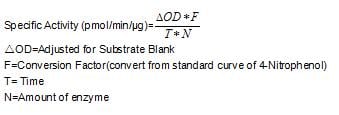 Bioactivity
(One unit of enzyme activity is defined as the 1ug of enzyme required to convert 1pmol of 4-Nitrophenyl acetate to 4-Nitrophenol in 1min at 37 degree C. The specific activity of recombinant Rat CES3 is 1200 pmol/min/ug.)
Bioactivity
(One unit of enzyme activity is defined as the 1ug of enzyme required to convert 1pmol of 4-Nitrophenyl acetate to 4-Nitrophenol in 1min at 37 degree C. The specific activity of recombinant Rat CES3 is 1200 pmol/min/ug.)
Carboxylesterase 3 (CES3), Active Protein (Cat# AAA153135)
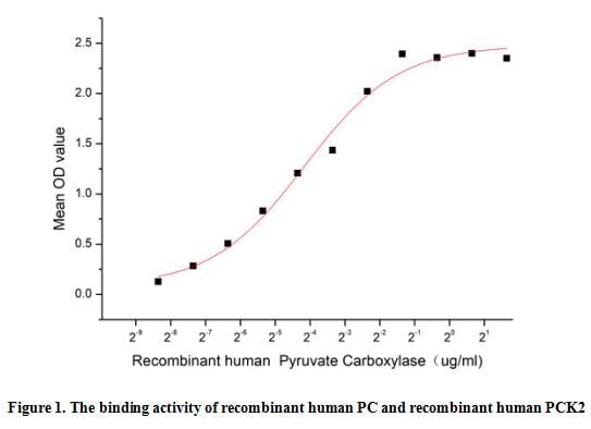 Bioactivity
(Pyruvate carboxylase (PC) is a mitochondrial, biotin-containing enzyme catalyzing the ATP-dependent synthesis of oxaloacetate from pyruvate and bicarbonate, with a critical anaplerotic role in sustaining the brain metabolism. Pyruvate carboxylase (PC) def)
Bioactivity
(Pyruvate carboxylase (PC) is a mitochondrial, biotin-containing enzyme catalyzing the ATP-dependent synthesis of oxaloacetate from pyruvate and bicarbonate, with a critical anaplerotic role in sustaining the brain metabolism. Pyruvate carboxylase (PC) def)
Pyruvate Carboxylase (PC), Active Protein (Cat# AAA153136)


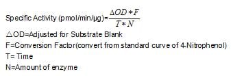 Bioactivity
(One unit of enzyme activity is defined as the 1ug of enzyme required to convert 1pmol of 4-Nitrophenyl acetate to 4-Nitrophenol in 1min at 37 degree C. The specific activity of recombinant human CES5A is 1300 pmol/min/ug.)
Bioactivity
(One unit of enzyme activity is defined as the 1ug of enzyme required to convert 1pmol of 4-Nitrophenyl acetate to 4-Nitrophenol in 1min at 37 degree C. The specific activity of recombinant human CES5A is 1300 pmol/min/ug.)
Carboxylesterase 5A (CES5A), Active Protein (Cat# AAA153140)

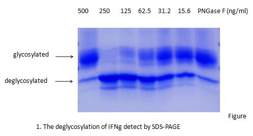 Bioactivity
(PNGase F (Peptide-N-glycosidase F) is a kind of enzymes for the deglycosylation of glycoproteins. The enzyme releases asparagine-linked oligosaccharides from glycoproteins and glycopeptides by hydrolyzing the amide of the asparagine (Asn) side chain. Thus)
Bioactivity
(PNGase F (Peptide-N-glycosidase F) is a kind of enzymes for the deglycosylation of glycoproteins. The enzyme releases asparagine-linked oligosaccharides from glycoproteins and glycopeptides by hydrolyzing the amide of the asparagine (Asn) side chain. Thus)
Peptide-N4-N-Acetyl-Beta-D-Glucosaminyl Asparagine Amidase F (PNGaseF), Active Protein (Cat# AAA153141)
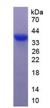
 Bioactivity
(Galectin 9 (GAL9) is a member of the beta-galactoside-binding galectin family. Galectin-9 is found outside of cells and may be exported by non-classical pathways. Galectin 9 exhibits a variety of biological activities, the majority of which have focused o)
Bioactivity
(Galectin 9 (GAL9) is a member of the beta-galactoside-binding galectin family. Galectin-9 is found outside of cells and may be exported by non-classical pathways. Galectin 9 exhibits a variety of biological activities, the majority of which have focused o)
Galectin 9 (GAL9), Active Protein (Cat# AAA153026)
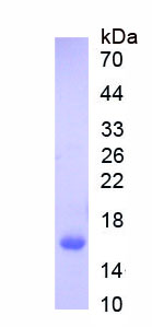
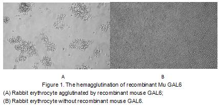 Bioactivity
(Galectin-6(GAL6) is a protein that in humans is encoded by the LGALS6 gene. The galectins constitute a large family of carbohydrate-binding proteins with specificity for N-acetyl-lactosamine-containing glycoproteins. At least 14 mammalian galectins, which)
Bioactivity
(Galectin-6(GAL6) is a protein that in humans is encoded by the LGALS6 gene. The galectins constitute a large family of carbohydrate-binding proteins with specificity for N-acetyl-lactosamine-containing glycoproteins. At least 14 mammalian galectins, which)
Galectin 6 (GAL6), Active Protein (Cat# AAA153030)
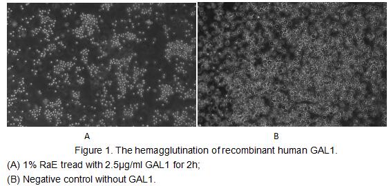
 Bioactivity
(Figure 2. The hemagglutination assay of GAL1 in V- bottom shaped 96-well microtiter plate.)
Bioactivity
(Figure 2. The hemagglutination assay of GAL1 in V- bottom shaped 96-well microtiter plate.)
Galectin 1 (GAL1), Active Protein (Cat# AAA153032)
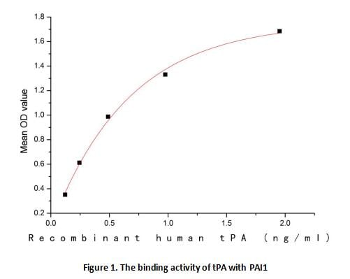 Bioactivity
(Plasminogen activators are serine proteases that catalyze the activation of plasmin via proteolytic cleavage of its zymogen form plasminogen. Plasmin is an important factor in fibrinolysis, the breakdown of fibrin polymers formed during blood clotting. Th)
Bioactivity
(Plasminogen activators are serine proteases that catalyze the activation of plasmin via proteolytic cleavage of its zymogen form plasminogen. Plasmin is an important factor in fibrinolysis, the breakdown of fibrin polymers formed during blood clotting. Th)
Tissue Plasminogen Activator (tPA), Active Protein (Cat# AAA153036)
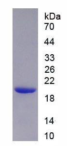
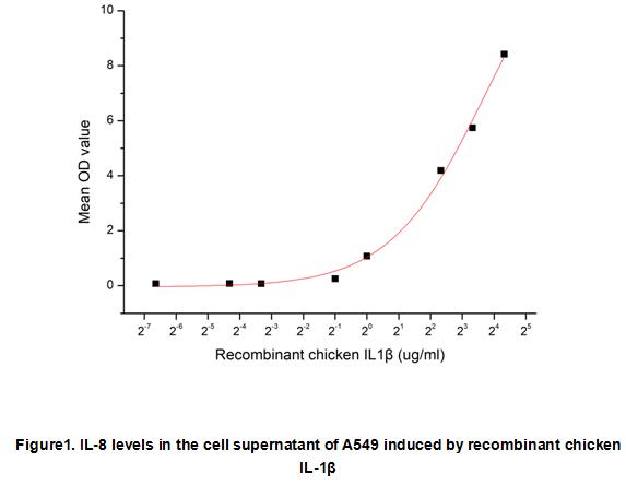
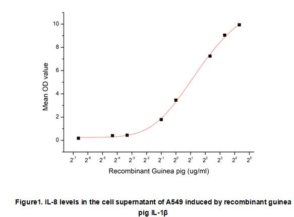
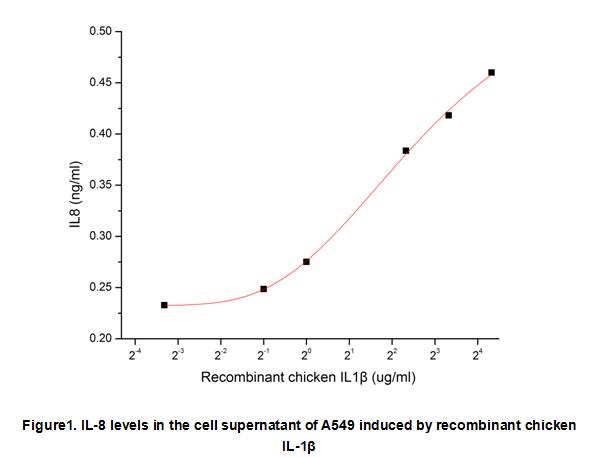 Bioactivity
(Interleukin 1 beta (IL-1beta) also known as leukocytic pyrogen, leukocytic endogenous mediator, mononuclear cell factor, lymphocyte activating factor and other names, is a member of the interleukin 1 family of cytokines. This cytokine is an important medi)
Bioactivity
(Interleukin 1 beta (IL-1beta) also known as leukocytic pyrogen, leukocytic endogenous mediator, mononuclear cell factor, lymphocyte activating factor and other names, is a member of the interleukin 1 family of cytokines. This cytokine is an important medi)
Interleukin 1 Beta (IL1b), Active Protein (Cat# AAA153042)

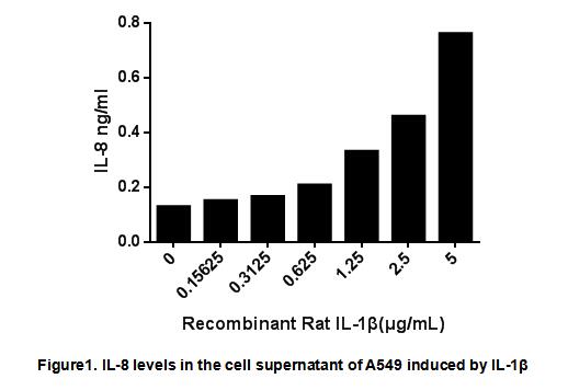
 Bioactivity
(Interleukin 1 beta (IL-1beta) also known as leukocytic pyrogen, leukocytic endogenous mediator, mononuclear cell factor, lymphocyte activating factor and other names, is a member of the interleukin 1 family of cytokines. This cytokine is an important medi)
Bioactivity
(Interleukin 1 beta (IL-1beta) also known as leukocytic pyrogen, leukocytic endogenous mediator, mononuclear cell factor, lymphocyte activating factor and other names, is a member of the interleukin 1 family of cytokines. This cytokine is an important medi)
Interleukin 1 Beta (IL1b), Active Protein (Cat# AAA153043)
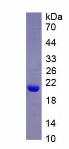
 Bioactivity
(Interleukin 1 beta (IL-1beta) also known as leukocytic pyrogen, leukocytic endogenous mediator, mononuclear cell factor, lymphocyte activating factor and other names, is a member of the interleukin 1 family of cytokines. This cytokine is an important medi)
Bioactivity
(Interleukin 1 beta (IL-1beta) also known as leukocytic pyrogen, leukocytic endogenous mediator, mononuclear cell factor, lymphocyte activating factor and other names, is a member of the interleukin 1 family of cytokines. This cytokine is an important medi)
Interleukin 1 Beta (IL1b), Active Protein (Cat# AAA153044)

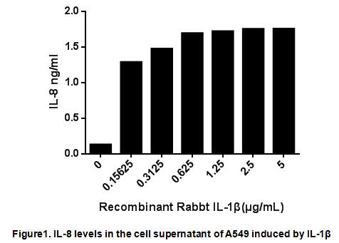
 Bioactivity
(Interleukin 1 beta (IL-1beta) also known as leukocytic pyrogen, leukocytic endogenous mediator, mononuclear cell factor, lymphocyte activating factor and other names, is a member of the interleukin 1 family of cytokines. This cytokine is an important medi)
Bioactivity
(Interleukin 1 beta (IL-1beta) also known as leukocytic pyrogen, leukocytic endogenous mediator, mononuclear cell factor, lymphocyte activating factor and other names, is a member of the interleukin 1 family of cytokines. This cytokine is an important medi)
Interleukin 1 Beta (IL1b), Active Protein (Cat# AAA153047)
 Bioactivity
(Interleukin 1 beta (IL-1beta) also known as leukocytic pyrogen, leukocytic endogenous mediator, mononuclear cell factor, lymphocyte activating factor and other names, is a member of the interleukin 1 family of cytokines. This cytokine is an important medi)
Bioactivity
(Interleukin 1 beta (IL-1beta) also known as leukocytic pyrogen, leukocytic endogenous mediator, mononuclear cell factor, lymphocyte activating factor and other names, is a member of the interleukin 1 family of cytokines. This cytokine is an important medi)
Interleukin 1 Beta (IL1b), Active Protein (Cat# AAA153048)
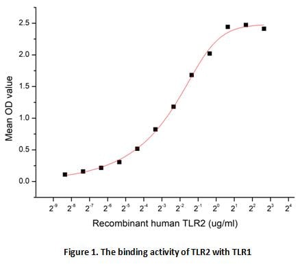 Bioactivity
(TLR2 is a member of TLR family which is type I transmembrane proteins with a large number of extracellular leucine-rich repeats (LRRs) and a cytoplasmic Toll/IL-1 receptor (TIR) domain. Human TLR2 is synthesized as a 784 amino acid precursor that contains)
Bioactivity
(TLR2 is a member of TLR family which is type I transmembrane proteins with a large number of extracellular leucine-rich repeats (LRRs) and a cytoplasmic Toll/IL-1 receptor (TIR) domain. Human TLR2 is synthesized as a 784 amino acid precursor that contains)
Toll Like Receptor 2 (TLR2), Active Protein (Cat# AAA153052)

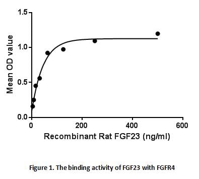 Bioactivity
(Fibroblast growth factor 23 or FGF23 is a member of the fibroblast growth factor (FGF) family which is responsible for phosphate and vitamin D metabolism. The main function of FGF23 seems to be regulation of phosphate concentration in plasma. FGF23 decrea)
Bioactivity
(Fibroblast growth factor 23 or FGF23 is a member of the fibroblast growth factor (FGF) family which is responsible for phosphate and vitamin D metabolism. The main function of FGF23 seems to be regulation of phosphate concentration in plasma. FGF23 decrea)
Fibroblast Growth Factor 23 (FGF23), Active Protein (Cat# AAA153055)
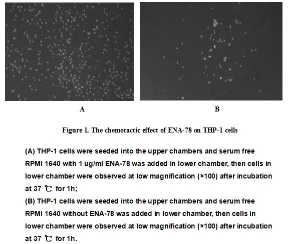

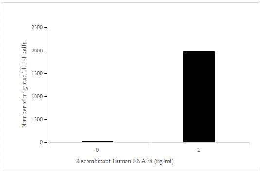 Bioactivity
(Figure 2. The chemotactic effect of ENA-78 on THP-1 cells)
Bioactivity
(Figure 2. The chemotactic effect of ENA-78 on THP-1 cells)
Epithelial Neutrophil Activating Peptide 78 (ENA78), Active Protein (Cat# AAA153062)
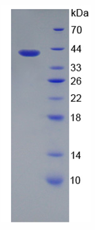
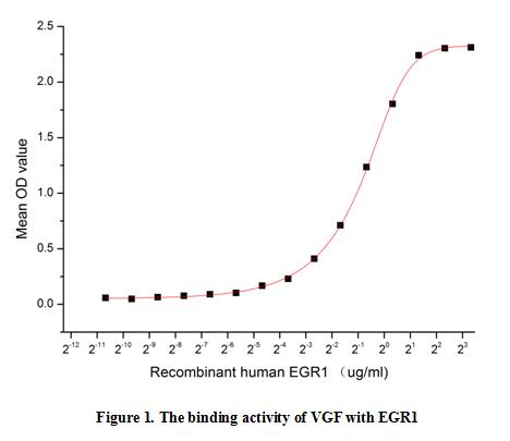 Bioactivity
(Neurosecretory protein VGF is specifically expressed in a subpopulation of neuroendocrine cells, and is upregulated by nerve growth factor. Human VGF precursor is 615 amino acids (aa) in length. It contains an 22 aa signal sequence plus a 593 aa mature re)
Bioactivity
(Neurosecretory protein VGF is specifically expressed in a subpopulation of neuroendocrine cells, and is upregulated by nerve growth factor. Human VGF precursor is 615 amino acids (aa) in length. It contains an 22 aa signal sequence plus a 593 aa mature re)
VGF Nerve Growth Factor Inducible (VGF), Active Protein (Cat# AAA153073)
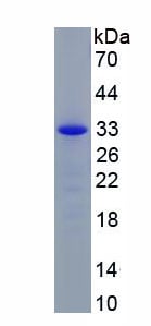
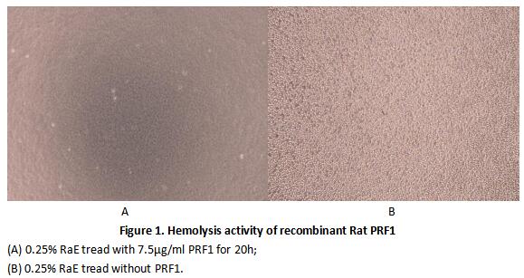 Bioactivity
(Perforin 1 (PRF1) is a pore forming cytolytic protein found in the granules of cytotoxic T lymphocytes (CTLs) and NK cells. Upon degranulation, perforin binds to the target cell's plasma membrane, and oligomerises in a Ca2 dependent manner to form pores o)
Bioactivity
(Perforin 1 (PRF1) is a pore forming cytolytic protein found in the granules of cytotoxic T lymphocytes (CTLs) and NK cells. Upon degranulation, perforin binds to the target cell's plasma membrane, and oligomerises in a Ca2 dependent manner to form pores o)
Perforin 1 (PRF1), Active Protein (Cat# AAA153081)
 Application Data
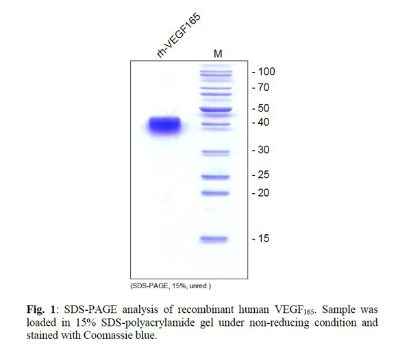
VEGF165, Active Protein (Cat# AAA79271)
 Bioactivity
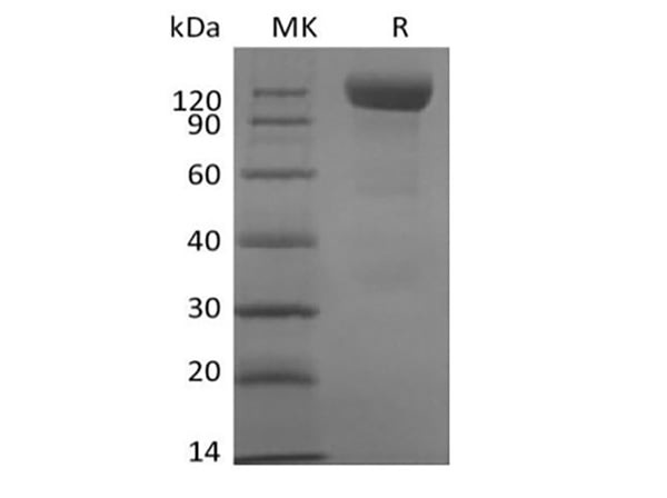
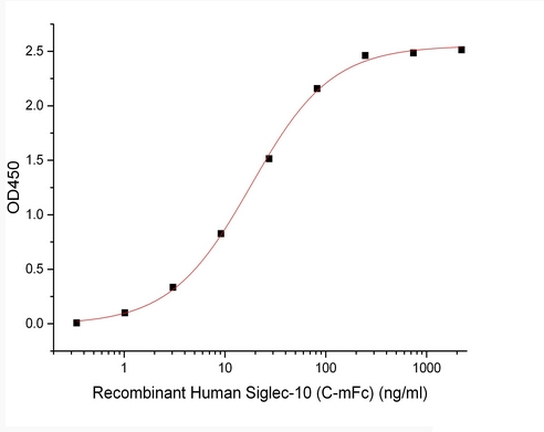
Siglec-10, Active Protein (Cat# AAA177946)
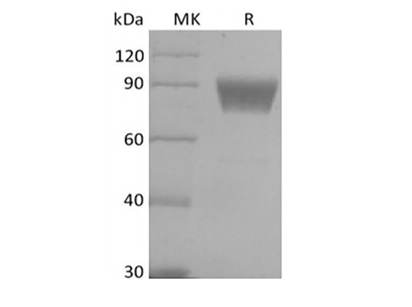
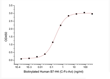 Bioactivity
B7 Homolog 4/B7-H4/VTCN1 Biotinylated, Active Protein (Cat# AAA177958)
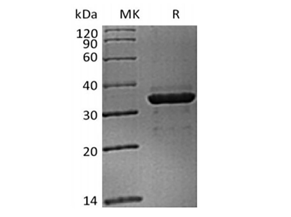
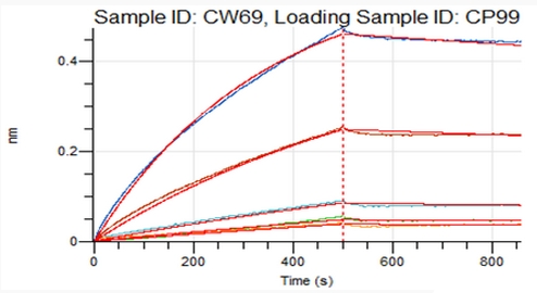 Bioactivity
FGL1, Active Protein (Cat# AAA177972)
FasL, Active Protein (Cat# AAA75199)
CTGF, Active Protein (Cat# AAA75454)
IL11, Active Protein (Cat# AAA75591)
BMP6, Active Protein (Cat# AAA75597)
 Application Data
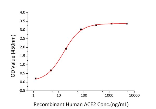
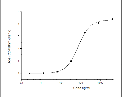
(Fig 2: Immobilized Recombinant 2019-nCoV Spike S1-His at 2ug/mL (100 uL/well) can bind Recombinant Human ACE2 with a linear range of 1.5-15 ng/mL.)
Application Data
(Fig 2: Immobilized Recombinant 2019-nCoV Spike S1-His at 2ug/mL (100 uL/well) can bind Recombinant Human ACE2 with a linear range of 1.5-15 ng/mL.)
COVID 19 Spike S1 Coronavirus, Active Protein (Cat# AAA78533)
 Application Data
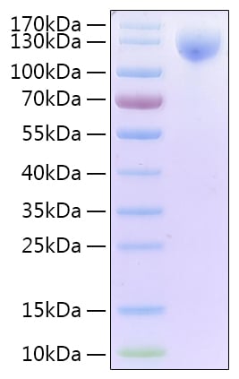
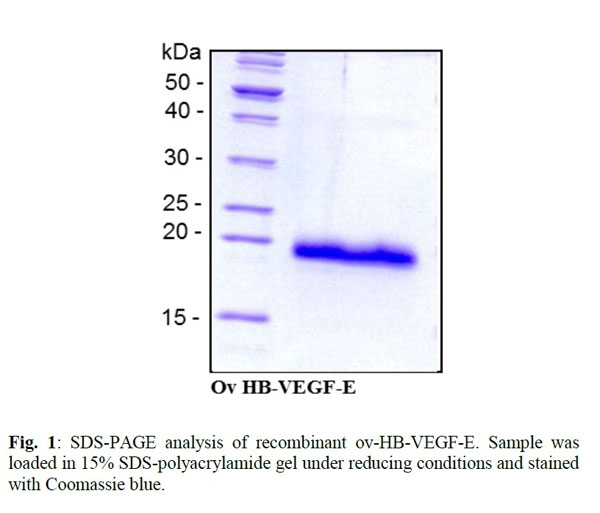
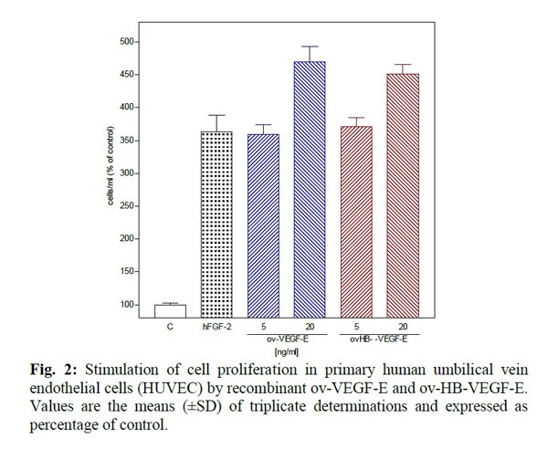
VEGF-E, Heparin-binding, Active Protein (Cat# AAA79256)
Granzyme B, Active Protein (Cat# AAA79267)
G-CSF, Active Protein (Cat# AAA79275)
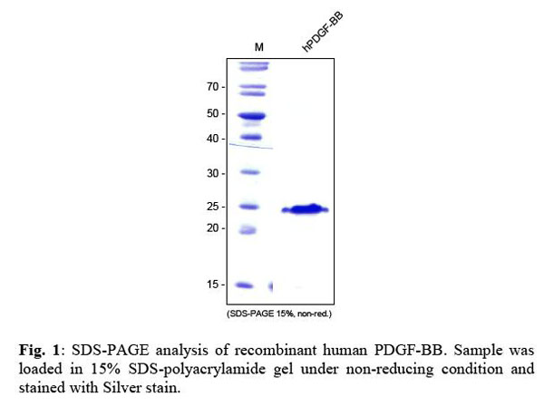
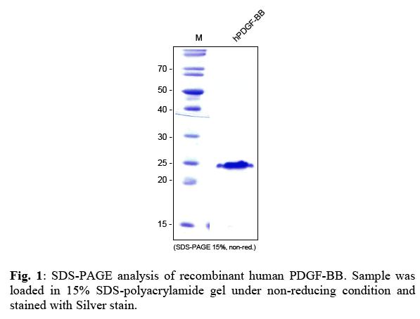
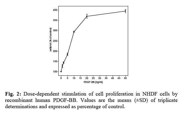
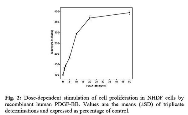 Application Data
PDGF-BB, Active Protein (Cat# AAA79279)
 Application Data
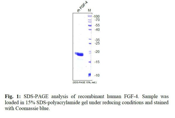
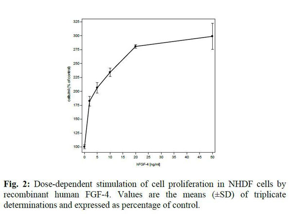
FGF-4, Active Protein (Cat# AAA79290)
 Application Data
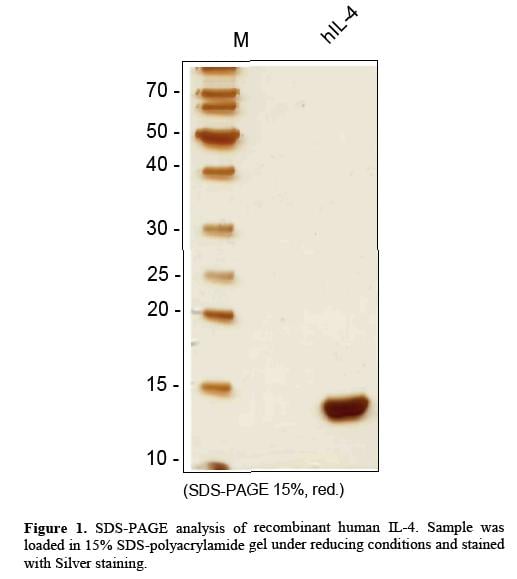
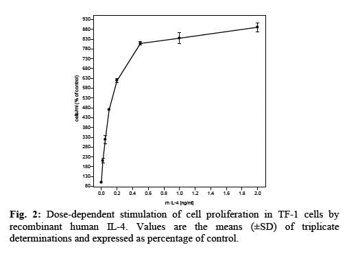
IL-4, Active Protein (Cat# AAA79122)
 Application Data
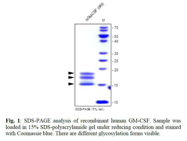
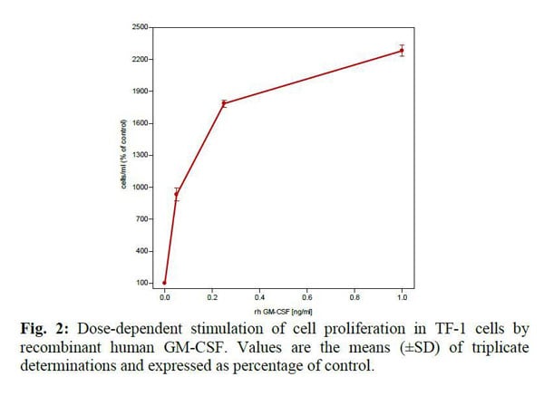
GM-CSF, Active Protein (Cat# AAA79138)
 Application Data
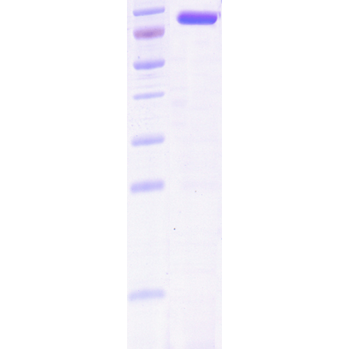
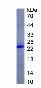
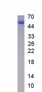
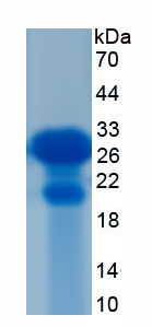
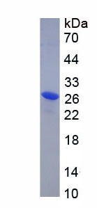
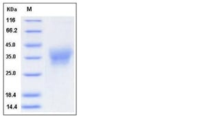
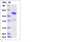
 SDS-PAGE
(SDS-PAGE of 73kDa Hsc70 protein)
SDS-PAGE
(SDS-PAGE of 73kDa Hsc70 protein)
Hsc70, Active Protein (Cat# AAA103775)
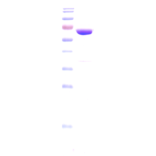 SDS-PAGE
(SDS-PAGE of 60kDa Hsp60 protein (AAA103776).)
SDS-PAGE
(SDS-PAGE of 60kDa Hsp60 protein (AAA103776).)
Hsp60, Active Protein (Cat# AAA103776)
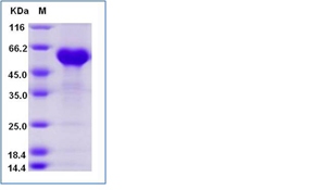
 Application Data
PDGF-BB, Active Protein (Cat# AAA79160)
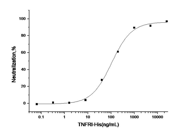
 Application Data
(Measured by its ability to inhibit TNF-alpha mediated cytotoxicity in L-929 mouse fibrosarcoma cells in the presence of the metabolic inhibitor actinomycin D. The ED50 for this effect is typically2-10ng/mL in the presence of 0.25 ng/mL recombinant human TNF-alpha.)
Application Data
(Measured by its ability to inhibit TNF-alpha mediated cytotoxicity in L-929 mouse fibrosarcoma cells in the presence of the metabolic inhibitor actinomycin D. The ED50 for this effect is typically2-10ng/mL in the presence of 0.25 ng/mL recombinant human TNF-alpha.)
TNFRSF1A, Active Protein (Cat# AAA257939)
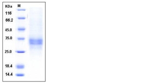
 Application Data
(Measured by its ability to inhibit TNFalpha-mediated cytotoxicity in L929 mouse fibrosarcoma cells in the presence of metabolic inhibitor actinomycin D. The ED50 for this effect is typically 0.2-1ug/mL in the presence of 0.25 ng/mL recombinant human TNFalpha.)
Application Data
(Measured by its ability to inhibit TNFalpha-mediated cytotoxicity in L929 mouse fibrosarcoma cells in the presence of metabolic inhibitor actinomycin D. The ED50 for this effect is typically 0.2-1ug/mL in the presence of 0.25 ng/mL recombinant human TNFalpha.)
TNFRSF1A, Active Protein (Cat# AAA257940)
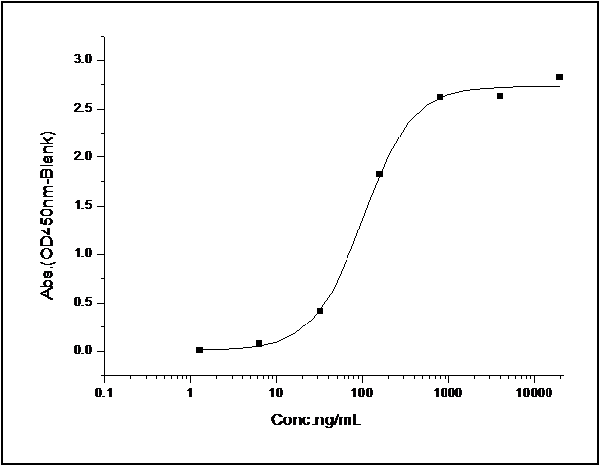 Application Data
(Measured by its binding ability in a functional ELISA. Immobilized human EFNB2 at 2 ug/ml (100 ul/well) can bind human EphB4 with a linear ranger of 1.56-12.5 ng/ml.)
Application Data
(Measured by its binding ability in a functional ELISA. Immobilized human EFNB2 at 2 ug/ml (100 ul/well) can bind human EphB4 with a linear ranger of 1.56-12.5 ng/ml.)
Ephrin B2, Active Protein (Cat# AAA257942)
 Application Data
Ephrin-A1, Active Protein (Cat# AAA257944)
What Are Active Proteins?
Proteins are large molecules made up of long chains of amino acids.
They will typically fold into a very particular 3-dimensional shape/conformation, that is sometimes referred to as their “native” form, which allows them to work properly in the body. For the purposes of product categorization, AAA Biotech will typically refer to proteins purified from their original animal host as being “native” proteins (this is to signify their difference compared to their “recombinant” or “synthetic” protein counterparts).
If a protein successfully folds into the correct shape, it is will typically display high fidelity characteristics to its original protein in its original animal host, and be classified as an active protein, as it will be able to function “normally” in most enzymatic or binding capacities. If it loses this shape, due to factors such as heat or strong chemicals (such as detergents), it becomes inactive and is no longer able to perform its basic functions. All of the proteins in this category are made under strict quality control, and they are active, pure, low in contaminants, and stable.
Most are stored as freeze-dried powders and come without extra tags, so they’re very close to the actual natural/native form.
Key Applications of Active Proteins
1. Scientific Research
- Aid in the study of how proteins function in the body
- Aid in understanding various disease processes
2. Drug Development
- Powerful tools to investigate how potential drugs interact with specific proteins
- Ideal for identifying drug targets
3. Cell Culture
- Are routinely utilized to support cell growth and function (e.g., using exogenous growth factors)
- Can be used to promote cellular development into specific types (differentiation)
4. Diagnostics
- Regularly utilized in tests to detect diseases or infections (e.g., COVID-19, cancer)
- Note: All products are strictly for research-use only (RUO).
5. Therapeutics
- Some active proteins are used directly as treatments (e.g., insulin, enzymes)
- Note: All products are strictly for research-use only (RUO).
6. Vaccine Development
- Used to create or test vaccines by mimicking parts of viruses or bacteria
7. Biochemical Assays
- They can facilitate the characterization of enzyme activity, binding strength, or protein interactions in lab tests
Why Buy Active Proteins from AAA Biotech?
- High biological activity – Verified to perform as expected or indicated on datasheet
- Strict quality control – We are confident in our active proteins’ reliability and consistency
- High purity & low endotoxin – Ideal for applications involving sensitive or precious samples/components
- Freeze-dried for stability – Long shelf life and straightforward storage
- Mostly tag-free – Closer to natural/native protein form
FAQ
1. What are active proteins used for in research?
Active proteins are used primarily in the study of how proteins function, in characterizing/discovering drug interactions, supporting cell growth, running biochemical assays, and in development of diagnostics or therapeutics.
2. How are AAA Biotech's active proteins validated?
AAA Biotech’s active proteins are validated through strict quality control and functional assays to ensure they are properly folded and active. “Active”, though, can be an ambiguous term, so if a specific “activity” or “binding” capability of a protein is of crucial interest to you, please inquire with us prior to purchase, and we will provide further details on how the “Active” modifier was determined to be applicable.
3. Are these proteins tested for biological activity?
Yes, all active proteins from AAA Biotech are tested to confirm they have the expected biological activity before being offered for use. Though, said “biological activity” can be either “enzymatic”, “binding”, or both.