Filters
▼Clonality
▼Type
▼Reactivity
▼Gene Name
▼Isotype
▼Host
▼Application
▼Clone
▼Active Proteins
AAA Biotech also known as AAA Bio or AAABio provides a variety of high-quality recombinant and natural/native proteins that are proven to work in a wide range of experiments. Explore our products to find the active protein that best fits your needs or experimental model.
Viewing 750-800 of 2567 product results
 Bioactivity
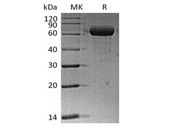
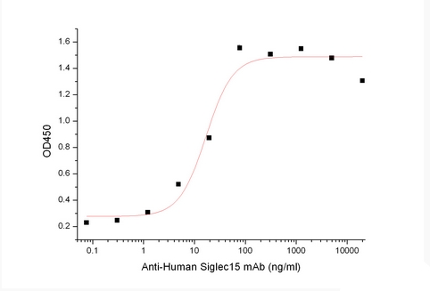
Sialic acid-binding Ig-like lectin 15/Siglec-15/CD33L3, Active Protein (Cat# AAA177935)
 Bioactivity
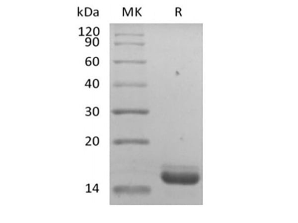
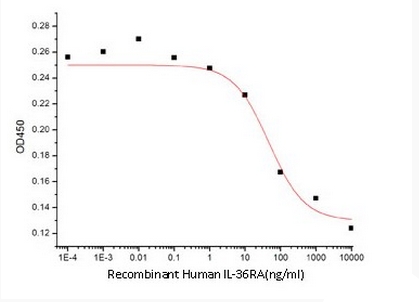
IL-36 Receptor Antagonist Protein/IL-36RN/IL-1F5, Active Protein (Cat# AAA177938)
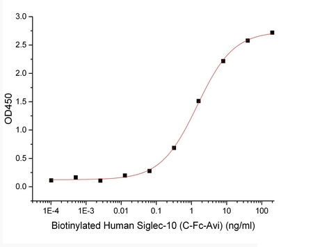 Bioactivity
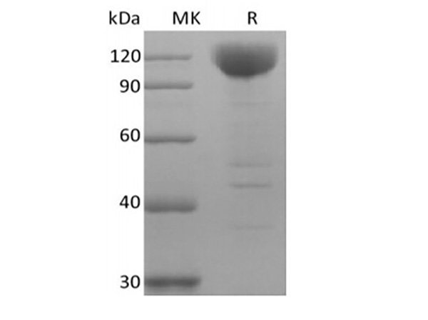
Siglec-10 Biotinylated, Active Protein (Cat# AAA177959)
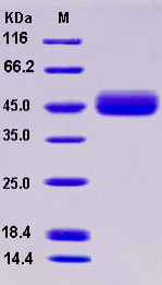 SDS-PAGE
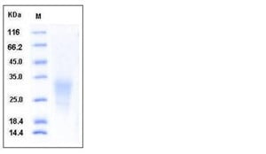
IL13RA2 / CD213A2, Active Protein (Cat# AAA173596)
 SDS-PAGE
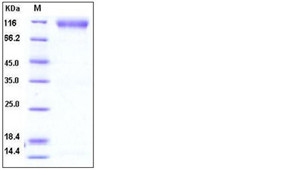
DAPK1 / DAP Kinase 1, Active Protein (Cat# AAA173507)

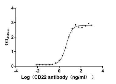 Bioactivity
B-Cell Receptor CD22 (CD22), Active Protein (Cat# AAA243725)

 Bioactivity
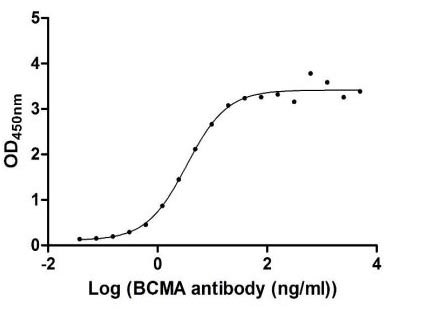
Tumor Necrosis Factor Receptor Superfamily Member 17 (TNFRSF17), Active Protein (Cat# AAA243734)
 Application Data
(Measured by its ability to inhibit BMP4-induced alkaline phosphatase production by MC3T3E1 mouse preosteoblast cells. The ED50 for this effect is 2.5-10 ug/mL.)
Application Data
(Measured by its ability to inhibit BMP4-induced alkaline phosphatase production by MC3T3E1 mouse preosteoblast cells. The ED50 for this effect is 2.5-10 ug/mL.)
BMPR1A/ALK-3, Active Protein (Cat# AAA257883)
 Application Data
(Measured by its ability to inhibit FGF-acidic (aFGF/FGF1) dependent proliferation of Balb/C 3T3 mouse fibroblasts. The ED50 for this effect is typically 0.5-2.5 ng/ml.)
Application Data
(Measured by its ability to inhibit FGF-acidic (aFGF/FGF1) dependent proliferation of Balb/C 3T3 mouse fibroblasts. The ED50 for this effect is typically 0.5-2.5 ng/ml.)
FGFR2, Active Protein (Cat# AAA257933)
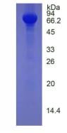
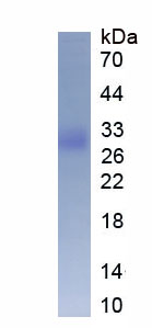
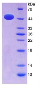
 SDS-PAGE
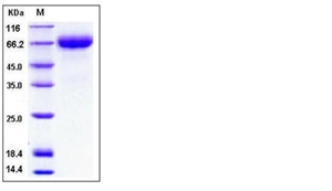
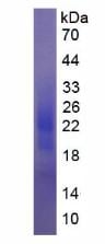
Hemopexin, Active Protein (Cat# AAA257938)

 Application Data
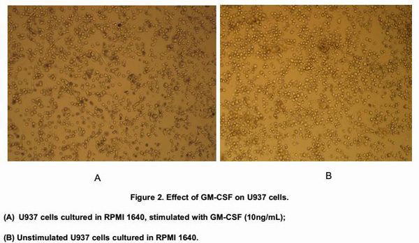
Colony Stimulating Factor 2, Granulocyte Macrophage (GMCSF), Active Protein (Cat# AAA146598)


 Application Data
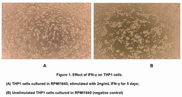
Interferon Gamma (IFNg), Active Protein (Cat# AAA146601)

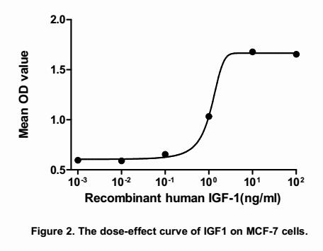 Application Data
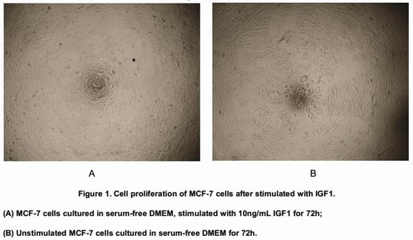
Insulin Like Growth Factor 1 (IGF1), Active Protein (Cat# AAA146604)
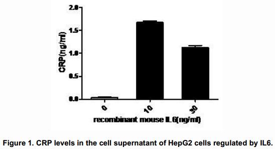
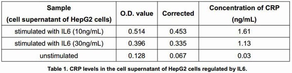

 Application Data
Interleukin 6 (IL6), Active Protein (Cat# AAA146610)

 Application Data
Tumor Necrosis Factor Alpha (TNFa), Active Protein (Cat# AAA146615)

 Application Data
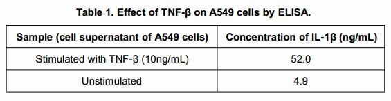
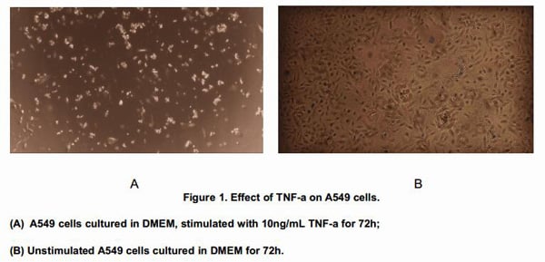
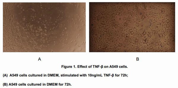
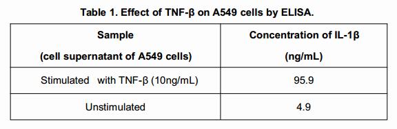
Tumor Necrosis Factor Beta (TNFb), Active Protein (Cat# AAA146616)
 Application Data
Osteopontin (OPN), Active Protein (Cat# AAA146622)
 WB (Western Blot)
(Western BlotSample: Recombinant HRG, Human;Antibody: Rabbit Anti-Human HRG Ab)
WB (Western Blot)
(Western BlotSample: Recombinant HRG, Human;Antibody: Rabbit Anti-Human HRG Ab)
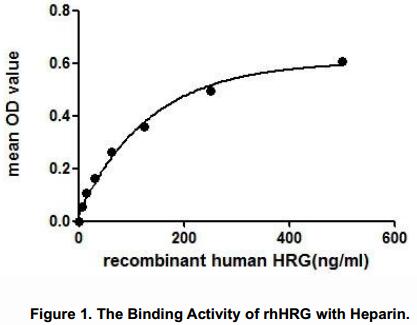
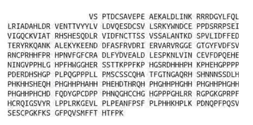
Histidine Rich Glycoprotein (HRG), Active Protein (Cat# AAA146623)
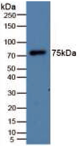
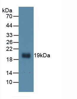
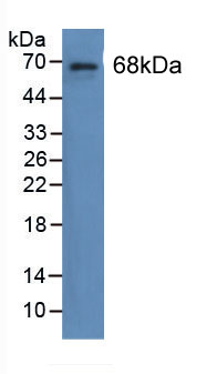
 WB (Western Blot)
(Figure 4. Western BlotSample: Recombinant LMTK3, Human;Antibody: Rabbit Anti-Human LMTK3 Ab)
WB (Western Blot)
(Figure 4. Western BlotSample: Recombinant LMTK3, Human;Antibody: Rabbit Anti-Human LMTK3 Ab)
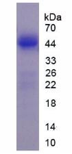
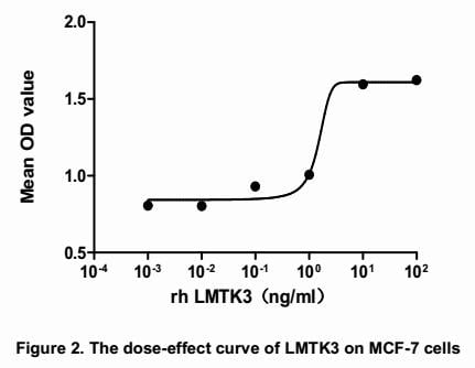
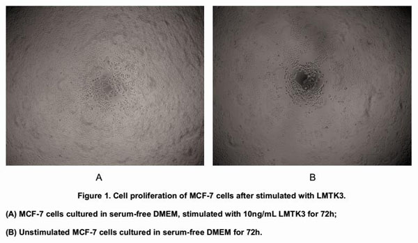
Lemur Tyrosine Kinase 3 (LMTK3), Active Protein (Cat# AAA146624)
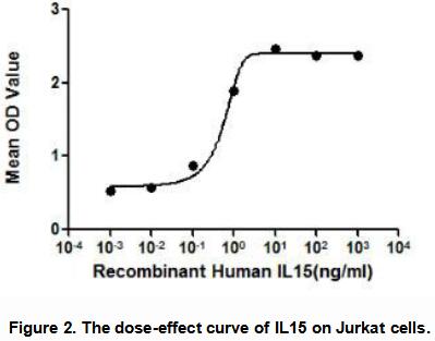

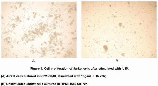
 Application Data
Interleukin 15 (IL15), Active Protein (Cat# AAA148129)

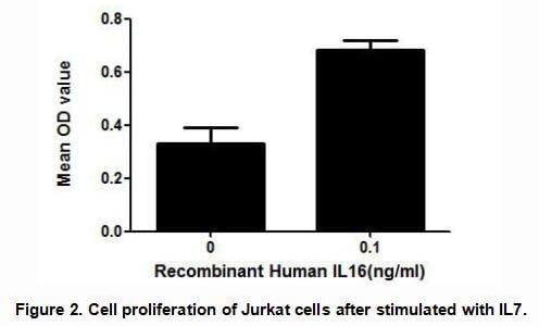 Application Data
Interleukin 16 (IL16), Active Protein (Cat# AAA148141)
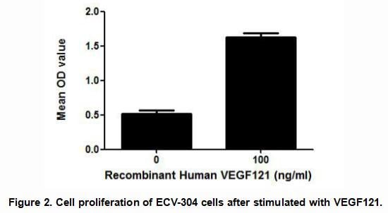

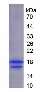
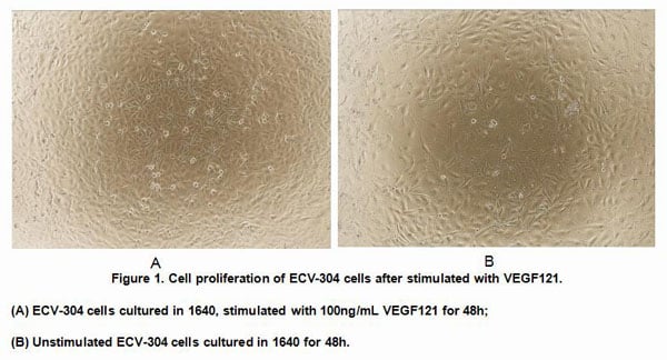
 Application Data
Vascular Endothelial Growth Factor 121 (VEGF121), Active Protein (Cat# AAA148143)
 WB (Western Blot)
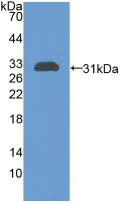
(Sample: Recombinant MMP9, Porcine; Antibody: Rabbit Anti-Porcine MMP9 Ab)
WB (Western Blot)
(Sample: Recombinant MMP9, Porcine; Antibody: Rabbit Anti-Porcine MMP9 Ab)
Matrix Metalloproteinase 9 (MMP9), Active Protein (Cat# AAA149237)
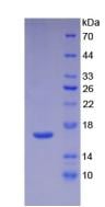
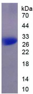
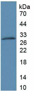
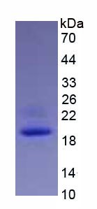
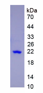
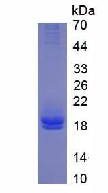 SDS-PAGE
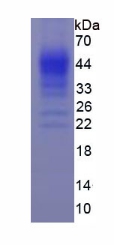
Interleukin 2 (IL2), Active Protein (Cat# AAA148221)
 Application Data
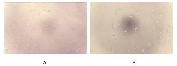
(Figure. Cell proliferation of Jurkat cells after stimulated with IL1a.Interleukin 1 alpha (IL1) also known as hematopoietin 1 is a cytokine of the interleukin 1 family that in humans is encoded by the IL1A gene. IL1 is produced mainly by activated macrophages, as well as neutrophils, epithelial cells, and endothelial cells. It possesses metabolic, physiological, haematopoietic activities, and plays one of the central roles in the regulation of the immune responses. It binds to the interleukin-1 receptor. It is on the pathway that activates tumor necrosis factor-alpha. To test the effect of IL1a on cell proliferation, Jurkat cells were seeded into triplicate wells of 96-well plates at a density of 2,000 cells/well with 2% serum standard 1640 including various concentrations of recombinant human IL1a. After incubated for 96h, cells were observed by inverted microscope and cell proliferation was measured by Cell Counting Kit-8 (CCK-8). Briefly, 10uL of CCK-8 solution was added to each well of the plate, then the absorbance at 450nm was measured using a microplate reader after incubating the plate for 1-4 hours at 37. Proliferation of Jurkat cells after incubation with IL1a for 96h observed by inverted microscope was shown in Figure 1. Cell viability was assessed by CCK-8 (Cell Counting Kit-8) assay after incubation with recombinant IL1a for 96h. The result was shown in Figure 2. It was obvious that IL1a significantly increased cell viability of Jurkat cells.(A) Jurkat cells cultured in 1640, stimulated with 10ng/mL IL1a for 96h; (B) Unstimulated Jurkat cells cultured in1640 for 96h.)
Application Data
(Figure. Cell proliferation of Jurkat cells after stimulated with IL1a.Interleukin 1 alpha (IL1) also known as hematopoietin 1 is a cytokine of the interleukin 1 family that in humans is encoded by the IL1A gene. IL1 is produced mainly by activated macrophages, as well as neutrophils, epithelial cells, and endothelial cells. It possesses metabolic, physiological, haematopoietic activities, and plays one of the central roles in the regulation of the immune responses. It binds to the interleukin-1 receptor. It is on the pathway that activates tumor necrosis factor-alpha. To test the effect of IL1a on cell proliferation, Jurkat cells were seeded into triplicate wells of 96-well plates at a density of 2,000 cells/well with 2% serum standard 1640 including various concentrations of recombinant human IL1a. After incubated for 96h, cells were observed by inverted microscope and cell proliferation was measured by Cell Counting Kit-8 (CCK-8). Briefly, 10uL of CCK-8 solution was added to each well of the plate, then the absorbance at 450nm was measured using a microplate reader after incubating the plate for 1-4 hours at 37. Proliferation of Jurkat cells after incubation with IL1a for 96h observed by inverted microscope was shown in Figure 1. Cell viability was assessed by CCK-8 (Cell Counting Kit-8) assay after incubation with recombinant IL1a for 96h. The result was shown in Figure 2. It was obvious that IL1a significantly increased cell viability of Jurkat cells.(A) Jurkat cells cultured in 1640, stimulated with 10ng/mL IL1a for 96h; (B) Unstimulated Jurkat cells cultured in1640 for 96h.)
Interleukin 1 Alpha, Active Protein (Cat# AAA150058)
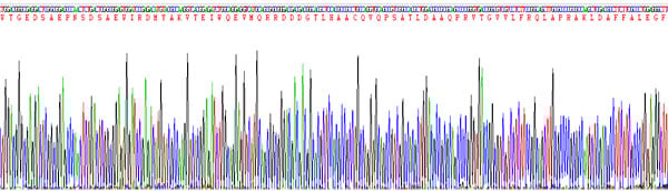
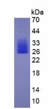
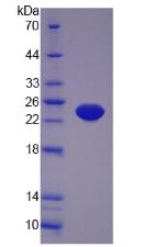
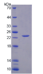
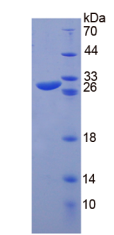
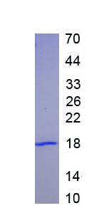
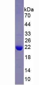
 SDS_PAGE
(Sample: Active recombinant SOD3, Human)
SDS_PAGE
(Sample: Active recombinant SOD3, Human)
Superoxide Dismutase 3, Extracellular, Active Protein (Cat# AAA150072)
 Application Data
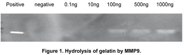
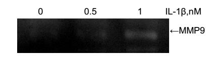
(IL-1 (Interleukin-1 beta) is a proinflammatory and immunoregulatory cytokine involved in a variety of cellular activities. It has been elucidated that IL-1 stimulation of cells activates MMP-9 (matrix metalloproteinases-9) secretion by the activation of the dual signalling pathways. Thus, a stimulation assay of IL-1 was conducted using 3T3 cell line, and the MMP-9 activity was detected through gel zymography. Briefly, 1×10⁶ 3T3 cells were cultured overnight in 5%DMEM, washed with serum-free medium and then stimulated with different concentrations of IL-1 for 20h, and cell lysates were collected to measure MMP-9 activity. Cell lysates samples were denatured by SDS loading buffer, electrophoresed through sodium dodecyl sulphate–polyacrylamide gel (SDS–PAGE; 10% gels) containing gelatin (1mg/mL) with nonreducing conditions. After renaturation, incubation and coomassiebrilliant blue (CBB)-stained, MMPs hydrolyzed gelatin nearby, indicated by the white binds on the gel. Result Increased MMP-9 activity in 3T3 cells due to the stimulation of IL-1 was shown in Figure 1.Figure. Activation of MMP-9 by IL-1.)
Application Data
(IL-1 (Interleukin-1 beta) is a proinflammatory and immunoregulatory cytokine involved in a variety of cellular activities. It has been elucidated that IL-1 stimulation of cells activates MMP-9 (matrix metalloproteinases-9) secretion by the activation of the dual signalling pathways. Thus, a stimulation assay of IL-1 was conducted using 3T3 cell line, and the MMP-9 activity was detected through gel zymography. Briefly, 1×10⁶ 3T3 cells were cultured overnight in 5%DMEM, washed with serum-free medium and then stimulated with different concentrations of IL-1 for 20h, and cell lysates were collected to measure MMP-9 activity. Cell lysates samples were denatured by SDS loading buffer, electrophoresed through sodium dodecyl sulphate–polyacrylamide gel (SDS–PAGE; 10% gels) containing gelatin (1mg/mL) with nonreducing conditions. After renaturation, incubation and coomassiebrilliant blue (CBB)-stained, MMPs hydrolyzed gelatin nearby, indicated by the white binds on the gel. Result Increased MMP-9 activity in 3T3 cells due to the stimulation of IL-1 was shown in Figure 1.Figure. Activation of MMP-9 by IL-1.)
Interleukin 1 Beta, Active Protein (Cat# AAA150093)
 Application Data
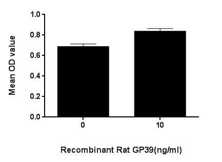
(Figure. Cell proliferation of spleen cells after stimulated with GP39.)
Application Data
(Figure. Cell proliferation of spleen cells after stimulated with GP39.)
Chitinase-3-like Protein 1, Active Protein (Cat# AAA150109)
 Bioactivity
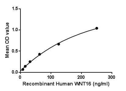
(Wingless-type MMTV integration site family, member 16 is a protein that in humans is encoded by the WNT16 gene. The WNT gene family consists of structurally related genes that encode secreted signaling proteins. These proteins have been implicated in oncogenesis and in several developmental processes, including regulation of cell fate and patterning during embryogenesis. This gene is a member of the WNT gene family. It contains two transcript variants diverging at the 5 termini. These two variants are proposed to be the products of separate promoters and not to be splice variants from a single promoter. Besides, Tubulin Beta 3 (TUBb3) has been identified as an interactor of WNT16, thus a binding ELISA assay was conducted to detect the interaction of recombinant human WNT16 and recombinant human TUBb3. Briefly, WNT16 were diluted serially in PBS, with 0.01% BSA (pH 7.4). Duplicate samples of 100L were then transferred to TUBb3-coated microtiter wells and incubated for 2h at 37. Wells were washed with PBST and incubated for 1h with anti-WNT16 pAb, then aspirated and washed 3 times. After incubation with HRP labelled secondary antibody, wells were aspirated and washed 3 times. With the addition of substrate solution, wells were incubated 15-25 minutes at 37. Finally, add 50uL stop solution to the wells and read at 450nm immediately. The binding activity of WNT16 and TUBb3 was shown in Figure 1, and this effect was in a dose dependent manner.Figure. The binding activity of WNT16 with TUBb3.)
Bioactivity
(Wingless-type MMTV integration site family, member 16 is a protein that in humans is encoded by the WNT16 gene. The WNT gene family consists of structurally related genes that encode secreted signaling proteins. These proteins have been implicated in oncogenesis and in several developmental processes, including regulation of cell fate and patterning during embryogenesis. This gene is a member of the WNT gene family. It contains two transcript variants diverging at the 5 termini. These two variants are proposed to be the products of separate promoters and not to be splice variants from a single promoter. Besides, Tubulin Beta 3 (TUBb3) has been identified as an interactor of WNT16, thus a binding ELISA assay was conducted to detect the interaction of recombinant human WNT16 and recombinant human TUBb3. Briefly, WNT16 were diluted serially in PBS, with 0.01% BSA (pH 7.4). Duplicate samples of 100L were then transferred to TUBb3-coated microtiter wells and incubated for 2h at 37. Wells were washed with PBST and incubated for 1h with anti-WNT16 pAb, then aspirated and washed 3 times. After incubation with HRP labelled secondary antibody, wells were aspirated and washed 3 times. With the addition of substrate solution, wells were incubated 15-25 minutes at 37. Finally, add 50uL stop solution to the wells and read at 450nm immediately. The binding activity of WNT16 and TUBb3 was shown in Figure 1, and this effect was in a dose dependent manner.Figure. The binding activity of WNT16 with TUBb3.)
Wingless Type MMTV Integration Site Family, Member 16, Active Protein (Cat# AAA150149)
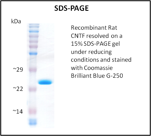
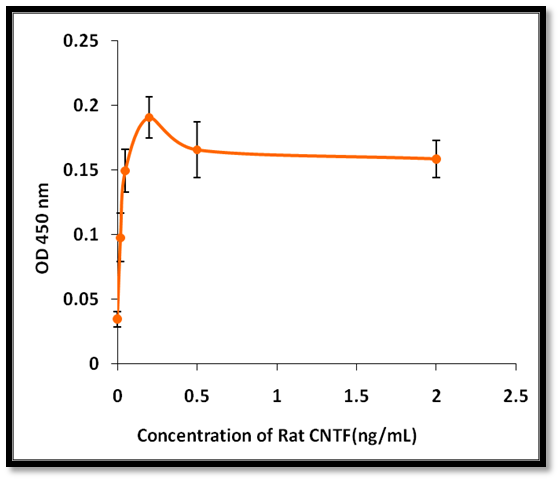 Application Data
CNTF, Active Protein (Cat# AAA214336)
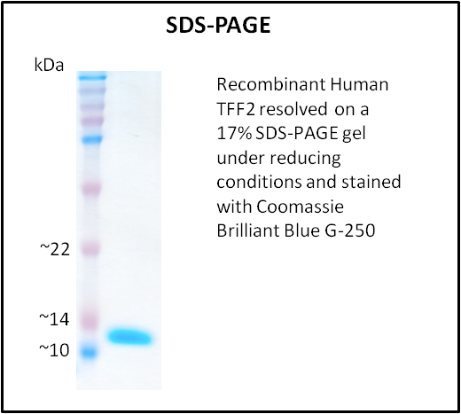
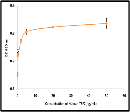 Application Data
TFF2, Active Protein (Cat# AAA214282)
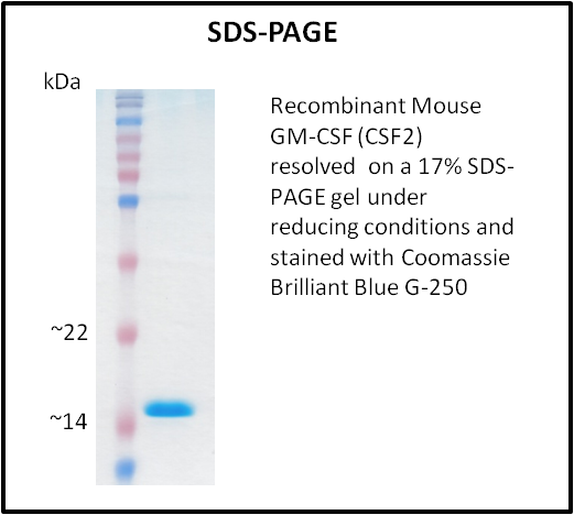
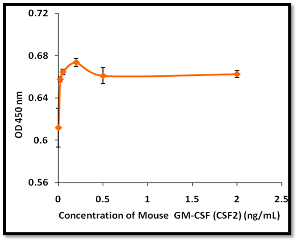 Application Data
GM-CSF (CSF2), Active Protein (Cat# AAA214310)
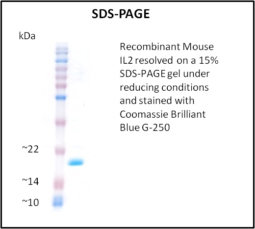
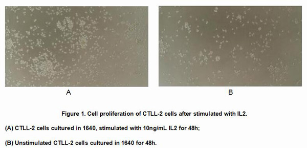
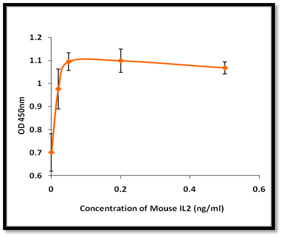 Application Data
IL2, Active Protein (Cat# AAA214316)
 Application Data
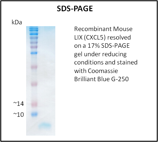
LIX (CXCL5), Active Protein (Cat# AAA214332)
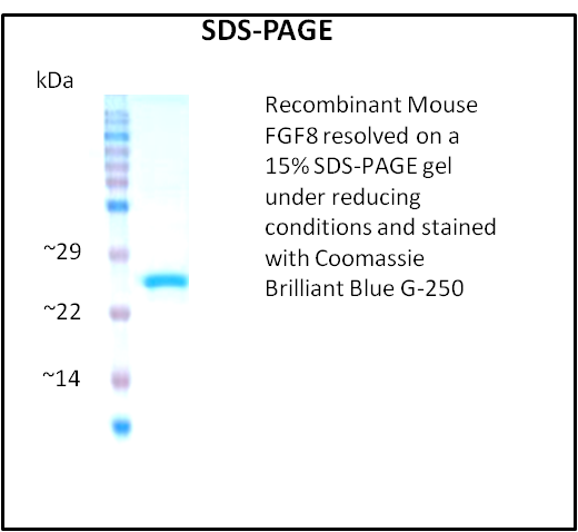
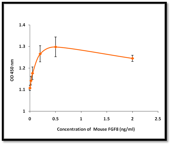 Application Data
FGF8, Active Protein (Cat# AAA214334)
 Bioactivity
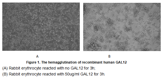
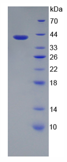
(Galectin-12 is a member of a family of mammalian lectins known as galectins. The galectins constitute a large family of carbohydrate-binding proteins that function in many systems both intracellularly and following secretion. Galectins contain either one or two carbohydrate recognition domains (CRR) which mediate recognition of N-acetyl-lactosamine-containing glycoproteins. Individual galectins differ in their tissue distribution and in their carbohydrate-binding specificities. Galectin-12 is predominantly expressed in adipose tissue and detected also in macrophages and other leukocytes. It plays an important role in cell-cell adhesion, cell-matrix interactions, macrophage activation, angiogenesis, metastasis, apoptosis. In this case, we chose rabbit erythrocyte (RaE) to assay its ability of agglutination. A general procedure for hemagglutination assay (or haemagglutination assay; HA) is as follows, two-fold dilute the recombinant Hu GAL12 with 0.9% sodium chloride injection, add 50uL a serial dilution of GAL12 to each well of a U or V-bottom shaped 96-well microtiter plate. The final well serves as a negative control with no GAL12, replace with 50uL 0.9% sodium chloride injection. Then add 50uL 1% rabbit erythrocyte to each well and mixed gently. The plate is incubated for 3 hours at room temperature. The results are shown in Figure 1. It was obvious that the minimal effective concentration of GAL12 is 0.781 ug/mL.)
Bioactivity
(Galectin-12 is a member of a family of mammalian lectins known as galectins. The galectins constitute a large family of carbohydrate-binding proteins that function in many systems both intracellularly and following secretion. Galectins contain either one or two carbohydrate recognition domains (CRR) which mediate recognition of N-acetyl-lactosamine-containing glycoproteins. Individual galectins differ in their tissue distribution and in their carbohydrate-binding specificities. Galectin-12 is predominantly expressed in adipose tissue and detected also in macrophages and other leukocytes. It plays an important role in cell-cell adhesion, cell-matrix interactions, macrophage activation, angiogenesis, metastasis, apoptosis. In this case, we chose rabbit erythrocyte (RaE) to assay its ability of agglutination. A general procedure for hemagglutination assay (or haemagglutination assay; HA) is as follows, two-fold dilute the recombinant Hu GAL12 with 0.9% sodium chloride injection, add 50uL a serial dilution of GAL12 to each well of a U or V-bottom shaped 96-well microtiter plate. The final well serves as a negative control with no GAL12, replace with 50uL 0.9% sodium chloride injection. Then add 50uL 1% rabbit erythrocyte to each well and mixed gently. The plate is incubated for 3 hours at room temperature. The results are shown in Figure 1. It was obvious that the minimal effective concentration of GAL12 is 0.781 ug/mL.)
Galectin 12 (GAL12), Active Protein (Cat# AAA152174)
 Bioactivity
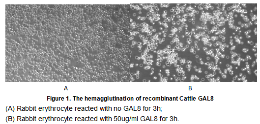
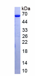
(Galectin 8 (GAL8), also known as prostate carcinoma tumor antigen 1 (PCTA1) in human, is a tandem repeat-type galectin. is a member of the lectin family, of which 14 mammalian galectins have been identified. It is also a member of the beta-galactoside-binding protein family that plays an important role in cell-cell adhesion, cell-matrix interactions, macrophage activation, angiogenesis, metastasis, apoptosis. In this case, we chose rabbit erythrocyte (RaE) to assay its ability of agglutination. A general procedure for hemagglutination assay (or haemagglutination assay; HA) is as follows, two-fold dilute the recombinant Bovine GAL8 with 0.9% sodium chloride injection, add 50uL a serial dilution of GAL8 to each well of a U or V-bottom shaped 96-well microtiter plate. The final well serves as a negative control with no GAL8, replace with 50uL 0.9% sodium chloride injection. Then add 50uL 1% rabbit erythrocyte to each well and mixed gently. The plate is incubated for 3 hours at room temperature. The results are shown in Figure 1. It was obvious that the minimal effective concentration of GAL8 is 0.781 ug/mL.)
Bioactivity
(Galectin 8 (GAL8), also known as prostate carcinoma tumor antigen 1 (PCTA1) in human, is a tandem repeat-type galectin. is a member of the lectin family, of which 14 mammalian galectins have been identified. It is also a member of the beta-galactoside-binding protein family that plays an important role in cell-cell adhesion, cell-matrix interactions, macrophage activation, angiogenesis, metastasis, apoptosis. In this case, we chose rabbit erythrocyte (RaE) to assay its ability of agglutination. A general procedure for hemagglutination assay (or haemagglutination assay; HA) is as follows, two-fold dilute the recombinant Bovine GAL8 with 0.9% sodium chloride injection, add 50uL a serial dilution of GAL8 to each well of a U or V-bottom shaped 96-well microtiter plate. The final well serves as a negative control with no GAL8, replace with 50uL 0.9% sodium chloride injection. Then add 50uL 1% rabbit erythrocyte to each well and mixed gently. The plate is incubated for 3 hours at room temperature. The results are shown in Figure 1. It was obvious that the minimal effective concentration of GAL8 is 0.781 ug/mL.)
Galectin 8 (GAL8), Active Protein (Cat# AAA152177)
 Bioactivity
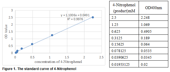
(carboxylesterase 1 (CES1) also known as Liver carboxylesterase 1 is a serine esterase and member of a large multigene carboxylesterase family. The protein Involved in the detoxification of xenobiotics and in the activation of ester and amide prodrugs. Hydrolyzes aromatic and aliphatic esters, but has no catalytic activity toward amides or a fatty acyl-CoA ester. Hydrolyzes the methyl ester group of cocaine to form benzoylecgonine. Thus, the recombinant human CES1 activity was measured by its ability to hydrolyze 4-Nitrophenyl acetate (4-NPA) to 4-Nitrophenol. The reaction was performed in 50 mM Tris, pH 7.5(Assay Buffer), initiated by addition 50 uL of various concentrations of CES1 (dilute by Assay Buffer) to 50 uL of 2 mM Substrate 4-NPA(100 mM stock in Acetone, dilute by deionized water). Incubated at 37 degree C for 10min, then read at a wavelength of 400 nm.)
Bioactivity
(carboxylesterase 1 (CES1) also known as Liver carboxylesterase 1 is a serine esterase and member of a large multigene carboxylesterase family. The protein Involved in the detoxification of xenobiotics and in the activation of ester and amide prodrugs. Hydrolyzes aromatic and aliphatic esters, but has no catalytic activity toward amides or a fatty acyl-CoA ester. Hydrolyzes the methyl ester group of cocaine to form benzoylecgonine. Thus, the recombinant human CES1 activity was measured by its ability to hydrolyze 4-Nitrophenyl acetate (4-NPA) to 4-Nitrophenol. The reaction was performed in 50 mM Tris, pH 7.5(Assay Buffer), initiated by addition 50 uL of various concentrations of CES1 (dilute by Assay Buffer) to 50 uL of 2 mM Substrate 4-NPA(100 mM stock in Acetone, dilute by deionized water). Incubated at 37 degree C for 10min, then read at a wavelength of 400 nm.)
Carboxylesterase 1 (CES1), Active Protein (Cat# AAA152179)
 Bioactivity
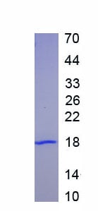
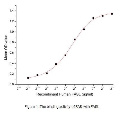
(FAS (Tumor necrosis factor receptor superfamily member 6) belongs to the tumor necrosis factor receptor superfamily. FAS contains a death domain, which has been shown to play a central role in the physiological regulation of programmed cell death. A bindi)
Bioactivity
(FAS (Tumor necrosis factor receptor superfamily member 6) belongs to the tumor necrosis factor receptor superfamily. FAS contains a death domain, which has been shown to play a central role in the physiological regulation of programmed cell death. A bindi)
Factor Related Apoptosis (FAS), Active Protein (Cat# AAA152950)
 Bioactivity
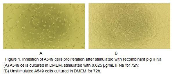
(Figure 2. Inhibition of A549 cells proliferation after stimulated with recombinant mouse IFN-alpha.)
Bioactivity
(Figure 2. Inhibition of A549 cells proliferation after stimulated with recombinant mouse IFN-alpha.)
Interferon Alpha (IFNa), Active Protein (Cat# AAA152953)
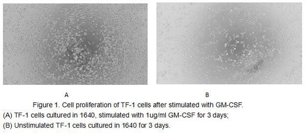
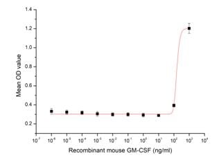 Bioactivity
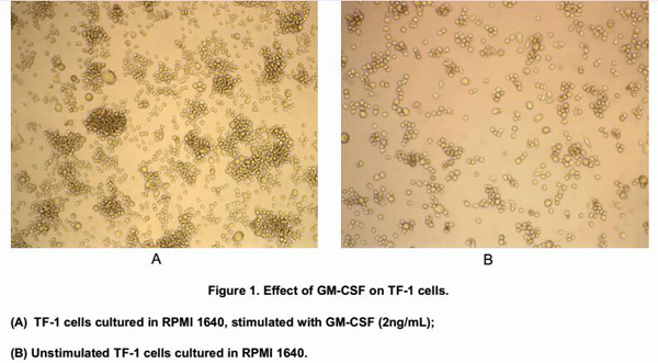
(Figure 2. Cell proliferation of TF-1 cells after stimulated with GM-CSF.)
Bioactivity
(Figure 2. Cell proliferation of TF-1 cells after stimulated with GM-CSF.)
Colony Stimulating Factor 2, Granulocyte Macrophage (GM-CSF), Active Protein (Cat# AAA152960)
 Bioactivity
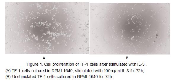
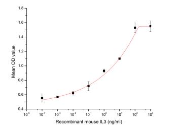
(Figure 2. The dose-effect curve of IL-3 on TF-1 cells)
Bioactivity
(Figure 2. The dose-effect curve of IL-3 on TF-1 cells)
Interleukin 3 (IL3), Active Protein (Cat# AAA152969)
 Bioactivity
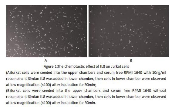
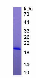
(Interleukin 8 (IL8 or chemokine (C-X-C motif) ligand 8, CXCL8) is a chemokine produced by macrophages and other cell types such as epithelial cells, airway smooth muscle cells and endothelial cells.Thus, chemotaxis assay used 24-well microchemotaxis syste)
Bioactivity
(Interleukin 8 (IL8 or chemokine (C-X-C motif) ligand 8, CXCL8) is a chemokine produced by macrophages and other cell types such as epithelial cells, airway smooth muscle cells and endothelial cells.Thus, chemotaxis assay used 24-well microchemotaxis syste)
Interleukin 8 (IL8), Active Protein (Cat# AAA152973)
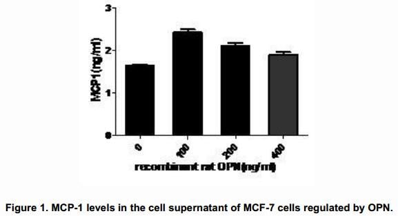
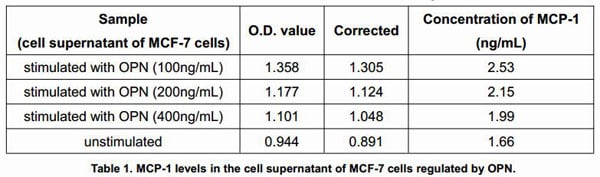
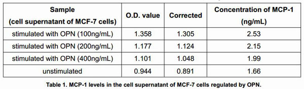
 Bioactivity
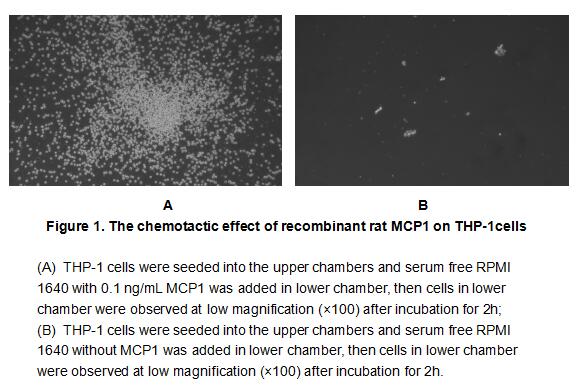
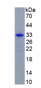
(Monocyte Chemotactic Protein 1 (MCP1), also known as C-C motif chemokine 2(CCL2), is a member of the beta (C-C) subfamily of chemokines that is a chemoattractant for monocytes and basophils. Rat CCL2 is secreted as a 14 kDa glycoprotein monomer but noncov)
Bioactivity
(Monocyte Chemotactic Protein 1 (MCP1), also known as C-C motif chemokine 2(CCL2), is a member of the beta (C-C) subfamily of chemokines that is a chemoattractant for monocytes and basophils. Rat CCL2 is secreted as a 14 kDa glycoprotein monomer but noncov)
Monocyte Chemotactic Protein 1 (MCP1), Active Protein (Cat# AAA152976)
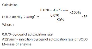 Bioactivity
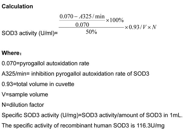
(Extracellular superoxide dismutase [Cu-Zn] is an enzyme that in humans is encoded by the SOD3 gene. This gene encodes a member of the superoxide dismutase (SOD) protein family. SODs are antioxidant enzymes that catalyze the dismutation of two superoxide r)
Bioactivity
(Extracellular superoxide dismutase [Cu-Zn] is an enzyme that in humans is encoded by the SOD3 gene. This gene encodes a member of the superoxide dismutase (SOD) protein family. SODs are antioxidant enzymes that catalyze the dismutation of two superoxide r)
Superoxide Dismutase 3, Extracellular (SOD3), Active Protein (Cat# AAA152980)

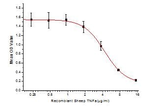 Bioactivity
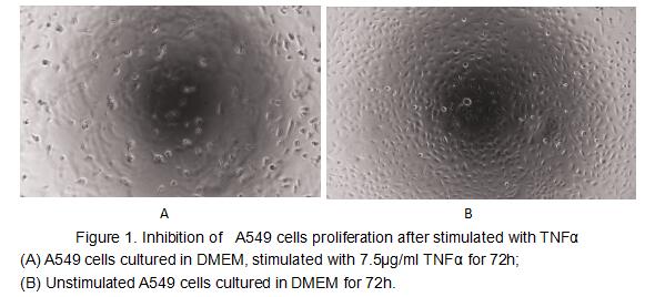
(Figure 2. Inhibition of A549 cells proliferation after stimulated with TNFalpha)
Bioactivity
(Figure 2. Inhibition of A549 cells proliferation after stimulated with TNFalpha)
Tumor Necrosis Factor Alpha (TNFa), Active Protein (Cat# AAA152989)
 Bioactivity
(Tumor necrosis factor (TNF, tumor necrosis factor alpha, TNFalpha, cachexin, or cachectin) is a cell signaling protein (cytokine) involved in systemic inflammation and is one of the cytokines that make up the acute phase reaction. The primary role of TNF)
Bioactivity
(Tumor necrosis factor (TNF, tumor necrosis factor alpha, TNFalpha, cachexin, or cachectin) is a cell signaling protein (cytokine) involved in systemic inflammation and is one of the cytokines that make up the acute phase reaction. The primary role of TNF)
Tumor Necrosis Factor Alpha (TNFa), Active Protein (Cat# AAA152994)
 Bioactivity
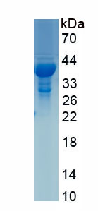
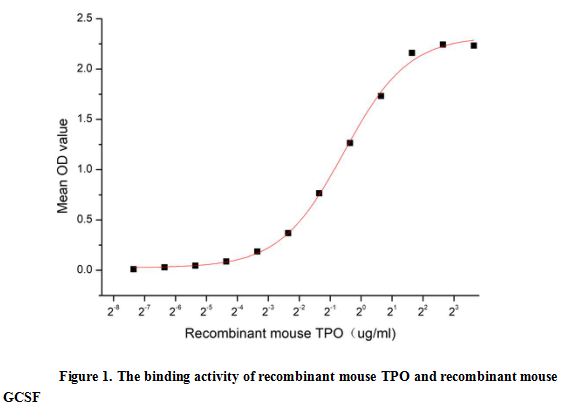
(Thrombopoietin (TPO) is a humoral growth factor that is necessary for megakaryocyte proliferation and maturation, as well as for thrombopoiesis.TPO can promote platelet production, aggregation, ECM adhesion, and activation. It is principally produced in t)
Bioactivity
(Thrombopoietin (TPO) is a humoral growth factor that is necessary for megakaryocyte proliferation and maturation, as well as for thrombopoiesis.TPO can promote platelet production, aggregation, ECM adhesion, and activation. It is principally produced in t)
Thrombopoietin (TPO), Active Protein (Cat# AAA152997)
 Bioactivity
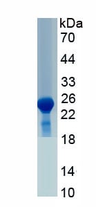
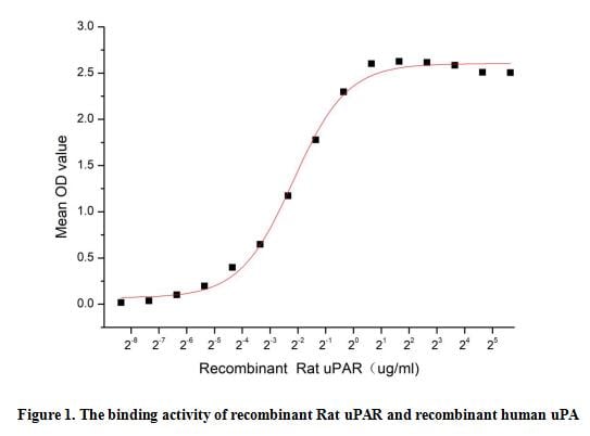
(Plasminogen Activator, Urokinase Receptor (uPAR) is the receptor for urokinase plasminogen activator and, given its role in localizing and promoting plasmin formation, likely influences many normal and pathological processes related to cell-surface plasmi)
Bioactivity
(Plasminogen Activator, Urokinase Receptor (uPAR) is the receptor for urokinase plasminogen activator and, given its role in localizing and promoting plasmin formation, likely influences many normal and pathological processes related to cell-surface plasmi)
Plasminogen Activator, Urokinase Receptor (uPAR), Active Protein (Cat# AAA152999)
 Bioactivity
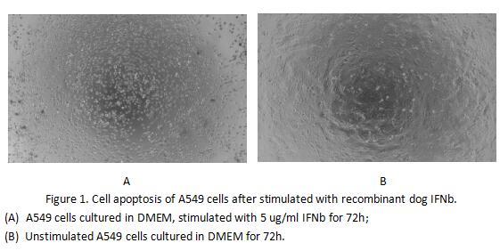
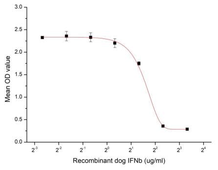
(Figure 2. Cell apoptosis of A549 cells after stimulated with recombinant dog IFNb.)
Bioactivity
(Figure 2. Cell apoptosis of A549 cells after stimulated with recombinant dog IFNb.)
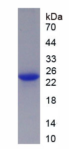
Interferon Beta (IFNb), Active Protein (Cat# AAA153002)
What Are Active Proteins?
Proteins are large molecules made up of long chains of amino acids.
They will typically fold into a very particular 3-dimensional shape/conformation, that is sometimes referred to as their “native” form, which allows them to work properly in the body. For the purposes of product categorization, AAA Biotech will typically refer to proteins purified from their original animal host as being “native” proteins (this is to signify their difference compared to their “recombinant” or “synthetic” protein counterparts).
If a protein successfully folds into the correct shape, it is will typically display high fidelity characteristics to its original protein in its original animal host, and be classified as an active protein, as it will be able to function “normally” in most enzymatic or binding capacities. If it loses this shape, due to factors such as heat or strong chemicals (such as detergents), it becomes inactive and is no longer able to perform its basic functions. All of the proteins in this category are made under strict quality control, and they are active, pure, low in contaminants, and stable.
Most are stored as freeze-dried powders and come without extra tags, so they’re very close to the actual natural/native form.
Key Applications of Active Proteins
1. Scientific Research
- Aid in the study of how proteins function in the body
- Aid in understanding various disease processes
2. Drug Development
- Powerful tools to investigate how potential drugs interact with specific proteins
- Ideal for identifying drug targets
3. Cell Culture
- Are routinely utilized to support cell growth and function (e.g., using exogenous growth factors)
- Can be used to promote cellular development into specific types (differentiation)
4. Diagnostics
- Regularly utilized in tests to detect diseases or infections (e.g., COVID-19, cancer)
- Note: All products are strictly for research-use only (RUO).
5. Therapeutics
- Some active proteins are used directly as treatments (e.g., insulin, enzymes)
- Note: All products are strictly for research-use only (RUO).
6. Vaccine Development
- Used to create or test vaccines by mimicking parts of viruses or bacteria
7. Biochemical Assays
- They can facilitate the characterization of enzyme activity, binding strength, or protein interactions in lab tests
Why Buy Active Proteins from AAA Biotech?
- High biological activity – Verified to perform as expected or indicated on datasheet
- Strict quality control – We are confident in our active proteins’ reliability and consistency
- High purity & low endotoxin – Ideal for applications involving sensitive or precious samples/components
- Freeze-dried for stability – Long shelf life and straightforward storage
- Mostly tag-free – Closer to natural/native protein form
FAQ
1. What are active proteins used for in research?
Active proteins are used primarily in the study of how proteins function, in characterizing/discovering drug interactions, supporting cell growth, running biochemical assays, and in development of diagnostics or therapeutics.
2. How are AAA Biotech's active proteins validated?
AAA Biotech’s active proteins are validated through strict quality control and functional assays to ensure they are properly folded and active. “Active”, though, can be an ambiguous term, so if a specific “activity” or “binding” capability of a protein is of crucial interest to you, please inquire with us prior to purchase, and we will provide further details on how the “Active” modifier was determined to be applicable.
3. Are these proteins tested for biological activity?
Yes, all active proteins from AAA Biotech are tested to confirm they have the expected biological activity before being offered for use. Though, said “biological activity” can be either “enzymatic”, “binding”, or both.