Filters
▼Clonality
▼Type
▼Reactivity
▼Gene Name
▼Isotype
▼Host
▼Application
▼Clone
▼Secondary Antibodies
At AAA Biotech also known as AAA Bio or AAABio, we provide a vast collection of high-quality, purified, conjugated and unconjugated secondary antibodies to use in various applications, such as Western Blotting, Flow Cytometry, Cell Imaging, or CUT & Tag.
Our secondary antibodies are available with a wide range of labels—such as HRP, AP, FITC, and biotin—to match your detection system and enhance signal sensitivity. Each antibody is rigorously tested to ensure high specificity and minimal cross-reactivity, delivering reliable performance across multiple species and assay types.
Whether you're conducting qualitative imaging or quantitative analysis, AAA Biotech has the right secondary antibody to support your research with consistency and precision. Our entire catalog of secondary antibodies is available for browsing through our main website.
Viewing 100-150 of 552 product results
Goat Anti-Rabbit IgM+IgG (H+L chain specific), Polyclonal Secondary Antibody (Cat# AAA78816)
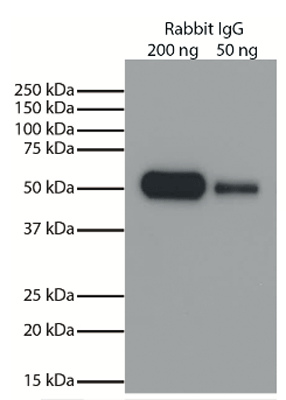 Application Data
(Purified rabbit IgG was resolved by electrophoresis, transferred to PDVF membrane, and visualized using Goat Anti-Rabbit IgG-HRP secondary antibody and chemiluminescent detection.)
Application Data
(Purified rabbit IgG was resolved by electrophoresis, transferred to PDVF membrane, and visualized using Goat Anti-Rabbit IgG-HRP secondary antibody and chemiluminescent detection.)
Goat anti Rabbit IgG, Polyclonal Secondary Antibody (Cat# AAA78820)
 ELISA
ELISA
Rabbit Anti-Mouse IgG (H+L), Polyclonal Secondary Antibody (Cat# AAA78844)
Goat anti Pig IgG (H + L) (HRP), Secondary Antibody (Cat# AAA75531)
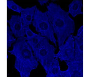 IF (Immunofluorescence)
(Immunofluorescence using hCEC cells, 1st Ab (anti-beta-Actin at 1/250) and 2nd Ab (anti-mouse AB27405 at 1/1,000); cells were fixed with methanol;)
IF (Immunofluorescence)
(Immunofluorescence using hCEC cells, 1st Ab (anti-beta-Actin at 1/250) and 2nd Ab (anti-mouse AB27405 at 1/1,000); cells were fixed with methanol;)
IgG, Polyclonal Secondary Antibody (Cat# AAA63217)
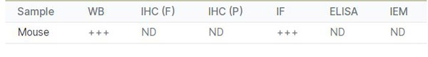
 IF (Immunofluorescence)
(Immunofluorescence using hCEC cells, 1st Ab (anti-beta-Actin at 1/250) and 2nd Ab (anti-mouse AB27633 at 1/1,000); cells were fixed with methanol;)
IF (Immunofluorescence)
(Immunofluorescence using hCEC cells, 1st Ab (anti-beta-Actin at 1/250) and 2nd Ab (anti-mouse AB27633 at 1/1,000); cells were fixed with methanol;)
IgG, Polyclonal Secondary Antibody (Cat# AAA63220)
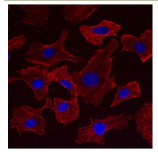
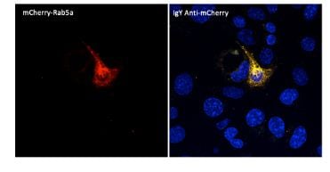 IF (Immunofluorescence)
(Immunofluorescence -Anti-mCherry Ab (AB9770) using hCEC cells transduced with mCherry-Rab5a; cells were fixed with methanol and mCherry Ab at 1/250; 2nd Ab goatAnti-IgY (AB307633) at 1:1,000;)
IF (Immunofluorescence)
(Immunofluorescence -Anti-mCherry Ab (AB9770) using hCEC cells transduced with mCherry-Rab5a; cells were fixed with methanol and mCherry Ab at 1/250; 2nd Ab goatAnti-IgY (AB307633) at 1:1,000;)
IgY, Polyclonal Secondary Antibody (Cat# AAA63225)
 DB (Dot Blot)
(Dot Blot analysis using Goat anti Rabbit IgG FITC and HRP conj ugated secondary antibody used to detect nanogram - picogram levels of rabbit IgG by dot blot on nitrocellulose membrane. 4 ul each of serial 1 in 4 dilutions of rabbit IgG were dotted on nitrocellulose and allowed to dry. Membrane was blocked in 3% BSA for 10 minutes dried for later use and rewetted with blocking buffer.)
DB (Dot Blot)
(Dot Blot analysis using Goat anti Rabbit IgG FITC and HRP conj ugated secondary antibody used to detect nanogram - picogram levels of rabbit IgG by dot blot on nitrocellulose membrane. 4 ul each of serial 1 in 4 dilutions of rabbit IgG were dotted on nitrocellulose and allowed to dry. Membrane was blocked in 3% BSA for 10 minutes dried for later use and rewetted with blocking buffer.)
Goat anti Rabbit IgG (H + L) (FITC), Secondary Antibody (Cat# AAA75483)
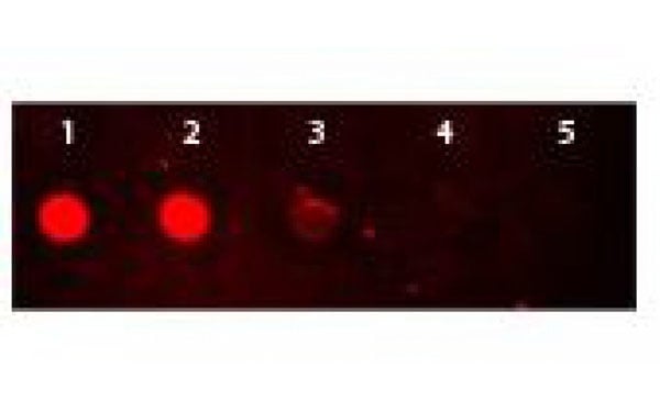 DB (Dot Blot)
(Dot Blot of Rhodamine Conjugated Goat-anti-Rabbit IgG. Antigen: Rabbit IgG. Load: Lane 1 - 50ng Lane 2 - 16.67ng Lane 3 - 5.56ng Lane 4 - 1.85ng Lane 5 - 0.62ng. Primary antibody: none. Secondary antibody: Rhodamine Conjugated Goat-a-Rabbit IgG secondary antibody at 1:1,000 for 60 min at RT. Block: MB-070 for 60 min at RT.)
DB (Dot Blot)
(Dot Blot of Rhodamine Conjugated Goat-anti-Rabbit IgG. Antigen: Rabbit IgG. Load: Lane 1 - 50ng Lane 2 - 16.67ng Lane 3 - 5.56ng Lane 4 - 1.85ng Lane 5 - 0.62ng. Primary antibody: none. Secondary antibody: Rhodamine Conjugated Goat-a-Rabbit IgG secondary antibody at 1:1,000 for 60 min at RT. Block: MB-070 for 60 min at RT.)
Goat anti Rabbit IgG (H + L) (rhodamine), Secondary Antibody (Cat# AAA75484)
Kappa Chain, Secondary Antibody (Cat# AAA75151)
 WB (Western Blot)
Goat anti Human IgG (H + L) (FITC), Secondary Antibody (Cat# AAA75619)
Goat anti Rabbit IgG (heavy and light chains), Polyclonal Secondary Antibody (Cat# AAA77558)
Goat anti Horse IgM (Fc specific), Polyclonal Secondary Antibody (Cat# AAA77564)
Rabbit anti Mouse IgA (Fc specific), Polyclonal Secondary Antibody (Cat# AAA77586)
Goat anti Human IgG (Fc specific) immunofixation, Polyclonal Secondary Antibody (Cat# AAA77595)
Inter-species cross-reactivity is a normal feature of antibodies to mammalian immunoglobulins, since homologous proteins of different species frequently share antigenic determinants. The degree of crossreactivity is also dependent on the conc
Goat anti Guinea Pig IgG2 (subclass specific), Polyclonal Secondary Antibody (Cat# AAA77605)
Goat anti Guinea Pig IgG (heavy and light chains), Polyclonal Secondary Antibody (Cat# AAA77612)
Sheep anti Human IgA (Fc specific), Polyclonal Secondary Antibody (Cat# AAA77615)
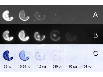
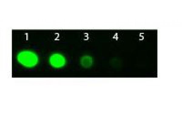 DB (Dot Blot)
(Antigen: Rabbit IgG. Load: Lane 1 - 100 ng Lane 2 - 33.3 ng Lane 3 - 11.1 ng Lane 4 - 3.70 ng Lane 5 - 1.23 ng. Goat Fab'2 anti-Rabbit IgG Antibody (FITC) at 1:1,000 for 1 HR at RT. Block for 1 HR at RT.)
DB (Dot Blot)
(Antigen: Rabbit IgG. Load: Lane 1 - 100 ng Lane 2 - 33.3 ng Lane 3 - 11.1 ng Lane 4 - 3.70 ng Lane 5 - 1.23 ng. Goat Fab'2 anti-Rabbit IgG Antibody (FITC) at 1:1,000 for 1 HR at RT. Block for 1 HR at RT.)
Goat anti Rabbit IgG (H + L) (Fab'2) (FITC), Secondary Antibody (Cat# AAA75502)
Goat anti Mouse IgM (PE) (mu chain specific), Secondary Antibody (Cat# AAA75522)
Donkey anti Goat IgG (H + L) (Cy3), Secondary Antibody (Cat# AAA75548)

 IEP (Immunoelectrophoresis)
IgG (H&L), Secondary Antibody (Cat# AAA79054)
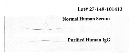
Cross Reactivity: Based on IEP, no reactivity is observed to non-immunoglobulin human serum immunoglobulins
Affinity purified using solid phase Human IgG.
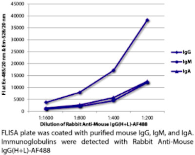
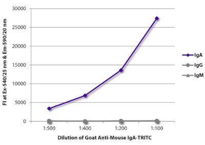 ELISA
(ELISA plate was coated with purified mouse IgA , IgG , and IgM. Immunoglobulins were detected with serially diluted Goat Anti-Mouse IgA-PE)
ELISA
(ELISA plate was coated with purified mouse IgA , IgG , and IgM. Immunoglobulins were detected with serially diluted Goat Anti-Mouse IgA-PE)
Goat Anti-Mouse IgA (alpha chain specific), Polyclonal Secondary Antibody (Cat# AAA78772)
 IF (Immunofluorescence)
(Product: Goat Anti-Mouse IgG2a-FITCAmount used: )
IF (Immunofluorescence)
(Product: Goat Anti-Mouse IgG2a-FITCAmount used: )
Goat Anti-Mouse IgG2a (gamma 2a chain specific), Polyclonal Secondary Antibody (Cat# AAA78780)
Goat F(ab')2 Anti-Mouse IgG2a (gamma 2a chain specific), Polyclonal Secondary Antibody (Cat# AAA78781)
Goat Anti-Mouse IgG2b (gamma 2b chain specific), Secondary Antibody (Cat# AAA78782)
Goat Anti-Mouse IgG3 (gamma 3 chain specific), Polyclonal Secondary Antibody (Cat# AAA78784)
 Application Data
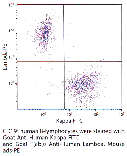
Goat Anti-Human kappa (kappa chain specific), Polyclonal Secondary Antibody (Cat# AAA78799)
Goat Anti-Human kappa (kappa chain specific), Polyclonal Secondary Antibody (Cat# AAA78801)
Goat Anti-Human lambda (lambda chain specific), Polyclonal Secondary Antibody (Cat# AAA78805)
Goat Anti-Human lambda (lambda chain specific), Polyclonal Secondary Antibody (Cat# AAA78806)
Goat Anti-Rat IgM (u chain specific), Polyclonal Secondary Antibody (Cat# AAA78808)
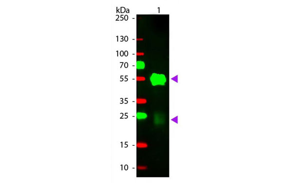
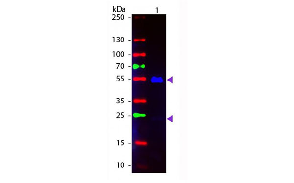
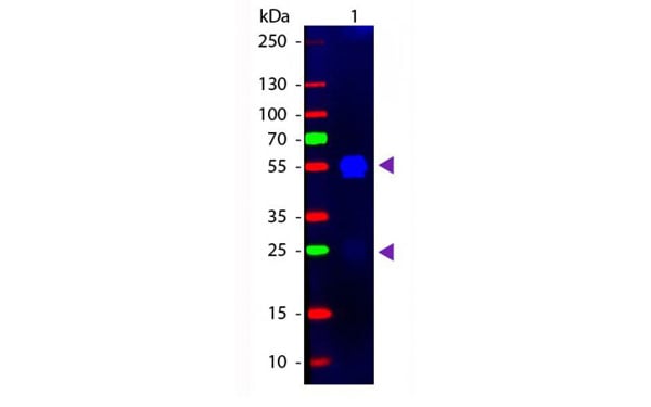
 WB (Western Blot)
(Total cell Iysates from Jurkat cells were resolved by electrophoresis, transferred to PVDF membrane, and probed with Rabbit Anti-Human DR5-UNlB . Proteins were visualized using Goat Anti-Rabbit IgG(H+L). Mouse/Human ads-HRP (AAA78823) secondary antibody and chemfluminescent detection.)
WB (Western Blot)
(Total cell Iysates from Jurkat cells were resolved by electrophoresis, transferred to PVDF membrane, and probed with Rabbit Anti-Human DR5-UNlB . Proteins were visualized using Goat Anti-Rabbit IgG(H+L). Mouse/Human ads-HRP (AAA78823) secondary antibody and chemfluminescent detection.)
Goat Anti-Rabbit IgG (H+L chain specific), Polyclonal Secondary Antibody (Cat# AAA78823)
Goat Anti-Rabbit IgG (H+L chain specific), Polyclonal Secondary Antibody (Cat# AAA78825)
Rabbit F(ab')2 Anti-Sheep IgG (H+L), Polyclonal Secondary Antibody (Cat# AAA78828)
Goat Anti-Porcine IgG (H+L), Polyclonal Secondary Antibody (Cat# AAA78830)
(may react with immunoglobulins from other species and the light chains of other porcine immunoglobulins)
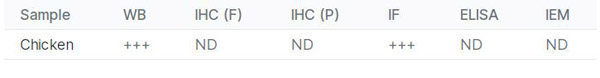
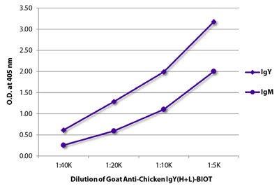 Application Data
(ELISA plate was coated with purified chicken IgY and IgM. Immunoglobulins were detected with Goat Anti-Chicken IgY(H+L)-BIOT)
Application Data
(ELISA plate was coated with purified chicken IgY and IgM. Immunoglobulins were detected with Goat Anti-Chicken IgY(H+L)-BIOT)
Goat Anti-Chicken IgY (H+L), Polyclonal Secondary Antibody (Cat# AAA78838)
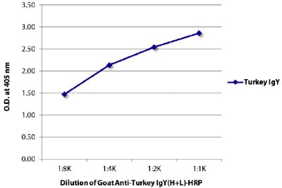 ELISA
(ELISA plate was coated with purified turkey IgY. Immunoglobulin was detected with Goat Anti-Turkey IgY (H+L)-HRP (AAA78839).)
ELISA
(ELISA plate was coated with purified turkey IgY. Immunoglobulin was detected with Goat Anti-Turkey IgY (H+L)-HRP (AAA78839).)
Goat Anti-Turkey IgY (H+L), Polyclonal Secondary Antibody (Cat# AAA78839)
Rabbit Anti-Mouse IgG (H+L), Polyclonal Secondary Antibody (Cat# AAA78846)
Rabbit Anti-Rat IgG (H+L), Polyclonal Secondary Antibody (Cat# AAA78847)
IgG2b, Monoclonal Secondary Antibody (Cat# AAA78867)
Mouse Anti-Hamster IgG1, Monoclonal Secondary Antibody (Cat# AAA78868)

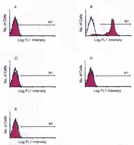
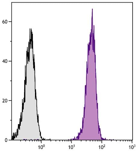 Application Data
(BALB/c mouse splenocytes were stained with Rat Anti-Mouse CD45-UNLB followed by Goat Fab Anti-Rat Ig, Mouse ads-FITC)
Application Data
(BALB/c mouse splenocytes were stained with Rat Anti-Mouse CD45-UNLB followed by Goat Fab Anti-Rat Ig, Mouse ads-FITC)
Fab Ig, Polyclonal Secondary Antibody (Cat# AAA78929)
Mouse (C3H/HeJ) Anti-Mouse H-2Kd/H-2Dd, Monoclonal Secondary Antibody (Cat# AAA78690)
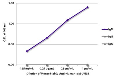 ELISA
(ELISA plate was coated with purified human IgM,IgG, and IgA. Immunoglobulins were detected with serially diluted Mouse F(ab')2 Anti-Human IgM-UNLB followed by Goat Anti-Mouse IgG(H+L), Human ads-HRP (AAA78714))
ELISA
(ELISA plate was coated with purified human IgM,IgG, and IgA. Immunoglobulins were detected with serially diluted Mouse F(ab')2 Anti-Human IgM-UNLB followed by Goat Anti-Mouse IgG(H+L), Human ads-HRP (AAA78714))
Mouse F(ab')2 Anti-Human IgM (u chain specific), Monoclonal Secondary Antibody (Cat# AAA78714)
Mouse Anti-Human IgG2 (gamma 2 chain specific), Monoclonal Secondary Antibody (Cat# AAA78724)
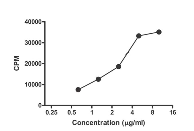
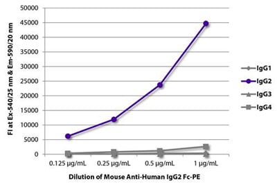 Application Data
(FLISA plate was coated with purified human IgG1, IgG2, IgG3, and IgG4. Immunoglobulins were detected with serially diluted Mouse Anti-Human IgG2 Fc-PE.)
Application Data
(FLISA plate was coated with purified human IgG1, IgG2, IgG3, and IgG4. Immunoglobulins were detected with serially diluted Mouse Anti-Human IgG2 Fc-PE.)
Mouse Anti-Human IgG2 (gamma 2 chain specific), Monoclonal Secondary Antibody (Cat# AAA78728)
Mouse Anti-Human IgA1 (alpha 1 chain specific), Monoclonal Secondary Antibody (Cat# AAA78731)
Mouse Anti-Human IgG4 (gamma 4 chain specific), Monoclonal Secondary Antibody (Cat# AAA78736)
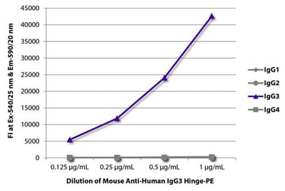 Application Data
(FLISA plate was coated with purified human IgG1, IgG2, IgG3, and IgG4. Immunoglobulins were detected with serially diluted Mouse Anti-Human IgG3 Hinge-PE)
Application Data
(FLISA plate was coated with purified human IgG1, IgG2, IgG3, and IgG4. Immunoglobulins were detected with serially diluted Mouse Anti-Human IgG3 Hinge-PE)
Mouse Anti-Human IgG3 (gamma 3 chain specific), Monoclonal Secondary Antibody (Cat# AAA78743)
What Are Secondary Antibodies?
Secondary antibodies are immunoglobulins that bind to primary antibodies, which are directly bound to the target antigen. They are conjugated with either an enzyme, fluorophore, or biotin, and they are almost always only targeting the constant region (Fc) of the primary antibody. As a result, this amplifies the signal in various assays and helps researchers detect, quantify, and visualize specific antigens in a complex biological sample.
Key Applications of Secondary Antibodies
- Western blotting for protein detection and quantification.
- ELISA (Enzyme-Linked Immunosorbent Assay) for antigen-antibody interaction studies and quantification.
- Immunohistochemistry (IHC) and immunofluorescence imaging for tissue and cell analysis.
- Flow cytometry for cell surface and intracellular marker detection.
- Immunoprecipitation (IP) and chromatin immunoprecipitation (ChIP) assays.
- Signal amplification in various immunoassays.
- Multiplex assays using fluorophore-conjugated secondary antibodies.
Advantages of Using Secondary Antibodies
-
High sensitivity
Multiple secondary antibodies can bind to a single primary antibody and amplify the signal. This is especially useful for detecting antigens even if they are in low concentrations.
-
Increased flexibility in assay design
Secondary antibodies come pre-conjugated with labels, such as enzymes, fluorophores, biotin, and more. This provides researchers with flexibility to use in multiple applications. They also support multiplexing for the simultaneous detection of multiple targets.
-
Cost-effective
Since the Fc domain remains constant within the same animal class, only one type of secondary antibody is needed to bind many types of primary antibodies. This reduces the cost of labeling multiple primary antibodies.
-
Better signal-to-noise ratio
Secondary antibodies minimize the background noise. As a result, they provide clearer and more accurate results.
-
Versatility
A single species- and isotype-specific secondary antibody can be used with any compatible primary antibody. This can help streamline reagent inventory and reduce cost and preparation time.
-
Easier detection
Unlike primary antibodies, secondary antibodies’ main characteristic is that they will almost always be partnered with a component that can be used for detection. This can make detection and visualization easy.
Why Buy Secondary Antibodies from AAA Biotech?
- Highly Validated: Most of our secondary antibodies are thoroughly tested to ensure they work reliably across different experiments.
- Versatile Applications: Our secondary antibodies can be used in a wide range of research techniques, including (but not limited to) immunocytochemistry (ICC), ELISA, immunofluorescence (IF), immunohistochemistry (IHC), flow cytometry (FC), immunoprecipitation (IP), and Western blotting (WB).
- Support for Rare Species: We offer secondary antibodies specifically designed for rare or less common species—something many other suppliers don’t provide.
- Affordable Prices: Our secondary antibodies are available at competitive prices to support all research budgets.
- Quick and Convenient Ordering: Simple online ordering process with responsive customer support.
- Privacy: We respect your privacy and ensure your information is protected at every step of the ordering process, and in all other communication with our team members.
FAQ
1. Can I reuse a secondary antibody?
Not recommended for critical experiments, as reusing may compromise sensitivity and specificity.
2. What is the difference between primary and secondary antibodies?
Primary antibodies bind directly to the antigen. Secondary antibodies bind to the primary and are usually labeled for detection.
3. How long do secondary antibodies last?
When stored properly (usually at –20°C or 4°C, depending on formulation), they can last for years. Always refer to the product datasheet.
4. What happens if you use too much secondary antibody?
Excess can increase background noise, leading to a poor signal-to-noise ratio. Titrate to find the optimal concentration.
5. Can secondary antibodies be monoclonal?
Yes. While many are polyclonal, monoclonal secondary antibodies are available for consistent batch-to-batch performance.
6. How to find secondary antibodies?
Use our catalog filters to search by host species, conjugate type, target species, and application.
7. How to store secondary antibodies?
Store at recommended temperatures away from light. Avoid repeated freeze-thaw cycles.