Filters
▼Clonality
▼Type
▼Reactivity
▼Gene Name
▼Isotype
▼Host
▼Application
▼Clone
▼Secondary Antibodies
At AAA Biotech also known as AAA Bio or AAABio, we provide a vast collection of high-quality, purified, conjugated and unconjugated secondary antibodies to use in various applications, such as Western Blotting, Flow Cytometry, Cell Imaging, or CUT & Tag.
Our secondary antibodies are available with a wide range of labels—such as HRP, AP, FITC, and biotin—to match your detection system and enhance signal sensitivity. Each antibody is rigorously tested to ensure high specificity and minimal cross-reactivity, delivering reliable performance across multiple species and assay types.
Whether you're conducting qualitative imaging or quantitative analysis, AAA Biotech has the right secondary antibody to support your research with consistency and precision. Our entire catalog of secondary antibodies is available for browsing through our main website.
Viewing 1-50 of 552 product results
Goat Anti-Mouse lambda (lambda chain specific), Secondary Antibody (Cat# AAA78777)
Goat Anti-Mouse kappa (kappa chain specific), Secondary Antibody (Cat# AAA78775)
Mouse anti Human IgG2 (Fc subclass specific), Monoclonal Secondary Antibody (Cat# AAA77535)
Mouse anti Human Bence Jones lambda (surface and hidden determinants), Monoclonal Secondary Antibody (Cat# AAA77534)
 IF (Immunofluorescence)
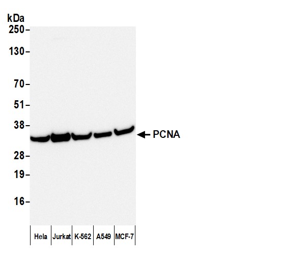
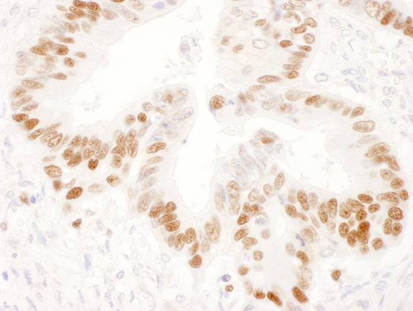
(Figure 3. IF analysis of NEFH using anti-NEFH antibody (MA1071).NEFH was detected in a paraffin-embedded section of human colon cancer tissue. Heat mediated antigen retrieval was performed in EDTA buffer (pH 8.0, epitope retrieval solution). The tissue section was blocked with 10% goat serum. The tissue section was then incubated with 5ug/mL mouse anti-NEFH Antibody (MA1071) overnight at 4 degree C. DyLight 647 Conjugated Goat Anti-Mouse IgG (AAA127949) was used as secondary antibody at 1:500 dilution and incubated for 30 minutes at 37 degree C. The section was counterstained with DAPI. Visualize using a fluorescence microscope and filter sets appropriate for the label used.)
IF (Immunofluorescence)
(Figure 3. IF analysis of NEFH using anti-NEFH antibody (MA1071).NEFH was detected in a paraffin-embedded section of human colon cancer tissue. Heat mediated antigen retrieval was performed in EDTA buffer (pH 8.0, epitope retrieval solution). The tissue section was blocked with 10% goat serum. The tissue section was then incubated with 5ug/mL mouse anti-NEFH Antibody (MA1071) overnight at 4 degree C. DyLight 647 Conjugated Goat Anti-Mouse IgG (AAA127949) was used as secondary antibody at 1:500 dilution and incubated for 30 minutes at 37 degree C. The section was counterstained with DAPI. Visualize using a fluorescence microscope and filter sets appropriate for the label used.)
IgG, Polyclonal Secondary Antibody (Cat# AAA127949)
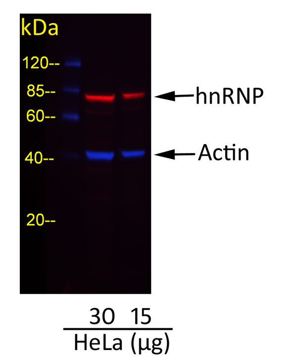 WB (Western Blot)
(Detection of Actin and hnRNP in HeLa Whole Cell Lysate. Primary Antibodies: cocktail of rabbit anti-Actin and mouse anti-hnRNP at 1 ug/ml each. Secondary Antibodies: cocktail of Dylight 488-conjugated goat anti-rabbit AAA210708 (AAA210708-5) (blue) and Dylight 680-conjugated goat anti-mouse (red) at 0.5 ug/ml each. Acquisition: Syngene G:Box, 6 seconds (blue) and 42 seconds (red).)
WB (Western Blot)
(Detection of Actin and hnRNP in HeLa Whole Cell Lysate. Primary Antibodies: cocktail of rabbit anti-Actin and mouse anti-hnRNP at 1 ug/ml each. Secondary Antibodies: cocktail of Dylight 488-conjugated goat anti-rabbit AAA210708 (AAA210708-5) (blue) and Dylight 680-conjugated goat anti-mouse (red) at 0.5 ug/ml each. Acquisition: Syngene G:Box, 6 seconds (blue) and 42 seconds (red).)
IgG Heavy and Light Chain Cross-Adsorbed, Polyclonal Secondary Antibody (Cat# AAA210708)
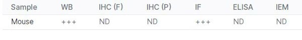
Minimum Reactivity: Human, Mouse, Rat, Chicken, Bovine, Horse, Pig
Mouse anti Human secretory component (free and bound), Monoclonal Secondary Antibody (Cat# AAA77530)
Mouse anti Human IgG2 (Fc subclass specific), Monoclonal Secondary Antibody (Cat# AAA77531)
 Application Data
Mouse Anti-Rat IgG2b (gamma 2b chain specific), Monoclonal Secondary Antibody (Cat# AAA78693)
Mouse Anti-Rat Kappa (kappa chain specific), Monoclonal Secondary Antibody (Cat# AAA78697)
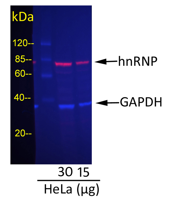 WB (Western Blot)
(Detection of GAPDH and hnRNP in HeLa Whole Cell Lysate. Primary Antibodies: cocktail of goat anti-GAPDH and mouse anti-hnRNP at 1 ug/ml each. Secondary Antibodies: cocktail of Dylight 488-conjugated rabbit anti-goat AAA213528 (AAA213528-2) (blue) and Dylight 680-conjugated rabbit anti-mouse (red) at 0.5 ug/ml each. Acquisition: Syngene G:Box, 52 seconds (blue) and 77 seconds (red).)
WB (Western Blot)
(Detection of GAPDH and hnRNP in HeLa Whole Cell Lysate. Primary Antibodies: cocktail of goat anti-GAPDH and mouse anti-hnRNP at 1 ug/ml each. Secondary Antibodies: cocktail of Dylight 488-conjugated rabbit anti-goat AAA213528 (AAA213528-2) (blue) and Dylight 680-conjugated rabbit anti-mouse (red) at 0.5 ug/ml each. Acquisition: Syngene G:Box, 52 seconds (blue) and 77 seconds (red).)
IgG Heavy and Light Chain, Polyclonal Secondary Antibody (Cat# AAA213528)
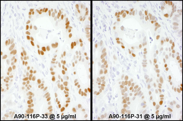
 WB (Western Blot)
(Detection of human PCNA by western blot with HRP-conjugated Goat anti-Mouse IgG Heavy and Light Chain Antibody. Samples: Whole cell lysate (50 ug) from Hela, Jurkat, K-562, A549, and MCF-7 cells prepared using NETN lysis buffer. Antibody: Mouse anti-PCNA Monoclonal Antibody [PC10] used for WB at 1:1000. Secondary: HRP-conjugated Goat anti-Mouse IgG Heavy and Light Chain Antibody (AAA213712). Detection: Chemiluminescence with an exposure time of 3 seconds.)
WB (Western Blot)
(Detection of human PCNA by western blot with HRP-conjugated Goat anti-Mouse IgG Heavy and Light Chain Antibody. Samples: Whole cell lysate (50 ug) from Hela, Jurkat, K-562, A549, and MCF-7 cells prepared using NETN lysis buffer. Antibody: Mouse anti-PCNA Monoclonal Antibody [PC10] used for WB at 1:1000. Secondary: HRP-conjugated Goat anti-Mouse IgG Heavy and Light Chain Antibody (AAA213712). Detection: Chemiluminescence with an exposure time of 3 seconds.)
IgG Heavy and Light Chain, Polyclonal Secondary Antibody (Cat# AAA213712)
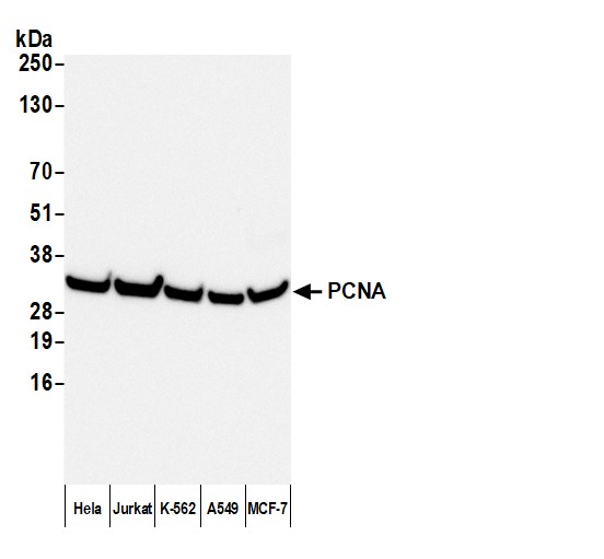 WB (Western Blot)
(Detection of human PCNA by western blot with HRP-conjugated Rabbit anti-Mouse IgG Heavy and Light Chain Antibody. Samples: Whole cell lysate (50 ug) from Hela, Jurkat, K-562, A549, and MCF-7 cells prepared using NETN lysis buffer. Antibody: Mouse anti-PCNA Monoclonal Antibody [PC10] used for WB at 1:1000. Secondary: HRP-conjugated Rabbit anti-Mouse IgG Heavy and Light Chain Antibody (AAA213713). Detection: Chemiluminescence with an exposure time of 3 seconds.)
WB (Western Blot)
(Detection of human PCNA by western blot with HRP-conjugated Rabbit anti-Mouse IgG Heavy and Light Chain Antibody. Samples: Whole cell lysate (50 ug) from Hela, Jurkat, K-562, A549, and MCF-7 cells prepared using NETN lysis buffer. Antibody: Mouse anti-PCNA Monoclonal Antibody [PC10] used for WB at 1:1000. Secondary: HRP-conjugated Rabbit anti-Mouse IgG Heavy and Light Chain Antibody (AAA213713). Detection: Chemiluminescence with an exposure time of 3 seconds.)
IgG Heavy and Light Chain, Polyclonal Secondary Antibody (Cat# AAA213713)
F(ab')2 IgM, Polyclonal Secondary Antibody (Cat# AAA213714)
IgM, Monoclonal Secondary Antibody (Cat# AAA174767)
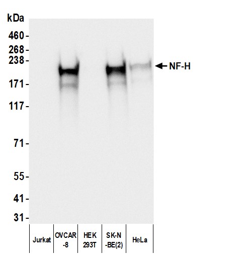 WB (Western Blot)
(Detection of human NF-H using mouse anti-Chicken IgY secondary antibody by western blot. Samples: Whole cell lysate (50 ug) from Jurkat, OVCAR-8, HEK293T, SK-N-BE(2), and HeLa cells prepared using NETN lysis buffer. Primary: Chicken anti-NF-H antibody. Secondary: mouse anti-Chicken IgY Light Chain monoclonal antibody [1Y-263] (AAA213527 lot 1) used at 1:1000. Tertiary: HRP-conjugated goat anti-mouse IgG . Detection: Chemiluminescence with an exposure time of 1 second.)
WB (Western Blot)
(Detection of human NF-H using mouse anti-Chicken IgY secondary antibody by western blot. Samples: Whole cell lysate (50 ug) from Jurkat, OVCAR-8, HEK293T, SK-N-BE(2), and HeLa cells prepared using NETN lysis buffer. Primary: Chicken anti-NF-H antibody. Secondary: mouse anti-Chicken IgY Light Chain monoclonal antibody [1Y-263] (AAA213527 lot 1) used at 1:1000. Tertiary: HRP-conjugated goat anti-mouse IgG . Detection: Chemiluminescence with an exposure time of 1 second.)
IgY Light Chain, Monoclonal Secondary Antibody (Cat# AAA213527)
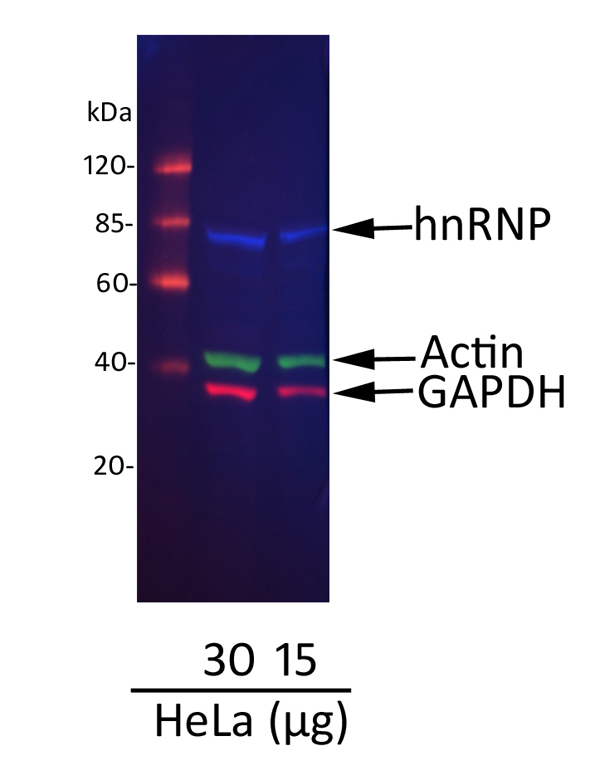 WB (Western Blot)
(Detection of hnRNP, Actin, and GAPDH in HeLa Whole Cell Lysate. Primary Antibodies: cocktail of mouse anti-hnRNP , rabbit anti-actin , and goat anti-GAPDH at 1 ug/ml each. Secondary Antibodies: cocktail of Dylight 488-conjugated donkey anti-mouse AAA213716 (AAA213716-5) (blue), Dylight 680-conjugated donkey anti-rabbit (green), and Dylight 800-conjugated donkey anti-goat A50-201D8 (A50-201D8-1) (red) at 0.5 ug/ml each. Acquisition: Syngene G:Box, 34 seconds (blue), 20 seconds (green), and 50 seconds (red).)
WB (Western Blot)
(Detection of hnRNP, Actin, and GAPDH in HeLa Whole Cell Lysate. Primary Antibodies: cocktail of mouse anti-hnRNP , rabbit anti-actin , and goat anti-GAPDH at 1 ug/ml each. Secondary Antibodies: cocktail of Dylight 488-conjugated donkey anti-mouse AAA213716 (AAA213716-5) (blue), Dylight 680-conjugated donkey anti-rabbit (green), and Dylight 800-conjugated donkey anti-goat A50-201D8 (A50-201D8-1) (red) at 0.5 ug/ml each. Acquisition: Syngene G:Box, 34 seconds (blue), 20 seconds (green), and 50 seconds (red).)
IgG Heavy and Light Chain Cross-Adsorbed, Polyclonal Secondary Antibody (Cat# AAA213716)
Minimum Reactivity: Human, Rat, Chicken, Sheep, Goat, Bovine, Rabbit
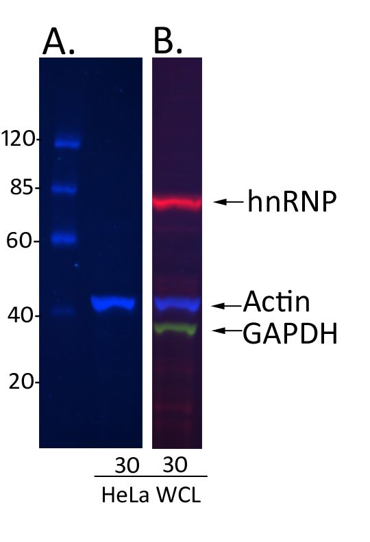 WB (Western Blot)
(Fluorescent western blot Analysis Demonstrating Diminished Cross-reactivity of donkey anti-rabbit IgG Cross-adsorbed Secondary Antibody with goat and mouse IgG. (A) HeLa whole cell lysate (30 ug) was blotted and incubated with a cocktail of mouse anti-hnRNP rabbit anti-actin and goat anti-GAPDH at 1 ug/ml each. For detection, the blot was incubated with 0.5 ug/ml donkey anti-rabbit IgG heavy and light chain cross-adsorbed antibody conjugated to Dylight 488 AAA210709. Donkey anti-rabbit IgG heavy and light chain cross-adsorbed antibody (AAA210709) specifically detected rabbit anti-actin (blue) and showed no cross-reactivity with the goat anti-GAPDH and mouse anti-hnRNP primary antibodies. (B) GAPDH, actin, and hnRNP were detected In a parallel strip incubated with the primary antibody cocktail used in (A), and a secondary antibody cocktail of Dylight 800-conjugated donkey anti-mouse (red), Dylight 488-conjugated donkey anti-rabbit AAA210709 (blue), and Dylight 488-conjugated donkey anti-goat (green) at 0.5 ug/ml each.)
WB (Western Blot)
(Fluorescent western blot Analysis Demonstrating Diminished Cross-reactivity of donkey anti-rabbit IgG Cross-adsorbed Secondary Antibody with goat and mouse IgG. (A) HeLa whole cell lysate (30 ug) was blotted and incubated with a cocktail of mouse anti-hnRNP rabbit anti-actin and goat anti-GAPDH at 1 ug/ml each. For detection, the blot was incubated with 0.5 ug/ml donkey anti-rabbit IgG heavy and light chain cross-adsorbed antibody conjugated to Dylight 488 AAA210709. Donkey anti-rabbit IgG heavy and light chain cross-adsorbed antibody (AAA210709) specifically detected rabbit anti-actin (blue) and showed no cross-reactivity with the goat anti-GAPDH and mouse anti-hnRNP primary antibodies. (B) GAPDH, actin, and hnRNP were detected In a parallel strip incubated with the primary antibody cocktail used in (A), and a secondary antibody cocktail of Dylight 800-conjugated donkey anti-mouse (red), Dylight 488-conjugated donkey anti-rabbit AAA210709 (blue), and Dylight 488-conjugated donkey anti-goat (green) at 0.5 ug/ml each.)
IgG Heavy and Light Chain Cross-Adsorbed, Polyclonal Secondary Antibody (Cat# AAA210709)
Minimum Reactivity: Human, Mouse, Rat, Chicken, Goat, Bovine, Pig
IgG, Secondary Antibody (Cat# AAA172622)
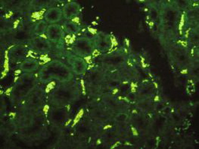
 IF (Immunofluorescence)
(Figure 1: FITC staining on IHC-P,Sample: Rat Testis TissuePrimary Ab: 3ug/mL Rabbit Anti-Rat H3 Ab (Catalog: )Second Ab: 1:500 Dilution of FITC-Linked Guinea pig Anti-Rabbit IgG Ab (Catalog: AAA146597))
IF (Immunofluorescence)
(Figure 1: FITC staining on IHC-P,Sample: Rat Testis TissuePrimary Ab: 3ug/mL Rabbit Anti-Rat H3 Ab (Catalog: )Second Ab: 1:500 Dilution of FITC-Linked Guinea pig Anti-Rabbit IgG Ab (Catalog: AAA146597))
IgG, Polyclonal Secondary Antibody (Cat# AAA146597)
Mouse anti Human IgG2 (Fc subclass specific), Monoclonal Secondary Antibody (Cat# AAA77533)
 IF (Immunofluorescence)
(Immunofluorescence Staining)
IF (Immunofluorescence)
(Immunofluorescence Staining)
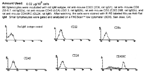
Mouse Anti-Rat IgM (u chain specific), Monoclonal Secondary Antibody (Cat# AAA78695)
Mouse Anti-Rat Kappa (kappa chain specific), Monoclonal Secondary Antibody (Cat# AAA78698)
IgG, Secondary Antibody (Cat# AAA172623)
IgG, Secondary Antibody (Cat# AAA172626)
IgG, Secondary Antibody (Cat# AAA173631)
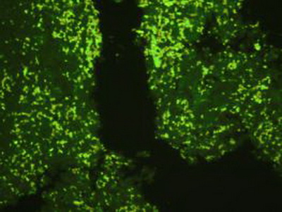
 IHC (Immunohistochemistry)
((Figure: FITC staining on IHC-P; Samples: Rat Lung Tissue.))
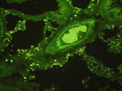
IHC (Immunohistochemistry)
((Figure: FITC staining on IHC-P; Samples: Rat Lung Tissue.))
IgG, Polyclonal Secondary Antibody (Cat# AAA146595)
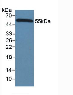
 WB (Western Blot)
((Figure. Used in Western Blot; Sample: Rabbit Serum.))
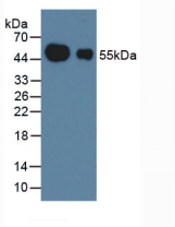
WB (Western Blot)
((Figure. Used in Western Blot; Sample: Rabbit Serum.))
Immunoglobulin G, Polyclonal Secondary Antibody (Cat# AAA147970)
Mouse Anti-Rat Kappa (kappa chain specific), Monoclonal Secondary Antibody (Cat# AAA78696)
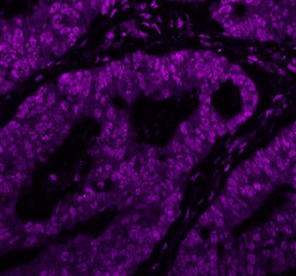
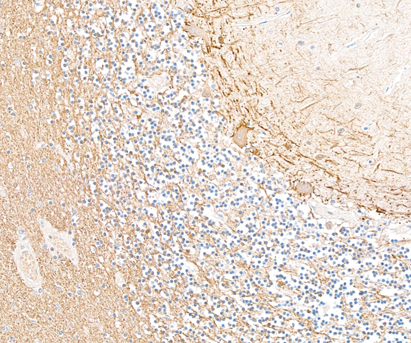
 IHC (Immunohiostchemistry)
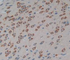
((Figure: DAB staining on IHC-P; Sample: Rat Brain Tissue.))
IHC (Immunohiostchemistry)
((Figure: DAB staining on IHC-P; Sample: Rat Brain Tissue.))
Guinea pig Anti-Rabbit IgG, Polyclonal Secondary Antibody (Cat# AAA145957)
IgG, Secondary Antibody (Cat# AAA172627)
 FLISA
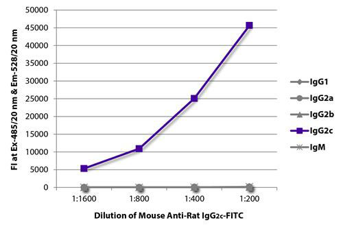
(FLISA plate was coated with purified rat IgG1, IgG2a, IgG2b, IgG2c, and IgM. Immunoglobulins were detected with serially diluted Mouse Anti-Rat IgG2c-FITC)
FLISA
(FLISA plate was coated with purified rat IgG1, IgG2a, IgG2b, IgG2c, and IgM. Immunoglobulins were detected with serially diluted Mouse Anti-Rat IgG2c-FITC)
Mouse Anti-Rat IgG2c (gamma 2c chain specific), Monoclonal Secondary Antibody (Cat# AAA78694)
Mouse anti Human IgG3 (G3m(U) allotype specific), Monoclonal Secondary Antibody (Cat# AAA77528)
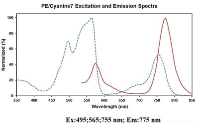
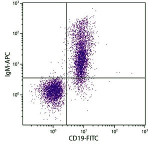
 FCM/FACS (Flow Cytometry)
(Human peripheral blood lymphocytes are stained with Anti-Human IgM Monoclonal Antibody(PE/Cyanine7 Conjugated)(filled gray histogram) or Mouse IgG1 Isotype Control PE/Cy7 (empty black histogram).)
FCM/FACS (Flow Cytometry)
(Human peripheral blood lymphocytes are stained with Anti-Human IgM Monoclonal Antibody(PE/Cyanine7 Conjugated)(filled gray histogram) or Mouse IgG1 Isotype Control PE/Cy7 (empty black histogram).)
IgM, Monoclonal Secondary Antibody (Cat# AAA174757)
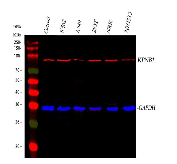
 WB (Western Blot)
((Figure: Used in Western Blot; Sample: Mouse Serum.))
WB (Western Blot)
((Figure: Used in Western Blot; Sample: Mouse Serum.))
IgG, Polyclonal Secondary Antibody (Cat# AAA146594)
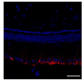
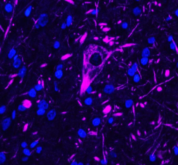
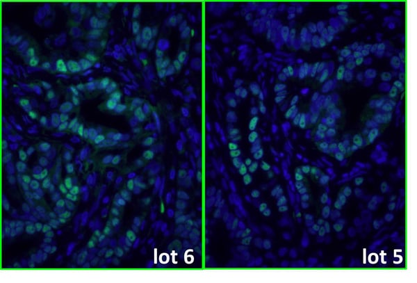
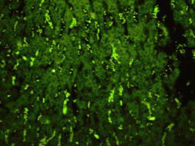
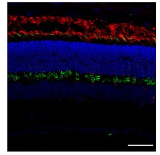
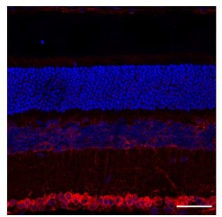 IS (Immunostaining)
(Immunostaining - C57BL/6J mouse retina label with: Red - 1st Ab GRP78 BiP (1/250); 2nd Ab goat anti-rat (AB28550, 1/1000), Blue - Nuclear staining (DAPI), scale bar = 40 um;)
IS (Immunostaining)
(Immunostaining - C57BL/6J mouse retina label with: Red - 1st Ab GRP78 BiP (1/250); 2nd Ab goat anti-rat (AB28550, 1/1000), Blue - Nuclear staining (DAPI), scale bar = 40 um;)
IgG, Polyclonal Secondary Antibody (Cat# AAA63222)
Goat anti Dog IgM (Fc specific), Polyclonal Secondary Antibody (Cat# AAA77540)
Goat anti Monkey IgA (Fc specific), Polyclonal Secondary Antibody (Cat# AAA77567)
Goat anti Dog IgA (Fc specific), Polyclonal Secondary Antibody (Cat# AAA77574)
Goat anti Human J chain of dimeric IgA, Polyclonal Secondary Antibody (Cat# AAA77598)
Rabbit anti Mouse IgG1 IgG2a IgG2b IgG3 (Fc specific), Polyclonal Secondary Antibody (Cat# AAA77603)
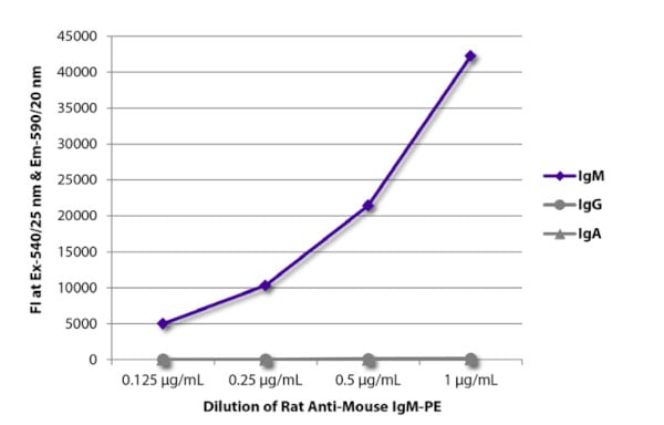 Application Data
(FLISA plate was coated with purified mouse IgM, IgG, and IgA. Immunoglobulins were detected with serially diluted Rat Anti-Mouse IgM-PE)
Application Data
(FLISA plate was coated with purified mouse IgM, IgG, and IgA. Immunoglobulins were detected with serially diluted Rat Anti-Mouse IgM-PE)
Rat Anti-Mouse IgM (u chain specific), Monoclonal Secondary Antibody (Cat# AAA78648)
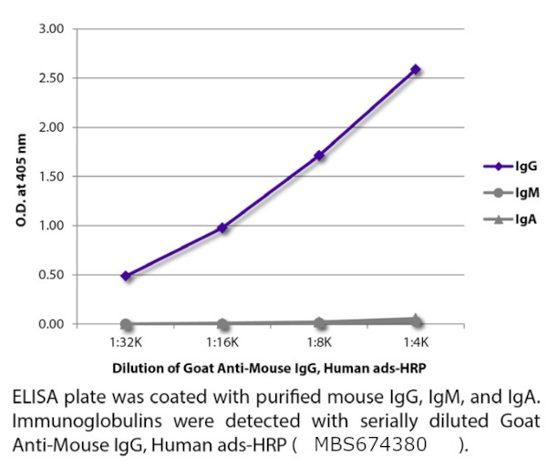
Goat Anti-Mouse IgM+IgG+IgA(H+L), Polyclonal Secondary Antibody (Cat# AAA78761)
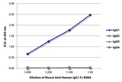 ELISA
(ELISA plate was coated with purified human IgG1, IgG2, IgG3, and IgG4. Immunoglobulins were detected with serilly diluted Mouse Anti-Human IgG1 Fc-BIMA followed by Streptavidin-HRP)
ELISA
(ELISA plate was coated with purified human IgG1, IgG2, IgG3, and IgG4. Immunoglobulins were detected with serilly diluted Mouse Anti-Human IgG1 Fc-BIMA followed by Streptavidin-HRP)
Rat Anti-Mouse IgG2b, Monoclonal Secondary Antibody (Cat# AAA78877)
 ELISA
ELISA
Goat Anti-Mouse IgG (gamma chain specific), Polyclonal Secondary Antibody (Cat# AAA78766)
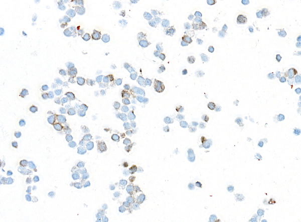
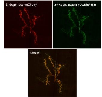 IF (Immunofluorescence)
(Immunofluorescence in Drosophila larvaenmJ muscle 6/7 expressing mCherry in neurons (DyGlutmcherry) using 1st AbAnti-mCherry at 1/1,000 and 2nd AbAnti-goat IgY conjugated to DyLight488 at 1/500;)
IF (Immunofluorescence)
(Immunofluorescence in Drosophila larvaenmJ muscle 6/7 expressing mCherry in neurons (DyGlutmcherry) using 1st AbAnti-mCherry at 1/1,000 and 2nd AbAnti-goat IgY conjugated to DyLight488 at 1/500;)
IgG, Polyclonal Secondary Antibody (Cat# AAA63210)
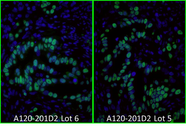
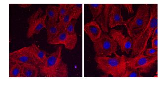 IF (Immunofluorescence)
(Immunofluorescence using hCEC cells, 1st Ab (anti-beta-Actin at 1/250) and 2nd Ab (anti-mouse AB27550 at 1/1,000); cells were fixed with methanol;)
IF (Immunofluorescence)
(Immunofluorescence using hCEC cells, 1st Ab (anti-beta-Actin at 1/250) and 2nd Ab (anti-mouse AB27550 at 1/1,000); cells were fixed with methanol;)
IgG, Polyclonal Secondary Antibody (Cat# AAA63219)
Sheep anti Human IgG4 (subclass specific), Polyclonal Secondary Antibody (Cat# AAA77553)
Inter-species cross-reactivity is a normal feature of antibodies to immunoglobulins, since Ig of different species frequently share antigenic determinants. Cross-reactivity of this antiSerum has not been tested in detail.
Rabbit anti Sheep IgM (Fc specific), Polyclonal Secondary Antibody (Cat# AAA77579)
What Are Secondary Antibodies?
Secondary antibodies are immunoglobulins that bind to primary antibodies, which are directly bound to the target antigen. They are conjugated with either an enzyme, fluorophore, or biotin, and they are almost always only targeting the constant region (Fc) of the primary antibody. As a result, this amplifies the signal in various assays and helps researchers detect, quantify, and visualize specific antigens in a complex biological sample.
Key Applications of Secondary Antibodies
- Western blotting for protein detection and quantification.
- ELISA (Enzyme-Linked Immunosorbent Assay) for antigen-antibody interaction studies and quantification.
- Immunohistochemistry (IHC) and immunofluorescence imaging for tissue and cell analysis.
- Flow cytometry for cell surface and intracellular marker detection.
- Immunoprecipitation (IP) and chromatin immunoprecipitation (ChIP) assays.
- Signal amplification in various immunoassays.
- Multiplex assays using fluorophore-conjugated secondary antibodies.
Advantages of Using Secondary Antibodies
-
High sensitivity
Multiple secondary antibodies can bind to a single primary antibody and amplify the signal. This is especially useful for detecting antigens even if they are in low concentrations.
-
Increased flexibility in assay design
Secondary antibodies come pre-conjugated with labels, such as enzymes, fluorophores, biotin, and more. This provides researchers with flexibility to use in multiple applications. They also support multiplexing for the simultaneous detection of multiple targets.
-
Cost-effective
Since the Fc domain remains constant within the same animal class, only one type of secondary antibody is needed to bind many types of primary antibodies. This reduces the cost of labeling multiple primary antibodies.
-
Better signal-to-noise ratio
Secondary antibodies minimize the background noise. As a result, they provide clearer and more accurate results.
-
Versatility
A single species- and isotype-specific secondary antibody can be used with any compatible primary antibody. This can help streamline reagent inventory and reduce cost and preparation time.
-
Easier detection
Unlike primary antibodies, secondary antibodies’ main characteristic is that they will almost always be partnered with a component that can be used for detection. This can make detection and visualization easy.
Why Buy Secondary Antibodies from AAA Biotech?
- Highly Validated: Most of our secondary antibodies are thoroughly tested to ensure they work reliably across different experiments.
- Versatile Applications: Our secondary antibodies can be used in a wide range of research techniques, including (but not limited to) immunocytochemistry (ICC), ELISA, immunofluorescence (IF), immunohistochemistry (IHC), flow cytometry (FC), immunoprecipitation (IP), and Western blotting (WB).
- Support for Rare Species: We offer secondary antibodies specifically designed for rare or less common species—something many other suppliers don’t provide.
- Affordable Prices: Our secondary antibodies are available at competitive prices to support all research budgets.
- Quick and Convenient Ordering: Simple online ordering process with responsive customer support.
- Privacy: We respect your privacy and ensure your information is protected at every step of the ordering process, and in all other communication with our team members.
FAQ
1. Can I reuse a secondary antibody?
Not recommended for critical experiments, as reusing may compromise sensitivity and specificity.
2. What is the difference between primary and secondary antibodies?
Primary antibodies bind directly to the antigen. Secondary antibodies bind to the primary and are usually labeled for detection.
3. How long do secondary antibodies last?
When stored properly (usually at –20°C or 4°C, depending on formulation), they can last for years. Always refer to the product datasheet.
4. What happens if you use too much secondary antibody?
Excess can increase background noise, leading to a poor signal-to-noise ratio. Titrate to find the optimal concentration.
5. Can secondary antibodies be monoclonal?
Yes. While many are polyclonal, monoclonal secondary antibodies are available for consistent batch-to-batch performance.
6. How to find secondary antibodies?
Use our catalog filters to search by host species, conjugate type, target species, and application.
7. How to store secondary antibodies?
Store at recommended temperatures away from light. Avoid repeated freeze-thaw cycles.