Filters
Clonality
Type
Reactivity
Gene Name
Isotype
Host
Application
Clone
2811 results for " C" - showing 1650-1700
 Standard Curve (Sample)
Hemojuvelin, ELISA Kit (Cat# AAA16529)
Full Name
Human Hemojuvelin ELISA Kit
Gene Names
HFE2; JH; HJV; RGMC; HFE2A
Reactivity
Human
Pricing
 Standard Curve (Sample)
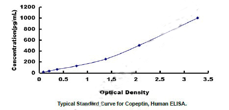
Copeptin (CPP), ELISA Kit (Cat# AAA20717)
Full Name
Human Copeptin (CPP) ELISA Kit
Reactivity
Human
Pricing
 Standard Curve (Sample)
Platelet Derived Growth Factor AA (PDGFAA), ELISA Kit (Cat# AAA13064)
Full Name
Porcine Platelet Derived Growth Factor AA (PDGFAA) ELISA kit
Gene Names
PDGFB; SIS; SSV; IBGC5; PDGF2; c-sis; PDGF-2
Reactivity
Porcine
Pricing
 Standard Curve (Sample)
baculoviral IAP repeat-containing 2, ELISA Kit (Cat# AAA18402)
Full Name
Human Baculoviral IAP repeat-containing protein 2, BIRC2 ELISA Kit
Gene Names
BIRC2; API1; MIHB; HIAP2; RNF48; cIAP1; Hiap-2; c-IAP1
Reactivity
Human
Pricing


 WB (Western Blot)
(NME2 monoclonal antibody (M08), clone 1F2. Western Blot analysis of NME2 expression in Raw 264.7.)
WB (Western Blot)
(NME2 monoclonal antibody (M08), clone 1F2. Western Blot analysis of NME2 expression in Raw 264.7.)
NME2, Monoclonal Antibody (Cat# AAA26035)
Full Name
NME2 (Non-Metastatic Cells 2, Protein (NM23B) Expressed in, MGC111212, NDPK-B, NDPKB, NM23-H2, NM23B, puf) (APC)
Gene Names
NME2; PUF; NDKB; NDPKB; NM23B; NDPK-B; NM23-H2
Applications
EIA, IF, WB
Purity
Purified
Pricing
 Standard Curve (Sample)
Tyrosine protein kinase Yes (YES1), ELISA Kit (Cat# AAA27167)
Full Name
Human Tyrosine protein kinase Yes (YES1) ELISA Kit
Gene Names
YES1; Yes; c-yes; HsT441; P61-YES
Reactivity
Human
Pricing


 WB (Western Blot)
(NME2 monoclonal antibody (M06), clone 1D3. Western Blot analysis of NME2 expression in NIH/3T3 (Cat # L018V1).)
WB (Western Blot)
(NME2 monoclonal antibody (M06), clone 1D3. Western Blot analysis of NME2 expression in NIH/3T3 (Cat # L018V1).)
NME2, Monoclonal Antibody (Cat# AAA26339)
Full Name
NME2 (Non-Metastatic Cells 2, Protein (NM23B) Expressed in, MGC111212, NDPK-B, NDPKB, NM23-H2, NM23B, puf) (FITC)
Gene Names
NME2; PUF; NDKB; NDPKB; NM23B; NDPK-B; NM23-H2
Applications
IF, IHC, WB
Purity
Purified
Pricing
 Standard Curve (Sample)
Intact Fibroblast Growth Factor 23, ELISA Kit (Cat# AAA17207)
Full Name
Human Intact Fibroblast Growth Factor 23 ELISA Kit
Gene Names
FGF23; ADHR; FGFN; HYPF; HPDR2; PHPTC
Reactivity
Human
Pricing
 Standard Curve (Sample)
LTF, ELISA Kit (Cat# AAA21718)
Full Name
Human LTF(Lactoferrin)ELISA Kit
Gene Names
LTF; LF; HLF2; GIG12; HEL110
Reactivity
Human
Pricing


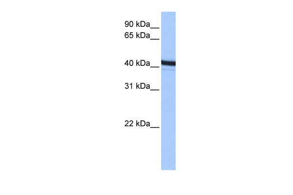
 WB (Western Blot)
(WB Suggested Anti-FER1L3 Antibody Titration: 0.2-1 ug/mlELISA Titer: 1:312500Positive Control: MCF7 cell lysate.MYOF is strongly supported by BioGPS gene expression data to be expressed in MCF7)
WB (Western Blot)
(WB Suggested Anti-FER1L3 Antibody Titration: 0.2-1 ug/mlELISA Titer: 1:312500Positive Control: MCF7 cell lysate.MYOF is strongly supported by BioGPS gene expression data to be expressed in MCF7)
FER1L3, Polyclonal Antibody (Cat# AAA23526)
Full Name
FER1L3 antibody - C-terminal region
Gene Names
MYOF; FER1L3
Reactivity
Cow, Dog, Guinea Pig, Horse, Human, Mouse, Rabbit, Rat, Zebrafish
Applications
WB
Purity
Affinity Purified
Pricing


 WB (Western Blot)
(WB Suggested Anti-FOXA1 antibody Titration: 1 ug/mLSample Type: Human liver)
WB (Western Blot)
(WB Suggested Anti-FOXA1 antibody Titration: 1 ug/mLSample Type: Human liver)
FOXA1, Polyclonal Antibody (Cat# AAA23415)
Full Name
FOXA1 antibody - C-terminal region
Gene Names
FOXA1; HNF3A; TCF3A
Reactivity
Cow, Dog, Guinea Pig, Human, Mouse, Rat, Yeast, Zebrafish
Applications
WB, IHC
Purity
Affinity Purified
Pricing


 IHC (Immunohistochemistry)
(Figure 10. IHC analysis of COX IV using anti-COX IV antibody (AAA19173).COX IV was detected in paraffin-embedded section of mouse kidney tissue. Heat mediated antigen retrieval was performed in citrate buffer (pH6, epitope retrieval solution) for 20 mins. The tissue section was blocked with 10% goat serum. The tissue section was then incubated with 2ug/ml rabbit anti-COX IV Antibody (AAA19173) overnight at 4 degree C. Biotinylated goat anti-rabbit IgG was used as secondary antibody and incubated for 30 minutes at 37 degree C. The tissue section was developed using Strepavidin-Biotin-Complex (SABC) with DAB as the chromogen.)
IHC (Immunohistochemistry)
(Figure 10. IHC analysis of COX IV using anti-COX IV antibody (AAA19173).COX IV was detected in paraffin-embedded section of mouse kidney tissue. Heat mediated antigen retrieval was performed in citrate buffer (pH6, epitope retrieval solution) for 20 mins. The tissue section was blocked with 10% goat serum. The tissue section was then incubated with 2ug/ml rabbit anti-COX IV Antibody (AAA19173) overnight at 4 degree C. Biotinylated goat anti-rabbit IgG was used as secondary antibody and incubated for 30 minutes at 37 degree C. The tissue section was developed using Strepavidin-Biotin-Complex (SABC) with DAB as the chromogen.)
COX IV, Polyclonal Antibody (Cat# AAA19173)
Full Name
Anti-COX IV Picoband Antibody
Gene Names
COX4I1; COX4; COXIV; COX4-1; COXIV-1; COX IV-1
Reactivity
Human, Mouse, Rat
No cross reactivity with other proteins.
No cross reactivity with other proteins.
Applications
EIA, IHC, WB
Pricing


 FCM (Flow Cytometry)
(Figure 7. Flow Cytometry analysis of A549 cells using anti-DR6/TNFRSF21 antibody (AAA19292).Overlay histogram showing A549 cells stained with AAA19292 (Blue line). The cells were blocked with 10% normal goat serum. And then incubated with rabbit anti-DR6/TNFRSF21 Antibody (AAA19292, 1μg/1x106 cells) for 30 min at 20 degree C. DyLight®488 conjugated goat anti-rabbit IgG (5-10μg/1x106 cells) was used as secondary antibody for 30 minutes at 20 degree C. Isotype control antibody (Green line) was rabbit IgG (1μg/1x106) used under the same conditions. Unlabelled sample (Red line) was also used as a control.)
FCM (Flow Cytometry)
(Figure 7. Flow Cytometry analysis of A549 cells using anti-DR6/TNFRSF21 antibody (AAA19292).Overlay histogram showing A549 cells stained with AAA19292 (Blue line). The cells were blocked with 10% normal goat serum. And then incubated with rabbit anti-DR6/TNFRSF21 Antibody (AAA19292, 1μg/1x106 cells) for 30 min at 20 degree C. DyLight®488 conjugated goat anti-rabbit IgG (5-10μg/1x106 cells) was used as secondary antibody for 30 minutes at 20 degree C. Isotype control antibody (Green line) was rabbit IgG (1μg/1x106) used under the same conditions. Unlabelled sample (Red line) was also used as a control.)
DR6/TNFRSF21, Polyclonal Antibody (Cat# AAA19292)
Full Name
Anti-DR6/TNFRSF21 Antibody
Gene Names
TNFRSF21; DR6; CD358; BM-018
Reactivity
Human, Mouse, Rat
Applications
WB, IHC-P, FC/FACS/FCM
Purity
Immunogen affinity purified.
Pricing
 Standard Curve (Sample)
Vitamin C (VC), ELISA Kit (Cat# AAA12744)
Full Name
Chicken Vitamin C (VC) ELISA Kit
Reactivity
Chicken
Pricing
 SDS-PAGE
C-type lectin domain family 18 member A (CLEC18A), Recombinant Protein (Cat# AAA18626)
Full Name
Recombinant Human C-type lectin domain family 18 member A (CLEC18A)
Gene Names
CLEC18A; MRCL; MRCL1; MRLP2
Purity
Greater or equal to 85% purity as determined by SDS-PAGE.
Pricing
 SDS-PAGE
CD45R0, Recombinant Protein (Cat# AAA22242)
Full Name
Recombinant Human CD45R0 (C-6His)
Gene Names
PTPRC; LCA; LY5; B220; CD45; L-CA; T200; CD45R; GP180
Purity
>95% as determined by reducing SDS-PAGE
Pricing
 Standard Curve (Sample)
C-type lectin domain family 3, member B, ELISA Kit (Cat# AAA18264)
Full Name
Mouse Tetranectin, CLEC3B ELISA Kit
Reactivity
Mouse
Pricing
 Standard Curve (Sample)
insulin-like growth factor 1 (somatomedin C), ELISA Kit (Cat# AAA15485)
Full Name
Chicken Insulin-like growth factors 1, IGF-1 ELISA Kit
Gene Names
IGF1; IGF-1; IGF-I
Reactivity
Chicken
Pricing
 Standard Curve (Sample)
Triiodothyronine (T3), ELISA Kit (Cat# AAA12487)
Full Name
Rat Triiodothyronine (T3) ELISA Kit
Gene Names
THRA; AR7; EAR7; ERBA; CHNG6; ERBA1; NR1A1; THRA1; THRA2; ERB-T-1; c-ERBA-1
Reactivity
Rat
Pricing
 Standard Curve (Sample)
vascular endothelial growth factor C, ELISA Kit (Cat# AAA15241)
Full Name
Mouse Vascular Endothelial cell Growth Factor C, VEGF-C ELISA Kit
Gene Names
Vegfc; VEGF-C; AW228853
Reactivity
Mouse
Pricing
 Standard Curve (Sample)
pyruvate kinase, muscle, ELISA Kit (Cat# AAA14969)
Full Name
Human Pyruvate Kinase, M2-PK ELISA Kit
Gene Names
PKM; PK3; TCB; OIP3; PKM2; CTHBP; THBP1; HEL-S-30
Reactivity
Human
Pricing
 SDS-PAGE
Apolipoprotein C-III (Apoc3), Recombinant Protein (Cat# AAA18490)
Full Name
Recombinant Mouse Apolipoprotein C-III (Apoc3)
Gene Names
Apoc3; apo-CIII; apoC-III
Purity
Greater or equal to 85% purity as determined by SDS-PAGE.
Pricing


 WB (Western Blot)
(CSK monoclonal antibody. Western Blot analysis of CSK expression in Hela NE.)
WB (Western Blot)
(CSK monoclonal antibody. Western Blot analysis of CSK expression in Hela NE.)
CSK, Monoclonal Antibody (Cat# AAA24474)
Full Name
CSK (Tyrosine-protein Kinase CSK, C-SRC Kinase, Protein-tyrosine Kinase CYL) APC
Reactivity
Human
Applications
EIA, IHC, IP, WB
Purity
Purified by Protein A Affinity Chromatography.
Pricing


 FCM (Flow Cytometry)
(Figure 10. Flow Cytometry analysis of U87 cells using anti-GNG2 antibody (AAA19312).Overlay histogram showing U87 cells stained with AAA19312 (Blue line). The cells were blocked with 10% normal goat serum. And then incubated with rabbit anti-GNG2 Antibody (AAA19312, 1μg/1x106 cells) for 30 min at 20 degree C. DyLight®488 conjugated goat anti-rabbit IgG (5-10μg/1x106 cells) was used as secondary antibody for 30 minutes at 20 degree C. Isotype control antibody (Green line) was rabbit IgG (1μg/1x106) used under the same conditions. Unlabelled sample (Red line) was also used as a control.)
FCM (Flow Cytometry)
(Figure 10. Flow Cytometry analysis of U87 cells using anti-GNG2 antibody (AAA19312).Overlay histogram showing U87 cells stained with AAA19312 (Blue line). The cells were blocked with 10% normal goat serum. And then incubated with rabbit anti-GNG2 Antibody (AAA19312, 1μg/1x106 cells) for 30 min at 20 degree C. DyLight®488 conjugated goat anti-rabbit IgG (5-10μg/1x106 cells) was used as secondary antibody for 30 minutes at 20 degree C. Isotype control antibody (Green line) was rabbit IgG (1μg/1x106) used under the same conditions. Unlabelled sample (Red line) was also used as a control.)
GNG2, Polyclonal Antibody (Cat# AAA19312)
Full Name
Anti-GNG2 Antibody
Reactivity
Human, Mouse, Rat
Applications
WB, IHC-P, FC/FACS/FCM, EIA
Purity
Immunogen affinity purified.
Pricing


 FCM (Flow Cytometry)
(Figure 9. Flow Cytometry analysis of 293T cells using anti-CDC45L antibody (AAA19360).Overlay histogram showing 293T cells stained with AAA19360 (Blue line). The cells were blocked with 10% normal goat serum. And then incubated with mouse anti- CDC45L Antibody (AAA19360, 1μg/1x106 cells) for 30 min at 20 degree C. DyLight®488 conjugated goat anti-mouse IgG (BA1126, 5-10μg/1x106 cells) was used as secondary antibody for 30 minutes at 20 degree C. Isotype control antibody (Green line) was mouse IgG (1μg/1x106) used under the same conditions. Unlabelled sample (Red line) was also used as a control.)
FCM (Flow Cytometry)
(Figure 9. Flow Cytometry analysis of 293T cells using anti-CDC45L antibody (AAA19360).Overlay histogram showing 293T cells stained with AAA19360 (Blue line). The cells were blocked with 10% normal goat serum. And then incubated with mouse anti- CDC45L Antibody (AAA19360, 1μg/1x106 cells) for 30 min at 20 degree C. DyLight®488 conjugated goat anti-mouse IgG (BA1126, 5-10μg/1x106 cells) was used as secondary antibody for 30 minutes at 20 degree C. Isotype control antibody (Green line) was mouse IgG (1μg/1x106) used under the same conditions. Unlabelled sample (Red line) was also used as a control.)
CDC45L, Monoclonal Antibody (Cat# AAA19360)
Full Name
Anti-CDC45L Antibody (monoclonal, 6H6)
Gene Names
CDC45; CDC45L; CDC45L2; PORC-PI-1
Reactivity
Human, Mouse, Rat
Applications
WB, IHC-P, FC/FACS/FCM
Purity
Immunogen affinity purified.
Pricing


 WB (Western Blot)
(Figure 1. Western blot analysis of CISD2 using anti-CISD2 antibody (AAA19311).Electrophoresis was performed on a 5-20% SDS-PAGE gel at 70V (Stacking gel) / 90V (Resolving gel) for 2-3 hours. The sample well of each lane was loaded with 50ug of sample under reducing conditions.Lane 1: human HEK293 whole cell lysatesLane 2: human HELA whole cell lysatesLane 3: human MCF-7 whole cell lysatesLane 4: monkey kidney tissue lysatesLane 5: human SW620 whole cell lysatesLane 6: human Raji whole cell lysatesLane 7: rat kidney tissue lysatesLane 8: mouse kidney tissue lysates.After Electrophoresis, proteins were transferred to a Nitrocellulose membrane at 150mA for 50-90 minutes. Blocked the membrane with 5% Non-fat Milk/ TBS for 1. 5 hour at RT. The membrane was incubated with rabbit anti-CISD2 antigen affinity purified polyclonal antibody (Catalog # AAA19311) at 0. 5 μg/mL overnight at 4 degree C, then washed with TBS-0. 1%Tween 3 times with 5 minutes each and probed with a goat anti-rabbit IgG-HRP secondary antibody at a dilution of 1:10000 for 1. 5 hour at RT. The signal is developed using an Enhanced Chemiluminescent detection (ECL) kit (Catalog # with Tanon 5200 system. A specific band was detected for CISD2 at approximately 15KD. The expected band size for CISD2 is at 15KD.)
WB (Western Blot)
(Figure 1. Western blot analysis of CISD2 using anti-CISD2 antibody (AAA19311).Electrophoresis was performed on a 5-20% SDS-PAGE gel at 70V (Stacking gel) / 90V (Resolving gel) for 2-3 hours. The sample well of each lane was loaded with 50ug of sample under reducing conditions.Lane 1: human HEK293 whole cell lysatesLane 2: human HELA whole cell lysatesLane 3: human MCF-7 whole cell lysatesLane 4: monkey kidney tissue lysatesLane 5: human SW620 whole cell lysatesLane 6: human Raji whole cell lysatesLane 7: rat kidney tissue lysatesLane 8: mouse kidney tissue lysates.After Electrophoresis, proteins were transferred to a Nitrocellulose membrane at 150mA for 50-90 minutes. Blocked the membrane with 5% Non-fat Milk/ TBS for 1. 5 hour at RT. The membrane was incubated with rabbit anti-CISD2 antigen affinity purified polyclonal antibody (Catalog # AAA19311) at 0. 5 μg/mL overnight at 4 degree C, then washed with TBS-0. 1%Tween 3 times with 5 minutes each and probed with a goat anti-rabbit IgG-HRP secondary antibody at a dilution of 1:10000 for 1. 5 hour at RT. The signal is developed using an Enhanced Chemiluminescent detection (ECL) kit (Catalog # with Tanon 5200 system. A specific band was detected for CISD2 at approximately 15KD. The expected band size for CISD2 is at 15KD.)
CISD2, Polyclonal Antibody (Cat# AAA19311)
Full Name
Anti-CISD2 Antibody
Gene Names
CISD2; ERIS; WFS2; ZCD2; NAF-1; Miner1
Reactivity
Human, Mouse, Rat, Monkey
Applications
WB, IHC-P, ICC, IF, FC/FACS/FCM, EIA
Purity
Immunogen affinity purified.
Pricing


 FCM (Flow Cytometry)
(Figure 7. Flow Cytometry analysis of RH35 cells using anti- EIF4A1 antibody (AAA19380).Overlay histogram showing RH35 cells stained with AAA19380 (Blue line). The cells were blocked with 10% normal goat serum. And then incubated with mouse anti-EIF4A1 Antibody (AAA19380, 1μg/1x106 cells) for 30 min at 20 degree C. DyLight®488 conjugated goat anti-mouse IgG (BA1126, 5-10μg/1x106 cells) was used as secondary antibody for 30 minutes at 20 degree C. Isotype control antibody (Green line) was mouse IgG (1μg/1x106) used under the same conditions. Unlabelled sample (Red line) was also used as a control.)
FCM (Flow Cytometry)
(Figure 7. Flow Cytometry analysis of RH35 cells using anti- EIF4A1 antibody (AAA19380).Overlay histogram showing RH35 cells stained with AAA19380 (Blue line). The cells were blocked with 10% normal goat serum. And then incubated with mouse anti-EIF4A1 Antibody (AAA19380, 1μg/1x106 cells) for 30 min at 20 degree C. DyLight®488 conjugated goat anti-mouse IgG (BA1126, 5-10μg/1x106 cells) was used as secondary antibody for 30 minutes at 20 degree C. Isotype control antibody (Green line) was mouse IgG (1μg/1x106) used under the same conditions. Unlabelled sample (Red line) was also used as a control.)
EIF4A1, Monoclonal Antibody (Cat# AAA19380)
Full Name
Anti-EIF4A1 Antibody (monoclonal, 11B8)
Gene Names
EIF4A1; DDX2A; EIF4A; EIF-4A; eIF4A-I; eIF-4A-I
Reactivity
Human, Mouse, Rat
Applications
WB, IHC-P, ICC, IF, FC/FACS/FCM
Purity
Immunogen affinity purified.
Pricing


 FCM (Flow Cytometry)
(Figure 13. Flow Cytometry analysis of MCF-7 cells using anti-EHD3 antibody (AAA19295).Overlay histogram showing MCF-7 cells stained with AAA19295 (Blue line). The cells were blocked with 10% normal goat serum. And then incubated with rabbit anti-EHD3 Antibody (AAA19295, 1μg/1x106 cells) for 30 min at 20 degree C. DyLight®488 conjugated goat anti-rabbit IgG (5-10μg/1x106 cells) was used as secondary antibody for 30 minutes at 20 degree C. Isotype control antibody (Green line) was rabbit IgG (1μg/1x106) used under the same conditions. Unlabelled sample (Red line) was also used as a control.)
FCM (Flow Cytometry)
(Figure 13. Flow Cytometry analysis of MCF-7 cells using anti-EHD3 antibody (AAA19295).Overlay histogram showing MCF-7 cells stained with AAA19295 (Blue line). The cells were blocked with 10% normal goat serum. And then incubated with rabbit anti-EHD3 Antibody (AAA19295, 1μg/1x106 cells) for 30 min at 20 degree C. DyLight®488 conjugated goat anti-rabbit IgG (5-10μg/1x106 cells) was used as secondary antibody for 30 minutes at 20 degree C. Isotype control antibody (Green line) was rabbit IgG (1μg/1x106) used under the same conditions. Unlabelled sample (Red line) was also used as a control.)
EHD3, Polyclonal Antibody (Cat# AAA19295)
Full Name
Anti-EHD3 Antibody
Gene Names
EHD2; PAST2
Reactivity
Human, Monkey, Mouse, Rat
Applications
WB, IHC-P, ICC, IF, FC/FACS/FCM
Purity
Immunogen affinity purified.
Pricing


 WB (Western Blot)
(FG Pancreatic Carcinoma Cell Lines stably expressing vector along (FG-V) the b3 integrin subunit (FG-b3) or a b3 truncation mutant (FG-759x). Src Mab (AAA28639) was diluted 1:500 in 1% BSA/TBST and incubated Overnight at 4 degree C. After washing 3x 5 min. with TBST the blots were incubated with 1:5000 Goat anti-mouse or Goat anti-rabbit secondary antibody for 1 hr at Room temperature. The blots were again washed 3x 5 min. with TBST and developed using ECL reagent.Data and protocol kindly provided by Dr. Weis of Cheresh Lab, UCSD.)
WB (Western Blot)
(FG Pancreatic Carcinoma Cell Lines stably expressing vector along (FG-V) the b3 integrin subunit (FG-b3) or a b3 truncation mutant (FG-759x). Src Mab (AAA28639) was diluted 1:500 in 1% BSA/TBST and incubated Overnight at 4 degree C. After washing 3x 5 min. with TBST the blots were incubated with 1:5000 Goat anti-mouse or Goat anti-rabbit secondary antibody for 1 hr at Room temperature. The blots were again washed 3x 5 min. with TBST and developed using ECL reagent.Data and protocol kindly provided by Dr. Weis of Cheresh Lab, UCSD.)
SRC, Monoclonal Antibody (Cat# AAA28639)
Full Name
SRC Antibody
Gene Names
SRC; ASV; SRC1; c-SRC; p60-Src
Reactivity
Human, mouse
Applications
WB, EIA, IF
Purity
This antibody is purified through a protein G column, followed by dialysis against PBS.
Pricing
 Standard Curve (Sample)
Interleukin 33, IL-33, ELISA Kit (Cat# AAA11316)
Full Name
Canine Interleukin 33, IL-33 ELISA Kit
Gene Names
IL33; DVS27; IL1F11; NF-HEV; NFEHEV; C9orf26
Reactivity
Canine
Pricing
 Standard Curve (Sample)
Anti Protein C Antibody, ELISA Kit (Cat# AAA27409)
Full Name
Human Anti Protein C Antibody ELISA Kit
Reactivity
Human
Pricing
 Standard Curve (Sample)
Protein Kinase C Delta, ELISA Kit (Cat# AAA17444)
Full Name
Human Protein Kinase C Delta ELISA Kit
Gene Names
PRKCD; MAY1; PKCD; ALPS3; CVID9; nPKC-delta
Reactivity
Human
Pricing
 Standard Curve (Sample)
Platelet Derived Growth Factor (PDGF), ELISA Kit (Cat# AAA12654)
Full Name
Porcine Platelet Derived Growth Factor (PDGF) ELISA kit
Gene Names
PDGFB; SIS; SSV; IBGC5; PDGF2; c-sis; PDGF-2
Reactivity
Porcine
Pricing
 Standard Curve (Sample)
Pulmonary Surfactant Associated Protein D, ELISA Kit (Cat# AAA17316)
Full Name
Human Pulmonary Surfactant Associated Protein D ELISA Kit
Gene Names
MBL2; MBL1; cMBl; collectin
Reactivity
Human
Pricing


 WB (Western Blot)
(WB Suggested Anti-ACTR1B Antibody Titration: 0.2-1 ug/mlPositive Control: HepG2 cell lysateACTR1B is supported by BioGPS gene expression data to be expressed in HepG2)
WB (Western Blot)
(WB Suggested Anti-ACTR1B Antibody Titration: 0.2-1 ug/mlPositive Control: HepG2 cell lysateACTR1B is supported by BioGPS gene expression data to be expressed in HepG2)
ACTR1B, Polyclonal Antibody (Cat# AAA23545)
Full Name
ACTR1B antibody - C-terminal region
Gene Names
ACTR1B; PC3; ARP1B; CTRN2
Reactivity
Cow, Dog, Guinea Pig, Horse, Human, Mouse, Rabbit, Rat, Zebrafish
Applications
WB
Purity
Affinity Purified
Pricing


 IF (Immunofluorescence)
(Figure 7. IF analysis of METTL1 using anti- METTL1 antibody (AAA19336).METTL1 was detected in immunocytochemical section of A431 cells. Enzyme antigen retrieval was performed using IHC enzyme antigen retrieval reagent for 15 mins. The cells were blocked with 10% goat serum. And then incubated with 5μg/mL rabbit anti-METTL1 Antibody (AAA19336) overnight at 4 degree C. DyLight®488 Conjugated Goat Anti-Rabbit IgG was used as secondary antibody at 1:100 dilution and incubated for 30 minutes at 37 degree C. The section was counterstained with DAPI. Visualize using a fluorescence microscope and filter sets appropriate for the label used.)
IF (Immunofluorescence)
(Figure 7. IF analysis of METTL1 using anti- METTL1 antibody (AAA19336).METTL1 was detected in immunocytochemical section of A431 cells. Enzyme antigen retrieval was performed using IHC enzyme antigen retrieval reagent for 15 mins. The cells were blocked with 10% goat serum. And then incubated with 5μg/mL rabbit anti-METTL1 Antibody (AAA19336) overnight at 4 degree C. DyLight®488 Conjugated Goat Anti-Rabbit IgG was used as secondary antibody at 1:100 dilution and incubated for 30 minutes at 37 degree C. The section was counterstained with DAPI. Visualize using a fluorescence microscope and filter sets appropriate for the label used.)
METTL1, Polyclonal Antibody (Cat# AAA19336)
Full Name
Anti-METTL1 Antibody
Gene Names
METTL1; TRM8; TRMT8; C12orf1; YDL201w
Reactivity
Human, Rat
Applications
WB, IHC-P, ICC, IF, EIA
Purity
Immunogen affinity purified.
Pricing
 Standard Curve (Sample)
CD115, ELISA Kit (Cat# AAA17901)
Full Name
Human CD115 ELISA Kit
Gene Names
CSF1R; FMS; CSFR; FIM2; HDLS; C-FMS; CD115; CSF-1R; M-CSF-R
Reactivity
Human
Applications
SE
Pricing
 Standard Curve (Sample)
platelet factor 4 variant 1, ELISA Kit (Cat# AAA18204)
Full Name
Human Platelet factor 4 variant, PF4V1 ELISA Kit
Gene Names
PF4V1; PF4A; CXCL4L1; CXCL4V1; PF4-ALT; SCYB4V1
Reactivity
Human
Pricing
 Standard Curve (Sample)
Cytochrome P450 2C19, CYP2C19, ELISA Kit (Cat# AAA19042)
Full Name
Human Cytochrome P450 2C19, CYP2C19 ELISA Kit
Gene Names
CYP2C19; CPCJ; CYP2C; P450C2C; CYPIIC17; CYPIIC19; P450IIC19
Reactivity
Human
Pricing
 Standard Curve (Sample)
Stromal cell derived factor 1beta, ELISA Kit (Cat# AAA16767)
Full Name
Porcine Stromal cell derived factor 1beta ELISA Kit
Gene Names
CXCL12; IRH; PBSF; SDF1; TLSF; TPAR1; SCYB12
Reactivity
Porcine
Pricing


 Application Data
(Detection limit for recombinant GST tagged UBE2C is ~0.03ng/ml as a capture antibody.)
Application Data
(Detection limit for recombinant GST tagged UBE2C is ~0.03ng/ml as a capture antibody.)
UBE2C, Monoclonal Antibody (Cat# AAA25580)
Full Name
UBE2C (Ubiquitin-conjugating Enzyme E2 C, UbcH10, Ubiquitin Carrier Protein C, Ubiquitin-protein Ligase C, dJ447F3.2) (HRP)
Gene Names
UBE2C; UBCH10; dJ447F3.2
Reactivity
Human
Applications
EIA, IHC, WB
Purity
Purified by Protein A Affinity Chromatography.
Pricing
 SDS-PAGE
KLK4, Active Protein (Cat# AAA27762)
Full Name
KLK4 Protein, Human, Recombinant (His Tag)
Gene Names
KLK4; ARM1; EMSP; PSTS; AI2A1; EMSP1; KLK-L1; PRSS17; MGC116827; MGC116828
Purity
>94% as determined by SDS-PAGE
Pricing

 SDS-PAGE
Ubiquitin, Recombinant Protein (Cat# AAA20969)
Full Name
Recombinant Ubiquitin (Ub)
Gene Names
UBC; HMG20
Reactivity
Homo sapiens (Human), Mus musculus (Mouse), Rattus norvegicus (Rat), Oryctolagus cuniculus (Rabbit), Canis familiaris; Canine (Dog), Sus scrofa; Porcine (Pig), Bos taurus; Bovine (Cattle), Equus caballus; Equine (Horse)
Applications
WB
Purity
> 95%
Pricing
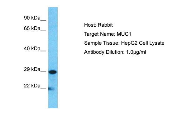


 WB (Western Blot)
(Host: RabbitTarget Name: MUC1Sample Type: HepG2Lane A: Primary AntibodyLane B: Primary Antibody + Blocking PeptidePrimary Antibody Concentration: 1ug/mlPeptide Concentration: 5ug/mlLysate Quantity: 25ug/lane/laneGel Concentration: 0.12)
WB (Western Blot)
(Host: RabbitTarget Name: MUC1Sample Type: HepG2Lane A: Primary AntibodyLane B: Primary Antibody + Blocking PeptidePrimary Antibody Concentration: 1ug/mlPeptide Concentration: 5ug/mlLysate Quantity: 25ug/lane/laneGel Concentration: 0.12)
MUC1, Polyclonal Antibody (Cat# AAA23492)
Full Name
MUC1 antibody - C-terminal region
Gene Names
MUC1; EMA; MCD; PEM; PUM; KL-6; MAM6; MCKD; PEMT; CD227; H23AG; MCKD1; MUC-1; ADMCKD; ADMCKD1; CA 15-3; MUC-1/X; MUC1/ZD; MUC-1/SEC
Reactivity
Cow, Dog, Guinea Pig, Horse, Human, Mouse, Rabbit, Rat, Pig
Applications
IHC, WB
Purity
Affinity Purified
Pricing


 DB (Dot Blot)
(Formalin-fixed and paraffin-embedded human cancer tissue reacted with the primary antibody, which was peroxidase-conjugated to the secondary antibody, followed by AEC staining. This data demonstrates the use of this antibody for immunohistochemistry; clinical relevance has not been evaluated. BC = breast carcinoma; HC = hepatocarcinoma.)
DB (Dot Blot)
(Formalin-fixed and paraffin-embedded human cancer tissue reacted with the primary antibody, which was peroxidase-conjugated to the secondary antibody, followed by AEC staining. This data demonstrates the use of this antibody for immunohistochemistry; clinical relevance has not been evaluated. BC = breast carcinoma; HC = hepatocarcinoma.)
Phospho-HIST1H3B3 (S10), Polyclonal Antibody (Cat# AAA28670)
Full Name
Phospho-HIST1H3B3 (S10) Antibody
Gene Names
HIST1H3A; H3/A; H3FA
Reactivity
Human
Applications
DB, EIA, IHC, WB
Purity
Peptide Affinity Purified Rabbit Polyclonal Antibody (Pab)
Pricing


 IHC (Immunohistochemistry)
(Figure 7. IHC analysis of CD105 using anti-CD105 antibody (AAA19165).CD105 was detected in paraffin-embedded section of rat spleen tissue. Heat mediated antigen retrieval was performed in citrate buffer (pH6, epitope retrieval solution) for 20 mins. The tissue section was blocked with 10% goat serum. The tissue section was then incubated with 1ug/ml rabbit anti-CD105 Antibody (AAA19165) overnight at 4 degree C. Biotinylated goat anti-rabbit IgG was used as secondary antibody and incubated for 30 minutes at 37 degree C. The tissue section was developed using Strepavidin-Biotin-Complex (SABC) with DAB as the chromogen.)
IHC (Immunohistochemistry)
(Figure 7. IHC analysis of CD105 using anti-CD105 antibody (AAA19165).CD105 was detected in paraffin-embedded section of rat spleen tissue. Heat mediated antigen retrieval was performed in citrate buffer (pH6, epitope retrieval solution) for 20 mins. The tissue section was blocked with 10% goat serum. The tissue section was then incubated with 1ug/ml rabbit anti-CD105 Antibody (AAA19165) overnight at 4 degree C. Biotinylated goat anti-rabbit IgG was used as secondary antibody and incubated for 30 minutes at 37 degree C. The tissue section was developed using Strepavidin-Biotin-Complex (SABC) with DAB as the chromogen.)
CD105, Polyclonal Antibody (Cat# AAA19165)
Full Name
Anti-CD105 Picoband Antibody
Gene Names
Eng; Endo; CD105; AI528660; AI662476; S-endoglin
Reactivity
Mouse, Rat
No cross reactivity with other proteins.
No cross reactivity with other proteins.
Applications
EIA, IHC, WB
Purity
Immunogen affinity purified
Pricing


 WB (Western Blot)
(WB Suggested Anti-SHH Antibody Titration: 0.2-1 ug/mlPositive Control: HepG2 cell lysate)
WB (Western Blot)
(WB Suggested Anti-SHH Antibody Titration: 0.2-1 ug/mlPositive Control: HepG2 cell lysate)
SHH, Polyclonal Antibody (Cat# AAA23516)
Full Name
SHH antibody - N-terminal region
Gene Names
SHH; TPT; HHG1; HLP3; HPE3; SMMCI; ShhNC; TPTPS; MCOPCB5
Reactivity
Predicted Reactivity: Cow, Dog, Goat, Guinea Pig, Horse, Human, Mouse, Rabbit, Rat, Zebrafish, Chicken (Tested Reactivity: Human, Mouse, Chicken)
Applications
IHC, WB
Purity
Affinity Purified
Pricing


 FCM (Flow Cytometry)
(Figure 11. Flow Cytometry analysis of U937 cells using anti-Coronin 1a/TACO/CORO1A antibody (AAA19291).Overlay histogram showing U937 cells stained with AAA19291 (Blue line). The cells were blocked with 10% normal goat serum. And then incubated with rabbit anti-Coronin 1a/TACO/CORO1A Antibody (AAA19291, 1μg/1x106 cells) for 30 min at 20 degree C. DyLight®488 conjugated goat anti-rabbit IgG (5-10μg/1x106 cells) was used as secondary antibody for 30 minutes at 20 degree C. Isotype control antibody (Green line) was rabbit IgG (1μg/1x106) used under the same conditions. Unlabelled sample (Red line) was also used as a control.)
FCM (Flow Cytometry)
(Figure 11. Flow Cytometry analysis of U937 cells using anti-Coronin 1a/TACO/CORO1A antibody (AAA19291).Overlay histogram showing U937 cells stained with AAA19291 (Blue line). The cells were blocked with 10% normal goat serum. And then incubated with rabbit anti-Coronin 1a/TACO/CORO1A Antibody (AAA19291, 1μg/1x106 cells) for 30 min at 20 degree C. DyLight®488 conjugated goat anti-rabbit IgG (5-10μg/1x106 cells) was used as secondary antibody for 30 minutes at 20 degree C. Isotype control antibody (Green line) was rabbit IgG (1μg/1x106) used under the same conditions. Unlabelled sample (Red line) was also used as a control.)
Coronin 1a/TACO/CORO1A, Polyclonal Antibody (Cat# AAA19291)
Full Name
Anti-Coronin 1a/TACO/CORO1A Antibody
Gene Names
CORO1A; p57; TACO; CLABP; HCORO1; CLIPINA
Reactivity
Human, Mouse, Rat
Applications
WB, IHC-P, FC/FACS/FCM, EIA
Purity
Immunogen affinity purified.
Pricing


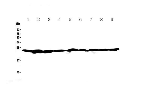 WB (Western Blot)
(Figure 1. Western blot analysis of Ran using anti-Ran antibody (AAA19131). Electrophoresis was performed on a 5-20% SDS-PAGE gel at 70V (Stacking gel) / 90V (Resolving gel) for 2-3 hours. The sample well of each lane was loaded with 50ug of sample under reducing conditions. Lane 1: rat brain tissue lysate,Lane 2: rat testis tissue lysate,Lane 3: rat thymus tissue lysate,Lane 4: mouse brain tissue lysate,Lane 5: mouse testis tissue lysate,Lane 6: mouse thymus tissue lysate,Lane 7: human A549 whole cell lysate,Lane 8: human 22RV1 whole cell lysate,Lane 9: human Hela whole cell lysate. After Electrophoresis, proteins were transferred to a Nitrocellulose membrane at 150mA for 50-90 minutes. Blocked the membrane with 5% Non-fat Milk/ TBS for 1.5 hour at RT. The membrane was incubated with rabbit anti-Ran antigen affinity purified polyclonal antibody at 0.5ug/mL overnight at 4 degree C, then washed with TBS-0.1%Tween 3 times with 5 minutes each and probed with a goat anti-rabbit IgG-HRP secondary antibody at a dilution of 1:10000 for 1.5 hour at RT. The signal is developed using an Enhanced Chemiluminescent detection (ECL) kit with Tanon 5200 system. A specific band was detected for Ran at approximately 24KD. The expected band size for Ran is at 24KD.)
WB (Western Blot)
(Figure 1. Western blot analysis of Ran using anti-Ran antibody (AAA19131). Electrophoresis was performed on a 5-20% SDS-PAGE gel at 70V (Stacking gel) / 90V (Resolving gel) for 2-3 hours. The sample well of each lane was loaded with 50ug of sample under reducing conditions. Lane 1: rat brain tissue lysate,Lane 2: rat testis tissue lysate,Lane 3: rat thymus tissue lysate,Lane 4: mouse brain tissue lysate,Lane 5: mouse testis tissue lysate,Lane 6: mouse thymus tissue lysate,Lane 7: human A549 whole cell lysate,Lane 8: human 22RV1 whole cell lysate,Lane 9: human Hela whole cell lysate. After Electrophoresis, proteins were transferred to a Nitrocellulose membrane at 150mA for 50-90 minutes. Blocked the membrane with 5% Non-fat Milk/ TBS for 1.5 hour at RT. The membrane was incubated with rabbit anti-Ran antigen affinity purified polyclonal antibody at 0.5ug/mL overnight at 4 degree C, then washed with TBS-0.1%Tween 3 times with 5 minutes each and probed with a goat anti-rabbit IgG-HRP secondary antibody at a dilution of 1:10000 for 1.5 hour at RT. The signal is developed using an Enhanced Chemiluminescent detection (ECL) kit with Tanon 5200 system. A specific band was detected for Ran at approximately 24KD. The expected band size for Ran is at 24KD.)
Ran, Polyclonal Antibody (Cat# AAA19131)
Full Name
Anti-Ran Picoband Antibody
Gene Names
RAN; TC4; Gsp1; ARA24
Reactivity
Human, Mouse, Rat
No cross reactivity with other proteins.
No cross reactivity with other proteins.
Applications
IHC, WB
Purity
Immunogen affinity purified
Pricing
 Standard Curve (Sample)
S100A12, ELISA Kit (Cat# AAA23759)
Full Name
Rat S100A12 ELISA Kit
Gene Names
S100A12; p6; CAGC; CGRP; MRP6; CAAF1; MRP-6; ENRAGE
Reactivity
Rat
Pricing