Filters
Clonality
Type
Reactivity
Gene Name
Isotype
Host
Application
Clone
2811 results for " C" - showing 1750-1800
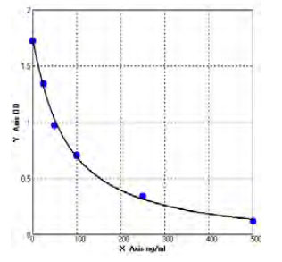
 Standard Curve (Sample)
Eotaxin, ELISA Kit (Cat# AAA12672)
Full Name
Porcine Eotaxin ELISA Kit
Gene Names
CCL11; SCYA11
Reactivity
Porcine
Pricing
 Standard Curve (Sample)
Cross linked C terminal Telopeptide of type I Collagen (CTX1), ELISA Kit (Cat# AAA17004)
Full Name
Monkey Cross linked C terminal Telopeptide of type I Collagen (CTX1) ELISA Kit
Reactivity
Monkey
Pricing
 Standard Curve (Sample)
Cross linked C terminal Telopeptide of type I Collagen (CTX1), ELISA Kit (Cat# AAA16095)
Full Name
Mouse Cross linked C terminal Telopeptide of type I Collagen (CTX1) ELISA Kit
Reactivity
Mouse
Pricing
 Standard Curve (Sample)
Cleaved CASP8 (Cleaved Caspase-8), ELISA Kit (Cat# AAA17768)
Full Name
Human Cleaved CASP8 (Cleaved Caspase-8)
Gene Names
CFLAR; CASH; FLIP; MRIT; CLARP; FLAME; Casper; FLAME1; c-FLIP; FLAME-1; I-FLICE; c-FLIPL; c-FLIPR; c-FLIPS; CASP8AP1
Reactivity
Human
Pricing
 Standard Curve (Sample)
Galectin-3-binding protein (LG3BP), ELISA Kit (Cat# AAA11336)
Full Name
Human Galectin-3-binding protein (LG3BP) ELISA Kit
Gene Names
Lgals3bp; mama; CyCAP; MAC2BP; Ppicap
Reactivity
Human
Pricing

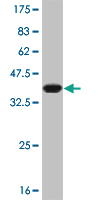
 SDS-PAGE
Mucin 5 Subtype AC (MUC5AC), Recombinant Protein (Cat# AAA20219)
Full Name
Recombinant Mucin 5 Subtype AC (MUC5AC)
Gene Names
Muc5ac; MGM; 2210005L13Rik
Reactivity
Mus musculus (Mouse)
Applications
WB, May be suitable for use in other assays to be determined by the end user.
Purity
> 90%
Pricing
 Standard Curve (Sample)
C-telopeptide of type II collagen, ELISA Kit (Cat# AAA16329)
Full Name
Guinea pig C-telopeptide of type II collagen ELISA Kit
Gene Names
COL2A1; AOM; ANFH; SEDC; STL1; COL11A3
Reactivity
Guinea Pig
Pricing

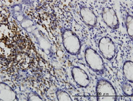
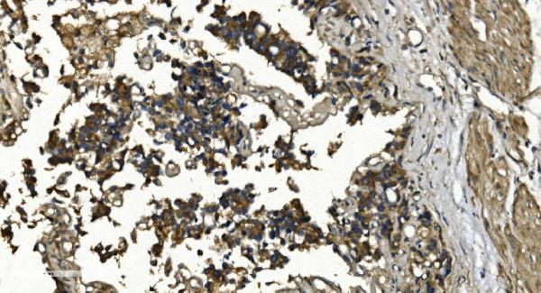
 IHC (Immunohistochemistry)
(Immunohistochemistry of paraffin-embedded Human colon cancer using CD192 Polyclonal Antibody at dilution of 1:20)
IHC (Immunohistochemistry)
(Immunohistochemistry of paraffin-embedded Human colon cancer using CD192 Polyclonal Antibody at dilution of 1:20)
CD192, Polyclonal Antibody (Cat# AAA22006)
Full Name
CD192 Polyclonal Antibody
Gene Names
CCR2; CKR2; CCR-2; CCR2A; CCR2B; CD192; CKR2A; CKR2B; CMKBR2; MCP-1-R; CC-CKR-2
Reactivity
Human, Mouse
Applications
EIA, WB
Purity
Affinity purification
Pricing
 SDS-PAGE
Outer membrane protein C, Recombinant Protein (Cat# AAA18709)
Full Name
Recombinant Escherichia coli (strain K12) Outer membrane protein C
Gene Names
ompC; butR; ECK2207; JW2203; meoA; par
Purity
Greater or equal to 85% purity as determined by SDS-PAGE.
Pricing


 WB (Western Blot)
(WB Suggested Anti-PHB Antibody Titration: 0.2-1 ug/mlPositive Control: Jurkat cell lysatePHB is supported by BioGPS gene expression data to be expressed in Jurkat)
WB (Western Blot)
(WB Suggested Anti-PHB Antibody Titration: 0.2-1 ug/mlPositive Control: Jurkat cell lysatePHB is supported by BioGPS gene expression data to be expressed in Jurkat)
PHB, Polyclonal Antibody (Cat# AAA23420)
Full Name
PHB antibody - C-terminal region
Gene Names
PHB; PHB1; HEL-215; HEL-S-54e
Reactivity
Cow, Dog, Guinea Pig, Horse, Human, Mouse, Rabbit, Rat, Zebrafish
Applications
WB, IHC
Purity
Affinity Purified
Pricing


 WB (Western Blot)
(WB Suggested Anti-YAP1 Antibody Titration: 1 ug/mlPositive Control: 721_B cell lysate)
WB (Western Blot)
(WB Suggested Anti-YAP1 Antibody Titration: 1 ug/mlPositive Control: 721_B cell lysate)
YAP1, Polyclonal Antibody (Cat# AAA23538)
Full Name
YAP1 antibody - C-terminal region
Gene Names
YAP1; YAP; YKI; COB1; YAP2; YAP65
Reactivity
Tested Reactivity: Human
Predicted Reactivity: Cow, Dog, Horse, Human, Mouse, Pig, Rabbit, Rat, Sheep, Zebrafish
Predicted Reactivity: Cow, Dog, Horse, Human, Mouse, Pig, Rabbit, Rat, Sheep, Zebrafish
Applications
IHC, WB
Purity
Affinity Purified
Pricing


 IHC (Immunohistchemistry)
(Figure 6. IHC analysis of NSF using anti-NSF antibody (AAA19136).NSF was detected in paraffin-embedded section of human mammary cancer tissue. Heat mediated antigen retrieval was performed in citrate buffer (pH6, epitope retrieval solution) for 20 mins. The tissue section was blocked with 10% goat serum. The tissue section was then incubated with 1ug/ml rabbit anti-NSF Antibody (AAA19136) overnight at 4 degree C. Biotinylated goat anti-rabbit IgG was used as secondary antibody and incubated for 30 minutes at 37 degree C. The tissue section was developed using Strepavidin-Biotin-Complex (SABC) with DAB as the chromogen.)
IHC (Immunohistchemistry)
(Figure 6. IHC analysis of NSF using anti-NSF antibody (AAA19136).NSF was detected in paraffin-embedded section of human mammary cancer tissue. Heat mediated antigen retrieval was performed in citrate buffer (pH6, epitope retrieval solution) for 20 mins. The tissue section was blocked with 10% goat serum. The tissue section was then incubated with 1ug/ml rabbit anti-NSF Antibody (AAA19136) overnight at 4 degree C. Biotinylated goat anti-rabbit IgG was used as secondary antibody and incubated for 30 minutes at 37 degree C. The tissue section was developed using Strepavidin-Biotin-Complex (SABC) with DAB as the chromogen.)
NSF, Polyclonal Antibody (Cat# AAA19136)
Full Name
Anti-NSF Picoband antibody
Gene Names
NSF; SKD2; SEC18
Reactivity
Human, Mouse, Rat
No cross reactivity with other proteins.
No cross reactivity with other proteins.
Applications
EIA, IHC, WB
Pricing


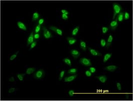
 IF (Immunofluorescence)
(Immunofluorescence analysis of U-2 OS cells using PFDN4 Polyclonal Antibody at dilution of 1:100. Blue: DAPI for nuclear staining.)
IF (Immunofluorescence)
(Immunofluorescence analysis of U-2 OS cells using PFDN4 Polyclonal Antibody at dilution of 1:100. Blue: DAPI for nuclear staining.)
PFDN4, Polyclonal Antibody (Cat# AAA22187)
Full Name
PFDN4 Polyclonal Antibody
Gene Names
PFDN4; C1; PFD4
Reactivity
Human, Mouse, Rat
Applications
IHC, IF
Purity
Affinity purification
Pricing
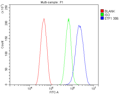


 FCM (Flow Cytometry)
(Figure 11. Flow Cytometry analysis of RH35 cells using anti- eRF1/ETF1 antibody (AAA19382).Overlay histogram showing RH35 cells stained with AAA19382 (Blue line). The cells were blocked with 10% normal goat serum. And then incubated with mouse anti-eRF1/ETF1 Antibody (AAA19382, 1μg/1x106 cells) for 30 min at 20 degree C. DyLight®488 conjugated goat anti-mouse IgG (BA1126, 5-10μg/1x106 cells) was used as secondary antibody for 30 minutes at 20 degree C. Isotype control antibody (Green line) was mouse IgG (1μg/1x106) used under the same conditions. Unlabelled sample (Red line) was also used as a control.)
FCM (Flow Cytometry)
(Figure 11. Flow Cytometry analysis of RH35 cells using anti- eRF1/ETF1 antibody (AAA19382).Overlay histogram showing RH35 cells stained with AAA19382 (Blue line). The cells were blocked with 10% normal goat serum. And then incubated with mouse anti-eRF1/ETF1 Antibody (AAA19382, 1μg/1x106 cells) for 30 min at 20 degree C. DyLight®488 conjugated goat anti-mouse IgG (BA1126, 5-10μg/1x106 cells) was used as secondary antibody for 30 minutes at 20 degree C. Isotype control antibody (Green line) was mouse IgG (1μg/1x106) used under the same conditions. Unlabelled sample (Red line) was also used as a control.)
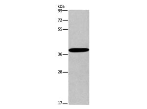
eRF1/ETF1, Monoclonal Antibody (Cat# AAA19382)
Full Name
Anti-eRF1/ETF1 Antibody (monoclonal, 3B6)
Gene Names
ETF1; ERF; RF1; ERF1; TB3-1; D5S1995; SUP45L1
Reactivity
Human, Mouse, Rat
Applications
WB, IHC-P, ICC, IF, FC/FACS/FCM
Purity
Immunogen affinity purified.
Pricing


 FCM (Flow Cytometry)
(Figure 8. Flow Cytometry analysis of THP-1 cells using anti-INPPL1 antibody (AAA19364).Overlay histogram showing THP-1 cells stained with AAA19364 (Blue line). The cells were blocked with 10% normal goat serum. And then incubated with mouse anti- INPPL1 Antibody (AAA19364, 1μg/1x106 cells) for 30 min at 20 degree C. DyLight®488 conjugated goat anti-mouse IgG (BA1126, 5-10μg/1x106 cells) was used as secondary antibody for 30 minutes at 20 degree C. Isotype control antibody (Green line) was mouse IgG (1μg/1x106) used under the same conditions. Unlabelled sample (Red line) was also used as a control.)
FCM (Flow Cytometry)
(Figure 8. Flow Cytometry analysis of THP-1 cells using anti-INPPL1 antibody (AAA19364).Overlay histogram showing THP-1 cells stained with AAA19364 (Blue line). The cells were blocked with 10% normal goat serum. And then incubated with mouse anti- INPPL1 Antibody (AAA19364, 1μg/1x106 cells) for 30 min at 20 degree C. DyLight®488 conjugated goat anti-mouse IgG (BA1126, 5-10μg/1x106 cells) was used as secondary antibody for 30 minutes at 20 degree C. Isotype control antibody (Green line) was mouse IgG (1μg/1x106) used under the same conditions. Unlabelled sample (Red line) was also used as a control.)
INPPL1, Monoclonal Antibody (Cat# AAA19364)
Full Name
Anti-INPPL1 Antibody (monoclonal, 8C13)
Gene Names
INPPL1; OPSMD; SHIP2
Reactivity
Human, Mouse, Rat
Applications
WB, IHC-P, ICC, IF, FC/FACS/FCM
Purity
Immunogen affinity purified.
Pricing
 Standard Curve (Sample)
FGF23, ELISA Kit (Cat# AAA17870)
Full Name
Human FGF23 ELISA Kit
Gene Names
FGF23; ADHR; FGFN; HYPF; HPDR2; PHPTC
Reactivity
Human
Applications
SE
Pricing
 Standard Curve (Sample)
insulin-like growth factor 1 (somatomedin C), ELISA Kit (Cat# AAA15145)
Full Name
Bovine Insulin-like growth factor 1, IGF-1 ELISA Kit
Gene Names
IGF1; IGF-1; IGF-I
Reactivity
Bovine
Pricing
 Standard Curve (Sample)
Lactoferrin, ELISA Kit (Cat# AAA16911)
Full Name
Sheep Lactoferrin ELISA Kit
Gene Names
LTF; LF; HLF2; GIG12; HEL110
Reactivity
Sheep
Pricing


 WB (Western Blot)
(DNAJC10 monoclonal antibody. Western Blot analysis of DNAJC10 expression in HeLa.)
WB (Western Blot)
(DNAJC10 monoclonal antibody. Western Blot analysis of DNAJC10 expression in HeLa.)
DNAJC10, Monoclonal Antibody (Cat# AAA24489)
Full Name
DNAJC10 (DnaJ Homolog Subfamily C Member 10, ER-resident Protein ERdj5, Macrothioredoxin, MTHr, ERDJ5, UNQ495/PRO1012, DKFZp434J1813, MGC104194) APC
Gene Names
DNAJC10; JPDI; MTHr; ERdj5; PDIA19
Reactivity
Human, Mouse, Rat
Applications
EIA, IF, IHC, WB
Purity
Purified by Protein A Affinity Chromatography.
Pricing

 IHC (Immunohistochemistry)
(Immunohistochemical analysis of Somatostatin staining in human brain formalin fixed paraffin embedded tissue section. The section was pre-treated using heat mediated antigen retrieval with sodium citrate buffer (pH 6.0). The section was then incubated with the antibody at room temperature and detected using an HRP conjugated compact polymer system. DAB was used as the chromogen. The section was then counterstained with haematoxylin and mounted with DPX.)
IHC (Immunohistochemistry)
(Immunohistochemical analysis of Somatostatin staining in human brain formalin fixed paraffin embedded tissue section. The section was pre-treated using heat mediated antigen retrieval with sodium citrate buffer (pH 6.0). The section was then incubated with the antibody at room temperature and detected using an HRP conjugated compact polymer system. DAB was used as the chromogen. The section was then counterstained with haematoxylin and mounted with DPX.)
Somatostatin, Polyclonal Antibody (Cat# AAA17844)
Full Name
Anti-Somatostatin Antibody
Gene Names
SST; SMST
Reactivity
Human, Mouse, Rat, Bovine, Chicken, Monkey, Pig, Sheep
Applications
WB, IHC
Purity
The antibody was purified by immunogen affinity chromatography.
Pricing
 Standard Curve (Sample)
amyloid beta peptide 1-42, ELISA Kit (Cat# AAA16487)
Full Name
Human amyloid beta peptide 1-42 ELISA Kit
Gene Names
App; Ag; Abpp; Adap; Cvap; Abeta; betaApp; E030013M08Rik
Reactivity
Human
Pricing


 IHC (Immunohistchemistry)
(Immunohistochemistry analysis using Mouse Anti-Hsp70 Monoclonal Antibody, Clone BB70. Tissue: hepatocytes. Species: Rat. Fixation: Paraffin Embedded. Primary Antibody: Mouse Anti-Hsp70 Monoclonal Antibody at 1:200. Liver sections were paraffin embedded. First pictures in series show two hours after exposure to stress, the second shows the control. Courtesy of: G. Matic, University of Belgrade, Serbia.)
IHC (Immunohistchemistry)
(Immunohistochemistry analysis using Mouse Anti-Hsp70 Monoclonal Antibody, Clone BB70. Tissue: hepatocytes. Species: Rat. Fixation: Paraffin Embedded. Primary Antibody: Mouse Anti-Hsp70 Monoclonal Antibody at 1:200. Liver sections were paraffin embedded. First pictures in series show two hours after exposure to stress, the second shows the control. Courtesy of: G. Matic, University of Belgrade, Serbia.)
Hsp70/Hsc70, Monoclonal Antibody (Cat# AAA17795)
Full Name
Hsp70/Hsc70 Antibody: RPE
Gene Names
HSPA2; HSP70
Reactivity
Human, Mouse, Rat, Sheep, Dog, Beluga, Bovine, Fish, Guinea pig, Scallop pig, Hamster, Rabbit, Chicken, Xenopus, Drosophila, Yeast
Applications
WB, IP, IHC
Pricing


 IHC (Immunohistochemistry)
(Figure 8. IHC analysis of VEGF Receptor 3 using anti-VEGF Receptor 3 antibody (AAA19146).VEGF Receptor 3 was detected in paraffin-embedded section of human mammary cancer tissue. Heat mediated antigen retrieval was performed in citrate buffer (pH6, epitope retrieval solution) for 20 mins. The tissue section was blocked with 10% goat serum. The tissue section was then incubated with 1ug/ml rabbit anti-VEGF Receptor 3 Antibody (AAA19146) overnight at 4 degree C. Biotinylated goat anti-rabbit IgG was used as secondary antibody and incubated for 30 minutes at 37 degree C. The tissue section was developed using Strepavidin-Biotin-Complex (SABC) with DAB as the chromogen.)
IHC (Immunohistochemistry)
(Figure 8. IHC analysis of VEGF Receptor 3 using anti-VEGF Receptor 3 antibody (AAA19146).VEGF Receptor 3 was detected in paraffin-embedded section of human mammary cancer tissue. Heat mediated antigen retrieval was performed in citrate buffer (pH6, epitope retrieval solution) for 20 mins. The tissue section was blocked with 10% goat serum. The tissue section was then incubated with 1ug/ml rabbit anti-VEGF Receptor 3 Antibody (AAA19146) overnight at 4 degree C. Biotinylated goat anti-rabbit IgG was used as secondary antibody and incubated for 30 minutes at 37 degree C. The tissue section was developed using Strepavidin-Biotin-Complex (SABC) with DAB as the chromogen.)
VEGF Receptor 3, Polyclonal Antibody (Cat# AAA19146)
Full Name
Anti-VEGF Receptor 3 Picoband Antibody
Gene Names
FLT4; PCL; FLT-4; FLT41; LMPH1A; VEGFR3; VEGFR-3
Reactivity
Human, Mouse, Rat
No cross reactivity with other proteins.
No cross reactivity with other proteins.
Applications
IHC, WB
Purity
Immunogen affinity purified
Pricing


 FCM (Flow Cytometry)
(Figure 9. Flow Cytometry analysis of U251 cells using anti-RAB1B antibody (AAA19296).Overlay histogram showing U251 cells stained with AAA19296 (Blue line). The cells were blocked with 10% normal goat serum. And then incubated with rabbit anti-RAB1B Antibody (AAA19296, 1μg/1x106 cells) for 30 min at 20 degree C. DyLight®488 conjugated goat anti-rabbit IgG (5-10μg/1x106 cells) was used as secondary antibody for 30 minutes at 20 degree C. Isotype control antibody (Green line) was rabbit IgG (1μg/1x106) used under the same conditions. Unlabelled sample (Red line) was also used as a control.)
FCM (Flow Cytometry)
(Figure 9. Flow Cytometry analysis of U251 cells using anti-RAB1B antibody (AAA19296).Overlay histogram showing U251 cells stained with AAA19296 (Blue line). The cells were blocked with 10% normal goat serum. And then incubated with rabbit anti-RAB1B Antibody (AAA19296, 1μg/1x106 cells) for 30 min at 20 degree C. DyLight®488 conjugated goat anti-rabbit IgG (5-10μg/1x106 cells) was used as secondary antibody for 30 minutes at 20 degree C. Isotype control antibody (Green line) was rabbit IgG (1μg/1x106) used under the same conditions. Unlabelled sample (Red line) was also used as a control.)
RAB1B, Polyclonal Antibody (Cat# AAA19296)
Full Name
Anti-RAB1B Antibody
Reactivity
Human, Mouse, Rat
Applications
WB, IHC-P, ICC, IF, FC/FACS/FCM
Purity
Immunogen affinity purified.
Pricing
 Standard Curve (Sample)
Transglutaminase, ELISA Kit (Cat# AAA15969)
Full Name
Mouse Transglutaminase ELISA Kit
Reactivity
Mouse
Pricing
 Standard Curve (Sample)
Cystatin C, ELISA Kit (Cat# AAA17536)
Full Name
Porcine Cystatin C ELISA Kit
Gene Names
CST3; ARMD11; HEL-S-2
Reactivity
Porcine
Pricing
 Standard Curve (Sample)
Interleukin 8, ELISA Kit (Cat# AAA17520)
Full Name
Human Interleukin 8 ELISA Kit
Gene Names
CXCL8; IL8; NAF; GCP1; LECT; LUCT; NAP1; GCP-1; LYNAP; MDNCF; MONAP; NAP-1
Reactivity
Human
Pricing
 Standard Curve (Sample)
apolipoprotein C-II, ELISA Kit (Cat# AAA18200)
Full Name
Mouse Apolipoprotein C-II, APOC2 ELISA Kit
Reactivity
Mouse
Pricing
 Standard Curve (Sample)
ATP-binding cassette sub-family C member 8 (ABCC8), ELISA Kit (Cat# AAA27090)
Full Name
Rat ATP-binding cassette sub-family C member 8 (ABCC8) ELISA Kit
Gene Names
Abcc8; Sur; Sur1
Reactivity
Rat
Pricing
 Standard Curve (Sample)
Platelet Factor 4, ELISA Kit (Cat# AAA16087)
Full Name
Human Platelet Factor 4 ELISA Kit
Gene Names
PF4; PF-4; CXCL4; SCYB4
Reactivity
Human
Pricing


 WB (Western Blot)
(NME2 monoclonal antibody (M08), clone 1F2. Western Blot analysis of NME2 expression in Raw 264.7 (Cat # L024V1).)
WB (Western Blot)
(NME2 monoclonal antibody (M08), clone 1F2. Western Blot analysis of NME2 expression in Raw 264.7 (Cat # L024V1).)
NME2, Monoclonal Antibody (Cat# AAA26567)
Full Name
NME2 (Non-Metastatic Cells 2, Protein (NM23B) Expressed in, MGC111212, NDPK-B, NDPKB, NM23-H2, NM23B, puf) (PE)
Gene Names
NME2; PUF; NDKB; NDPKB; NM23B; NDPK-B; NM23-H2
Applications
EIA, IF, WB
Purity
Purified
Pricing


 Application Data
(Detection limit for recombinant GST tagged TFAP4 is approximately 0.1ng/ml as a capture antibody.)
Application Data
(Detection limit for recombinant GST tagged TFAP4 is approximately 0.1ng/ml as a capture antibody.)
TFAP4, Monoclonal Antibody (Cat# AAA26599)
Full Name
TFAP4 (Transcription Factor AP-4 (Activating Enhancer Binding Protein 4), AP-4, bHLHc41) (PE)
Gene Names
TFAP4; AP-4; bHLHc41
Applications
IF, IHC, WB
Purity
Purified
Pricing


 WB (Western Blot)
(CSK monoclonal antibody. Western Blot analysis of CSK expression in Hela NE.)
WB (Western Blot)
(CSK monoclonal antibody. Western Blot analysis of CSK expression in Hela NE.)
CSK, Monoclonal Antibody (Cat# AAA25066)
Full Name
CSK (Tyrosine-protein Kinase CSK, C-SRC Kinase, Protein-tyrosine Kinase CYL) (FITC)
Reactivity
Human
Applications
EIA, IHC, IP, WB
Purity
Purified by Protein A Affinity Chromatography.
Pricing
 Standard Curve (Sample)
TOM1-Like Protein 2 (TOM1L2), ELISA Kit (Cat# AAA31483)
Full Name
Human TOM1-Like Protein 2 (TOM1L2) ELISA Kit
Reactivity
Human
Pricing


 WB (Western Blot)
(DNAJC10 monoclonal antibody. Western Blot analysis of DNAJC10 expression in HeLa.)
WB (Western Blot)
(DNAJC10 monoclonal antibody. Western Blot analysis of DNAJC10 expression in HeLa.)
DNAJC10, Monoclonal Antibody (Cat# AAA25081)
Full Name
DNAJC10 (DnaJ Homolog Subfamily C Member 10, ER-resident Protein ERdj5, Macrothioredoxin, MTHr, ERDJ5, UNQ495/PRO1012, DKFZp434J1813, MGC104194) (FITC)
Gene Names
DNAJC10; JPDI; MTHr; ERdj5; PDIA19
Reactivity
Human, Mouse, Rat
Applications
EIA, IF, IHC, WB
Purity
Purified by Protein A Affinity Chromatography.
Pricing
 Standard Curve (Sample)
Proto-oncogene c-Fos, ELISA Kit (Cat# AAA23339)
Full Name
Mouse Proto-oncogene c-Fos ELISA Kit
Gene Names
Fos; cFos; c-fos; D12Rfj1
Reactivity
Mouse
Pricing
 Standard Curve (Sample)
Cross Linked C-Telopeptide Of Type II Collagen (CTXII), ELISA Kit (Cat# AAA20563)
Full Name
Human Cross Linked C-Telopeptide Of Type II Collagen (CTXII) ELISA Kit
Reactivity
Human
Pricing


 FCM (Flow Cytometry)
(Figure 7. Flow Cytometry analysis of MCF-7 cells using anti-RGS6 antibody (AAA19308).Overlay histogram showing MCF-7 cells stained with AAA19308 (Blue line). The cells were blocked with 10% normal goat serum. And then incubated with rabbit anti-RGS6 Antibody (AAA19308, 1μg/1x106 cells) for 30 min at 20 degree C. DyLight®488 conjugated goat anti-rabbit IgG (5-10μg/1x106 cells) was used as secondary antibody for 30 minutes at 20 degree C. Isotype control antibody (Green line) was rabbit IgG (1μg/1x106) used under the same conditions. Unlabelled sample (Red line) was also used as a control.)
FCM (Flow Cytometry)
(Figure 7. Flow Cytometry analysis of MCF-7 cells using anti-RGS6 antibody (AAA19308).Overlay histogram showing MCF-7 cells stained with AAA19308 (Blue line). The cells were blocked with 10% normal goat serum. And then incubated with rabbit anti-RGS6 Antibody (AAA19308, 1μg/1x106 cells) for 30 min at 20 degree C. DyLight®488 conjugated goat anti-rabbit IgG (5-10μg/1x106 cells) was used as secondary antibody for 30 minutes at 20 degree C. Isotype control antibody (Green line) was rabbit IgG (1μg/1x106) used under the same conditions. Unlabelled sample (Red line) was also used as a control.)
RGS6, Polyclonal Antibody (Cat# AAA19308)
Full Name
Anti-RGS6 Antibody
Gene Names
RGS6; GAP
Reactivity
Human, Mouse, Rat
Applications
WB, IHC-P, FC/FACS/FCM, EIA
Purity
Immunogen affinity purified.
Pricing
 Standard Curve (Sample)
cluster of differentiation 45 (CD45), ELISA Kit (Cat# AAA27241)
Full Name
Human cluster of differentiation 45 (CD45) ELISA Kit
Gene Names
PTPRC; LCA; LY5; B220; CD45; L-CA; T200; CD45R; GP180
Reactivity
Human
Pricing


 WB (Western Blot)
(USF2 monoclonal antibody (M03), clone 6A9. Western Blot analysis of USF2 expression in PC-12 (Cat # L012V1).)
WB (Western Blot)
(USF2 monoclonal antibody (M03), clone 6A9. Western Blot analysis of USF2 expression in PC-12 (Cat # L012V1).)
USF2, Monoclonal Antibody (Cat# AAA26109)
Full Name
USF2 (Upstream Transcription Factor 2, c-fos Interacting, FIP, bHLHb12) (APC)
Gene Names
USF2; FIP; bHLHb12
Applications
IF, WB
Purity
Purified
Pricing
 Standard Curve (Sample)
C telopeptide of type II collagen, ELISA Kit (Cat# AAA16461)
Full Name
Human C telopeptide of type II collagen ELISA Kit
Gene Names
COL2A1; AOM; ANFH; SEDC; STL1; COL11A3
Reactivity
Human
Pricing


 DB (Dot Blot)
(Dot blot analysis using Rabbit Anti-Amyloid Fibrils (OC) Polyclonal Antibody (SPC-507). Tissue: Cell lysates. Species: Human. Primary Antibody: Rabbit Anti-Amyloid Fibrils (OC) Polyclonal Antibody (SPC-507) at 1:500, 1:5000. Beta Amyloid HEPES-NaCl aggregation, showing 1:500 (L) and 1:5000 (R) time lapse dot blot.)
DB (Dot Blot)
(Dot blot analysis using Rabbit Anti-Amyloid Fibrils (OC) Polyclonal Antibody (SPC-507). Tissue: Cell lysates. Species: Human. Primary Antibody: Rabbit Anti-Amyloid Fibrils (OC) Polyclonal Antibody (SPC-507) at 1:500, 1:5000. Beta Amyloid HEPES-NaCl aggregation, showing 1:500 (L) and 1:5000 (R) time lapse dot blot.)
Amyloid Fibrils (OC), Polyclonal Antibody (Cat# AAA17804)
Full Name
Amyloid Fibrils (OC) Antibody
Reactivity
Human. Potentially mouse and rat based on species homology.
Applications
IP, ICC, IHC, EIA, WB, DB
Pricing
 Standard Curve (Sample)
Ubiquitin, ELISA Kit (Cat# AAA16333)
Full Name
Human Ubiquitin ELISA Kit
Gene Names
RPS27A; UBC; S27A; CEP80; UBA80; HEL112; UBCEP1; UBCEP80
Reactivity
Human
Pricing
 SDS-PAGE
Parvalbumin beta, Recombinant Protein (Cat# AAA18634)
Full Name
Recombinant Gadus morhua subsp. callarias Parvalbumin beta
Purity
Greater or equal to 85% purity as determined by SDS-PAGE.
Pricing

 WB (Western Blot)
(Western blot analysis of extracts from COLO205 cells using Src (Ab-418) antibody and Src (phospho-Tyr418) antibody.)
WB (Western Blot)
(Western blot analysis of extracts from COLO205 cells using Src (Ab-418) antibody and Src (phospho-Tyr418) antibody.)
Src, Antibody (Cat# AAA17987)
Full Name
Src (Phospho-Tyr418) Antibody
Gene Names
SRC; ASV; SRC1; THC6; c-SRC; p60-Src
Reactivity
Human, Mouse, Rat
Applications
WB, IHC
Purity
Affinity-purified from rabbit antiserum by affinity-chromatography using epitope-specific phosphopeptide. The antibody against non-phosphopeptide was removed by chromatography using non-phosphopeptide corresponding to the phosphorylation site.
Pricing


 WB (Western Blot)
(WB Suggested Anti-STK3 Antibody Titration: 5.0ug/mlPositive Control: HepG2 cell lysate)
WB (Western Blot)
(WB Suggested Anti-STK3 Antibody Titration: 5.0ug/mlPositive Control: HepG2 cell lysate)
STK3, Polyclonal Antibody (Cat# AAA23535)
Full Name
STK3 antibody - C-terminal region
Gene Names
STK3; KRS1; MST2
Reactivity
Cow, Dog, Guinea Pig, Horse, Human, Mouse, Rabbit, Rat, Zebrafish
Applications
IHC, WB
Purity
Protein A purified
Pricing


 FCM (Flow Cytometry)
(Figure 6. Flow Cytometry analysis of THP-1 cells using anti-PRMT8 antibody (AAA19321).Overlay histogram showing THP-1 cells stained with AAA19321 (Blue line). The cells were blocked with 10% normal goat serum. And then incubated with rabbit anti-PRMT8 Antibody (AAA19321,1μg/1x106 cells) for 30 min at 20 degree C. DyLight®488 conjugated goat anti-rabbit IgG (5-10μg/1x106 cells) was used as secondary antibody for 30 minutes at 20 degree C. Isotype control antibody (Green line) was rabbit IgG (1μg/1x106) used under the same conditions. Unlabelled sample (Red line) was also used as a control.)
FCM (Flow Cytometry)
(Figure 6. Flow Cytometry analysis of THP-1 cells using anti-PRMT8 antibody (AAA19321).Overlay histogram showing THP-1 cells stained with AAA19321 (Blue line). The cells were blocked with 10% normal goat serum. And then incubated with rabbit anti-PRMT8 Antibody (AAA19321,1μg/1x106 cells) for 30 min at 20 degree C. DyLight®488 conjugated goat anti-rabbit IgG (5-10μg/1x106 cells) was used as secondary antibody for 30 minutes at 20 degree C. Isotype control antibody (Green line) was rabbit IgG (1μg/1x106) used under the same conditions. Unlabelled sample (Red line) was also used as a control.)
PRMT8, Polyclonal Antibody (Cat# AAA19321)
Full Name
Anti-PRMT8 Antibody
Gene Names
PRMT8; HRMT1L3; HRMT1L4
Reactivity
Human, Rat
Applications
WB, IHC-P, FC/FACS/FCM, EIA
Purity
Immunogen affinity purified.
Pricing


 FCM (Flow Cytometry)
(Figure 12. Flow Cytometry analysis of U937 cells using anti-DYNLL1/PIN antibody (AAA19276).Overlay histogram showing U937 cells stained with AAA19276 (Blue line). The cells were blocked with 10% normal goat serum. And then incubated with rabbit anti-DYNLL1/PIN Antibody (AAA19276, 1μg/1x106 cells) for 30 min at 20 degree C. DyLight®488 conjugated goat anti-rabbit IgG (5-10μg/1x106 cells) was used as secondary antibody for 30 minutes at 20 degree C. Isotype control antibody (Green line) was rabbit IgG (1μg/1x106) used under the same conditions. Unlabelled sample (Red line) was also used as a control.)
FCM (Flow Cytometry)
(Figure 12. Flow Cytometry analysis of U937 cells using anti-DYNLL1/PIN antibody (AAA19276).Overlay histogram showing U937 cells stained with AAA19276 (Blue line). The cells were blocked with 10% normal goat serum. And then incubated with rabbit anti-DYNLL1/PIN Antibody (AAA19276, 1μg/1x106 cells) for 30 min at 20 degree C. DyLight®488 conjugated goat anti-rabbit IgG (5-10μg/1x106 cells) was used as secondary antibody for 30 minutes at 20 degree C. Isotype control antibody (Green line) was rabbit IgG (1μg/1x106) used under the same conditions. Unlabelled sample (Red line) was also used as a control.)
DYNLL1/PIN, Polyclonal Antibody (Cat# AAA19276)
Full Name
Anti-DYNLL1/PIN Antibody
Gene Names
DYNLL1; LC8; PIN; DLC1; DLC8; LC8a; DNCL1; hdlc1; DNCLC1
Reactivity
Human, Mouse, Rat
Applications
WB, IHC-P, ICC, IF, FC/FACS/FCM, EIA
Purity
Immunogen affinity purified.
Pricing
 Standard Curve (Sample)
Lactoferrin (LTF), ELISA Kit (Cat# AAA13076)
Full Name
Chicken Lactoferrin (LTF) ELISA Kit
Gene Names
LTF; LF; HLF2; GIG12; HEL110
Reactivity
Chicken
Pricing


 ICC (Immunocytochemistry)
(ICC staining PRTN3 in SH-SY5Y cells (green). The nuclear counter stain is DAPI (blue). Cells were fixed in paraformaldehyde, permeabilised with 0.25% Triton X100/PBS.)
ICC (Immunocytochemistry)
(ICC staining PRTN3 in SH-SY5Y cells (green). The nuclear counter stain is DAPI (blue). Cells were fixed in paraformaldehyde, permeabilised with 0.25% Triton X100/PBS.)
PRTN3, Polyclonal Antibody (Cat# AAA29923)
Full Name
PRTN3 Antibody
Gene Names
PRTN3; MBN; MBT; NP4; P29; PR3; ACPA; AGP7; NP-4; PR-3; CANCA; C-ANCA
Reactivity
Human, Mouse
Applications
WB, ICC, IHC
Purity
ProA affinity purified
Pricing