Filters
Clonality
Type
Reactivity
Gene Name
Isotype
Host
Application
Clone
2811 results for " C" - showing 1700-1750
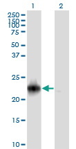
 SDS-PAGE
Gasdermin-C (GSDMC), Recombinant Protein (Cat# AAA18751)
Full Name
Recombinant Human Gasdermin-C (GSDMC)
Gene Names
GSDMC; MLZE
Purity
Greater or equal to 85% purity as determined by SDS-PAGE.
Pricing
 Standard Curve (Sample)
Tight junction protein ZO-1, ELISA Kit (Cat# AAA22701)
Full Name
Porcine Tight junction protein ZO-1 (TJP1) ELISA Kit
Gene Names
TJP1; ZO-1
Reactivity
Porcine
Pricing
 Standard Curve (Sample)
Endothelin-1, ELISA Kit (Cat# AAA23217)
Full Name
Human Endothelin-1 ELISA Kit
Gene Names
EDN1; ET1; QME; PPET1; ARCND3; HDLCQ7
Reactivity
Human
Pricing
 Standard Curve (Sample)
Galactocerebrosidase, ELISA Kit (Cat# AAA17311)
Full Name
Human Galactocerebrosidase ELISA Kit
Reactivity
Human
Pricing
 Standard Curve (Sample)
insulin-like growth factor 1 (somatomedin C), ELISA Kit (Cat# AAA15142)
Full Name
Pig insulin-like growth factors-1, IGF-1 ELISA Kit
Gene Names
IGF1; IGF-I; Npt2B
Reactivity
Pig
Pricing
 Standard Curve (Sample)
C Reactive Protein, ELISA Kit (Cat# AAA16884)
Full Name
Monkey C Reactive Protein ELISA Kit
Gene Names
CRP; PTX1
Reactivity
Monkey
Pricing


 IHC (Immunohistchemistry)
(Immunoperoxidase of monoclonal antibody to USF2 on formalin-fixed paraffin-embedded human esophagus. [antibody concentration 1 ug/ml])
IHC (Immunohistchemistry)
(Immunoperoxidase of monoclonal antibody to USF2 on formalin-fixed paraffin-embedded human esophagus. [antibody concentration 1 ug/ml])
USF2, Monoclonal Antibody (Cat# AAA26361)
Full Name
USF2 (Upstream Transcription Factor 2, c-fos Interacting, FIP, bHLHb12) (FITC)
Gene Names
USF2; FIP; bHLHb12
Applications
IF, IHC, WB
Purity
Purified
Pricing
 Standard Curve (Sample)
Intact Fibroblast Growth Factor 23, ELISA Kit (Cat# AAA17145)
Full Name
Mouse Intact Fibroblast Growth Factor 23 ELISA Kit
Gene Names
FGF23; ADHR; FGFN; HYPF; HPDR2; PHPTC
Reactivity
Mouse
Pricing


 WB (Western Blot)
(RRAS2 monoclonal antibody Western Blot analysis of RRAS2 expression in A-431.)
WB (Western Blot)
(RRAS2 monoclonal antibody Western Blot analysis of RRAS2 expression in A-431.)
RRAS2, Monoclonal Antibody (Cat# AAA14777)
Full Name
RRAS2 (Ras-related Protein R-Ras2, Ras-like Protein TC21, Teratocarcinoma Oncogene, TC21)
Gene Names
RRAS2; TC21
Reactivity
Human, Mouse, Rat
Applications
EL/EIA, WB, IHC, IF
Purity
Affinity Purified
Purified by Protein A affinity chromatography.
Purified by Protein A affinity chromatography.
Pricing


 Application Data
(Detection limit for recombinant GST tagged UBE2C is ~0.03ng/ml as a capture antibody.)
Application Data
(Detection limit for recombinant GST tagged UBE2C is ~0.03ng/ml as a capture antibody.)
UBE2C, Monoclonal Antibody (Cat# AAA24991)
Full Name
UBE2C (Ubiquitin-conjugating Enzyme E2 C, UbcH10, Ubiquitin Carrier Protein C, Ubiquitin-protein Ligase C, dJ447F3.2) (Biotin)
Gene Names
UBE2C; UBCH10; dJ447F3.2
Reactivity
Human
Applications
EIA, IF, IHC, WB
Purity
Purified by Protein A Affinity Chromatography.
Pricing


 IHC (Immunohistchemistry)
(Figure 6. IHC analysis of AMD1 using anti-AMD1 antibody (AAA19177).AMD1 was detected in paraffin-embedded section of human mammary cancer tissue. Heat mediated antigen retrieval was performed in citrate buffer (pH6, epitope retrieval solution) for 20 mins. The tissue section was blocked with 10% goat serum. The tissue section was then incubated with 1ug/ml rabbit anti-AMD1 Antibody (AAA19177) overnight at 4 degree C. Biotinylated goat anti-rabbit IgG was used as secondary antibody and incubated for 30 minutes at 37 degree C. The tissue section was developed using Strepavidin-Biotin-Complex (SABC) with DAB as the chromogen.)
IHC (Immunohistchemistry)
(Figure 6. IHC analysis of AMD1 using anti-AMD1 antibody (AAA19177).AMD1 was detected in paraffin-embedded section of human mammary cancer tissue. Heat mediated antigen retrieval was performed in citrate buffer (pH6, epitope retrieval solution) for 20 mins. The tissue section was blocked with 10% goat serum. The tissue section was then incubated with 1ug/ml rabbit anti-AMD1 Antibody (AAA19177) overnight at 4 degree C. Biotinylated goat anti-rabbit IgG was used as secondary antibody and incubated for 30 minutes at 37 degree C. The tissue section was developed using Strepavidin-Biotin-Complex (SABC) with DAB as the chromogen.)
AMD1/Adometdc, Polyclonal Antibody (Cat# AAA19177)
Full Name
Anti-AMD1/Adometdc Antibody
Gene Names
AMD1; AMD; SAMDC; ADOMETDC
Reactivity
Human, Mouse, Rat
No cross reactivity with other proteins.
No cross reactivity with other proteins.
Applications
IHC, WB
Purity
Immunogen affinity purified
Pricing


 IF (Immunofluorescence)
(Immunofluorescence Analysis of PFA fixed U87 cells labeling VCL-Monospecific Mouse Monoclonal Antibody (VCL/3617) followed by Goat anti-mouse IgG-CF488 (Green).)
IF (Immunofluorescence)
(Immunofluorescence Analysis of PFA fixed U87 cells labeling VCL-Monospecific Mouse Monoclonal Antibody (VCL/3617) followed by Goat anti-mouse IgG-CF488 (Green).)
Vinculin, Monoclonal Antibody (Cat# AAA23953)
Full Name
Vinculin (Marker of Age-related Macular Degeneration)
Gene Names
VCL; MV; MVCL; CMD1W; CMH15; HEL114
Reactivity
Human, Mouse, Rat, Cow, Pig, Rabbit, Frog, Fish, Bird
Applications
FC/FACS, IF, IHC
Purity
Purified Ab with BSA and Azide at 200ug/ml or Purified Ab with BSA and Azide at 200ug/ml or Purified Ab WITHOUT BSA and Azide at 1.0mg/ml
Pricing


 FCM (Flow Cytometry)
(Figure 6. Flow Cytometry analysis of A431 cells using anti-TMPRSS3 antibody (AAA19289).Overlay histogram showing A431 cells stained with AAA19289 (Blue line). The cells were blocked with 10% normal goat serum. And then incubated with rabbit anti-TMPRSS3 Antibody (AAA19289, 1μg/1x106 cells) for 30 min at 20 degree C. DyLight®488 conjugated goat anti-rabbit IgG (5-10μg/1x106 cells) was used as secondary antibody for 30 minutes at 20 degree C. Isotype control antibody (Green line) was rabbit IgG (1μg/1x106) used under the same conditions. Unlabelled sample (Red line) was also used as a control.)
FCM (Flow Cytometry)
(Figure 6. Flow Cytometry analysis of A431 cells using anti-TMPRSS3 antibody (AAA19289).Overlay histogram showing A431 cells stained with AAA19289 (Blue line). The cells were blocked with 10% normal goat serum. And then incubated with rabbit anti-TMPRSS3 Antibody (AAA19289, 1μg/1x106 cells) for 30 min at 20 degree C. DyLight®488 conjugated goat anti-rabbit IgG (5-10μg/1x106 cells) was used as secondary antibody for 30 minutes at 20 degree C. Isotype control antibody (Green line) was rabbit IgG (1μg/1x106) used under the same conditions. Unlabelled sample (Red line) was also used as a control.)
TMPRSS3, Polyclonal Antibody (Cat# AAA19289)
Full Name
Anti-TMPRSS3 Antibody
Gene Names
TMPRSS3; DFNB8; DFNB10; ECHOS1; TADG12
Reactivity
Human, Mouse, Rat
Applications
WB, IHC-P, FC/FACS/FCM, EIA
Purity
Immunogen affinity purified.
Pricing


 FCM (Flow Cytometry)
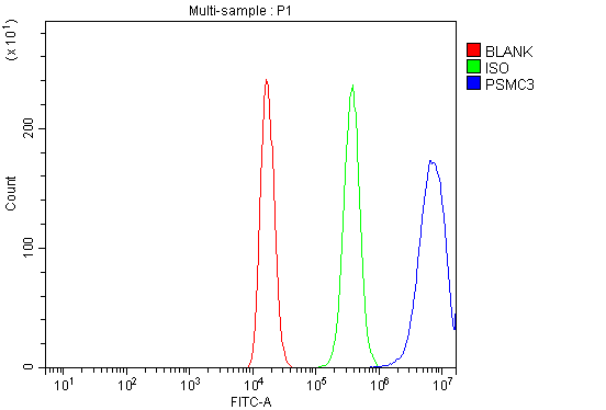
(Figure 7. Flow Cytometry analysis of 293T cells using anti-TBP-1/PSMC3 antibody (AAA19314).Overlay histogram showing 293T cells stained with AAA19314 (Blue line). The cells were blocked with 10% normal goat serum. And then incubated with rabbit anti-TBP-1/PSMC3 Antibody (AAA19314, 1μg/1x106 cells) for 30 min at 20 degree C. DyLight®488 conjugated goat anti-rabbit IgG (5-10μg/1x106 cells) was used as secondary antibody for 30 minutes at 20 degree C. Isotype control antibody (Green line) was rabbit IgG (1μg/1x106) used under the same conditions. Unlabelled sample (Red line) was also used as a control.)
FCM (Flow Cytometry)
(Figure 7. Flow Cytometry analysis of 293T cells using anti-TBP-1/PSMC3 antibody (AAA19314).Overlay histogram showing 293T cells stained with AAA19314 (Blue line). The cells were blocked with 10% normal goat serum. And then incubated with rabbit anti-TBP-1/PSMC3 Antibody (AAA19314, 1μg/1x106 cells) for 30 min at 20 degree C. DyLight®488 conjugated goat anti-rabbit IgG (5-10μg/1x106 cells) was used as secondary antibody for 30 minutes at 20 degree C. Isotype control antibody (Green line) was rabbit IgG (1μg/1x106) used under the same conditions. Unlabelled sample (Red line) was also used as a control.)
TBP-1/PSMC3, Polyclonal Antibody (Cat# AAA19314)
Full Name
Anti-TBP-1/PSMC3 Antibody
Gene Names
PSMC3; TBP1
Reactivity
Human, Mouse, Rat
Applications
WB, IHC-P, FC/FACS/FCM, EIA
Purity
Immunogen affinity purified.
Pricing


 FCM (Flow Cytometry)
(Figure 6. Flow Cytometry analysis of U937 cells using anti-BDH1 antibody (AAA19328).Overlay histogram showing U937 cells stained with AAA19328 (Blue line). The cells were blocked with 10% normal goat serum. And then incubated with rabbit anti-BDH1 Antibody (AAA19328,1μg/1x106 cells) for 30 min at 20 degree C. DyLight®488 conjugated goat anti-rabbit IgG (5-10μg/1x106 cells) was used as secondary antibody for 30 minutes at 20 degree C. Isotype control antibody (Green line) was rabbit IgG (1μg/1x106) used under the same conditions. Unlabelled sample (Red line) was also used as a control.)
FCM (Flow Cytometry)
(Figure 6. Flow Cytometry analysis of U937 cells using anti-BDH1 antibody (AAA19328).Overlay histogram showing U937 cells stained with AAA19328 (Blue line). The cells were blocked with 10% normal goat serum. And then incubated with rabbit anti-BDH1 Antibody (AAA19328,1μg/1x106 cells) for 30 min at 20 degree C. DyLight®488 conjugated goat anti-rabbit IgG (5-10μg/1x106 cells) was used as secondary antibody for 30 minutes at 20 degree C. Isotype control antibody (Green line) was rabbit IgG (1μg/1x106) used under the same conditions. Unlabelled sample (Red line) was also used as a control.)
BDH1, Polyclonal Antibody (Cat# AAA19328)
Full Name
Anti-BDH1 Antibody
Gene Names
BDH1; BDH; SDR9C1
Reactivity
Human, Rat
Applications
WB, IHC-P, ICC, IF, FC/FACS/FCM, EIA
Purity
Immunogen affinity purified.
Pricing
 Standard Curve (Sample)
Cross Linked C-Telopeptide Of Type I Collagen (CTX-I), ELISA Kit (Cat# AAA12644)
Full Name
Mouse Cross Linked C-Telopeptide Of Type I Collagen (CTX-I) ELISA Kit
Reactivity
Mouse
Pricing
 Standard Curve (Sample)
PIIICP, ELISA Kit (Cat# AAA21560)
Full Name
Mouse PIIICP (c-terminal Procollagen III propeptide) ELISA Kit
Reactivity
Mouse
Pricing
 Standard Curve (Sample)
Vitamin C, ELISA Kit (Cat# AAA16838)
Full Name
Chicken Vitamin C ELISA Kit
Reactivity
Chicken
Pricing
 Standard Curve (Sample)
MCP1, ELISA Kit (Cat# AAA17863)
Full Name
Rat MCP1 ELISA Kit
Gene Names
Ccl2; MCP-1; Scya2; Sigje
Reactivity
Rat
Applications
SE
Pricing
 Standard Curve (Sample)
SDF-1, ELISA Kit (Cat# AAA21894)
Full Name
Rat SDF-1 (Stromal Cell Derived Factor 1) ELISA Kit
Gene Names
cxcl12; sdf1; sdf-1; xSDF-1; xSDF-1alpha
Reactivity
Rat
Pricing
 Standard Curve (Sample)
Macrophage inflammatory protein-3alpha, ELISA Kit (Cat# AAA16047)
Full Name
Rabbit Macrophage inflammatory protein-3alpha ELISA Kit
Gene Names
Ccl20; CKb4; LARC; ST38; MIP3A; MIP-3A; Scya20; MIP-3[a]; exodus-1
Reactivity
Rabbit
Pricing


 Application Data
(Detection limit for recombinant GST tagged TFAP4 is approximately 0.1ng/ml as a capture antibody.)
Application Data
(Detection limit for recombinant GST tagged TFAP4 is approximately 0.1ng/ml as a capture antibody.)
TFAP4, Monoclonal Antibody (Cat# AAA26334)
Full Name
TFAP4 (Transcription Factor AP-4 (Activating Enhancer Binding Protein 4), AP-4, bHLHc41) (FITC)
Gene Names
TFAP4; AP-4; bHLHc41
Applications
IF, IHC, WB
Purity
Purified
Pricing


 Application Data
(Proximity Ligation Analysis of protein-protein interactions between PDGFRB and PLCG1. Mahlavu cells were stained with anti-PDGFRB rabbit purified polyclonal 1:600 and anti-PLCG1 mouse monoclonal antibody 1:100. Each red dot represents the detection of protein-protein interaction complex, and nuclei were counterstained with DAPI (blue).)
Application Data
(Proximity Ligation Analysis of protein-protein interactions between PDGFRB and PLCG1. Mahlavu cells were stained with anti-PDGFRB rabbit purified polyclonal 1:600 and anti-PLCG1 mouse monoclonal antibody 1:100. Each red dot represents the detection of protein-protein interaction complex, and nuclei were counterstained with DAPI (blue).)
Phospholipase C, gamma 1, Monoclonal Antibody (Cat# AAA25793)
Full Name
Phospholipase C, gamma 1 (Phospholipase C-gamma-1, PLCgamma1, PLC-gamma-1, PLCg1, NCKAP3, 1-Phosphatidylinositol-4,5-bisphosphate Phosphodiesterase gamma-1, Phospholipase C-II, PLC-II, Phosphoinositide Phospholipase C-gamma-1, PLC1, PLC148, PLC-148) (PE)
Gene Names
PLCG1; PLC1; NCKAP3; PLC-II; PLC148; PLCgamma1
Reactivity
Human
Applications
EIA, IF, WB
Purity
Purified by Protein A Affinity Chromatography.
Pricing


 WB (Western Blot)
(Western blot analysis of Human Lysates showing detection of Hsp90 protein using Mouse Anti-Hsp90 Monoclonal Antibody, Clone H9010. Primary Antibody: Mouse Anti-Hsp90 Monoclonal Antibody at 1:1000. Comparison of clone H9010 behavior with Hsp90 human beta (1) and Hsp90 human alpha (2). Courtesy of: David Toft, Mayo Clinic.)
WB (Western Blot)
(Western blot analysis of Human Lysates showing detection of Hsp90 protein using Mouse Anti-Hsp90 Monoclonal Antibody, Clone H9010. Primary Antibody: Mouse Anti-Hsp90 Monoclonal Antibody at 1:1000. Comparison of clone H9010 behavior with Hsp90 human beta (1) and Hsp90 human alpha (2). Courtesy of: David Toft, Mayo Clinic.)
Hsp90, Monoclonal Antibody (Cat# AAA17794)
Full Name
Hsp90 Antibody: HRP
Gene Names
HSP90AB1; HSP84; HSPC2; HSPCB; D6S182; HSP90B
Reactivity
Human (beta-specific), Rabbit (Beta Specific), Chicken (Alpha/Beta), Rat, Canine
Applications
WB, IP, EIA, IHC, IF
Pricing


 FCM (Flow Cytometry)
(Figure 10. Flow Cytometry analysis of A549 cells using anti- U2AF65/U2AF2 antibody (AAA19375).Overlay histogram showing A549 cells stained with AAA19375 (Blue line). The cells were blocked with 10% normal goat serum. And then incubated with mouse anti-U2AF65/U2AF2 Antibody (AAA19375, 1μg/1x106 cells) for 30 min at 20 degree C. DyLight®488 conjugated goat anti-mouse IgG (BA1126, 5-10μg/1x106 cells) was used as secondary antibody for 30 minutes at 20 degree C. Isotype control antibody (Green line) was mouse IgG (1μg/1x106) used under the same conditions. Unlabelled sample (Red line) was also used as a control.)
FCM (Flow Cytometry)
(Figure 10. Flow Cytometry analysis of A549 cells using anti- U2AF65/U2AF2 antibody (AAA19375).Overlay histogram showing A549 cells stained with AAA19375 (Blue line). The cells were blocked with 10% normal goat serum. And then incubated with mouse anti-U2AF65/U2AF2 Antibody (AAA19375, 1μg/1x106 cells) for 30 min at 20 degree C. DyLight®488 conjugated goat anti-mouse IgG (BA1126, 5-10μg/1x106 cells) was used as secondary antibody for 30 minutes at 20 degree C. Isotype control antibody (Green line) was mouse IgG (1μg/1x106) used under the same conditions. Unlabelled sample (Red line) was also used as a control.)
U2AF65/U2AF2, Monoclonal Antibody (Cat# AAA19375)
Full Name
Anti-U2AF65/U2AF2 Antibody (monoclonal, 10F4)
Gene Names
U2AF2; U2AF65
Reactivity
Human, Mouse, Rat
Applications
WB, IHC-P, ICC, IF, FC/FACS/FCM
Purity
Immunogen affinity purified.
Pricing


 FCM (Flow Cytometry)
(Figure 7. Flow Cytometry analysis of HELA cells using anti-DNAJC10 antibody (AAA19320).Overlay histogram showing HELA cells stained with AAA19320 (Blue line). The cells were blocked with 10% normal goat serum. And then incubated with rabbit anti-DNAJC10 Antibody (AAA19320, 1μg/1x106 cells) for 30 min at 20 degree C. DyLight®488 conjugated goat anti-rabbit IgG (5-10μg/1x106 cells) was used as secondary antibody for 30 minutes at 20 degree C. Isotype control antibody (Green line) was rabbit IgG (1μg/1x106) used under the same conditions. Unlabelled sample (Red line) was also used as a control.)
FCM (Flow Cytometry)
(Figure 7. Flow Cytometry analysis of HELA cells using anti-DNAJC10 antibody (AAA19320).Overlay histogram showing HELA cells stained with AAA19320 (Blue line). The cells were blocked with 10% normal goat serum. And then incubated with rabbit anti-DNAJC10 Antibody (AAA19320, 1μg/1x106 cells) for 30 min at 20 degree C. DyLight®488 conjugated goat anti-rabbit IgG (5-10μg/1x106 cells) was used as secondary antibody for 30 minutes at 20 degree C. Isotype control antibody (Green line) was rabbit IgG (1μg/1x106) used under the same conditions. Unlabelled sample (Red line) was also used as a control.)
DNAJC10, Polyclonal Antibody (Cat# AAA19320)
Full Name
Anti-DNAJC10 Antibody
Gene Names
DNAJC10; JPDI; MTHr; ERdj5; PDIA19
Reactivity
Human, Mouse, Rat
Applications
WB, IHC-P, ICC, IF, FC/FACS/FCM, EIA
Purity
Immunogen affinity purified.
Pricing
 Standard Curve (Sample)
Interleukin-18 binding prorein (IL-18BP), ELISA Kit (Cat# AAA12483)
Full Name
Porcine Interleukin-18 binding prorein (IL-18BP) ELISA Kit
Gene Names
IL18BP; IL18BPa
Reactivity
Porcine
Pricing
 Standard Curve (Sample)
Phospholipase A2 Receptor 1 (PLA2R1), ELISA Kit (Cat# AAA22378)
Full Name
Human Phospholipase A2 Receptor 1 (PLA2R1) ELISA Kit
Gene Names
PLA2R1; PLA2R; PLA2-R; PLA2IR; CLEC13C; PLA2G1R
Reactivity
Human
Pricing
 Standard Curve (Sample)
chemokine (C-X-C motif) ligand 17 (CXCL17), ELISA Kit (Cat# AAA18141)
Full Name
Human chemokine (C-X-C motif) ligand 17, CXCL17 ELISA Kit
Gene Names
CXCL17; DMC; VCC1; Dcip1; VCC-1; UNQ473
Reactivity
Human
Pricing
 Standard Curve (Sample)
proteinase 3, ELISA Kit (Cat# AAA15272)
Full Name
Human proteinase 3, PR3 ELISA Kit
Gene Names
PRTN3; MBN; MBT; NP4; P29; PR3; ACPA; AGP7; NP-4; PR-3; CANCA; C-ANCA
Reactivity
Human
Pricing
 Standard Curve (Sample)
interleukin 8, ELISA Kit (Cat# AAA15249)
Full Name
Bovine Interleukin 8, IL-8 ELISA Kit
Gene Names
CXCL8; IL8; IL-8
Reactivity
Bovine
Pricing
 Standard Curve (Sample)
CCL4L1, ELISA Kit (Cat# AAA21728)
Full Name
Human CCL4L1 (Chemokine C-C-Motif Ligand 4 Like Protein 1) ELISA Kit
Gene Names
CCL4L2; CCL4L; SCYA4L; AT744.2; SCYQ4L2
Reactivity
Human
Pricing


 WB (Western Blot)
(DNAJC10 monoclonal antibody. Western Blot analysis of DNAJC10 expression in HeLa.)
WB (Western Blot)
(DNAJC10 monoclonal antibody. Western Blot analysis of DNAJC10 expression in HeLa.)
DNAJC10, Monoclonal Antibody (Cat# AAA14783)
Full Name
DNAJC10 (DnaJ Homolog Subfamily C Member 10, ER-resident Protein ERdj5, Macrothioredoxin, MTHr, ERDJ5, UNQ495/PRO1012, DKFZp434J1813, MGC104194)
Gene Names
DNAJC10; JPDI; MTHr; ERdj5; PDIA19
Reactivity
Human, Mouse, Rat
Applications
EL/EIA, WB, IHC, IF
Purity
Affinity Purified
Purified by Protein A affinity chromatography.
Purified by Protein A affinity chromatography.
Pricing


 IHC (Immunohistchemistry)
(Figure 6. IHC analysis of TSPAN12 using anti-TSPAN12 antibody (AAA19174).TSPAN12 was detected in paraffin-embedded section of human rectal cancer tissue. Heat mediated antigen retrieval was performed in citrate buffer (pH6, epitope retrieval solution) for 20 mins. The tissue section was blocked with 10% goat serum. The tissue section was then incubated with 1ug/ml rabbit anti-TSPAN12 Antibody (AAA19174) overnight at 4 degree C. Biotinylated goat anti-rabbit IgG was used as secondary antibody and incubated for 30 minutes at 37 degree C. The tissue section was developed using Strepavidin-Biotin-Complex (SABC) with DAB as the chromogen.)
IHC (Immunohistchemistry)
(Figure 6. IHC analysis of TSPAN12 using anti-TSPAN12 antibody (AAA19174).TSPAN12 was detected in paraffin-embedded section of human rectal cancer tissue. Heat mediated antigen retrieval was performed in citrate buffer (pH6, epitope retrieval solution) for 20 mins. The tissue section was blocked with 10% goat serum. The tissue section was then incubated with 1ug/ml rabbit anti-TSPAN12 Antibody (AAA19174) overnight at 4 degree C. Biotinylated goat anti-rabbit IgG was used as secondary antibody and incubated for 30 minutes at 37 degree C. The tissue section was developed using Strepavidin-Biotin-Complex (SABC) with DAB as the chromogen.)
TSPAN12, Polyclonal Antibody (Cat# AAA19174)
Full Name
Anti-TSPAN12 Picoband antibody
Gene Names
TSPAN12; EVR5; NET2; NET-2; TM4SF12
Reactivity
Human, Mouse, Rat
No cross reactivity with other proteins.
No cross reactivity with other proteins.
Applications
EIA, IHC, WB
Pricing
 Standard Curve (Sample)
c-Fos, ELISA Kit (Cat# AAA16494)
Full Name
Rat c-Fos ELISA Kit
Gene Names
FOS; p55; AP-1; C-FOS
Reactivity
Rat
Pricing
 Standard Curve (Sample)
Insulin like Growth Factor1, ELISA Kit (Cat# AAA16820)
Full Name
Monkey Insulin like Growth Factor1 ELISA Kit
Gene Names
IGF1; IGFI; IGF-I; IGF1A
Reactivity
Monkey
Pricing
 Standard Curve (Sample)
C-type mannose receptor 2, ELISA Kit (Cat# AAA17527)
Full Name
Human C-type mannose receptor 2 ELISA Kit
Gene Names
MRC2; CD280; UPARAP; CLEC13E; ENDO180
Reactivity
Human
Pricing
 Standard Curve (Sample)
Interleukin 8, ELISA Kit (Cat# AAA17704)
Full Name
Chicken Interleukin 8 ELISA Kit
Gene Names
CXCL8; IL8; NAF; GCP1; LECT; LUCT; NAP1; GCP-1; LYNAP; MDNCF; MONAP; NAP-1
Reactivity
Chicken
Pricing


 WB (Western Blot)
(NME2 monoclonal antibody (M08), clone 1F2. Western Blot analysis of NME2 expression in Raw 264.7 (Cat # L024V1).)
WB (Western Blot)
(NME2 monoclonal antibody (M08), clone 1F2. Western Blot analysis of NME2 expression in Raw 264.7 (Cat # L024V1).)
NME2, Monoclonal Antibody (Cat# AAA26301)
Full Name
NME2 (Non-Metastatic Cells 2, Protein (NM23B) Expressed in, MGC111212, NDPK-B, NDPKB, NM23-H2, NM23B, puf) (FITC)
Gene Names
NME2; PUF; NDKB; NDPKB; NM23B; NDPK-B; NM23-H2
Applications
EIA, IF, WB
Purity
Purified
Pricing
 Standard Curve (Sample)
(Fig.1. Human Sarcolipin (SLN) Standard Curve.)
Standard Curve (Sample)
(Fig.1. Human Sarcolipin (SLN) Standard Curve.)
Sarcolipin (SLN), ELISA Kit (Cat# AAA31484)
Full Name
Human Sarcolipin (SLN) ELISA Kit
Reactivity
Human
Pricing


 IHC (Immunohistchemistry)
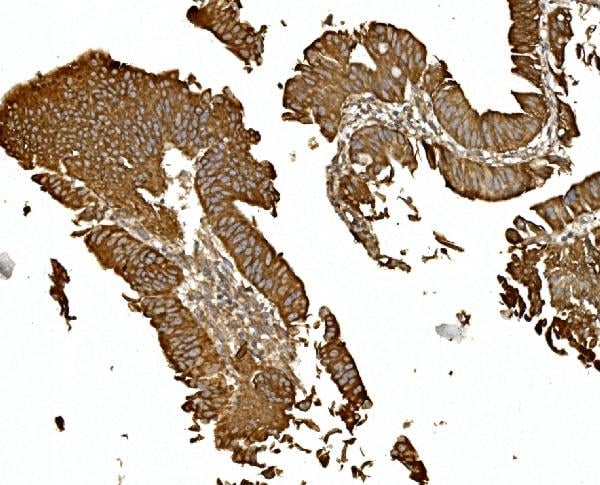
(Figure 6. IHC analysis of LGALS3BP using anti-LGALS3BP antibody (AAA19163).LGALS3BP was detected in paraffin-embedded section of mouse small intestine tissue. Heat mediated antigen retrieval was performed in citrate buffer (pH6, epitope retrieval solution) for 20 mins. The tissue section was blocked with 10% goat serum. The tissue section was then incubated with 2ug/ml rabbit anti-LGALS3BP Antibody (AAA19163) overnight at 4 degree C. Biotinylated goat anti-rabbit IgG was used as secondary antibody and incubated for 30 minutes at 37 degree C. The tissue section was developed using Strepavidin-Biotin-Complex (SABC) with DAB as the chromogen.)
IHC (Immunohistchemistry)
(Figure 6. IHC analysis of LGALS3BP using anti-LGALS3BP antibody (AAA19163).LGALS3BP was detected in paraffin-embedded section of mouse small intestine tissue. Heat mediated antigen retrieval was performed in citrate buffer (pH6, epitope retrieval solution) for 20 mins. The tissue section was blocked with 10% goat serum. The tissue section was then incubated with 2ug/ml rabbit anti-LGALS3BP Antibody (AAA19163) overnight at 4 degree C. Biotinylated goat anti-rabbit IgG was used as secondary antibody and incubated for 30 minutes at 37 degree C. The tissue section was developed using Strepavidin-Biotin-Complex (SABC) with DAB as the chromogen.)
LGALS3BP, Polyclonal Antibody (Cat# AAA19163)
Full Name
Anti-LGALS3BP Picoband Antibody
Reactivity
Human, Mouse
No cross reactivity with other proteins.
No cross reactivity with other proteins.
Applications
IHC, WB
Pricing


 FCM (Flow Cytometry)
(Figure 6. Flow Cytometry analysis of K562 cells using anti-D2AP antibody (M01756). Overlay histogram showing K562 cells stained with M01756 (Blue line).The cells were blocked with 10% normal goat serum. And then incubated with mouse anti-D2AP Antibody (M01756, 1ug/1x106 cells) for 30 min at 20 degree C. DyLight488 conjugated goat anti-mouse IgG (BA1126, 5-10ug/1x106 cells) was used as secondary antibody for 30 minutes at 20 degree C. Isotype control antibody (Green line) was mouse IgG (1ug/1x106) used under the same conditions. Unlabelled sample (Red line) was also used as a control.)
FCM (Flow Cytometry)
(Figure 6. Flow Cytometry analysis of K562 cells using anti-D2AP antibody (M01756). Overlay histogram showing K562 cells stained with M01756 (Blue line).The cells were blocked with 10% normal goat serum. And then incubated with mouse anti-D2AP Antibody (M01756, 1ug/1x106 cells) for 30 min at 20 degree C. DyLight488 conjugated goat anti-mouse IgG (BA1126, 5-10ug/1x106 cells) was used as secondary antibody for 30 minutes at 20 degree C. Isotype control antibody (Green line) was mouse IgG (1ug/1x106) used under the same conditions. Unlabelled sample (Red line) was also used as a control.)
CD2AP, Monoclonal Antibody (Cat# AAA19185)
Full Name
Anti-CD2AP Antibody (monoclonal, 5F8)
Gene Names
CD2AP; CMS
Reactivity
Human, Mouse, Rat
Applications
WB, IHC, FC/FACS
Purity
Immunogen Affinity Purified
Pricing


 FCM (Flow Cytometry)
(Figure 8. Flow Cytometry analysis of A549 cells using anti-Transketolase/TKT antibody (AAA19368).Overlay histogram showing A549 cells stained with AAA19368 (Blue line). The cells were blocked with 10% normal goat serum. And then incubated with mouse anti- Transketolase/TKT Antibody (AAA19368, 1μg/1x106 cells) for 30 min at 20 degree C. DyLight®488 conjugated goat anti-mouse IgG (BA1126, 5-10μg/1x106 cells) was used as secondary antibody for 30 minutes at 20 degree C. Isotype control antibody (Green line) was mouse IgG (1μg/1x106) used under the same conditions. Unlabelled sample (Red line) was also used as a control.)
FCM (Flow Cytometry)
(Figure 8. Flow Cytometry analysis of A549 cells using anti-Transketolase/TKT antibody (AAA19368).Overlay histogram showing A549 cells stained with AAA19368 (Blue line). The cells were blocked with 10% normal goat serum. And then incubated with mouse anti- Transketolase/TKT Antibody (AAA19368, 1μg/1x106 cells) for 30 min at 20 degree C. DyLight®488 conjugated goat anti-mouse IgG (BA1126, 5-10μg/1x106 cells) was used as secondary antibody for 30 minutes at 20 degree C. Isotype control antibody (Green line) was mouse IgG (1μg/1x106) used under the same conditions. Unlabelled sample (Red line) was also used as a control.)
Transketolase/TKT, Monoclonal Antibody (Cat# AAA19368)
Full Name
Anti-Transketolase/TKT Antibody (monoclonal, 2I3)
Gene Names
TKT; TK; TKT1; HEL107
Reactivity
Human, Mouse, Rat
Applications
WB, IHC-P, ICC, IF, FC/FACS/FCM
Purity
Immunogen affinity purified.
Pricing
 Standard Curve (Sample)
Apolipoprotein C2, ELISA Kit (Cat# AAA16085)
Full Name
Mouse Apolipoprotein C2 ELISA Kit
Gene Names
APOC2; APO-CII; APOC-II
Reactivity
Mouse
Pricing
 Standard Curve (Sample)
Renalase, ELISA Kit (Cat# AAA17500)
Full Name
Human Renalase ELISA Kit
Gene Names
RNLS; C10orf59; RENALASE
Reactivity
Human
Pricing
 Standard Curve (Sample)
CCL5, ELISA Kit (Cat# AAA17858)
Full Name
Rat CCL5 ELISA Kit
Gene Names
Ccl5; Scya5; Rantes
Reactivity
Rat
Applications
SE
Pricing
 WB (Western Blot)
(WB Suggested Anti-CTRC Antibody Titration: 0.2-1 ug/mlELISA Titer: 1:312500Positive Control: Human Liver)
WB (Western Blot)
(WB Suggested Anti-CTRC Antibody Titration: 0.2-1 ug/mlELISA Titer: 1:312500Positive Control: Human Liver)
CTRC, Polyclonal Antibody (Cat# AAA23430)
Full Name
CTRC antibody - N-terminal region
Gene Names
CTRC; CLCR; ELA4
Reactivity
Tested Species Reactivity: Human!!Predicted Species Reactivity: Human, Mouse, Rat, Cow, Dog, Pig, Rabbit
Applications
WB
Purity
Affinity Purified
Pricing
 Standard Curve (Sample)
met proto-oncogene (hepatocyte growth factor receptor), ELISA Kit (Cat# AAA15709)
Full Name
Mouse Hepatocyte Growth Factor Receptor, C-MET/HGFR ELISA Kit
Gene Names
Met; HGF; HGFR; Par4; c-Met; AI838057
Reactivity
Mouse
Pricing


 IHC (Immunohistchemistry)
(Figure 6. IHC analysis of Thrombopoietin using anti- Thrombopoietin antibody (AAA19166).Thrombopoietin was detected in paraffin-embedded section of rat kidney tissues. Heat mediated antigen retrieval was performed in citrate buffer (pH6, epitope retrieval solution) for 20 mins. The tissue section was blocked with 10% goat serum. The tissue section was then incubated with 1ug/ml rabbit anti- Thrombopoietin Antibody (AAA19166) overnight at 4 degree C. Biotinylated goat anti-rabbit IgG was used as secondary antibody and incubated for 30 minutes at 37 degree C. The tissue section was developed using Strepavidin-Biotin-Complex (SABC) with DAB as the chromogen.)
IHC (Immunohistchemistry)
(Figure 6. IHC analysis of Thrombopoietin using anti- Thrombopoietin antibody (AAA19166).Thrombopoietin was detected in paraffin-embedded section of rat kidney tissues. Heat mediated antigen retrieval was performed in citrate buffer (pH6, epitope retrieval solution) for 20 mins. The tissue section was blocked with 10% goat serum. The tissue section was then incubated with 1ug/ml rabbit anti- Thrombopoietin Antibody (AAA19166) overnight at 4 degree C. Biotinylated goat anti-rabbit IgG was used as secondary antibody and incubated for 30 minutes at 37 degree C. The tissue section was developed using Strepavidin-Biotin-Complex (SABC) with DAB as the chromogen.)
Thrombopoietin, Polyclonal Antibody (Cat# AAA19166)
Full Name
Anti-Thrombopoietin Picoband Antibody
Gene Names
Thpo; Ml; Tpo; Mgdf; Mpllg
Reactivity
Mouse, Rat
No cross reactivity with other proteins
No cross reactivity with other proteins
Applications
EIA, IHC, WB
Purity
Immunogen affinity purified
Pricing
 Standard Curve (Sample)
Nardilysin, NRD1, ELISA Kit (Cat# AAA19043)
Full Name
Human Nardilysin, NRD1 ELISA Kit
Reactivity
Human
Pricing