Filters
Clonality
Type
Reactivity
Gene Name
Isotype
Host
Application
Clone
2811 results for " C" - showing 1850-1900
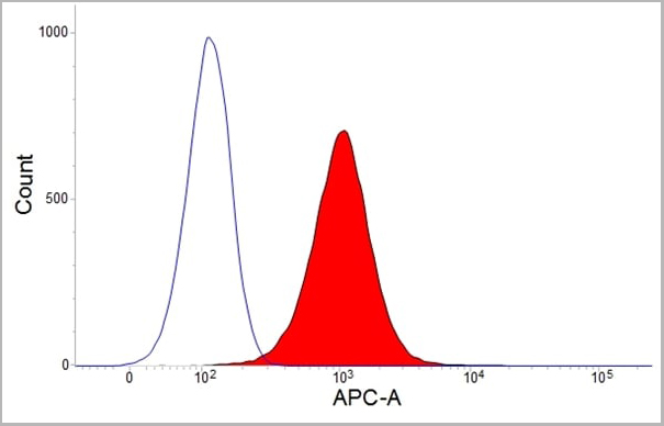
 Application Data
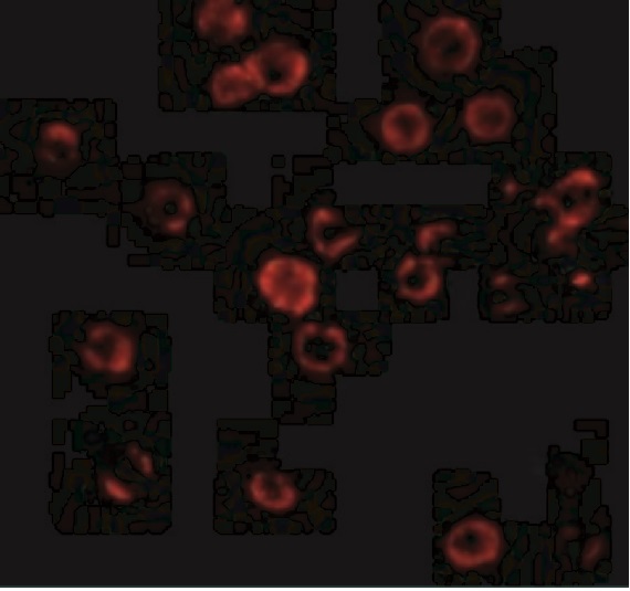
(Staining of human peripheral blood granulocytes with Mouse anti Human CD16:Alexa Fluor 647 (AAA11921))
Application Data
(Staining of human peripheral blood granulocytes with Mouse anti Human CD16:Alexa Fluor 647 (AAA11921))
CD16, Monoclonal Antibody (Cat# AAA11921)
Full Name
MOUSE ANTI HUMAN CD16
Gene Names
FCGR3B; CD16; FCG3; CD16b; FCGR3; FCR-10; FCRIII; FCRIIIb
Applications
Flow Cytometry, Immunoprecipitation
Pricing
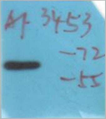
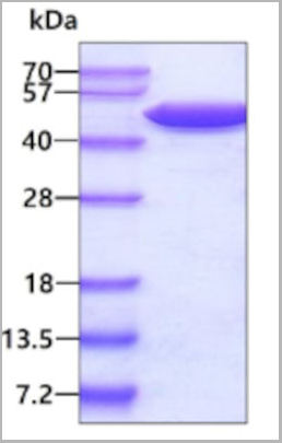
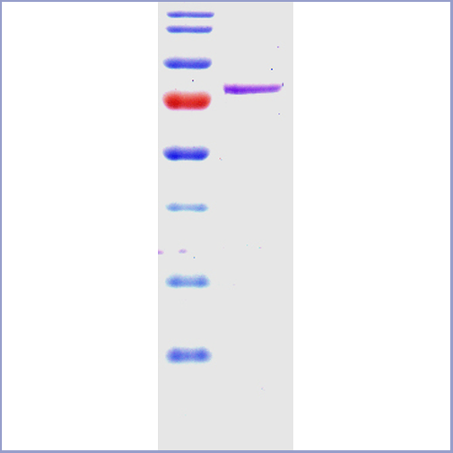 SDS-PAGE
(SDS-PAGE of 78 kDa Grp78 (BiP) protein (AAA17807))
SDS-PAGE
(SDS-PAGE of 78 kDa Grp78 (BiP) protein (AAA17807))
Grp78 (Bip), Active Protein (Cat# AAA17807)
Full Name
Recombinant Human GRP78 (Bip)
Gene Names
HSPA5; BIP; MIF2; GRP78; HEL-S-89n
Applications
Western Blot
Purity
Affinity Purified
Pricing
Chlamydia trachomatis, Polyclonal Antibody (Cat# AAA14333)
Full Name
Chlamydia trachomatis antibody
Reactivity
To be determined by end-user.
Applications
Immunofluorescence, Immunohistochemistry, Western Blot
Purity
> 95% pure
Pricing
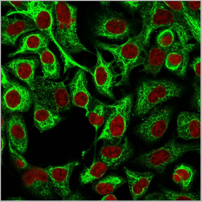
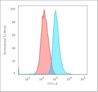 FCM (Flow Cytometry)
(Flow Cytometric Analysis of trypsinized MeOH-fixed HeLa cells using Cytokeratin 14 Mouse Monoclonal Antibody (LL002) followed by Goat anti-Mouse IgG-CF488 (Blue); Isotype Control (Red).)
FCM (Flow Cytometry)
(Flow Cytometric Analysis of trypsinized MeOH-fixed HeLa cells using Cytokeratin 14 Mouse Monoclonal Antibody (LL002) followed by Goat anti-Mouse IgG-CF488 (Blue); Isotype Control (Red).)
Cytokeratin 14 (KRT14), Monoclonal Antibody (Cat# AAA13813)
Full Name
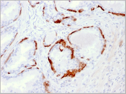
Cytokeratin 14 (KRT14) (Squamous Cell Marker) Mouse Monoclonal Antibody
Gene Names
KRT14; K14; NFJ; CK14; EBS3; EBS4
Reactivity
Human
Applications
Flow Cytometry, Immunofluorescence, Immunohistochemistry
Pricing
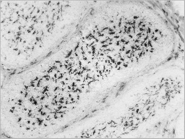
 Application Data
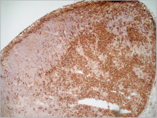
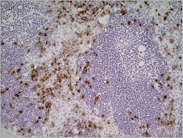
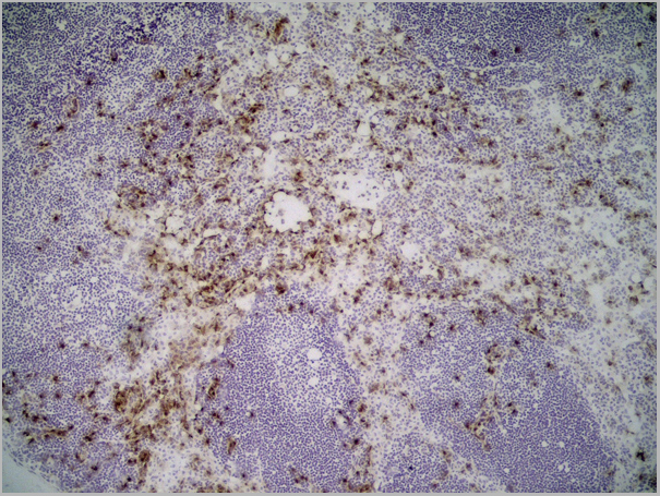
(Immunoperoxidase staining of rat lymph node cryosection with Mouse anti Rat CD169, clone ED3 followed by horseradish peroxidase conjugated Goat anti Mouse IgG2a as a detection reagent. High power)
Application Data
(Immunoperoxidase staining of rat lymph node cryosection with Mouse anti Rat CD169, clone ED3 followed by horseradish peroxidase conjugated Goat anti Mouse IgG2a as a detection reagent. High power)
CD169, Monoclonal Antibody (Cat# AAA11972)
Full Name
MOUSE ANTI RAT CD169
Applications
Immunohistochemistry, Flow Cytometry, Immunofluorescence, Immunoprecipitation
Pricing
Interleukin 6 Receptor, gp80, non-neutralizing, Monoclonal Antibody (Cat# AAA14678)
Full Name
Interleukin 6 Receptor, gp80, non-neutralizing (CD126, IL-6R)
Applications
Flow Cytometry
Purity
Purified
Pricing
ApoH, Native Protein (Cat# AAA14369)
Full Name
ApoH protein
Gene Names
APOH; BG; B2G1; B2GP1
Purity
> 96% pure
Pricing
Synaptosomal Protein 25kDa (SNAP-25) (a.a. 183-197) cleaved, Monoclonal Antibody (Cat# AAA13384)
Full Name
MAb to SNAP-25, cleaved
Applications
Immunohistochemistry
Purity
Product is sterile filtered.
Pricing
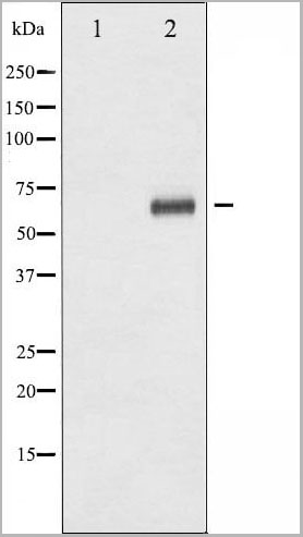
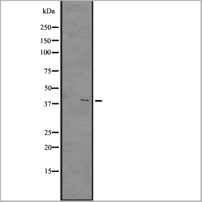
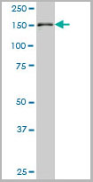
 WB (Western Blot)
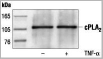
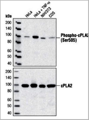
(Western blot analysis of extracts from HeLa, C6 and NIH/3T3 cells, using AAA14717.)
WB (Western Blot)
(Western blot analysis of extracts from HeLa, C6 and NIH/3T3 cells, using AAA14717.)
c-Phospholipase A2, Polyclonal Antibody (Cat# AAA14717)
Full Name
c-Phospholipase A2 (Cytosolic PLA2, cPLA2)
Reactivity
Human
Applications
Western Blot, Immunoprecipitation
Purity
Purified by Protein A and peptide affinity chromatography
Pricing
TRACP5a, Monoclonal Antibody (Cat# AAA14610)
Full Name
TRACP5a, Human, mAb 220
Applications
Immunoassay, Immunoprecipitation, Immunohistochemistry, Western Blot
Purity
Protein G
Pricing
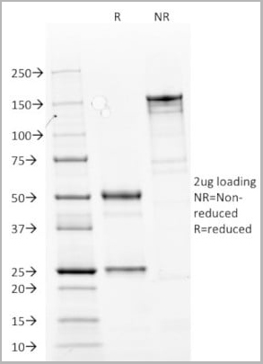
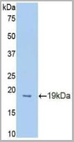
 SDS-PAGE
(SDS-PAGE analysis of recombinant IL-33 fragment on Coomassie Blue-stained 4-20% gradient gel.)
SDS-PAGE
(SDS-PAGE analysis of recombinant IL-33 fragment on Coomassie Blue-stained 4-20% gradient gel.)
IL-33, Recombinant Protein (Cat# AAA10966)
Full Name
IL-33 Recombinant Protein
Gene Names
IL33; DVS27; IL1F11; NF-HEV; NFEHEV; C9orf26
Applications
Western Blot
Purity
~95%
Pricing
CKMB, Recombinant Protein (Cat# AAA14392)
Full Name
CKMB Type II protein
Purity
> 99% pure by SDS-PAGE
Pricing
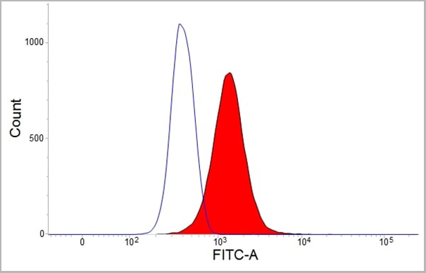
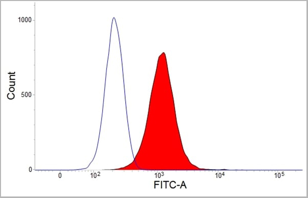
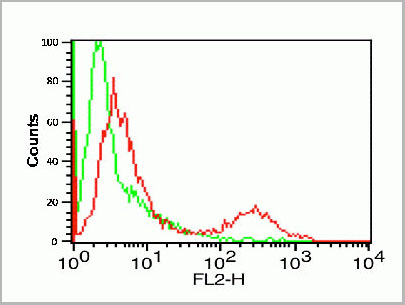
 FCM (Flow Cytometry)
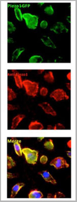
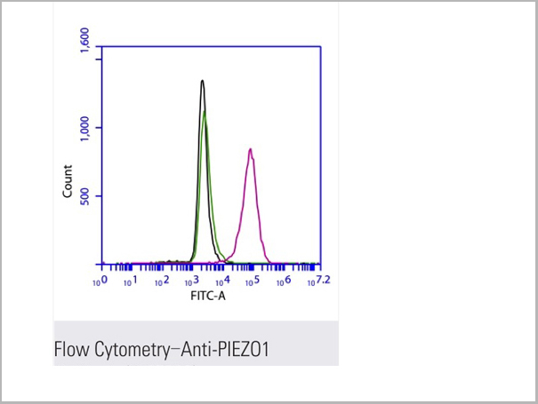
(Flow cytometric analysis of 2% paraformaldehyde-fixed THP1 (Human acute monocytic leukemia cell line) cells labeling PIEZO1 with AAA13800 at 1/200 dilution (red) compared with a mouse monoclonal IgG isotype control (black) and an unlabelled control (cells without incubation with primary antibody, green). Goat anti-mouse IgG (FITC) at 1/300 dilution was used as the secondary antibody.)
FCM (Flow Cytometry)
(Flow cytometric analysis of 2% paraformaldehyde-fixed THP1 (Human acute monocytic leukemia cell line) cells labeling PIEZO1 with AAA13800 at 1/200 dilution (red) compared with a mouse monoclonal IgG isotype control (black) and an unlabelled control (cells without incubation with primary antibody, green). Goat anti-mouse IgG (FITC) at 1/300 dilution was used as the secondary antibody.)
PIEZO1, Monoclonal Antibody (Cat# AAA13800)
Full Name
PIEZO1 Mouse mAb
Gene Names
PIEZO1; DHS; Mib; LMPH3; FAM38A
Reactivity
Homo Sapiens
Applications
Immunoprecipitation, Immunofluorescence, Flow Cytometry
Pricing
 IHC (Immunohistochemistry)
(DAB staining on IHC-P;Sample: Mouse Kidney Tissue;Primary Ab: 20ug/ml Rabbit Anti-Mouse EPCR AntibodySecond Ab: 2ug/mL HRP-Linked Caprine Anti-Rabbit IgG Polyclonal Antibody (Immunohistochemistry))
IHC (Immunohistochemistry)
(DAB staining on IHC-P;Sample: Mouse Kidney Tissue;Primary Ab: 20ug/ml Rabbit Anti-Mouse EPCR AntibodySecond Ab: 2ug/mL HRP-Linked Caprine Anti-Rabbit IgG Polyclonal Antibody (Immunohistochemistry))
Protein C Receptor, Endothelial (PROCR), Polyclonal Antibody (Cat# AAA20114)
Full Name
Polyclonal Antibody to Protein C Receptor, Endothelial (PROCR)
Gene Names
Procr; Ccca; Epcr; Ccd41; AI325044
Reactivity
Mouse
Applications
WB, IHC, ICC, IP
Purity
Antigen-specific affinity chromatography followed by Protein A affinity chromatography
Pricing

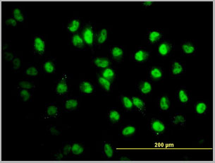
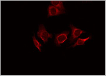
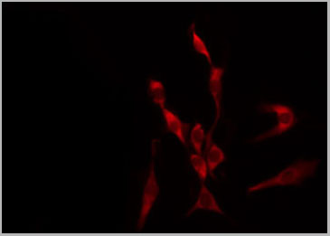 IF (Immunofluorescence)
(AAA31072 staining HeLa by IF/ICC. The sample were fixed with PFA and permeabilized in 0.1% Triton X-100, then blocked in 10% serum for 45 minutes at 25 degree C. The primary antibody was diluted at 1/200 and incubated with the sample for 1 hour at 37 degree C. An Alexa Fluor 594 conjugated goat anti-rabbit IgG (H+L) Ab, diluted at 1/600, was used as the secondary antibody.)
IF (Immunofluorescence)
(AAA31072 staining HeLa by IF/ICC. The sample were fixed with PFA and permeabilized in 0.1% Triton X-100, then blocked in 10% serum for 45 minutes at 25 degree C. The primary antibody was diluted at 1/200 and incubated with the sample for 1 hour at 37 degree C. An Alexa Fluor 594 conjugated goat anti-rabbit IgG (H+L) Ab, diluted at 1/600, was used as the secondary antibody.)
LKB1, Polyclonal Antibody (Cat# AAA31072)
Full Name
Phospho-LKB1 (Ser428) Antibody
Gene Names
STK11; PJS; LKB1; hLKB1
Reactivity
Human, Mouse
Applications
Western Blot, Immunohistochemistry, Immunofluorescence, Immunocytochemistry
Purity
From purified rabbit serum by affinity purification via sequential chromatography on phospho-and non-phospho-peptide affinity columns.
Pricing
Amyloid beta peptide, Polyclonal Antibody (Cat# AAA13671)
Full Name
Goat anti-Amyloid beta peptide (aa1-16) Antibody
Gene Names
APP; AAA; AD1; PN2; ABPP; APPI; CVAP; ABETA; PN-II; CTFgamma
Reactivity
Tested: Human
Expected from sequence similarity: Human, Dog, Pig, Cow
Expected from sequence similarity: Human, Dog, Pig, Cow
Applications
Peptide ELISA, Western Blot
Purity
Purified from goat serum by ammonium sulphate precipitation followed by antigen affinity chromatography using the immunizing peptide.
Pricing
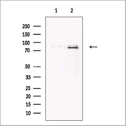
 WB (Western Blot)
(Western blot analysis of A2M expression in A375 (A), LO2 (B), PC12 (C) whole cell lysates.)
WB (Western Blot)
(Western blot analysis of A2M expression in A375 (A), LO2 (B), PC12 (C) whole cell lysates.)
A2M, Polyclonal Antibody (Cat# AAA27800)
Full Name
Anti-A2M Antibody
Gene Names
A2M; A2MD; CPAMD5; FWP007; S863-7
Reactivity
Human, Rat, Monkey
Applications
Western Blot
Purity
The antibody was purified by immunogen affinity chromatography.
Pricing
 WB (Western Blot)
(Western blot analysis of GPR84 expression in A431 whole cell lysates)
WB (Western Blot)
(Western blot analysis of GPR84 expression in A431 whole cell lysates)
GPR84, Polyclonal Antibody (Cat# AAA31105)
Full Name
GPR84 Antibody
Gene Names
GPR84; EX33; GPCR4
Reactivity
Human, Mouse
Applications
Western Blot, Immunohistochemistry
Purity
The antiserum was purified by peptide affinity chromatography using SulfoLinkTM Coupling Resin (Thermo Fisher Scientific).
Pricing
Ferritin, Monoclonal Antibody (Cat# AAA13374)
Full Name
MAb to Human Ferritin
Reactivity
Human
Applications
Immunoassay
Purity
>=95% pure (SDS-PAGE). Protein G chromatography
Pricing
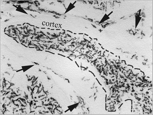
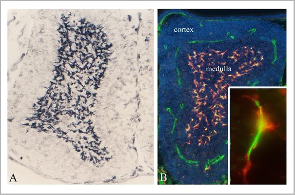
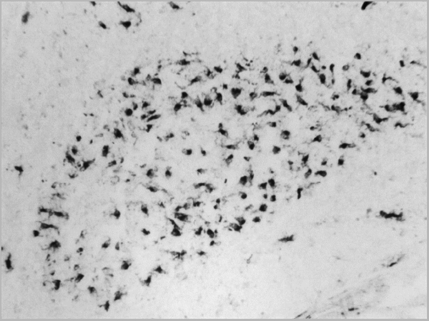 Application Data
(Mouse anti Chicken CSF1R (CD115) immunohistochemical staining of guinea fowl, Agelastes meleagredisbursa of Fabricius.Clone ROS-AV170 specifically stains the bursal secretory dendritic cells (BSDC) located in the medulla. Clone ROS-AV170 also recognizes some CSF1R+ve macrophages in the interfollicular connective tissue. Image courtesy of Dr Nandor Nagy, Semmelweis University, Hungary)
Application Data
(Mouse anti Chicken CSF1R (CD115) immunohistochemical staining of guinea fowl, Agelastes meleagredisbursa of Fabricius.Clone ROS-AV170 specifically stains the bursal secretory dendritic cells (BSDC) located in the medulla. Clone ROS-AV170 also recognizes some CSF1R+ve macrophages in the interfollicular connective tissue. Image courtesy of Dr Nandor Nagy, Semmelweis University, Hungary)
CSF1R, Monoclonal Antibody (Cat# AAA12252)
Full Name
MOUSE ANTI CHICKEN CSF1R
Gene Names
CSF1R; FMS; CSFR; FIM2; HDLS; C-FMS; CD115; CSF-1R; M-CSF-R
Reactivity
Chicken
Applications
Immunohistochemistry, Flow Cytometry, Immunofluorescence
Pricing
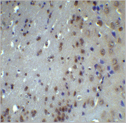
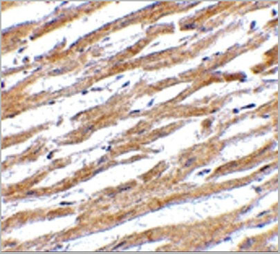
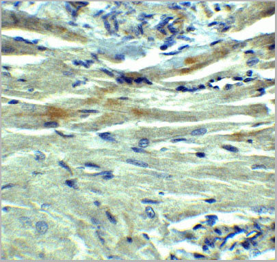
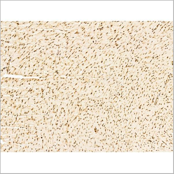
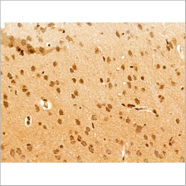
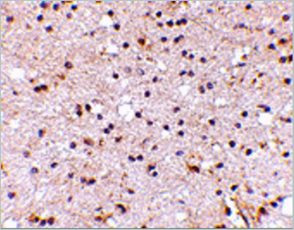
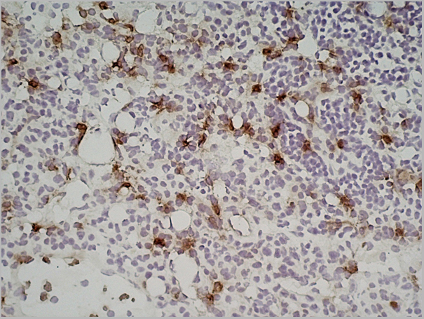
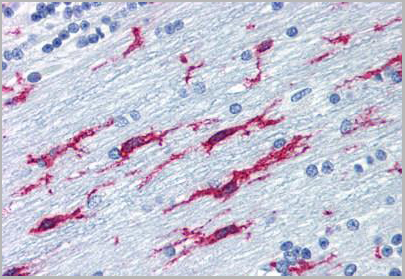
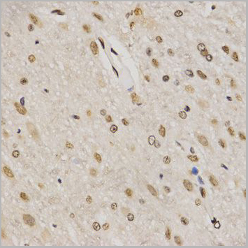
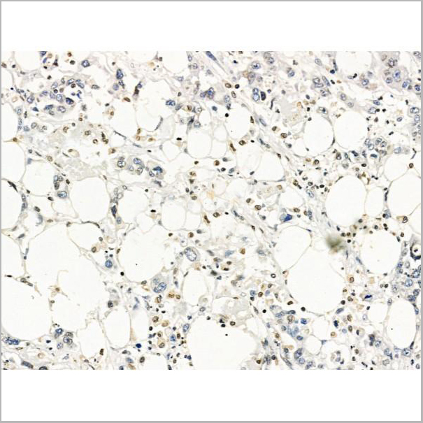
 IHC (Immunohistochemistry)
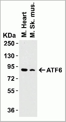
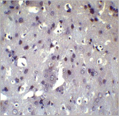
(Figure 8 Immunohistochemistry Validation of ATF6 in Rat Brain Tissue Immunohistochemical analysis of paraffin-embedded Rat brain tissue using anti-ATF6 antibody (AAA10937) at 5 ug/ml. Tissue was fixed with formaldehyde and blocked with 10% serum for 1 h at RT; antigen retrieval was by heat mediation with a citrate buffer (pH6). Samples were incubated with primary antibody overnight at 4°C. A goat anti-rabbit IgG H&L (HRP) at 1/250 was used as secondary. Counter stained with Hematoxylin.)
IHC (Immunohistochemistry)
(Figure 8 Immunohistochemistry Validation of ATF6 in Rat Brain Tissue Immunohistochemical analysis of paraffin-embedded Rat brain tissue using anti-ATF6 antibody (AAA10937) at 5 ug/ml. Tissue was fixed with formaldehyde and blocked with 10% serum for 1 h at RT; antigen retrieval was by heat mediation with a citrate buffer (pH6). Samples were incubated with primary antibody overnight at 4°C. A goat anti-rabbit IgG H&L (HRP) at 1/250 was used as secondary. Counter stained with Hematoxylin.)
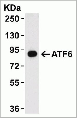
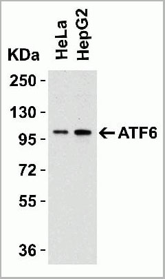
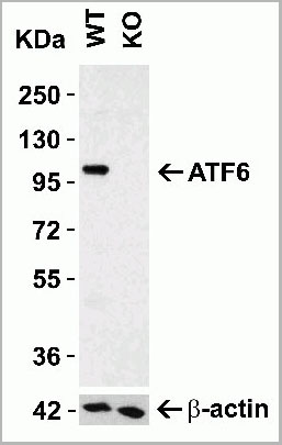
ATF6, Polyclonal Antibody (Cat# AAA10937)
Full Name
ATF6 Antibody
Gene Names
ATF6; ATF6A
Reactivity
Human, Mouse, Rat
Applications
Western Blot, Immunohistochemistry, Immunofluorescence
Purity
ATF6 Antibody is affinity chromatography purified via peptide column.
Pricing
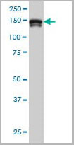 WB (Western Blot)
(Western blot of STAG2 expression in HeLa nuclear extract with STAG2 monoclonal antibody, clone 3C6.)
WB (Western Blot)
(Western blot of STAG2 expression in HeLa nuclear extract with STAG2 monoclonal antibody, clone 3C6.)
STAG2, Monoclonal Antibody (Cat# AAA12354)
Full Name
Mouse Monoclonal [clone 3C6] (IgG1,k) to Human STAG2
Gene Names
STAG2; SA2; SA-2; SCC3B; bA517O1.1
Reactivity
Human, Mouse
Applications
Western Blot, Immunohistochemistry, Immunofluorescence
Purity
Purified from ascites by Protein A
Pricing
Clostridium botulinum Type A Neurotoxin Complex, Antibody (Cat# AAA13408)
Full Name
Rabbit anti C. botulinum Neurotoxin Type A
Applications
Dot Blot, Western Blot
Purity
Protein G Sepharose Chromatography
Pricing
CD45, Monoclonal Antibody (Cat# AAA20023)
Full Name
Anti-Hu CD45 Purified
Gene Names
PTPRC; LCA; LY5; B220; CD45; L-CA; T200; CD45R; GP180
Reactivity
Human
Applications
Western Blot, Flow Cytometry
Purity
Purified by Protein-A Affinity Chromatography
Pricing
Platelet Factor 4, Monoclonal Antibody (Cat# AAA14278)
Full Name
Anti-Human Platelet Factor 4, Purified (Clone: RTO) (Mouse IgG2b)
Gene Names
PF4V1; PF4A; CXCL4L1; CXCL4V1; PF4-ALT; SCYB4V1
Reactivity
Human
Applications
Western Blot
Pricing
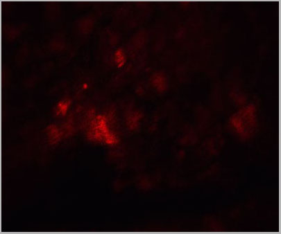
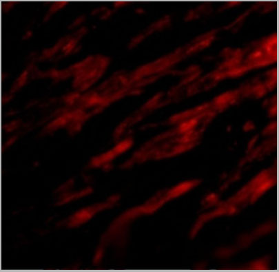
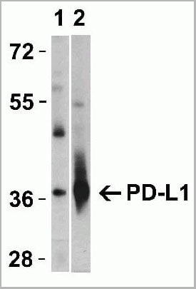
 IF (Immunofluorescence)
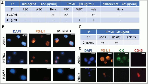
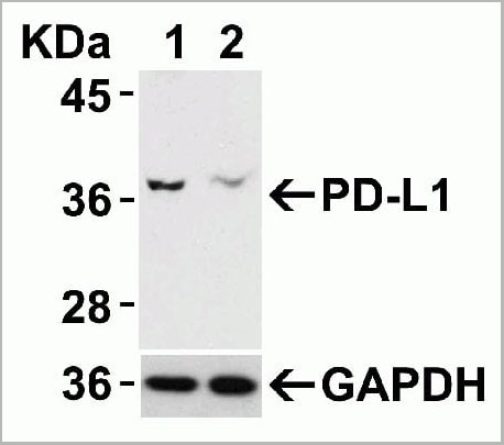
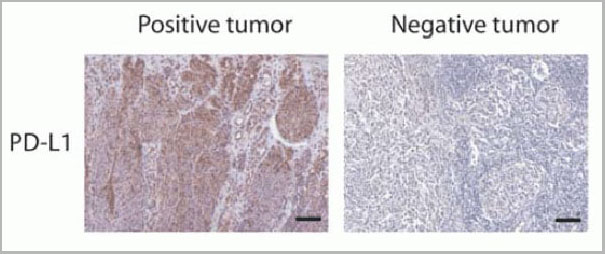
(Figure 12 Immunohistochemistry Validation of PD-L1in Human Tumors (Gadiot et al., 2011)Immunohistochemical analysis of patient tumors labeling PD-L1 with anti-PD-L1 antibodies (AAA10941). Several anti-PD-L1 antibodies were tested for staining, “Only 1 antibody gave no background staining and was competitively blocked by the addition of PD-L1Fc protein (AAA10941)”.)
IF (Immunofluorescence)
(Figure 12 Immunohistochemistry Validation of PD-L1in Human Tumors (Gadiot et al., 2011)Immunohistochemical analysis of patient tumors labeling PD-L1 with anti-PD-L1 antibodies (AAA10941). Several anti-PD-L1 antibodies were tested for staining, “Only 1 antibody gave no background staining and was competitively blocked by the addition of PD-L1Fc protein (AAA10941)”.)
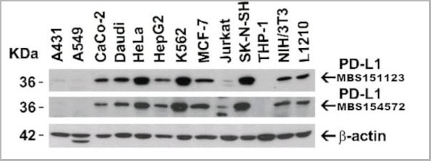
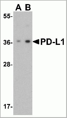
PDL-1, Polyclonal Antibody (Cat# AAA10941)
Full Name
PDL-1 Antibody
Gene Names
CD274; B7-H; B7H1; PDL1; PD-L1; PDCD1L1; PDCD1LG1
Applications
Western Blot, Immunohistochemistry, Immunofluorescence, Flow Cytometry
Purity
PD-L1 Antibody is affinity chromatography purified via peptide column.
Pricing
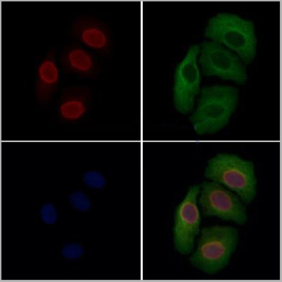
 IF (Immunofluorescence)
(AAA31248 staining Hela cells by IF/ICC. The samples were fixed with PFA and permeabilized in 0.1% Triton X-100,then blocked in 10% serum for 45 minutes at 25°C. Samples were then incubated with primary Ab(AAA31248 1:200) and mouse anti-beta tubulin Ab(T0023 1:200) for 1 hour at 37°C. An AlexaFluor594 conjugated goat anti-rabbit IgG(H+L) Ab(Red) and an AlexaFluor488 conjugated goat anti-mouse IgG(H+L) Ab(Green) were used as the secondary antibody.The nuclear counter stain is DAPI(blue).)
IF (Immunofluorescence)
(AAA31248 staining Hela cells by IF/ICC. The samples were fixed with PFA and permeabilized in 0.1% Triton X-100,then blocked in 10% serum for 45 minutes at 25°C. Samples were then incubated with primary Ab(AAA31248 1:200) and mouse anti-beta tubulin Ab(T0023 1:200) for 1 hour at 37°C. An AlexaFluor594 conjugated goat anti-rabbit IgG(H+L) Ab(Red) and an AlexaFluor488 conjugated goat anti-mouse IgG(H+L) Ab(Green) were used as the secondary antibody.The nuclear counter stain is DAPI(blue).)
PDE1C, Polyclonal Antibody (Cat# AAA31248)
Full Name
PDE1C Antibody
Gene Names
PDE1C; Hcam3; DFNA74; hCam-3; cam-PDE 1C
Reactivity
Human, Mouse, Rat
Predicted Reactivity: Pig(88%), Horse(100%), Rabbit(88%), Dog(100%)
Predicted Reactivity: Pig(88%), Horse(100%), Rabbit(88%), Dog(100%)
Applications
ELISA
Purity
The antiserum was purified by peptide affinity chromatography using SulfoLink Coupling Resin (Thermo Fisher Scientific).
Pricing
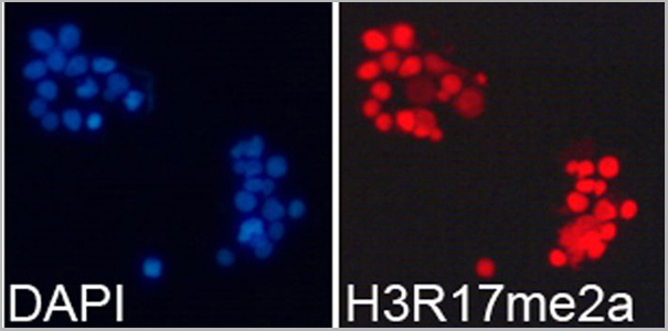
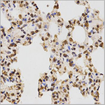
 IHC (Immunohistchemistry)
(Immunohistochemistry of paraffin-emebedded human placenta using H3R17me2a antibody (AAA10654) at dilution of 1:100 (40x lens).)
IHC (Immunohistchemistry)
(Immunohistochemistry of paraffin-emebedded human placenta using H3R17me2a antibody (AAA10654) at dilution of 1:100 (40x lens).)
H3R17me2a, Antibody (Cat# AAA10654)
Full Name
Histone H3R17me2a Polyclonal Antibody
Gene Names
HIST1H3J; H3/j; H3FJ
Reactivity
Human, Mouse, Rat, Other (Wide Range)
Applications
Western Blot, Immunohistochemistry, Immunofluorescence, Immunoprecipitation
Purity
Affinity Purification
Pricing
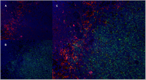
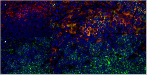
 Application Data
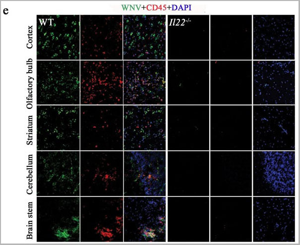
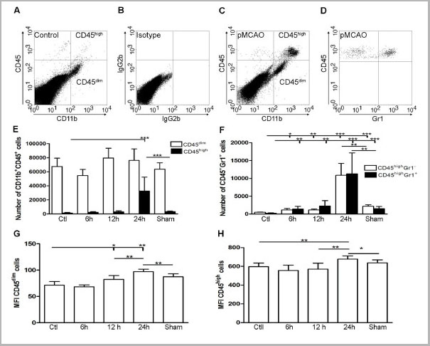
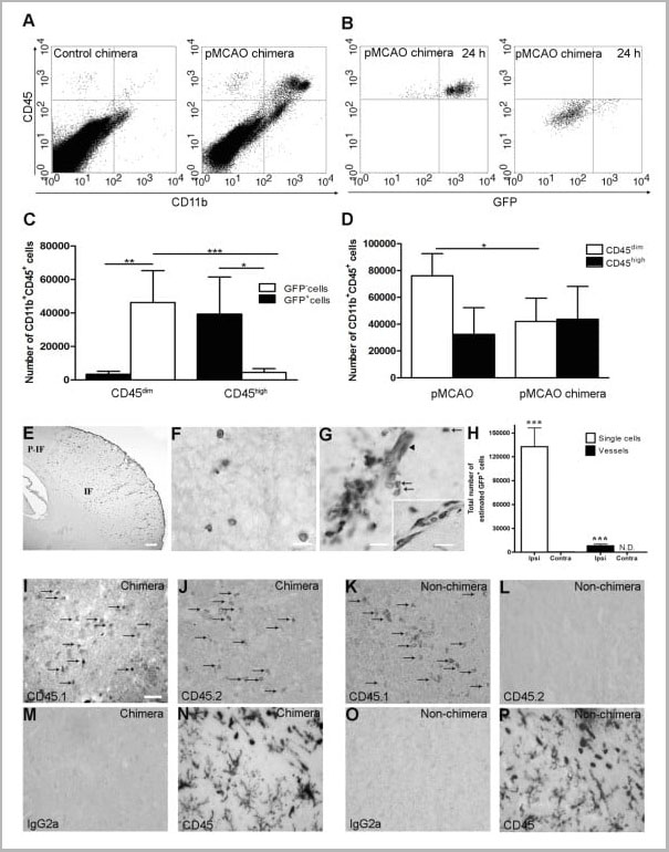
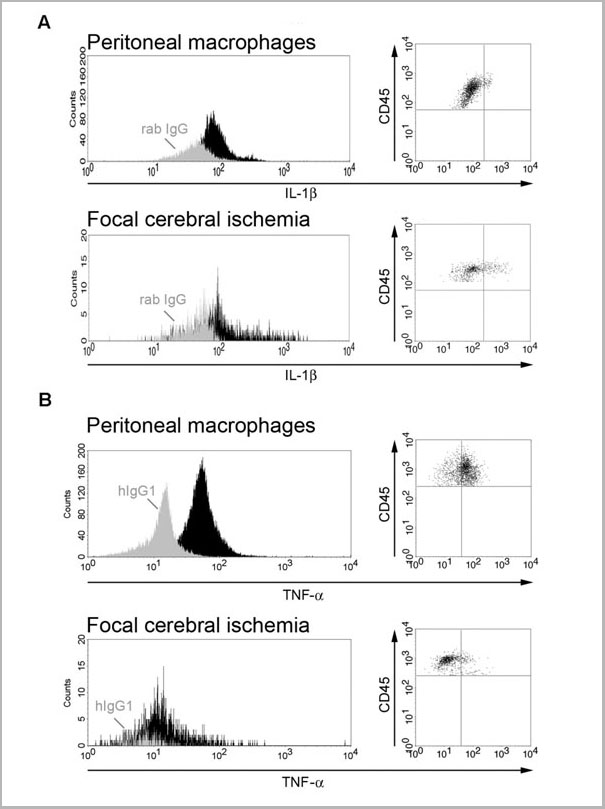
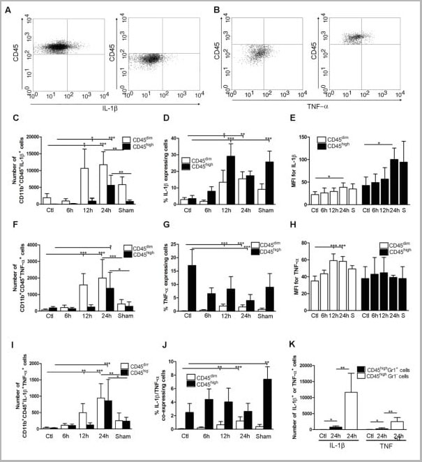
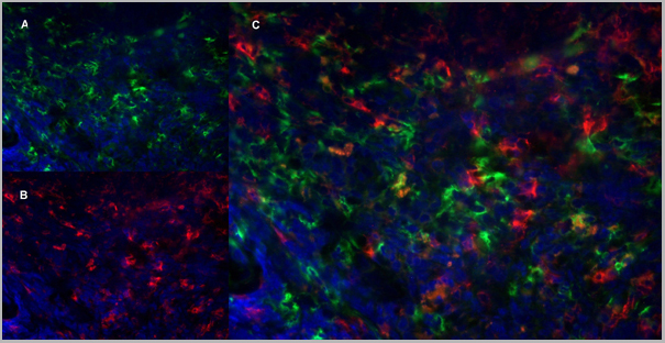
(Published customer image: Cytokine expression in segregated populations of cells following stroke. (A, B) Dot plots showing CD11b+CD45high macrophages/granulocytes (upper right quadrants) and CD11b+CD45dim microglia (bottom right quadrants) expressing IL-1beta (A) or TNF-a (B). (C-J) Bar graphs showing numbers and proportions of IL-1beta (C, D), TNF-a (F, G) and IL-1beta/TNF-a co-expressing (I, J) CD11b+CD45dim microglia and CD11b+CD45high macrophages/granulocytes in unmanipulated control mice (n = 10), in mice 6 (n = 7), 12 (n = 7), or 24 hours after pMCAO (n = 10), and in sham-operated mice 24 hours after pMCAO (n = 7). (E, H) Comparison of the MFI values for IL-1beta (E) and TNF-a (H) in viable CD11b+CD45dim microglia and CD11b+CD45high macrophages/granulocytes in unmanipulated mice, in mice 6, 12, or 24 hours after pMCAO, and in sham-operated mice 24 hours after pMCAO. Macrophages/granulocytes express significantly more IL-1beta than do microglial in unmanipulated mice, in mice 6, 12, or 24 hours after pMCAO, and in sham-operated mice 24 hours after pMCAO (E), whereas microglial cells express significantly higher levels of TNF-a than do macrophages/granulocytes at 12 h and 24 hours, and in sham-operated mice 24 hours after pMCAO (H). (K) CD11b+CD45highGr1- macrophages and not CD11b+CD45highGr1+ granulocytes are the main producers of IL-1beta and TNF-a 24 hours after pMCAO. *P < 0.05, **P < 0.01, and ***P < 0.001.From: http://www.jneuroinflammation.com/content/5/1/46.)
Application Data
(Published customer image: Cytokine expression in segregated populations of cells following stroke. (A, B) Dot plots showing CD11b+CD45high macrophages/granulocytes (upper right quadrants) and CD11b+CD45dim microglia (bottom right quadrants) expressing IL-1beta (A) or TNF-a (B). (C-J) Bar graphs showing numbers and proportions of IL-1beta (C, D), TNF-a (F, G) and IL-1beta/TNF-a co-expressing (I, J) CD11b+CD45dim microglia and CD11b+CD45high macrophages/granulocytes in unmanipulated control mice (n = 10), in mice 6 (n = 7), 12 (n = 7), or 24 hours after pMCAO (n = 10), and in sham-operated mice 24 hours after pMCAO (n = 7). (E, H) Comparison of the MFI values for IL-1beta (E) and TNF-a (H) in viable CD11b+CD45dim microglia and CD11b+CD45high macrophages/granulocytes in unmanipulated mice, in mice 6, 12, or 24 hours after pMCAO, and in sham-operated mice 24 hours after pMCAO. Macrophages/granulocytes express significantly more IL-1beta than do microglial in unmanipulated mice, in mice 6, 12, or 24 hours after pMCAO, and in sham-operated mice 24 hours after pMCAO (E), whereas microglial cells express significantly higher levels of TNF-a than do macrophages/granulocytes at 12 h and 24 hours, and in sham-operated mice 24 hours after pMCAO (H). (K) CD11b+CD45highGr1- macrophages and not CD11b+CD45highGr1+ granulocytes are the main producers of IL-1beta and TNF-a 24 hours after pMCAO. *P < 0.05, **P < 0.01, and ***P < 0.001.From: http://www.jneuroinflammation.com/content/5/1/46.)
CD45, Monoclonal Antibody (Cat# AAA11896)
Full Name
RAT ANTI MOUSE CD45
Gene Names
Ptprc; loc; B220; Cd45; L-CA; Ly-5; T200; CD45R; Lyt-4
Applications
Immunohistochemistry, Flow Cytometry, Immunofluorescence, Immunoprecipitation
Pricing
 IF (Immunofluorescence)
(Immunofluorescent analysis of Ephrin A5 staining in Hela cells. Formalin-fixed cells were permeabilized with 0.1% Triton X-100 in TBS for 5-10 minutes and blocked with 3% BSA-PBS for 30 minutes at room temperature. Cells were probed with the primary antibody in 3% BSA-PBS and incubated overnight at 4°C in a hidified chamber. Cells were washed with PBST and incubated with a DyLight 594-conjugated secondary antibody (red) in PBS at room temperature in the dark. DAPI was used to stain the cell nuclei (blue).)
IF (Immunofluorescence)
(Immunofluorescent analysis of Ephrin A5 staining in Hela cells. Formalin-fixed cells were permeabilized with 0.1% Triton X-100 in TBS for 5-10 minutes and blocked with 3% BSA-PBS for 30 minutes at room temperature. Cells were probed with the primary antibody in 3% BSA-PBS and incubated overnight at 4°C in a hidified chamber. Cells were washed with PBST and incubated with a DyLight 594-conjugated secondary antibody (red) in PBS at room temperature in the dark. DAPI was used to stain the cell nuclei (blue).)
Ephrin A5, Polyclonal Antibody (Cat# AAA27798)
Full Name
Anti-Ephrin A5 Antibody
Gene Names
EFNA5; AF1; EFL5; RAGS; EPLG7; GLC1M; LERK7
Reactivity
Human, Mouse, Rat, Bovine, Chicken, Rabbit
Applications
Western Blot, Immunohistochemistry, Immunofluorescence, Immunocytochemistry, Flow Cytometry
Purity
The antibody was purified by immunogen affinity chromatography.
Pricing
Hepatitis C Virus, Polyclonal Antibody (Cat# AAA14328)
Full Name
Hepatitis C Virus antibody
Applications
Western Blot
Pricing
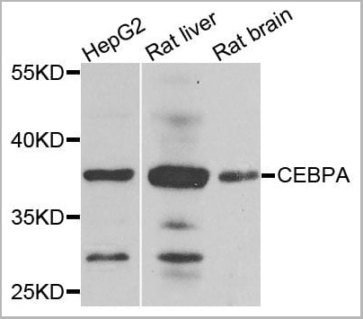
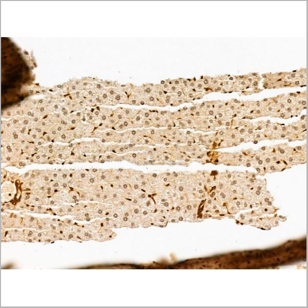
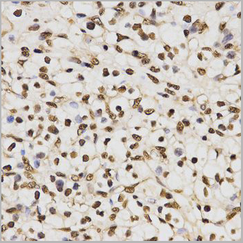
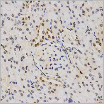
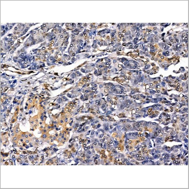
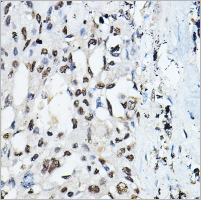 IHC (Immunohistchemistry)
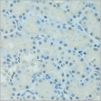
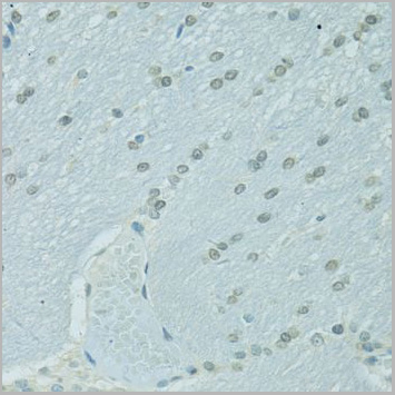
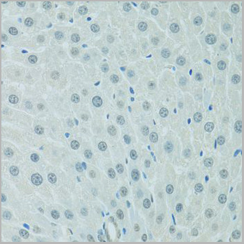
(Immunohistochemistry of paraffin-embedded Human lung cancer using CEBPA Rabbit pAb (AAA10634) at dilution of 1:100 (40x lens).Perform high pressure antigen retrieval with 10 mM citrate buffer pH 6.0 before commencing with IHC staining protocol.)
IHC (Immunohistchemistry)
(Immunohistochemistry of paraffin-embedded Human lung cancer using CEBPA Rabbit pAb (AAA10634) at dilution of 1:100 (40x lens).Perform high pressure antigen retrieval with 10 mM citrate buffer pH 6.0 before commencing with IHC staining protocol.)
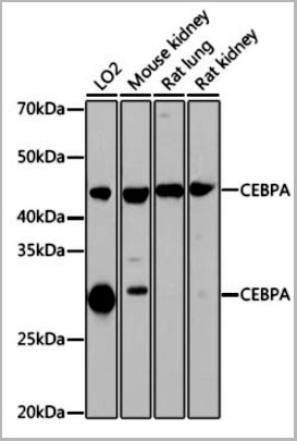
C/EBPalpha, Polyclonal Antibody (Cat# AAA10634)
Full Name
C/EBPalpha
Gene Names
CEBPA; CEBP; C/EBP-alpha
Applications
Western Blot, Immunohistochemistry
Purity
Affinity purification
Pricing
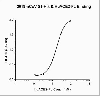 Application Data
(Binding data of recombinant 2019-nCoV Spike Protein (C-6His) with ACE2 receptor.)
Application Data
(Binding data of recombinant 2019-nCoV Spike Protein (C-6His) with ACE2 receptor.)
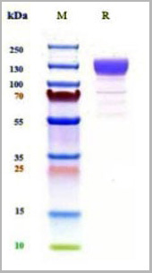
COVID 19 Spike S Protein Coronavirus, Recombinant Protein (Cat# AAA27945)
Full Name
Recombinant 2019-nCoV Spike Protein (C-6His)
Applications
Immunogen, Calibrator, Standard
Purity
Purity: Greater than 95% as determined by reducing SDS-PAGE.
Purification: Affinity purification chromatography.
Purification: Affinity purification chromatography.
Pricing
 SDS-PAGE
(3ug by SDS-PAGE under reducing condition and visualized by coomassie blue stain.)
SDS-PAGE
(3ug by SDS-PAGE under reducing condition and visualized by coomassie blue stain.)
Alpha-enolase, Active Protein (Cat# AAA11794)
Full Name
Alpha-enolase, 1-434aa, Human, E Coli (Bioactivity Validated)
Gene Names
ENO1; NNE; PPH; MPB1; ENO1L1; HEL-S-17
Applications
Enzyme Activity, SDS-PAGE
Purity
> 95% by SDS-PAGE
Pricing
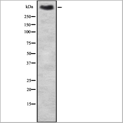 WB (Western Blot)
(Western blot analysis of MUC2 using HT-29 whole cell lysates)
WB (Western Blot)
(Western blot analysis of MUC2 using HT-29 whole cell lysates)
MUC2, Polyclonal Antibody (Cat# AAA31137)
Full Name
MUC2 Antibody
Gene Names
MUC2; MLP; SMUC; MUC-2
Reactivity
Human, Mouse, Rat
Applications
Western Blot, Immunohistochemistry, Immunofluorescence, Immunocytochemistry
Purity
The antiserum was purified by peptide affinity chromatography using SulfoLink™ Coupling Resin (Thermo Fisher Scientific).
Pricing
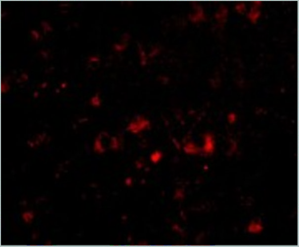
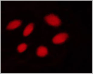
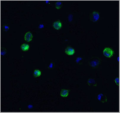
 IF (Immunofluorescence)
(Immunofluorescent analysis of 4% paraformaldehyde-fixed mouse brain issue labeling NPTX2 with AAA10933 at 20ug/mL, followed by goat anti-rabbit IgG secondaryantibody at 1/500 dilution (red) and DAPI staining (blue).)
IF (Immunofluorescence)
(Immunofluorescent analysis of 4% paraformaldehyde-fixed mouse brain issue labeling NPTX2 with AAA10933 at 20ug/mL, followed by goat anti-rabbit IgG secondaryantibody at 1/500 dilution (red) and DAPI staining (blue).)
NPTX2, Polyclonal Antibody (Cat# AAA10933)
Full Name
NPTX2 Antibody
Gene Names
NPTX2; NP2; NARP; NP-II
Reactivity
Human, Mouse, Rat
Applications
Western Blot, Immunohistochemistry, Immunofluorescence
Purity
NPTX2 Antibody is affinity chromatography purified via peptide column.
Pricing
C-telopeptide of type I collagen, ELISA Kit (Cat# AAA13203)
Full Name
Human C-telopeptide of type I collagen (ICTP) ELISA Kit
Reactivity
Human
Pricing
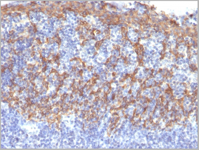
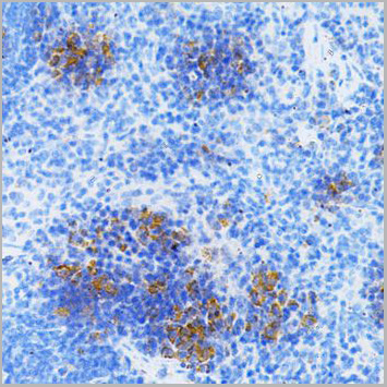
 IHC (Immunohistochemistry)
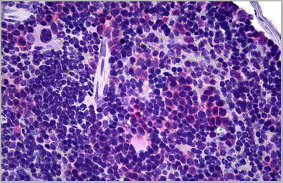
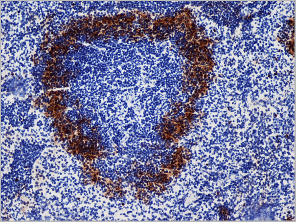
(Anti-TCR Gamma/Delta antibody IHC of mouse spleen. Immunohistochemistry of formalin-fixed, paraffin-embedded tissue after heat-induced antigen retrieval. Antibody dilution 1:50.)
IHC (Immunohistochemistry)
(Anti-TCR Gamma/Delta antibody IHC of mouse spleen. Immunohistochemistry of formalin-fixed, paraffin-embedded tissue after heat-induced antigen retrieval. Antibody dilution 1:50.)
TCR Gamma+TCR Delta, Monoclonal Antibody (Cat# AAA12360)
Full Name
Armenian Hamster Monoclonal [clone GL-3] (IgG) to Mouse TCR Gamma+TCR Delta
Reactivity
Mouse
Applications
Immunohistochemistry, Flow Cytometry
Purity
Protein G Purified
Pricing
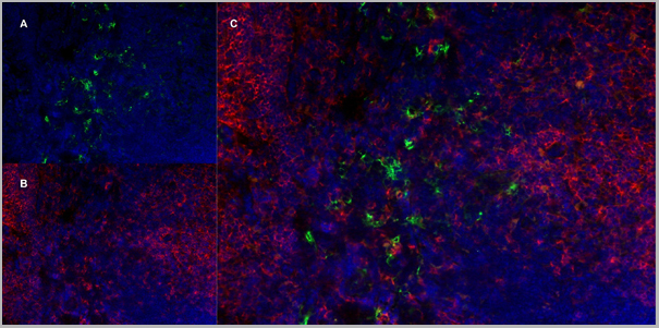
 Application Data
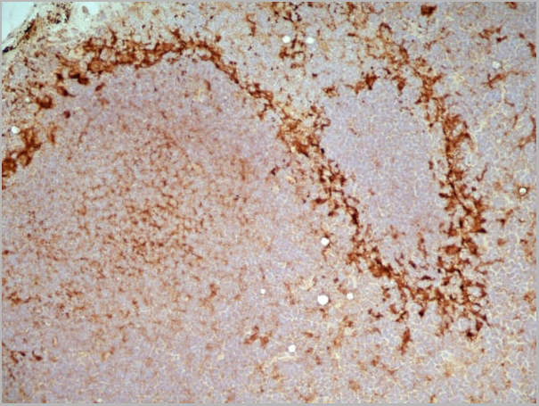
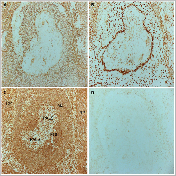
(Frozen mouse spleen stained with Rat anti Mouse CD169)
Application Data
(Frozen mouse spleen stained with Rat anti Mouse CD169)
CD169, Monoclonal Antibody (Cat# AAA12207)
Full Name
RAT ANTI MOUSE CD169
Gene Names
Siglec1; Sn; Cd169; Siglec-1
Applications
Immunohistochemistry, Flow Cytometry, Immunofluorescence
Pricing
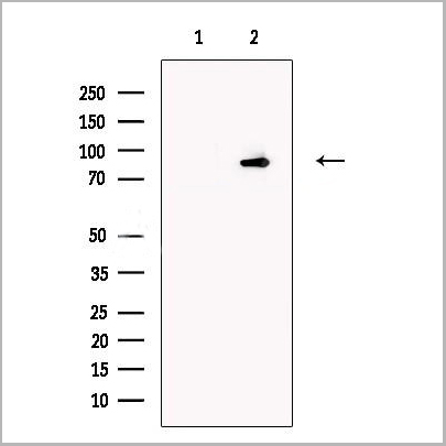
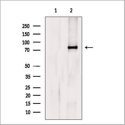
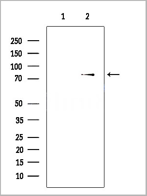
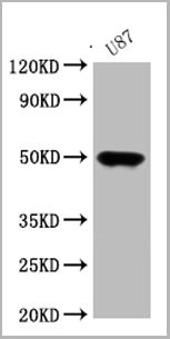 WB (Western Blot)
(Western BlotPositive WB detected in: U87 whole cell lysateAll lanes: ATAT1 antibody at 1:1000SecondaryGoat polyclonal to rabbit IgG at 1/50000 dilutionPredicted band size: 45, 46, 47, 38, 37, 36, 34 kDaObserved band size: 47 kDa)
WB (Western Blot)
(Western BlotPositive WB detected in: U87 whole cell lysateAll lanes: ATAT1 antibody at 1:1000SecondaryGoat polyclonal to rabbit IgG at 1/50000 dilutionPredicted band size: 45, 46, 47, 38, 37, 36, 34 kDaObserved band size: 47 kDa)
ATAT1, Polyclonal Antibody (Cat# AAA27001)
Full Name
ATAT1 Antibody
Gene Names
ATAT1; TAT; MEC17; C6orf134; Nbla00487; alpha-TAT; alpha-TAT1
Reactivity
Human
Applications
Western Blot
Purity
>95%, Protein G purified
Pricing
PLA2R1, Blocking Peptide (Cat# AAA13680)
Full Name
PLA2R1 Immunizing Peptide
Gene Names
PLA2R1; PLA2R; PLA2-R; PLA2IR; CLEC13C; PLA2G1R
Pricing
collagen, type I, alpha 2, Polyclonal Antibody (Cat# AAA15910)
Full Name
Rabbit anti-human collagen, type I, alpha 2 polyclonal Antibody
Gene Names
COL1A2; OI4
Reactivity
Human, Mouse, Rat
Applications
Western Blot, Immunohistochemistry
Purity
Antigen Affinity Purified
Pricing

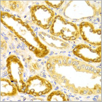
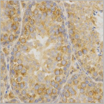
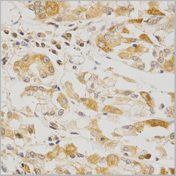

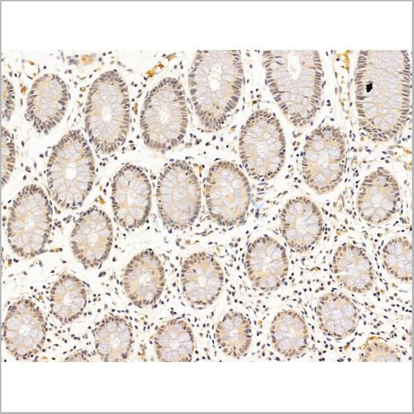
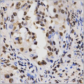
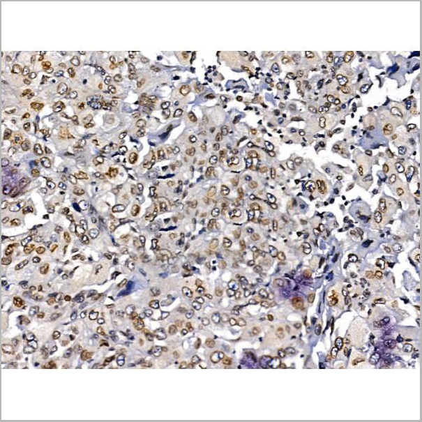
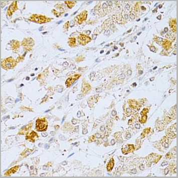 IHC (Immunohistchemistry)
(Immunohistochemistry of paraffin-embedded human stomach using OLR1 Antibody at dilution of 1:200 (40x lens).)
IHC (Immunohistchemistry)
(Immunohistochemistry of paraffin-embedded human stomach using OLR1 Antibody at dilution of 1:200 (40x lens).)
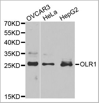
OLR1, Polyclonal Antibody (Cat# AAA10753)
Full Name
OLR1 Polyclonal Antibody
Gene Names
OLR1; LOX1; LOXIN; SLOX1; CLEC8A; SCARE1
Reactivity
Human
Applications
Western Blot, Immunohistochemistry
Purity
Affinity Purification
Pricing
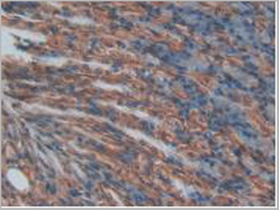
 IHC (Immunohistochemistry)
(Anti-ITGB2 / CD18 antibody IHC of human brain, cerebellum. Immunohistochemistry of formalin-fixed, paraffin-embedded tissue after heat-induced antigen retrieval. Antibody concentration 10 ug/ml.)
IHC (Immunohistochemistry)
(Anti-ITGB2 / CD18 antibody IHC of human brain, cerebellum. Immunohistochemistry of formalin-fixed, paraffin-embedded tissue after heat-induced antigen retrieval. Antibody concentration 10 ug/ml.)
ITGB2 / MAC-1 / CD18, Monoclonal Antibody (Cat# AAA12341)
Full Name
Mouse Monoclonal [clone MEM-48] (IgG1) to Human ITGB2 / MAC-1 / CD18
Gene Names
ITGB2; LAD; CD18; MF17; MFI7; LCAMB; LFA-1; MAC-1
Reactivity
Human
Applications
Immunohistochemistry, Western Blot, Immunoprecipitation, Flow Cytometry, Functional Assay
Purity
Purified by Protein A affinity chromatography.
>95% by SDS-PAGE.
>95% by SDS-PAGE.
Pricing
GNAT3, Antibody (Cat# AAA13665)
Full Name
Goat anti-GNAT3 Antibody
Gene Names
GNAT3; GDCA
Reactivity
Tested: Human; Expected from sequence similarity: Human, Mouse, Rat, Dog
Applications
Peptide ELISA, Immunohistochemistry, Western Blot
Purity
Purified from goat serum by ammonium sulphate precipitation followed by antigen affinity chromatography using the immunizing peptide.
Pricing

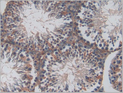
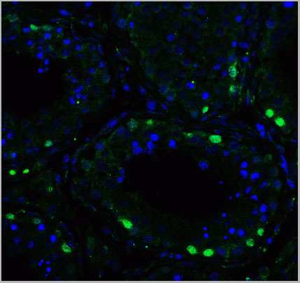
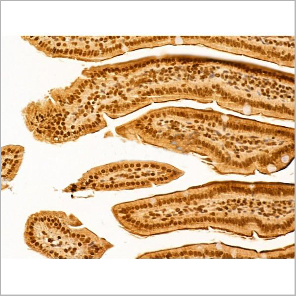
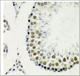
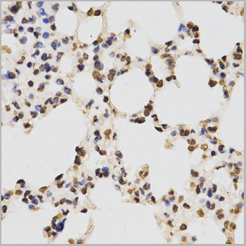
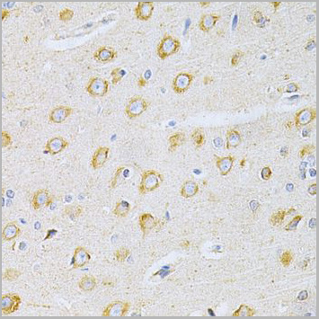
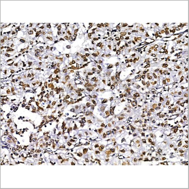
 IHC (Immunohistochemistry)
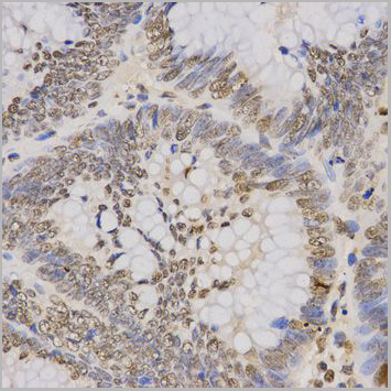
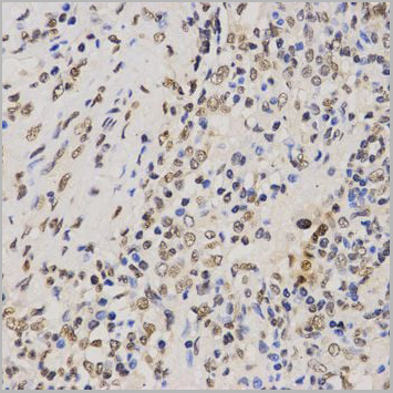
(Immunohistochemistry of paraffin-embedded mouse testis using TriMethyl-Histone H3-K79 antibody (AAA10656) at dilution of 1:200 (40x lens).)
IHC (Immunohistochemistry)
(Immunohistochemistry of paraffin-embedded mouse testis using TriMethyl-Histone H3-K79 antibody (AAA10656) at dilution of 1:200 (40x lens).)
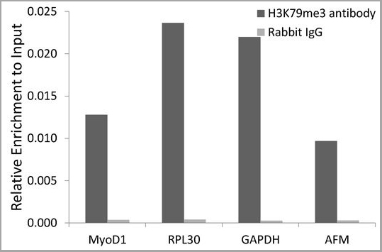
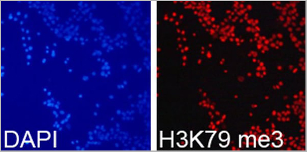
H3K79me3, Antibody (Cat# AAA10656)
Full Name
Histone H3K79me3 Polyclonal Antibody
Gene Names
HIST3H3; H3t; H3.4; H3/g; H3FT
Applications
Western Blot, Immunohistochemistry, Immunofluorescence, Immunoprecipitation, Chromatin Immunoprecipitation, Chromatin Immunoprecipitation
Purity
Affinity Purification
Pricing
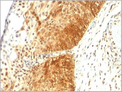
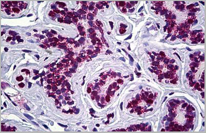
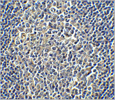
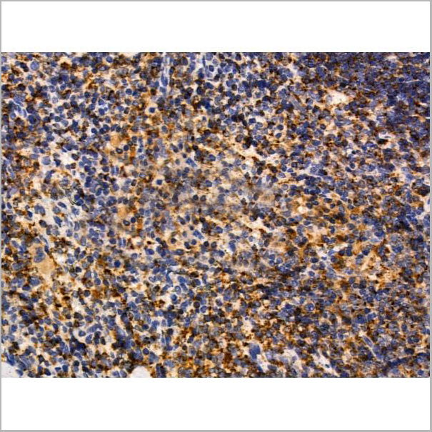
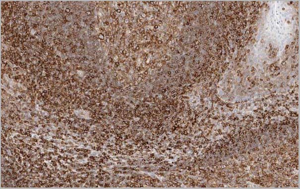 Application Data
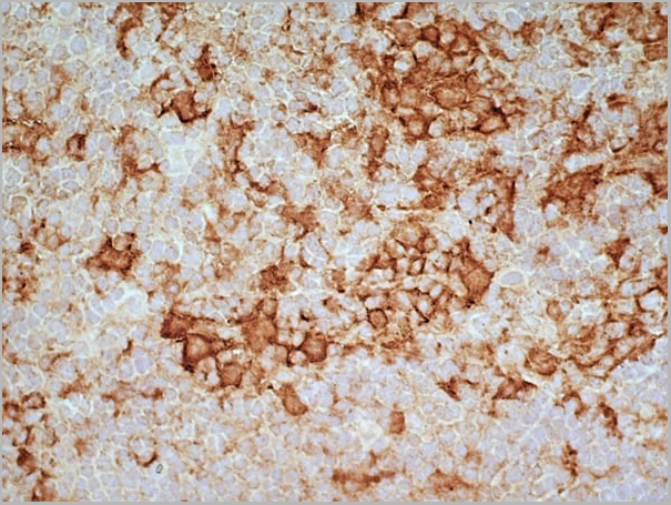
(Figure 1. Immunostaining of human tonsil tissue in paraffin section with HC10 (dilution 1:20.000).)
Application Data
(Figure 1. Immunostaining of human tonsil tissue in paraffin section with HC10 (dilution 1:20.000).)
HLA Class I Heavy Chain, Monoclonal Antibody (Cat# AAA14567)
Full Name
Mouse anti Human HLA Class I Heavy Chain (Restricted expression)
Reactivity
Human
Applications
Flow Cytometry, Immunocytochemistry, Immunohistochemistry, Immunoprecipitation, Western Blot
Pricing
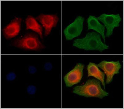
 IF (Immunofluorescence)
(AAA31214 staining Hela cells by IF/ICC. The samples were fixed with PFA and permeabilized in 0.1% Triton X-100,then blocked in 10% serum for 45 minutes at 25°C. Samples were then incubated with primary Ab(AAA31214 1:200) and mouse anti-beta tubulin Ab(T0023 1:200) for 1 hour at 37°C. An AlexaFluor594 conjugated goat anti-rabbit IgG(H+L) Ab(Red) and an AlexaFluor488 conjugated goat anti-mouse IgG(H+L) Ab(Green) were used as the secondary antibody.The nuclear counter stain is DAPI(blue).)
IF (Immunofluorescence)
(AAA31214 staining Hela cells by IF/ICC. The samples were fixed with PFA and permeabilized in 0.1% Triton X-100,then blocked in 10% serum for 45 minutes at 25°C. Samples were then incubated with primary Ab(AAA31214 1:200) and mouse anti-beta tubulin Ab(T0023 1:200) for 1 hour at 37°C. An AlexaFluor594 conjugated goat anti-rabbit IgG(H+L) Ab(Red) and an AlexaFluor488 conjugated goat anti-mouse IgG(H+L) Ab(Green) were used as the secondary antibody.The nuclear counter stain is DAPI(blue).)
MUM1, Polyclonal Antibody (Cat# AAA31214)
Full Name
MUM1 Antibody
Gene Names
PWWP3A; MUM1; MUM-1; EXPAND1; HSPC211
Reactivity
Human, Mouse, Rat, Monkey
Predicted Reactivity: Pig(100%), Bovine(100%), Horse(88%), Sheep(100%)
Predicted Reactivity: Pig(100%), Bovine(100%), Horse(88%), Sheep(100%)
Applications
ELISA
Purity
The antiserum was purified by peptide affinity chromatography using SulfoLink Coupling Resin (Thermo Fisher Scientific).
Pricing
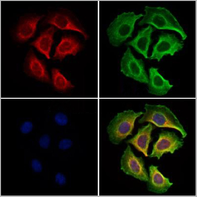
 IF (Immunofluorescence)
(AAA31138 staining Hela cells by IF/ICC. The samples were fixed with PFA and permeabilized in 0.1% Triton X-100, then blocked in 10% serum for 45 minutes at 25 degree C. Samples were then incubated with primary Ab(AAA31138) and mouse anti-beta tubulin Ab for 1 hour at 37 degree C. An AlexaFluor594 conjugated goat anti-rabbit IgG(H+L) Ab(Red) and an AlexaFluor488 conjugated goat anti-mouse IgG(H+L) Ab(Green) were used as the secondary Ab. The nuclear counter stain is DAPI (blue).)
IF (Immunofluorescence)
(AAA31138 staining Hela cells by IF/ICC. The samples were fixed with PFA and permeabilized in 0.1% Triton X-100, then blocked in 10% serum for 45 minutes at 25 degree C. Samples were then incubated with primary Ab(AAA31138) and mouse anti-beta tubulin Ab for 1 hour at 37 degree C. An AlexaFluor594 conjugated goat anti-rabbit IgG(H+L) Ab(Red) and an AlexaFluor488 conjugated goat anti-mouse IgG(H+L) Ab(Green) were used as the secondary Ab. The nuclear counter stain is DAPI (blue).)
Prestin, Polyclonal Antibody (Cat# AAA31138)
Full Name
Prestin Antibody
Gene Names
SLC26A5; PRES; DFNB61
Reactivity
Human, Mouse, Rat
Applications
Western Blot, Immunohistochemistry, Immunofluorescence
Purity
Peptide affinity chromatography using SulfoLink Coupling Resin (Thermo Fisher Scientific).
Pricing
Mouse IgE, BALB/c Isotype Control, Monoclonal Isotype Control (Cat# AAA14917)
Full Name
Mouse IgE-UNLB
Applications
ELISA; Flow Cytometry; Western Blot
Pricing