Filters
▼Clonality
▼Type
▼Reactivity
▼Gene Name
▼Isotype
▼Host
▼Application
▼Clone
▼Phospho Antibodies
Phospho-specific antibodies’ typical purpose is to enable researchers to detect changes in proteins. They will exclusively bind to the amino acid sequence on a protein that has been phosphorylated (which is both a physical & chemical change) and do not bind to the same amino acid sequence on said protein if it lacks said phosphorylation. This aids in being able to clearly see and understand the data produced from this particular protein modification.
Viewing 3600-3650 of 5297 product results
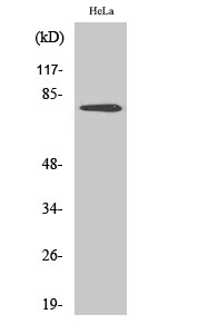
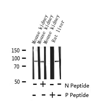
 WB (Western Blot)
(Western blot analysis of extracts from Hela cells, untreated (-) or treated, 1:5000. Secondary antibody was diluted at 1:20000)
WB (Western Blot)
(Western blot analysis of extracts from Hela cells, untreated (-) or treated, 1:5000. Secondary antibody was diluted at 1:20000)
Histone H1, ELISA Kit (Cat# AAA319064)
 WB (Western Blot)
(Western blot analysis of extracts from Hela cells, untreated (-) or treated, 1:2000. Secondary antibody was diluted at 1:20000 cells nucleus extracted by Minute TM Cytoplasmic and Nuclear Fractionation kit (SC-003,Inventbiotech,MN,USA).)
WB (Western Blot)
(Western blot analysis of extracts from Hela cells, untreated (-) or treated, 1:2000. Secondary antibody was diluted at 1:20000 cells nucleus extracted by Minute TM Cytoplasmic and Nuclear Fractionation kit (SC-003,Inventbiotech,MN,USA).)
Histone H3, ELISA Kit (Cat# AAA319068)
 WB (Western Blot)
(Western blot analysis of extracts from Hela cells, untreated (-) or treated, 1:2000. Secondary antibody was diluted at 1:20000 cells nucleus extracted by Minute TM Cytoplasmic and Nuclear Fractionation kit (SC-003,Inventbiotech,MN,USA).)
WB (Western Blot)
(Western blot analysis of extracts from Hela cells, untreated (-) or treated, 1:2000. Secondary antibody was diluted at 1:20000 cells nucleus extracted by Minute TM Cytoplasmic and Nuclear Fractionation kit (SC-003,Inventbiotech,MN,USA).)
Histone H3, ELISA Kit (Cat# AAA319072)
 WB (Western Blot)
(Western blot analysis of extracts from Hela cells, untreated (-) or treated, 1:2000. Secondary antibody was diluted at 1:20000 cells nucleus extracted by Minute TM Cytoplasmic and Nuclear Fractionation kit (SC-003,Inventbiotech,MN,USA).)
WB (Western Blot)
(Western blot analysis of extracts from Hela cells, untreated (-) or treated, 1:2000. Secondary antibody was diluted at 1:20000 cells nucleus extracted by Minute TM Cytoplasmic and Nuclear Fractionation kit (SC-003,Inventbiotech,MN,USA).)
Histone H3, ELISA Kit (Cat# AAA319074)
 WB (Western Blot)
(Western blot analysis of extracts from Hela cells, untreated (-) or treated, 1:2000. Secondary antibody was diluted at 1:20000 cells nucleus extracted by Minute TM Cytoplasmic and Nuclear Fractionation kit (SC-003,Inventbiotech,MN,USA).)
WB (Western Blot)
(Western blot analysis of extracts from Hela cells, untreated (-) or treated, 1:2000. Secondary antibody was diluted at 1:20000 cells nucleus extracted by Minute TM Cytoplasmic and Nuclear Fractionation kit (SC-003,Inventbiotech,MN,USA).)
Histone H3, ELISA Kit (Cat# AAA319075)
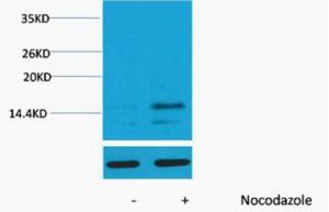
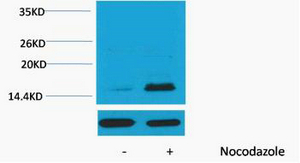
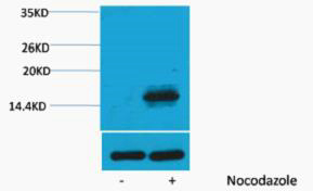
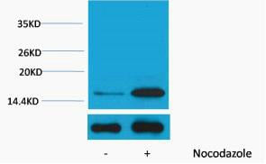
 WB (Western Blot)
(Western Blot analysis of Hela treated or untreated by LPS lysis, using primary antibody at 1:1000 dilution. Secondary antibody)
WB (Western Blot)
(Western Blot analysis of Hela treated or untreated by LPS lysis, using primary antibody at 1:1000 dilution. Secondary antibody)
PRK1, Polyclonal Antibody (Cat# AAA319478)
 WB (Western Blot)
(Western blot analysis of lysates from K562 cells, primary antibody was diluted at 1:1000, 4 degree over night)
WB (Western Blot)
(Western blot analysis of lysates from K562 cells, primary antibody was diluted at 1:1000, 4 degree over night)
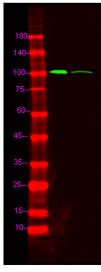
Prdx1, Polyclonal Antibody (Cat# AAA319486)
 WB (Western Blot)
(Western Blot analysis of various cells using Phospho-GSK3beta (S9) Polyclonal Antibody diluted at 1:500)
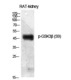
WB (Western Blot)
(Western Blot analysis of various cells using Phospho-GSK3beta (S9) Polyclonal Antibody diluted at 1:500)
GSK3beta, ELISA Kit (Cat# AAA319079)
 WB (Western Blot)
(Western Blot analysis of various cell lysis. Primary Antibody was diluted at 1:1000. Secondary antibody(was diluted at 1:10000)
WB (Western Blot)
(Western Blot analysis of various cell lysis. Primary Antibody was diluted at 1:1000. Secondary antibody(was diluted at 1:10000)
ERalpha, ELISA Kit (Cat# AAA319082)
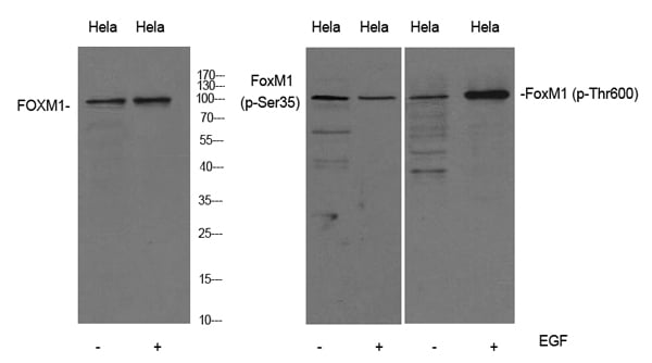 WB (Western Blot)
(Western Blot analysis of Hela cell lysis treated or untreated by EGF 20ng/ml 30'.. Primary Antibody was diluted at 1:1000. Secondary antibody( was diluted at 1:10000)
WB (Western Blot)
(Western Blot analysis of Hela cell lysis treated or untreated by EGF 20ng/ml 30'.. Primary Antibody was diluted at 1:1000. Secondary antibody( was diluted at 1:10000)
FoxM1, ELISA Kit (Cat# AAA319086)
 WB (Western Blot)
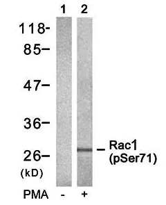
(Western blot analysis of Rac1(Phospho-Ser71) antibody in 293 cells lysates, cell treated or untreated with PMA 125ng/ml 30', 4 degree C over night, secondary antibody(cat: SA0002 was diluted at 1:10000, 37 degree C 1hour.)
WB (Western Blot)
(Western blot analysis of Rac1(Phospho-Ser71) antibody in 293 cells lysates, cell treated or untreated with PMA 125ng/ml 30', 4 degree C over night, secondary antibody(cat: SA0002 was diluted at 1:10000, 37 degree C 1hour.)
Rac1/cdc42, ELISA Kit (Cat# AAA319088)
 WB (Western Blot)
(Western blot analysis of lysates from SH-SY5Y cells, primary antibody was diluted at 1:1000, 4 degree C over night)
WB (Western Blot)
(Western blot analysis of lysates from SH-SY5Y cells, primary antibody was diluted at 1:1000, 4 degree C over night)
LDHA, ELISA Kit (Cat# AAA319093)
 WB (Western Blot)
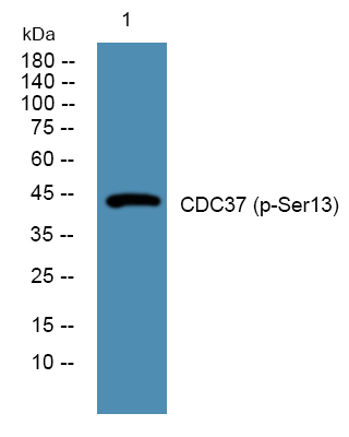
(Western blot analysis of lysates from K562 cells, primary antibody was diluted at 1:1000, 4 degree C over night)
WB (Western Blot)
(Western blot analysis of lysates from K562 cells, primary antibody was diluted at 1:1000, 4 degree C over night)
CDC37, ELISA Kit (Cat# AAA319097)
 WB (Western Blot)
(Western blot analysis of extracts from Hela cells, 1:2000. Secondary antibody was diluted at 1:20000 cells nucleus extracted by Minute TM Cytoplasmic and Nuclear Fractionation kit (SC-003,Inventbiotech,MN,USA).)
WB (Western Blot)
(Western blot analysis of extracts from Hela cells, 1:2000. Secondary antibody was diluted at 1:20000 cells nucleus extracted by Minute TM Cytoplasmic and Nuclear Fractionation kit (SC-003,Inventbiotech,MN,USA).)
Histone H2A.X, ELISA Kit (Cat# AAA319108)
 WB (Western Blot)
(Western blot analysis of lysates from KB cells, primary antibody was diluted at 1:1000, 4 degree C over night)
WB (Western Blot)
(Western blot analysis of lysates from KB cells, primary antibody was diluted at 1:1000, 4 degree C over night)
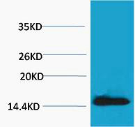
NPM, ELISA Kit (Cat# AAA319114)
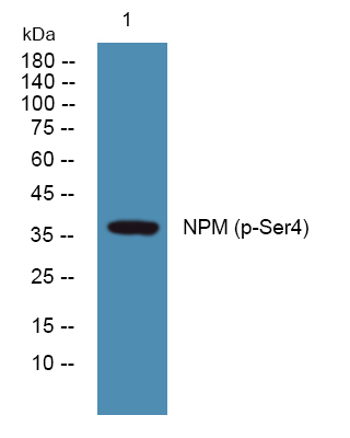
 WB (Western Blot)
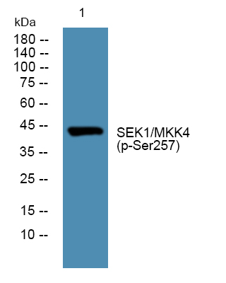
(Western blot analysis of lysates from DU145 cells, primary antibody was diluted at 1:1000, 4 degree C over night)
WB (Western Blot)
(Western blot analysis of lysates from DU145 cells, primary antibody was diluted at 1:1000, 4 degree C over night)
SEK1/MKK4, ELISA Kit (Cat# AAA319121)
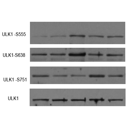 WB (Western Blot)
(Western Blot analysis of various cell lysis(provided by our customer). Primary Antibody was diluted at 1:1000. Secondary antibody(was diluted at 1:10000)
WB (Western Blot)
(Western Blot analysis of various cell lysis(provided by our customer). Primary Antibody was diluted at 1:1000. Secondary antibody(was diluted at 1:10000)
ULK1, ELISA Kit (Cat# AAA319132)
 WB (Western Blot)
(Western Blot analysis of HELA cells using Antibody diluted at 500. Secondary antibody was diluted at 1:20000)
WB (Western Blot)
(Western Blot analysis of HELA cells using Antibody diluted at 500. Secondary antibody was diluted at 1:20000)
Vimentin, ELISA Kit (Cat# AAA319133)
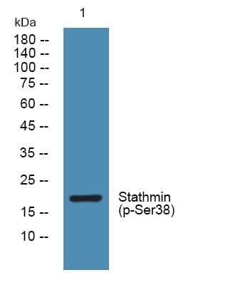 WB (Western Blot)
(Western blot analysis of lysates from K562 cells, primary antibody was diluted at 1:1000, 4 degree C over night)
WB (Western Blot)
(Western blot analysis of lysates from K562 cells, primary antibody was diluted at 1:1000, 4 degree C over night)
Stathmin, ELISA Kit (Cat# AAA319135)
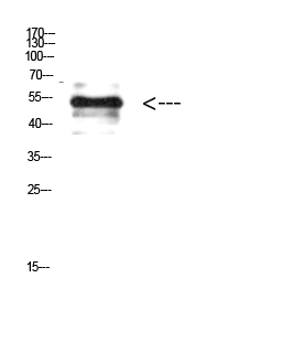
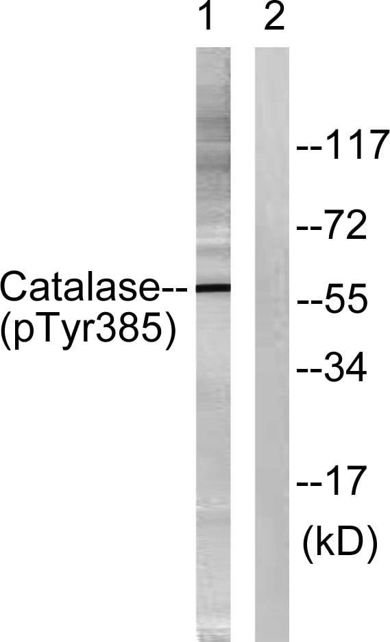 WB (Western Blot)
(Western blot analysis of lysates from Jurkat cells, using Catalase (Phospho-Tyr385) Antibody. The lane on the right is blocked with the phospho peptide.)
WB (Western Blot)
(Western blot analysis of lysates from Jurkat cells, using Catalase (Phospho-Tyr385) Antibody. The lane on the right is blocked with the phospho peptide.)
Catalase, ELISA Kit (Cat# AAA319136)
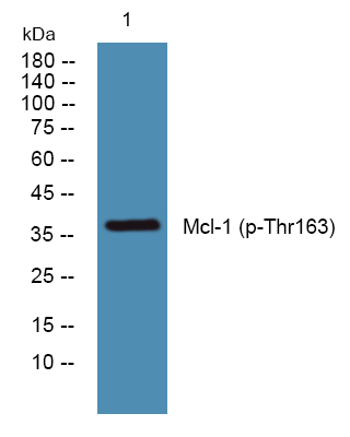 WB (Western Blot)
(Western blot analysis of lysates from SH-SY5Y cells, primary antibody was diluted at 1:1000, 4 degree C over night)
WB (Western Blot)
(Western blot analysis of lysates from SH-SY5Y cells, primary antibody was diluted at 1:1000, 4 degree C over night)
Mcl-1, ELISA Kit (Cat# AAA319139)
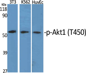 WB (Western Blot)
(Western Blot analysis of various cells using Phospho-Akt1 (T450) Polyclonal Antibody diluted at 1:1000)
WB (Western Blot)
(Western Blot analysis of various cells using Phospho-Akt1 (T450) Polyclonal Antibody diluted at 1:1000)
Akt1, ELISA Kit (Cat# AAA319157)
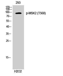 WB (Western Blot)
(Western Blot analysis of 293 cells using Phospho-MSK2 (T568) Polyclonal Antibody cells nucleus extracted by Minute TM Cytoplasmic and Nuclear Fractionation kit (SC-003,Inventbiotech,MN,USA).)
WB (Western Blot)
(Western Blot analysis of 293 cells using Phospho-MSK2 (T568) Polyclonal Antibody cells nucleus extracted by Minute TM Cytoplasmic and Nuclear Fractionation kit (SC-003,Inventbiotech,MN,USA).)
MSK2, ELISA Kit (Cat# AAA319258)
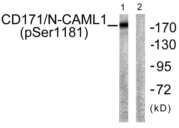 WB (Western Blot)
(Western blot analysis of lysates from K562 cells, using CD171/N-CAML1 (Phospho-Ser1181) Antibody. The lane on the right is blocked with the phospho peptide.)
WB (Western Blot)
(Western blot analysis of lysates from K562 cells, using CD171/N-CAML1 (Phospho-Ser1181) Antibody. The lane on the right is blocked with the phospho peptide.)
NCAM-L1, ELISA Kit (Cat# AAA319261)
 WB (Western Blot)
(Western Blot analysis of HepG2 cells using Phospho-Nur77 (S351) Polyclonal Antibody cells nucleus extracted by Minute TM Cytoplasmic and Nuclear Fractionation kit (SC-003,Inventbiotech,MN,USA).)
WB (Western Blot)
(Western Blot analysis of HepG2 cells using Phospho-Nur77 (S351) Polyclonal Antibody cells nucleus extracted by Minute TM Cytoplasmic and Nuclear Fractionation kit (SC-003,Inventbiotech,MN,USA).)
Nur77, ELISA Kit (Cat# AAA319266)
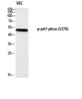 WB (Western Blot)
(Western blot analysis of VEC using p-p47-phox (S370) antibody.)
WB (Western Blot)
(Western blot analysis of VEC using p-p47-phox (S370) antibody.)
p47-phox, ELISA Kit (Cat# AAA319268)
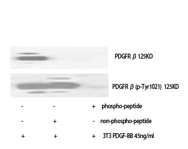 WB (Western Blot)
(Western Blot analysis of various cells using Phospho-PDGFR-beta (Y1021) Polyclonal Antibody)
WB (Western Blot)
(Western Blot analysis of various cells using Phospho-PDGFR-beta (Y1021) Polyclonal Antibody)
PDGFR-alpha, ELISA Kit (Cat# AAA319272)
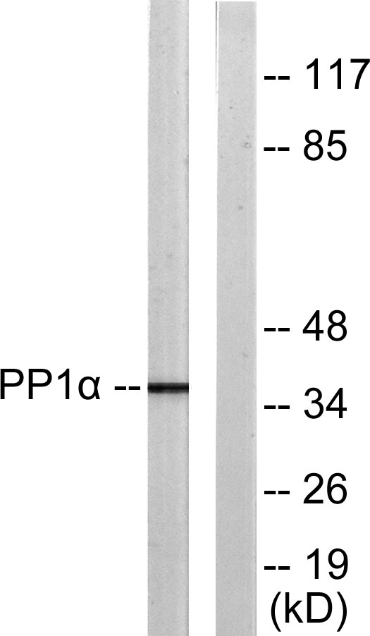 WB (Western Blot)
(Western blot analysis of PP1-alpha (Phospho-Thr320) Antibody. The lane on the right is blocked with the PP1-alpha (Phospho-Thr320) peptide.)
WB (Western Blot)
(Western blot analysis of PP1-alpha (Phospho-Thr320) Antibody. The lane on the right is blocked with the PP1-alpha (Phospho-Thr320) peptide.)
PP1alpha, ELISA Kit (Cat# AAA319275)
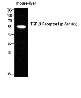 WB (Western Blot)
(Western Blot analysis of MOUSE-LIVER cells using Phospho-TGFbeta RI (S165) Polyclonal Antibody diluted at 1:1000)
WB (Western Blot)
(Western Blot analysis of MOUSE-LIVER cells using Phospho-TGFbeta RI (S165) Polyclonal Antibody diluted at 1:1000)
TGFbeta RI, ELISA Kit (Cat# AAA319280)
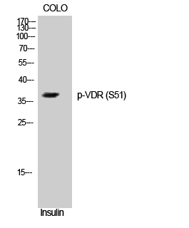 WB (Western Blot)
(Western Blot analysis of COLO cells using Phospho-VDR (S51) Polyclonal Antibody cells nucleus extracted by Minute TM Cytoplasmic and Nuclear Fractionation kit (SC-003,Inventbiotech,MN,USA).)
WB (Western Blot)
(Western Blot analysis of COLO cells using Phospho-VDR (S51) Polyclonal Antibody cells nucleus extracted by Minute TM Cytoplasmic and Nuclear Fractionation kit (SC-003,Inventbiotech,MN,USA).)
VDR, ELISA Kit (Cat# AAA319283)
 WB (Western Blot)
(Western Blot analysis of various cells using Phospho-Btk (Y551) Polyclonal Antibody)
WB (Western Blot)
(Western Blot analysis of various cells using Phospho-Btk (Y551) Polyclonal Antibody)
Btk, ELISA Kit (Cat# AAA319288)
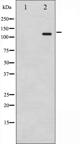
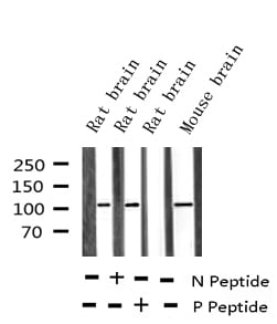
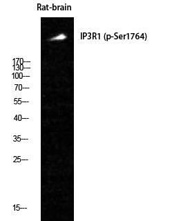 WB (Western Blot)
(Western Blot analysis of Rat-brain cells using Phospho-IP3R-I (S1764) Polyclonal Antibody)
WB (Western Blot)
(Western Blot analysis of Rat-brain cells using Phospho-IP3R-I (S1764) Polyclonal Antibody)
IP3R-I, ELISA Kit (Cat# AAA319289)
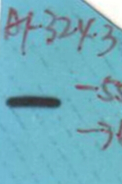
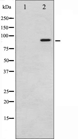
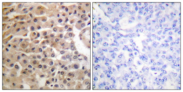
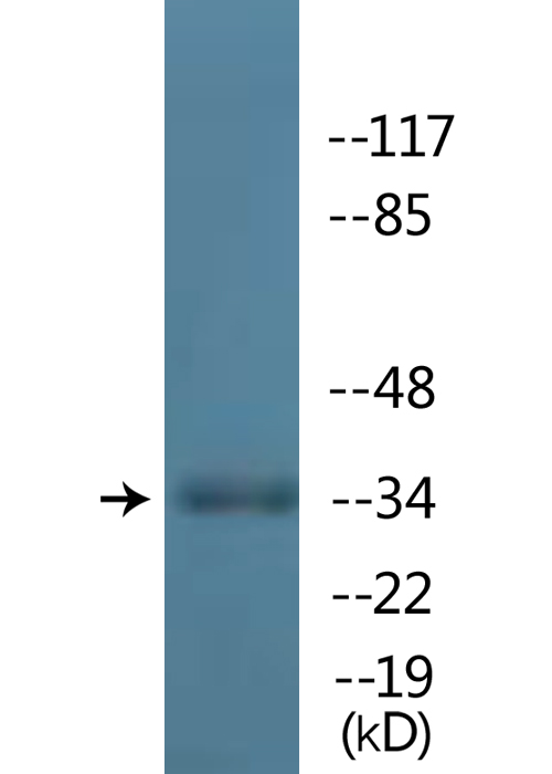
 WB (Western Blot)
(Western blot analysis of Phospho-Retinoblastoma (Ser795) expression in various lysates)
WB (Western Blot)
(Western blot analysis of Phospho-Retinoblastoma (Ser795) expression in various lysates)
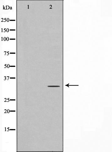
Retinoblastoma, Polyclonal Antibody (Cat# AAA321484)
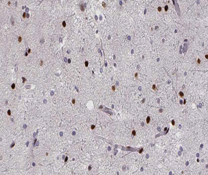
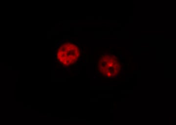
 IF (Immunofluorescence)
(AAA321535 staining HeLa by IF/ICC. The sample were fixed with PFA and permeabilized in 0.1% Triton X-100, then blocked in 10% serum for 45 minutes at 25 degree C. The primary antibody was diluted at 1/200 and incubated with the sample for 1 hour at 37 degree C. An Alexa Fluor 594 conjugated goat anti-rabbit IgG (H+L) Ab, diluted at 1/600, was used as the secondary antibody.)
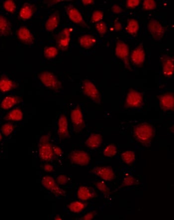
IF (Immunofluorescence)
(AAA321535 staining HeLa by IF/ICC. The sample were fixed with PFA and permeabilized in 0.1% Triton X-100, then blocked in 10% serum for 45 minutes at 25 degree C. The primary antibody was diluted at 1/200 and incubated with the sample for 1 hour at 37 degree C. An Alexa Fluor 594 conjugated goat anti-rabbit IgG (H+L) Ab, diluted at 1/600, was used as the secondary antibody.)
RFA2, Polyclonal Antibody (Cat# AAA321535)
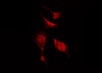
 IF (Immunofluorescence)
(AAA321579 staining K562 by IF/ICC. The sample were fixed with PFA and permeabilized in 0.1% Triton X-100, then blocked in 10% serum for 45 minutes at 25 degree C. The primary antibody was diluted at 1/200 and incubated with the sample for 1 hour at 37 degree C. An Alexa Fluor 594 conjugated goat anti-rabbit IgG (H+L) Ab, diluted at 1/600, was used as the secondary antibody.)
IF (Immunofluorescence)
(AAA321579 staining K562 by IF/ICC. The sample were fixed with PFA and permeabilized in 0.1% Triton X-100, then blocked in 10% serum for 45 minutes at 25 degree C. The primary antibody was diluted at 1/200 and incubated with the sample for 1 hour at 37 degree C. An Alexa Fluor 594 conjugated goat anti-rabbit IgG (H+L) Ab, diluted at 1/600, was used as the secondary antibody.)
MKP-1/2, Polyclonal Antibody (Cat# AAA321579)

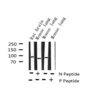
 WB (Western Blot)
(Western blot analysis of Phospho-Catenin beta (Tyr489)expression in various lysates)
WB (Western Blot)
(Western blot analysis of Phospho-Catenin beta (Tyr489)expression in various lysates)
Catenin beta, Polyclonal Antibody (Cat# AAA321596)
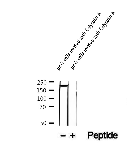 WB (Western Blot)
(Western blot analysis of extracts of pc-3 cells treated with Calyculin A, using Phospho-KIF1B (Ser1487) Antibody.)
WB (Western Blot)
(Western blot analysis of extracts of pc-3 cells treated with Calyculin A, using Phospho-KIF1B (Ser1487) Antibody.)
KIF1B, Polyclonal Antibody (Cat# AAA321341)
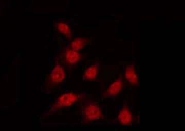
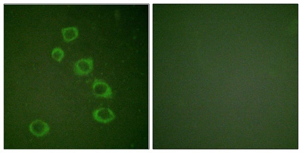
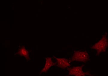 IF (Immunofluorescence)
(AAA321438 staining HuvEc by IF/ICC. The sample were fixed with PFA and permeabilized in 0.1% Triton X-100, then blocked in 10% serum for 45 minutes at 25 degree C. The primary antibody was diluted at 1/200 and incubated with the sample for 1 hour at 37 degree C. An Alexa Fluor 594 conjugated goat anti-rabbit IgG (H+L) Ab, diluted at 1/600, was used as the secondary antibody.)
IF (Immunofluorescence)
(AAA321438 staining HuvEc by IF/ICC. The sample were fixed with PFA and permeabilized in 0.1% Triton X-100, then blocked in 10% serum for 45 minutes at 25 degree C. The primary antibody was diluted at 1/200 and incubated with the sample for 1 hour at 37 degree C. An Alexa Fluor 594 conjugated goat anti-rabbit IgG (H+L) Ab, diluted at 1/600, was used as the secondary antibody.)
p95/NBS1, Polyclonal Antibody (Cat# AAA321438)
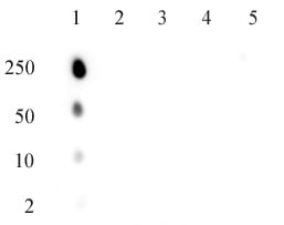 DB (Dot Blot)
(AbFlex ATM phospho Ser1981 antibody specifity Dot blot analysis was used to confirm the specificity of ATM phospho Ser1981 antibody. Single-stranded DNA oligonucleotides (amount of oligo in nanograms listed on the left side of the blot) were spotted on to a positively charged nylon membrane and blotted with antibody (0.5 ug/ml dilution). 1: ATM phospho serine 1981 (SLAFEEG(Sph)QSTTISS) 2: H2AX phospho serine 139 (CKATQA[Sph]QEY) 3: ATM unmodified serine 1981 (SLAFEEGSQSTTISS) 4: NBS phospho serine 343 (CPGPSL[Sph]QGVS) 5: BRCA1 phospho serine 1423 (LEQHG[Sph]QSNSC))
DB (Dot Blot)
(AbFlex ATM phospho Ser1981 antibody specifity Dot blot analysis was used to confirm the specificity of ATM phospho Ser1981 antibody. Single-stranded DNA oligonucleotides (amount of oligo in nanograms listed on the left side of the blot) were spotted on to a positively charged nylon membrane and blotted with antibody (0.5 ug/ml dilution). 1: ATM phospho serine 1981 (SLAFEEG(Sph)QSTTISS) 2: H2AX phospho serine 139 (CKATQA[Sph]QEY) 3: ATM unmodified serine 1981 (SLAFEEGSQSTTISS) 4: NBS phospho serine 343 (CPGPSL[Sph]QGVS) 5: BRCA1 phospho serine 1423 (LEQHG[Sph]QSNSC))
ATM phospho Ser1981, Antibody (Cat# AAA60273)
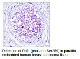
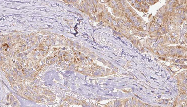
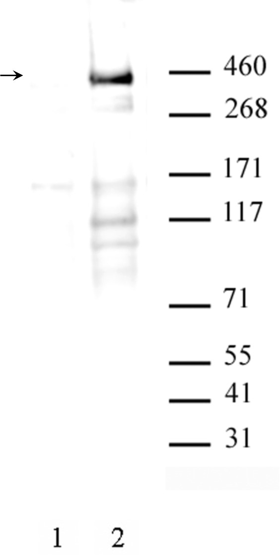
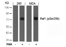 Application Data
Raf1, Polyclonal Antibody (Cat# AAA47790)
Phospho Tou protein, ELISA Kit (Cat# AAA85047)
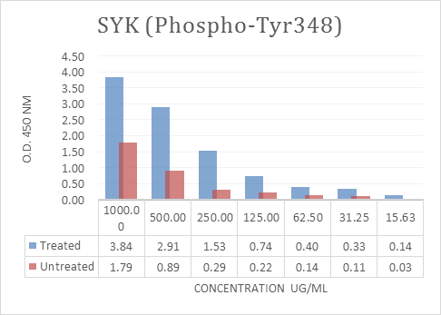
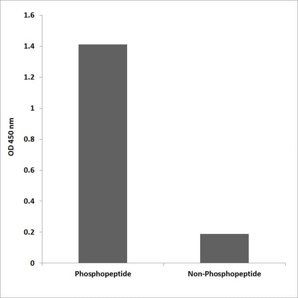
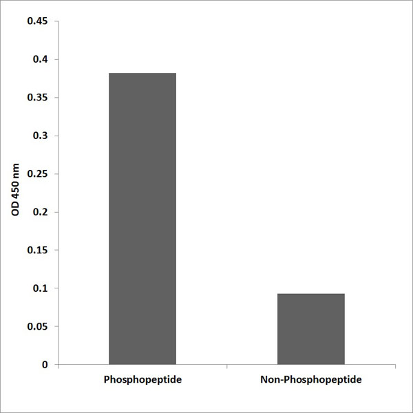
 ELISA
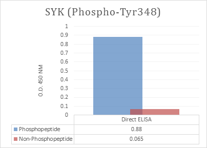
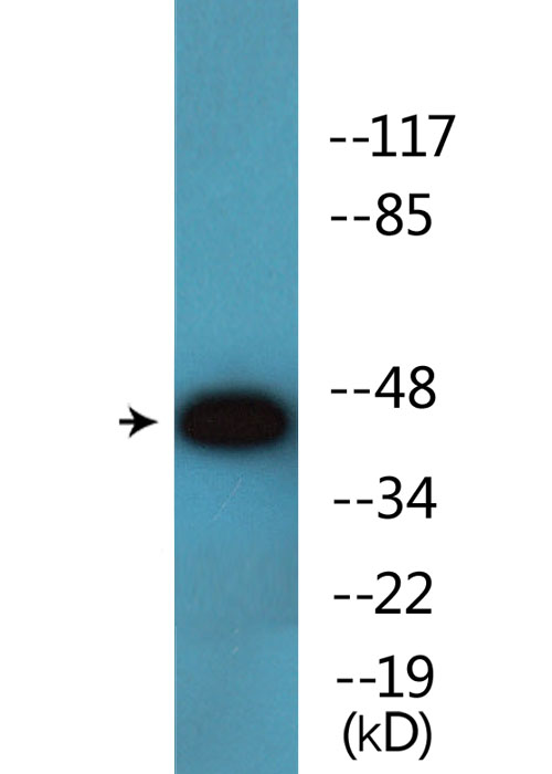
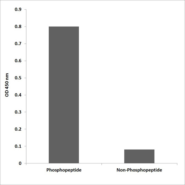
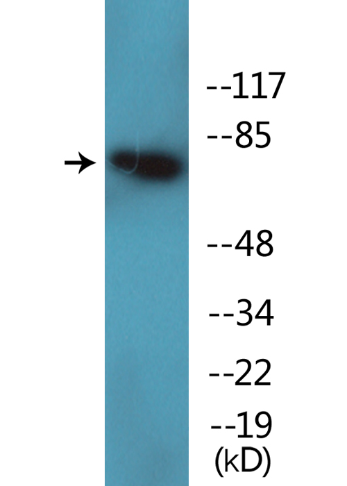
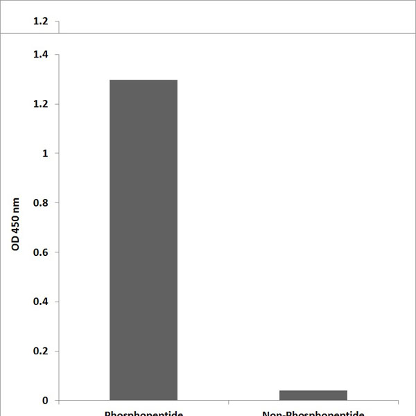
(Enzyme-Linked Immunosorbent Assay (ELISA) for immunogen phosphor-peptide (left) and non-phospho peptide (right), using Anti-SYK (Phospho-Tyr348) Antibody.)
ELISA
(Enzyme-Linked Immunosorbent Assay (ELISA) for immunogen phosphor-peptide (left) and non-phospho peptide (right), using Anti-SYK (Phospho-Tyr348) Antibody.)
SYK, ELISA Kit (Cat# AAA318613)
 ELISA
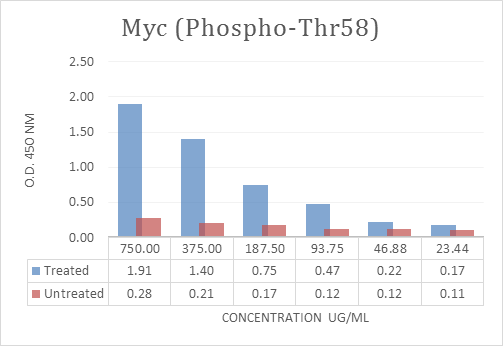
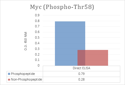
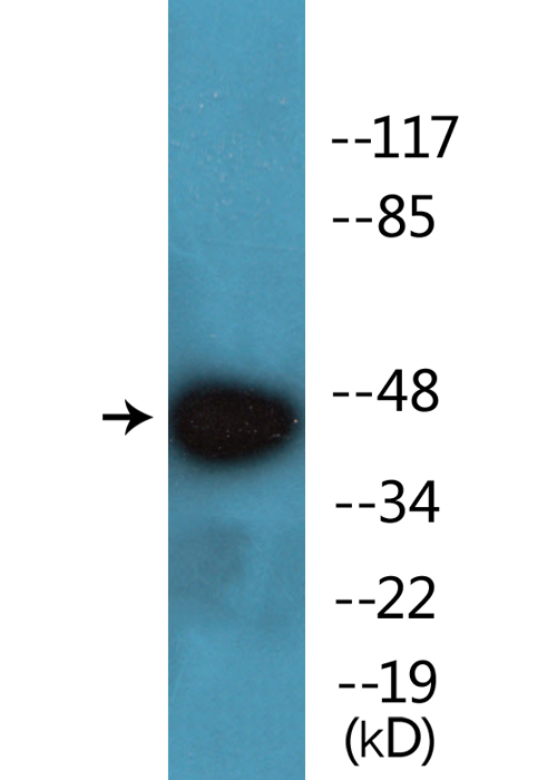
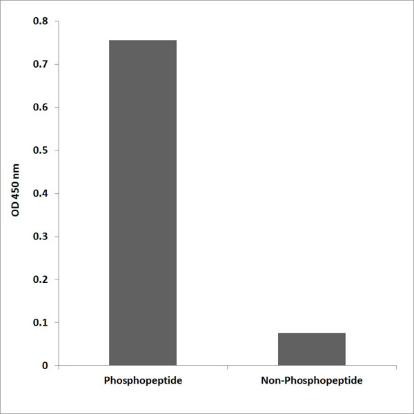
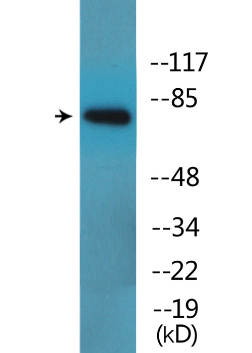
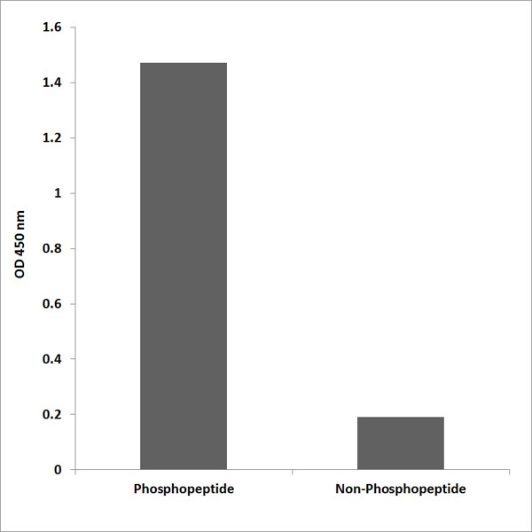
(Enzyme-Linked Immunosorbent Assay (ELISA) for immunogen phosphor-peptide (left) and non-phospho peptide (right), using Anti-Myc (Phospho-Thr58) Antibody.)
ELISA
(Enzyme-Linked Immunosorbent Assay (ELISA) for immunogen phosphor-peptide (left) and non-phospho peptide (right), using Anti-Myc (Phospho-Thr58) Antibody.)
Myc, ELISA Kit (Cat# AAA318625)
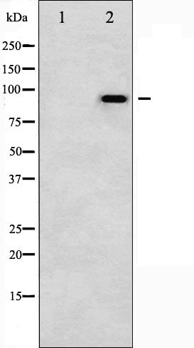
 WB (Western Blot)
(Western Blot analysis of various cells using Phospho-Neu (Y1221/Y1222) Polyclonal Antibody)
WB (Western Blot)
(Western Blot analysis of various cells using Phospho-Neu (Y1221/Y1222) Polyclonal Antibody)
Neu, Polyclonal Antibody (Cat# AAA318639)
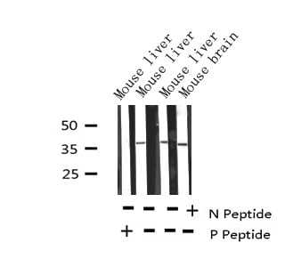
 WB (Western Blot)
(Western blot analysis of lysates from HepG2 cells treated with TNF 20ng/ml 5', using p47 phox (Phospho-Ser345) Antibody.)
WB (Western Blot)
(Western blot analysis of lysates from HepG2 cells treated with TNF 20ng/ml 5', using p47 phox (Phospho-Ser345) Antibody.)
p47 phox, Polyclonal Antibody (Cat# AAA318368)
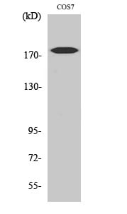
 WB (Western Blot)
(Western blot analysis of lysates from COS7 cells treated with PMA 125ng/ml 30', using PKA-R2 beta (Phospho-Ser113) Antibody.)
WB (Western Blot)
(Western blot analysis of lysates from COS7 cells treated with PMA 125ng/ml 30', using PKA-R2 beta (Phospho-Ser113) Antibody.)
PKA-R2beta, Polyclonal Antibody (Cat# AAA318374)
 WB (Western Blot)
(Western blot analysis of lysates from Jurkat cells treated with EGF 200ng/ml 15', using PKC theta (Phospho-Ser695) Antibody.)
WB (Western Blot)
(Western blot analysis of lysates from Jurkat cells treated with EGF 200ng/ml 15', using PKC theta (Phospho-Ser695) Antibody.)
PKC theta, Polyclonal Antibody (Cat# AAA318375)
 WB (Western Blot)
(Western blot analysis of lysates from COS7 cells treated with serum 20% 15', using SP1 (Phospho-Thr739) Antibody.)
WB (Western Blot)
(Western blot analysis of lysates from COS7 cells treated with serum 20% 15', using SP1 (Phospho-Thr739) Antibody.)
SP1, Polyclonal Antibody (Cat# AAA318388)
 WB (Western Blot)
(Western blot analysis of lysates from Jurkat cells treated with PMA 125ng/ml 30', using TAL-1 (Phospho-Ser122) Antibody.)
WB (Western Blot)
(Western blot analysis of lysates from Jurkat cells treated with PMA 125ng/ml 30', using TAL-1 (Phospho-Ser122) Antibody.)
TAL-1, Polyclonal Antibody (Cat# AAA318395)
 WB (Western Blot)
(Western blot analysis of lysates from HepG2 cells treated with Ca2+ 40uM 30', using Integrin beta1 (Phospho-Thr789) Antibody.)
WB (Western Blot)
(Western blot analysis of lysates from HepG2 cells treated with Ca2+ 40uM 30', using Integrin beta1 (Phospho-Thr789) Antibody.)
Integrin beta1, Polyclonal Antibody (Cat# AAA318330)
What Are Phospho Antibodies?
Protein phosphorylation is a process where a phosphate group is added to certain amino acid residues of a protein – usually serine (S), threonine (T), or tyrosine (Y) - by enzymes called kinases. This process is integral in controlling cellular signaling, cellular growth, and other biological functions.
Our catalog includes a wide range of phospho-specific antibodies that can accurately detect this important marker. They perform strongly in widely-used laboratory applications such as Western blot, flow cytometry, immunohistochemistry, and immunofluorescence microscopy. We value your trust in us and are committed to providing top-quality products and services. All of our antibodies are guaranteed to work for the applications and species indicated on our website & associated product pages.
What Are The Key Applications of Phospho Antibodies?
1. Western Blotting
One of the first steps a researcher can take in utilizing these phospho-specific antibodies, is to check if the antibody works using a technique referred to as “Western blot”. For those unfamiliar, Western Blot aids in showing whether the protein that the antibody recognizes is appearing at the correct/expected size. These phospho-specific antibodies should also be able to detect changes in the target protein’s phosphorylation (on/off state) when cells are stimulated in certain ways.
2. Staining of Fixed Cells (Immunocytochemistry)
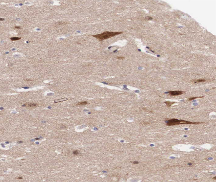
Another routine use of these phospho-specific antibodies, is to test if the antibody is able to demonstrate similar performance when used on fixed cells (intact cells that have been preserved) as it did in the Western blot tests. It is an important aspect in many cases to confirm that the antibody works in actual intact cell samples. Ideally, the method used for cellular fixation should be the same as what is used in pathology labs (like using 10% formalin). To check if the antibody works well in tissue sections (FFPE), researchers will often test it on fixed cells that are processed similar to tissue samples.
3. Specificity Tests Using Peptides
In order to make sure that the antibody is only binding to the right target:
- Laboratory technicians will mix the antibody with phospho-peptides (short segments of the protein containing the phosphate group modification).
- If the antibody signal disappears, it is confirmation that it is binding to the correct phosphorylated location.
- A more robust test is to use both the phosphorylated and non-phosphorylated (dephosphorylated) versions of the protein. The antibody should react only with the phosphorylated one.
- Another method sometimes utilized is to treat the sample with an enzyme, such as alkaline phosphatase, that specifically removes phosphate groups. If the antibody signal disappears after this, it also confirms specificity.
4. Genetic Confirmation
As a final step, scientists can genetically manipulate the nucleotide sequence and alter the target protein by removing the exact site where phosphorylation happens. If the antibody no longer appears to detect the modified protein, it is strong evidence supporting the antibody being specific for that phosphorylated site.
Why Buy Phospho Antibodies Through Us?
- The production laboratory adheres to strict and consistent protocols prior to releasing any of these phospho-specific antibodies:
- Standard methods and proper controls in all tests to ensure high quality.
- These antibodies are tested and validated in different cell types and species.
- High quality control criterion to ensure each batch is consistent, so you will obtain reliable results every time.
FAQ
1. What Are Phospho-Specific Antibodies?
Phospho-specific antibodies are made to detect proteins only when they have a phosphate group linked to a specific amino acid residue. This empowers scientists understand if a protein is "turned on" or active, based on its phosphorylation state.
2. How to Detect Phosphorylated Proteins in a Western Blot?
To find out if a protein is phosphorylated using Western blot:
- Use a phospho-specific antibody that binds only to the phosphorylated form of the protein.
- You can also use a “regular” antibody for the same amino acid sequence of the protein that the phospho-specific antibody is binding to (but in this case, this antibody will not bind if there is a phosphate group present) in order to compare how much of it is phosphorylated versus how much is non-phosphorylated (or “total” protein, if the “normal” antibody’s epitopes are non-phospho-site-specific).
3. How to Choose the Best Antibody?
Here are some simple tips to help you pick the right antibody:
- Know your target
- Match your sample characteristics
- Confirm the intended use is appropriate
- Check “host” and “type”
- Check the “quality” of the presented data/images
- Appraise whether the available validation meets your needs