Filters
▼Clonality
▼Type
▼Reactivity
▼Gene Name
▼Isotype
▼Host
▼Application
▼Clone
▼Phospho Antibodies
Phospho-specific antibodies’ typical purpose is to enable researchers to detect changes in proteins. They will exclusively bind to the amino acid sequence on a protein that has been phosphorylated (which is both a physical & chemical change) and do not bind to the same amino acid sequence on said protein if it lacks said phosphorylation. This aids in being able to clearly see and understand the data produced from this particular protein modification.
Viewing 3650-3700 of 5297 product results

 WB (Western Blot)
(Western blot analysis of lysates from HT29 cells treated with INSULIN 0.01U/ML 15', using IRF-3 (Phospho-Ser385) Antibody.)
WB (Western Blot)
(Western blot analysis of lysates from HT29 cells treated with INSULIN 0.01U/ML 15', using IRF-3 (Phospho-Ser385) Antibody.)
IRF-3, Polyclonal Antibody (Cat# AAA318331)
 WB (Western Blot)
(Western blot analysis of lysates from HepG2 cells treated with EGF 200ng/ml 30', using IRF-3 (Phospho-Ser386) Antibody.)
WB (Western Blot)
(Western blot analysis of lysates from HepG2 cells treated with EGF 200ng/ml 30', using IRF-3 (Phospho-Ser386) Antibody.)
IRF-3, Polyclonal Antibody (Cat# AAA318332)
 WB (Western Blot)
(Western blot analysis of lysates from HepG2 cells treated with Na3VO4 0.3mM 40', using JAK2 (Phospho-Tyr931) Antibody.)
WB (Western Blot)
(Western blot analysis of lysates from HepG2 cells treated with Na3VO4 0.3mM 40', using JAK2 (Phospho-Tyr931) Antibody.)
JAK2, Polyclonal Antibody (Cat# AAA318338)
 WB (Western Blot)
(Western blot analysis of lysates from Jurkat cells treated with IFN 2500U/ML 30', using LCK (Phospho-Ser59) Antibody.)
WB (Western Blot)
(Western blot analysis of lysates from Jurkat cells treated with IFN 2500U/ML 30', using LCK (Phospho-Ser59) Antibody.)
LCK, Polyclonal Antibody (Cat# AAA318343)
 WB (Western Blot)
(Western blot analysis of lysates from Jurkat cells, using Lck (Phospho-Tyr393) Antibody.)
WB (Western Blot)
(Western blot analysis of lysates from Jurkat cells, using Lck (Phospho-Tyr393) Antibody.)
Lck, Polyclonal Antibody (Cat# AAA318345)
 WB (Western Blot)
(Western blot analysis of lysates from COS7 cells treated with UV 15', using MAPKAPK-2 (Phospho-Thr222) Antibody.)
WB (Western Blot)
(Western blot analysis of lysates from COS7 cells treated with UV 15', using MAPKAPK-2 (Phospho-Thr222) Antibody.)
MAPKAPK-2, Polyclonal Antibody (Cat# AAA318352)
 WB (Western Blot)
(Western blot analysis of lysates from HeLa cells treated with nocodazole 1ug/ml 18h, using NPM (Phospho-Thr234) Antibody.)
WB (Western Blot)
(Western blot analysis of lysates from HeLa cells treated with nocodazole 1ug/ml 18h, using NPM (Phospho-Thr234) Antibody.)
NPM, Polyclonal Antibody (Cat# AAA318365)
 WB (Western Blot)
(Western blot analysis of lysates from HepG2 cells treated with serum 20% 15', using OSR1 (Phospho-Thr185) Antibody.)
WB (Western Blot)
(Western blot analysis of lysates from HepG2 cells treated with serum 20% 15', using OSR1 (Phospho-Thr185) Antibody.)
OSR1, Polyclonal Antibody (Cat# AAA318366)
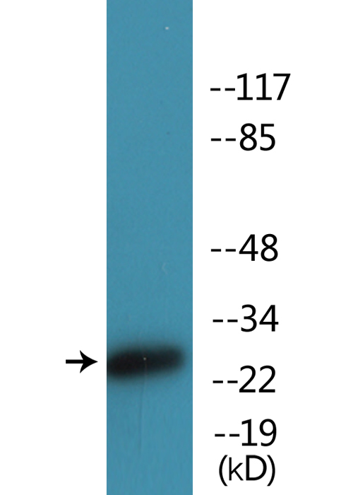
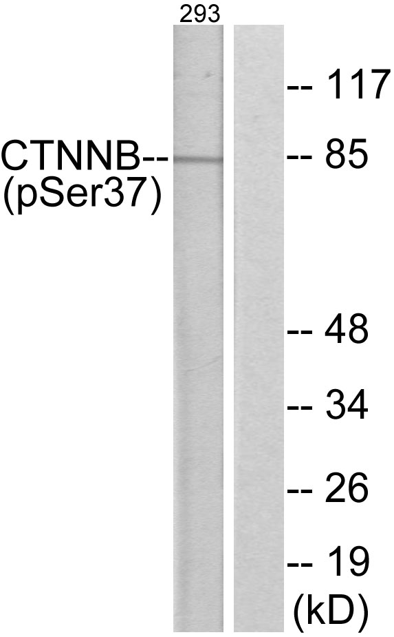 WB (Western Blot)
(Western blot analysis of lysates from 293 cells, using Catenin-beta (Phospho-Ser37) Antibody. The lane on the right is blocked with the phospho peptide.)
WB (Western Blot)
(Western blot analysis of lysates from 293 cells, using Catenin-beta (Phospho-Ser37) Antibody. The lane on the right is blocked with the phospho peptide.)
Catenin-beta, Polyclonal Antibody (Cat# AAA316466)
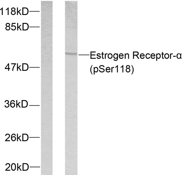 WB (Western Blot)
(Western blot analysis of lysates from MCF7 cells treated with Estradiol, using Estrogen Receptor-alpha (Phospho-Ser118) Antibody. The lane on the left is blocked with the phospho peptide.)
WB (Western Blot)
(Western blot analysis of lysates from MCF7 cells treated with Estradiol, using Estrogen Receptor-alpha (Phospho-Ser118) Antibody. The lane on the left is blocked with the phospho peptide.)
Estrogen Receptor-alpha, Polyclonal Antibody (Cat# AAA316491)
 WB (Western Blot)
(Western blot analysis of lysates from RAW264.7 cells and 293 cells, using p38 MAPK (Phospho-Tyr182) Antibody. The lane on the right is blocked with the phospho peptide.)
WB (Western Blot)
(Western blot analysis of lysates from RAW264.7 cells and 293 cells, using p38 MAPK (Phospho-Tyr182) Antibody. The lane on the right is blocked with the phospho peptide.)
p38 MAPK, Polyclonal Antibody (Cat# AAA316554)
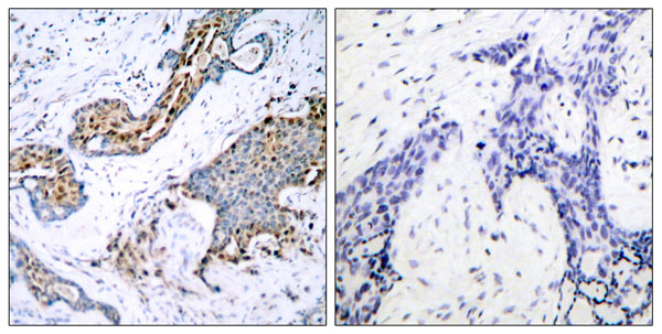
 IHC (Immunohiostchemistry)
(Immunohistochemistry analysis of paraffin-embedded human colon carcinoma, using 53BP1 (Phospho-Ser25) Antibody. The picture on the right is blocked with the phospho peptide.)
IHC (Immunohiostchemistry)
(Immunohistochemistry analysis of paraffin-embedded human colon carcinoma, using 53BP1 (Phospho-Ser25) Antibody. The picture on the right is blocked with the phospho peptide.)
53BP1, Polyclonal Antibody (Cat# AAA316345)
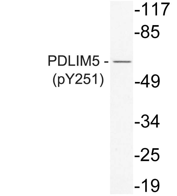 WB (Western Blot)
(Western blot analysis of lysate from K562 cells, uisng phospho-PDLIM5 (Phospho-Tyr251) antibody.)
WB (Western Blot)
(Western blot analysis of lysate from K562 cells, uisng phospho-PDLIM5 (Phospho-Tyr251) antibody.)
PDLIM5, Polyclonal Antibody (Cat# AAA318268)
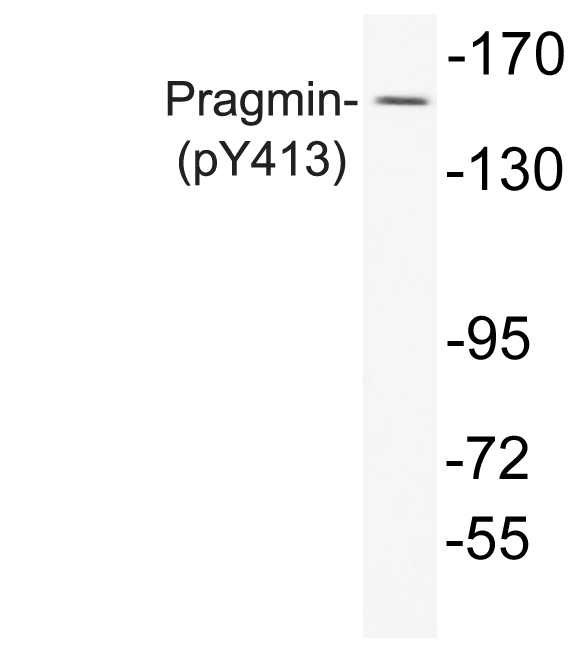 WB (Western Blot)
(Western blot analysis of lysate from A549 cells, using phospho-Pragmin (Phospho-Tyr413) antibody.)
WB (Western Blot)
(Western blot analysis of lysate from A549 cells, using phospho-Pragmin (Phospho-Tyr413) antibody.)
Pragmin, Polyclonal Antibody (Cat# AAA318270)

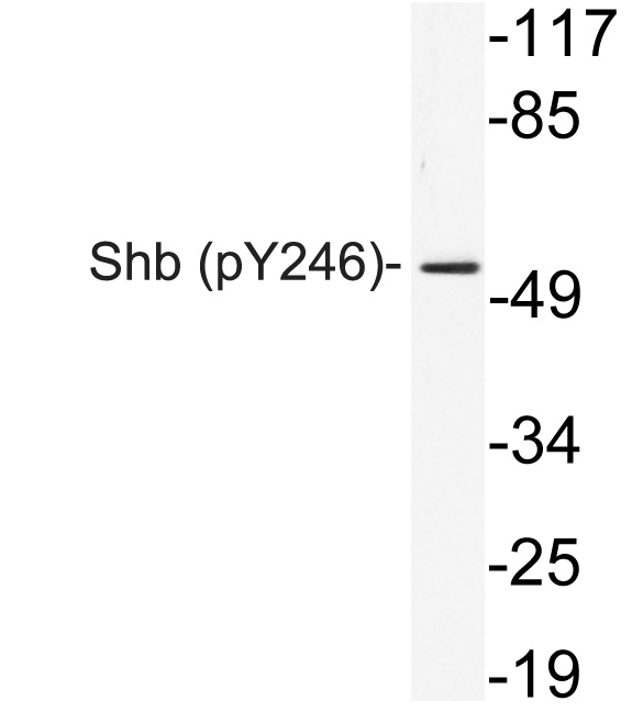 WB (Western Blot)
(Western blot analysis of lysate from 293 cells, using phospho-Serhb (Phospho-Tyr246) antibody.)
WB (Western Blot)
(Western blot analysis of lysate from 293 cells, using phospho-Serhb (Phospho-Tyr246) antibody.)
Shb, Polyclonal Antibody (Cat# AAA318272)
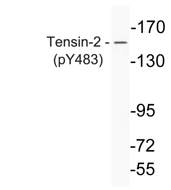 WB (Western Blot)
(Western blot analysis of lysate from K562 cells, using phospho-Thrensin-2 (Phospho-Tyr483) antibody.)
WB (Western Blot)
(Western blot analysis of lysate from K562 cells, using phospho-Thrensin-2 (Phospho-Tyr483) antibody.)
Tensin-2, Polyclonal Antibody (Cat# AAA318275)
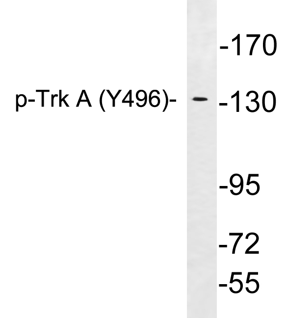 WB (Western Blot)
(Western blot analysis of lysates from Jurkat cells treated with starved, using phospho-Thrrk A (Phospho-Tyr496) antibody.)
WB (Western Blot)
(Western blot analysis of lysates from Jurkat cells treated with starved, using phospho-Thrrk A (Phospho-Tyr496) antibody.)
Trk A, Polyclonal Antibody (Cat# AAA318276)
 WB (Western Blot)
(Western blot analysis of lysates from COS7 cells treated with insulin 0.01U/ML 15', using 53BP1 (Phospho-Ser6) Antibody.)
WB (Western Blot)
(Western blot analysis of lysates from COS7 cells treated with insulin 0.01U/ML 15', using 53BP1 (Phospho-Ser6) Antibody.)
53BP1, Polyclonal Antibody (Cat# AAA318280)

 WB (Western Blot)
(Western blot analysis of lysates from COS7 cells treated with EGF 200ng/ml 30', using ABL1 (Phospho-Thr735) Antibody.)
WB (Western Blot)
(Western blot analysis of lysates from COS7 cells treated with EGF 200ng/ml 30', using ABL1 (Phospho-Thr735) Antibody.)
ABL1, Polyclonal Antibody (Cat# AAA318281)
 WB (Western Blot)
(Western blot analysis of lysates from HepG2 cells treated with PMA 125ng/ml 30', using AML1 (Phospho-Ser435) Antibody.)
WB (Western Blot)
(Western blot analysis of lysates from HepG2 cells treated with PMA 125ng/ml 30', using AML1 (Phospho-Ser435) Antibody.)
AML1, Polyclonal Antibody (Cat# AAA318284)
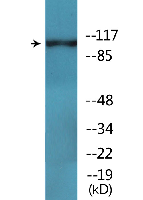
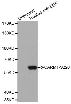
 WB (Western Blot)
(Western blot analysis of lysates from 293 cells treated with EGF 200ng/ml 30', using Breast Tumor Kinase (Phospho-Tyr447) Antibody.)
WB (Western Blot)
(Western blot analysis of lysates from 293 cells treated with EGF 200ng/ml 30', using Breast Tumor Kinase (Phospho-Tyr447) Antibody.)
Breast Tumor Kinase, Polyclonal Antibody (Cat# AAA318292)
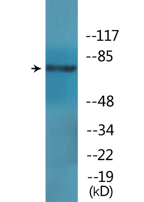
 WB (Western Blot)
(Western blot analysis of lysates from HepG2 cells treated with EGF 200ng/ml 5', using Caveolin 2 (Phospho-Tyr27) Antibody.)
WB (Western Blot)
(Western blot analysis of lysates from HepG2 cells treated with EGF 200ng/ml 5', using Caveolin 2 (Phospho-Tyr27) Antibody.)
Caveolin 2, Polyclonal Antibody (Cat# AAA318296)

 WB (Western Blot)
(Western blot analysis of lysates from HepG2 cells treated with nocodazole 1ug/ml 16h, using CD28 (Phospho-Tyr218) Antibody.)
WB (Western Blot)
(Western blot analysis of lysates from HepG2 cells treated with nocodazole 1ug/ml 16h, using CD28 (Phospho-Tyr218) Antibody.)
CD28, Polyclonal Antibody (Cat# AAA318297)
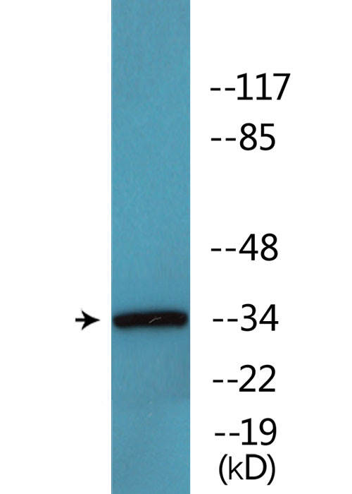 WB (Western Blot)
(Western blot analysis of lysates from HeLa cells, using CDC2 (Phospho-Thr161) Antibody.)
WB (Western Blot)
(Western blot analysis of lysates from HeLa cells, using CDC2 (Phospho-Thr161) Antibody.)
CDC2, Polyclonal Antibody (Cat# AAA318299)

 WB (Western Blot)
(Western blot analysis of lysates from K562 and HUVEC cells treated with PMA, using C-RAF (Phospho-Ser642) Antibody.)
WB (Western Blot)
(Western blot analysis of lysates from K562 and HUVEC cells treated with PMA, using C-RAF (Phospho-Ser642) Antibody.)
C-RAF, Polyclonal Antibody (Cat# AAA318302)
 WB (Western Blot)
(Western blot analysis of lysates from COS7 cells treated with EGF 200ng/ml 30', using EGFR (Phospho-Tyr1069) Antibody.)
WB (Western Blot)
(Western blot analysis of lysates from COS7 cells treated with EGF 200ng/ml 30', using EGFR (Phospho-Tyr1069) Antibody.)
EGFR, Polyclonal Antibody (Cat# AAA318307)
 WB (Western Blot)
(Western blot analysis of lysates from COS7 cells treated with EGF 200ng/ml 30', using FLT3 (Phospho-Tyr599) Antibody.)
WB (Western Blot)
(Western blot analysis of lysates from COS7 cells treated with EGF 200ng/ml 30', using FLT3 (Phospho-Tyr599) Antibody.)
FLT3, Polyclonal Antibody (Cat# AAA318315)

 WB (Western Blot)
(Western blot analysis of lysates from HepG2 cells treated with Na3VO4 0.3mM 40', using FLT3 (Phospho-Tyr969) Antibody.)
WB (Western Blot)
(Western blot analysis of lysates from HepG2 cells treated with Na3VO4 0.3mM 40', using FLT3 (Phospho-Tyr969) Antibody.)
FLT3, Polyclonal Antibody (Cat# AAA318317)
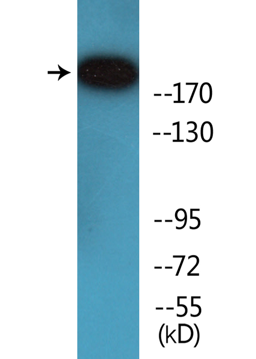
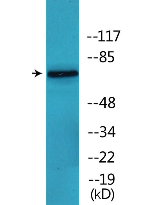
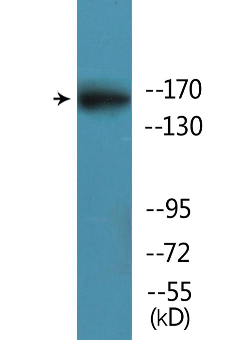 WB (Western Blot)
(Western blot analysis of lysates from mouse brain, using GRIN2B (Phospho-Ser1303) Antibody.)
WB (Western Blot)
(Western blot analysis of lysates from mouse brain, using GRIN2B (Phospho-Ser1303) Antibody.)
GRIN2B, Polyclonal Antibody (Cat# AAA318320)

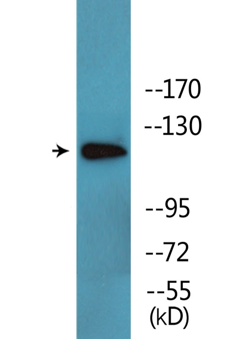
 WB (Western Blot)
(Western blot analysis of lysates from HT29 cells treated with insulin 0.01U/ml 15', using GRK2 (Phospho-Ser685) Antibody.)
WB (Western Blot)
(Western blot analysis of lysates from HT29 cells treated with insulin 0.01U/ml 15', using GRK2 (Phospho-Ser685) Antibody.)
GRK2, Polyclonal Antibody (Cat# AAA318321)

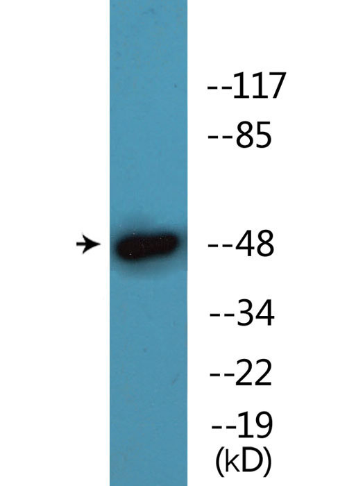
 WB (Western Blot)
(Western blot analysis of lysates from LOVO cells, using HDAC2 (Phospho-Ser394) Antibody.)
WB (Western Blot)
(Western blot analysis of lysates from LOVO cells, using HDAC2 (Phospho-Ser394) Antibody.)
HDAC2, Polyclonal Antibody (Cat# AAA318322)
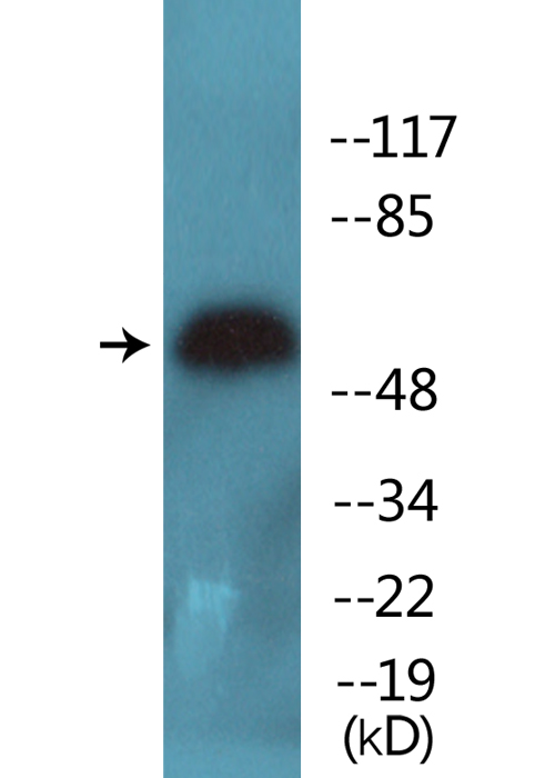
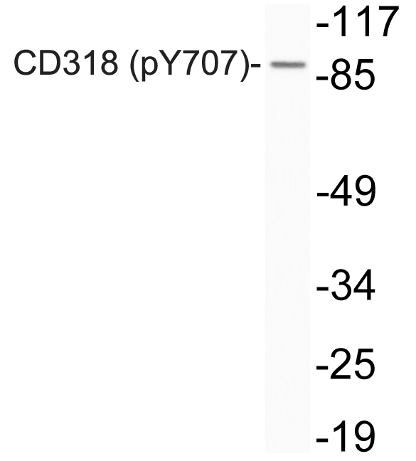 WB (Western Blot)
(Western blot analysis of lysates from Jurkat cells, using phospho-CD318 (Phospho-Tyr707) antibody.)
WB (Western Blot)
(Western blot analysis of lysates from Jurkat cells, using phospho-CD318 (Phospho-Tyr707) antibody.)
CD318, Polyclonal Antibody (Cat# AAA318259)
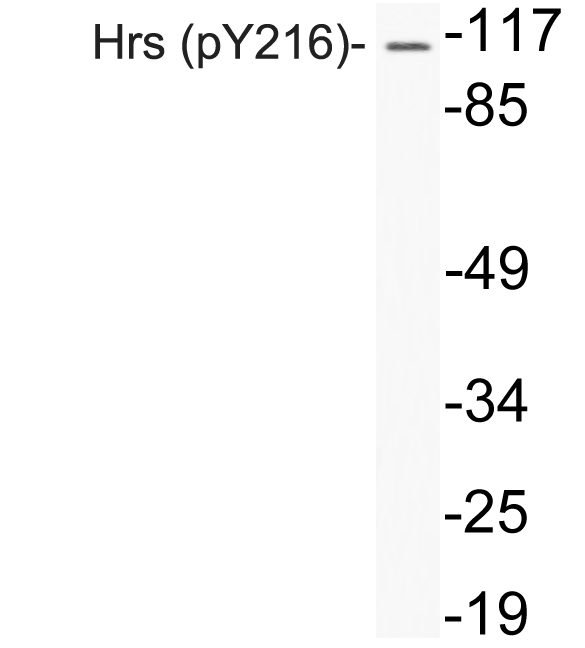 WB (Western Blot)
(Western blot analysis of lysates from A549 cells, using phospho-Hrs (Phospho-Tyr216) antibody.)
WB (Western Blot)
(Western blot analysis of lysates from A549 cells, using phospho-Hrs (Phospho-Tyr216) antibody.)
Hrs, Polyclonal Antibody (Cat# AAA318264)
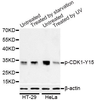
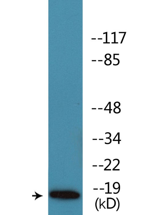
 IP (Immunoprecipitation)
(Immunoprecipitation analysis of 200ug extracts of HT-29 cells treated by starvation using 2.5ug Phospho-CDK1-Y15 antibody. Western blot was performed from the immunoprecipitate using Phospho-CDK1-Y15 antibody at a dilition of 1:1000.)
IP (Immunoprecipitation)
(Immunoprecipitation analysis of 200ug extracts of HT-29 cells treated by starvation using 2.5ug Phospho-CDK1-Y15 antibody. Western blot was performed from the immunoprecipitate using Phospho-CDK1-Y15 antibody at a dilition of 1:1000.)
CDK1-Y15, Antibody (Cat# AAA36660)
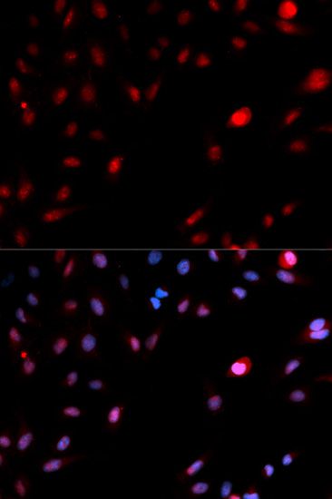
 IF (Immunofluorescence)
(Immunofluorescence analysis of U2OS cells using Phospho-Jun-S243 antibody. Blue: DAPI for nuclear staining.)
IF (Immunofluorescence)
(Immunofluorescence analysis of U2OS cells using Phospho-Jun-S243 antibody. Blue: DAPI for nuclear staining.)
c-Jun-S243, Antibody (Cat# AAA36767)
 IF (Immunofluorescence)
(Immunofluorescence analysis of MCF-7 cells using Phospho-FOS-T232 antibody. Blue: DAPI for nuclear staining.)
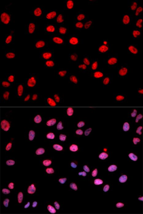
IF (Immunofluorescence)
(Immunofluorescence analysis of MCF-7 cells using Phospho-FOS-T232 antibody. Blue: DAPI for nuclear staining.)
FOS-T232, Antibody (Cat# AAA36769)
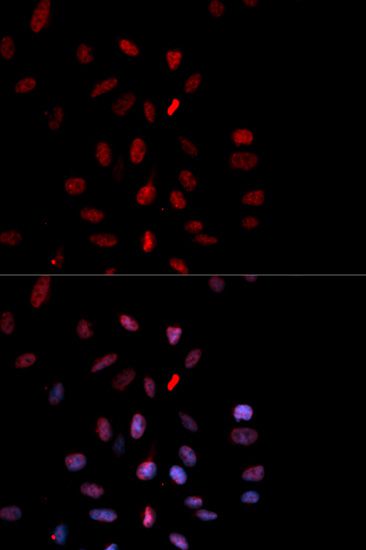
 IF (Immunofluorescence)
(Immunofluorescence staining of methanol-fixed HeLa cells using Phospho-CARM1-S228 antibody.)
IF (Immunofluorescence)
(Immunofluorescence staining of methanol-fixed HeLa cells using Phospho-CARM1-S228 antibody.)
CARM1-S228, Antibody (Cat# AAA37357)
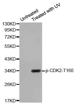
 IHC (Immunohiostchemistry)
(Immunohistochemistry of paraffin-embedded human breast carcinoma using Phospho-CDK2-T160 antibody.)
IHC (Immunohiostchemistry)
(Immunohistochemistry of paraffin-embedded human breast carcinoma using Phospho-CDK2-T160 antibody.)
CDK2-T160, Antibody (Cat# AAA37358)
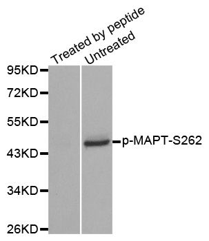
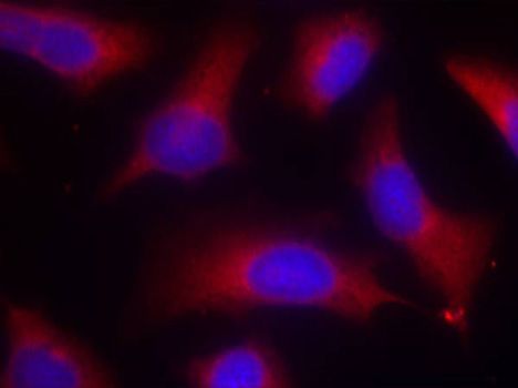 IF (Immunofluorescence)
(Immunofluorescence staining of methanol-fixed HeLa cells using Phospho-MAPT-S262 antibody.)
IF (Immunofluorescence)
(Immunofluorescence staining of methanol-fixed HeLa cells using Phospho-MAPT-S262 antibody.)
MAPT-S262, Antibody (Cat# AAA37417)
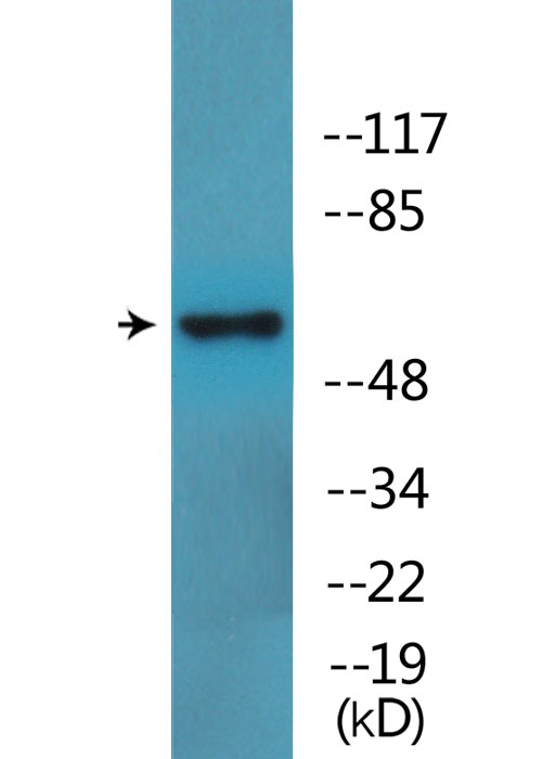
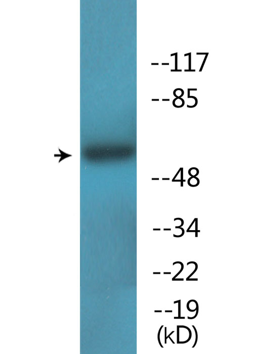
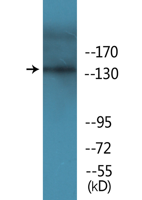
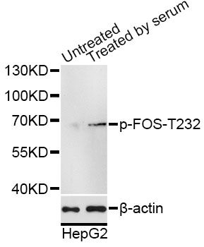
 WB (Western Blot)
(Western blot analysis of Phospho-Cdk1/2 (T14) expression in HeLa cell lysate treated with hydroxyurea (AAA124529).Electrophoresis was performed on a 5-20% SDS-PAGE gel at 70V (Stacking gel) / 90V (Resolving gel) for 2-3 hours. The sample well of each lane was loaded with 50ug of sample under reducing conditions.After Electrophoresis, proteins were transferred to a Nitrocellulose membrane at 150mA for 50-90 minutes. Blocked the membrane with 5% Non-fat Milk/ TBS for 1.5 hour at RT. The membrane was incubated with rabbit anti-CDK1 monoclonal antibody overnight at 4 degree C, then washed with TBS-0.1%Tween 3 times with 5 minutes each and probed with a goat anti-rabbit IgG-HRP secondary antibody at a dilution of 1:10000 for 1.5 hour at RT. The signal is developed using an Enhanced Chemiluminescent detection (ECL) kit with Tanon 5200 system. A specific band was detected for CDK1)
WB (Western Blot)
(Western blot analysis of Phospho-Cdk1/2 (T14) expression in HeLa cell lysate treated with hydroxyurea (AAA124529).Electrophoresis was performed on a 5-20% SDS-PAGE gel at 70V (Stacking gel) / 90V (Resolving gel) for 2-3 hours. The sample well of each lane was loaded with 50ug of sample under reducing conditions.After Electrophoresis, proteins were transferred to a Nitrocellulose membrane at 150mA for 50-90 minutes. Blocked the membrane with 5% Non-fat Milk/ TBS for 1.5 hour at RT. The membrane was incubated with rabbit anti-CDK1 monoclonal antibody overnight at 4 degree C, then washed with TBS-0.1%Tween 3 times with 5 minutes each and probed with a goat anti-rabbit IgG-HRP secondary antibody at a dilution of 1:10000 for 1.5 hour at RT. The signal is developed using an Enhanced Chemiluminescent detection (ECL) kit with Tanon 5200 system. A specific band was detected for CDK1)
Cdk1/2, Monoclonal Antibody (Cat# AAA124529)
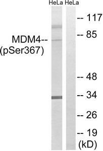
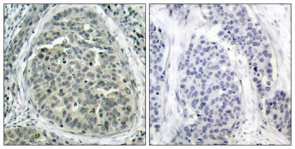
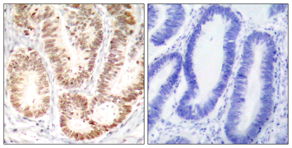
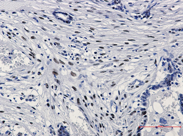
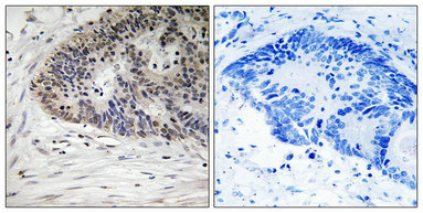 IHC (Immunohiostchemistry)
(Immunohistochemistry analysis of paraffin-embedded human colon carcinoma tissue using MDM4 (Phospho-Ser367) antibody. The picture on the right is treated with the synthesized peptide.)
IHC (Immunohiostchemistry)
(Immunohistochemistry analysis of paraffin-embedded human colon carcinoma tissue using MDM4 (Phospho-Ser367) antibody. The picture on the right is treated with the synthesized peptide.)
MDM4, Polyclonal Antibody (Cat# AAA243246)
 IF (Immunofluorescence)
(Immunofluorescence staining of methanol-fixed A549 cells using NMDAR1 (Phospho-Ser890) Antibody.)
IF (Immunofluorescence)
(Immunofluorescence staining of methanol-fixed A549 cells using NMDAR1 (Phospho-Ser890) Antibody.)
GRIN1, Polyclonal Antibody (Cat# AAA243193)


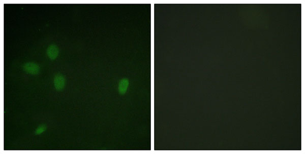
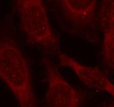
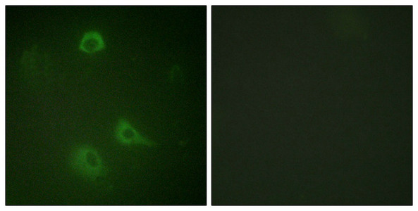
 IF (Immunofluorescence)
(Immunofluorescence staining of methanol-fixed Hela cells showing cytoplasmic, centrosomal and nuclear staining using p70 S6 Kinase(Phospho-Ser424) Antibody.)
IF (Immunofluorescence)
(Immunofluorescence staining of methanol-fixed Hela cells showing cytoplasmic, centrosomal and nuclear staining using p70 S6 Kinase(Phospho-Ser424) Antibody.)
RPS6KB1, Polyclonal Antibody (Cat# AAA243316)
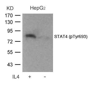
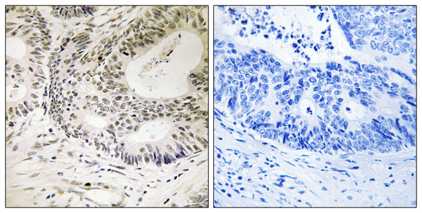
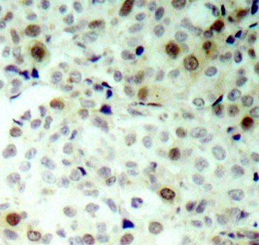
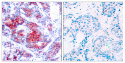 IHC (Immunohiostchemistry)
(Immunohistochemical analysis of paraffin-embedded human breast carcinoma tissue using STAT4(Phospho-Tyr693) Antibody(left) or the same antibody preincubated with blocking peptide(right).)
IHC (Immunohiostchemistry)
(Immunohistochemical analysis of paraffin-embedded human breast carcinoma tissue using STAT4(Phospho-Tyr693) Antibody(left) or the same antibody preincubated with blocking peptide(right).)
STAT4, Polyclonal Antibody (Cat# AAA243056)
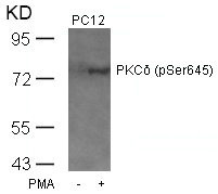
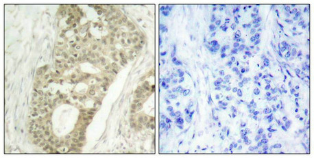 IHC (Immunohiostchemistry)
(Immunohistochemical analysis of paraffin-embedded human breast carcinoma tissue using PKCd(Phospho-Ser645) Antibody(left) or the same antibody preincubated with blocking peptide(right).)
IHC (Immunohiostchemistry)
(Immunohistochemical analysis of paraffin-embedded human breast carcinoma tissue using PKCd(Phospho-Ser645) Antibody(left) or the same antibody preincubated with blocking peptide(right).)
PRKCD, Polyclonal Antibody (Cat# AAA243143)
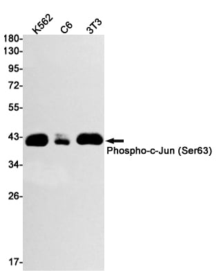
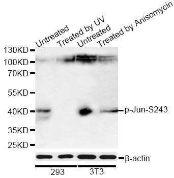
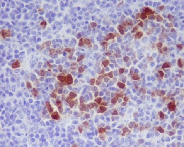
 IHC (Immunohiostchemistry)
(Immunohistochemistry of c-Jun (phospho S63) in paraffin-embedded Human breast cancer tissue using c-Jun (phospho S63) Rabbit mAb at dilution 1/20)
IHC (Immunohiostchemistry)
(Immunohistochemistry of c-Jun (phospho S63) in paraffin-embedded Human breast cancer tissue using c-Jun (phospho S63) Rabbit mAb at dilution 1/20)
c Jun, Monoclonal Antibody (Cat# AAA314291)
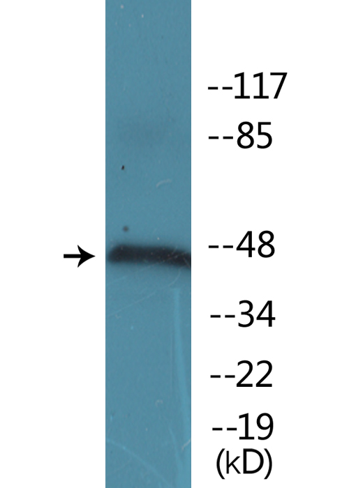
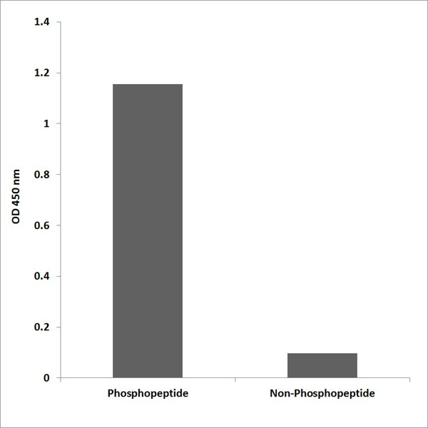
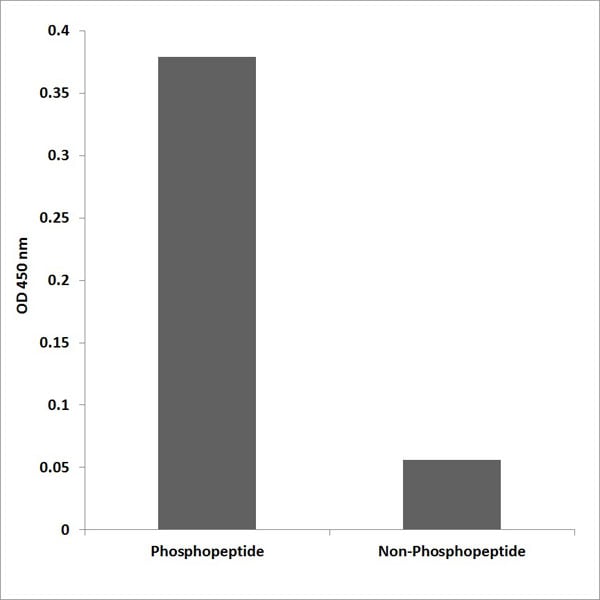
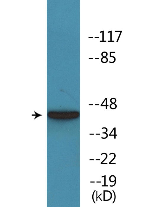
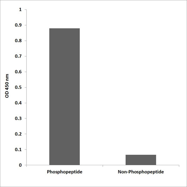
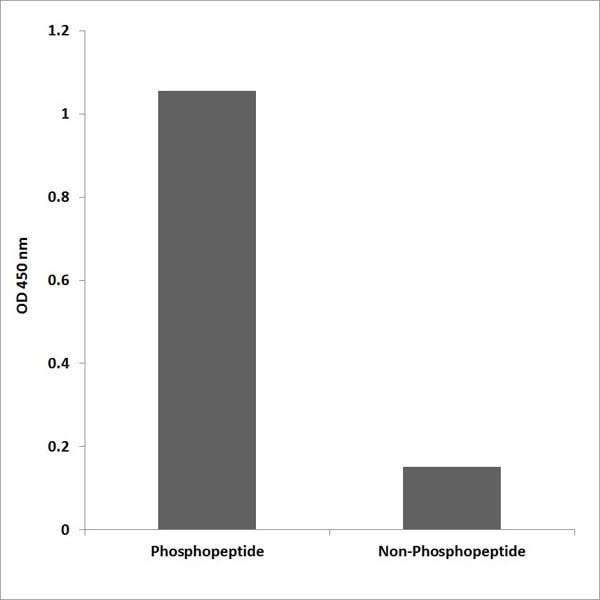
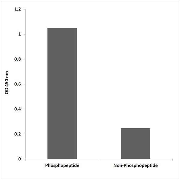
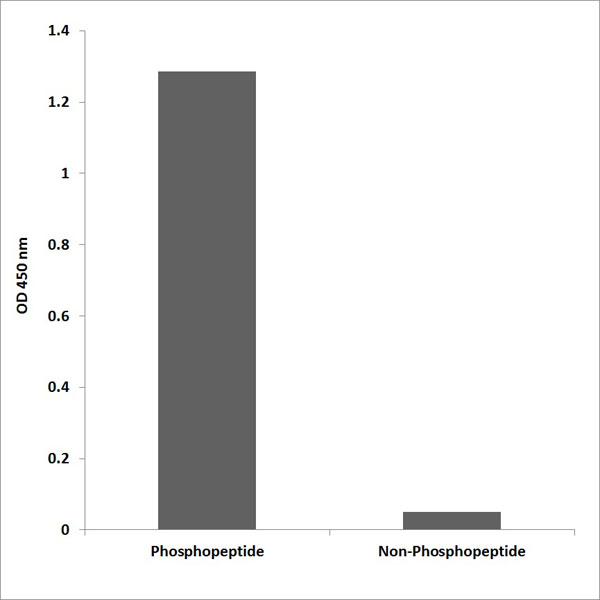
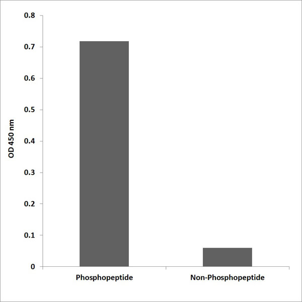
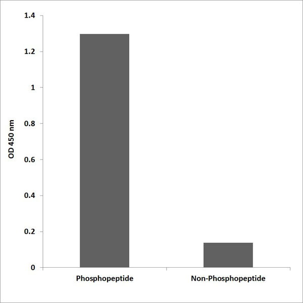
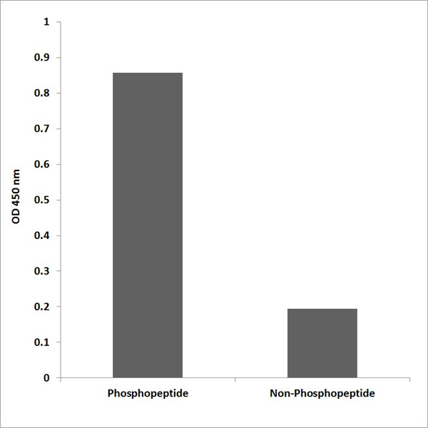
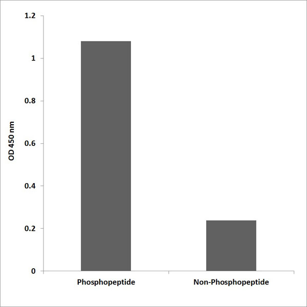
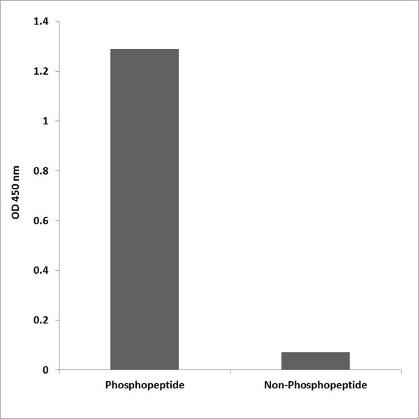
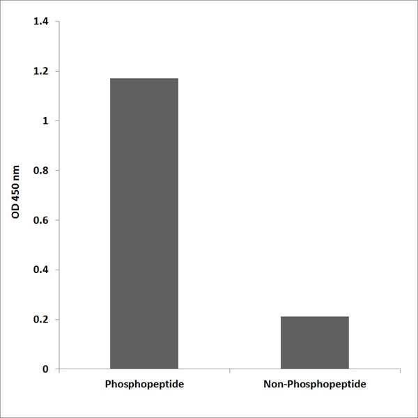
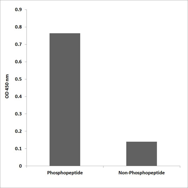
 DB (Dot Blot)
(Dot blot analysis of anti-Phospho-HIPK2-pY361 Antibody (AAA288429) on nitrocellulose membrane. 50ng of Phospho-peptide or Non Phospho-peptide per dot were adsorbed. Antibody working concentrations are 0.5ug per ml.)
DB (Dot Blot)
(Dot blot analysis of anti-Phospho-HIPK2-pY361 Antibody (AAA288429) on nitrocellulose membrane. 50ng of Phospho-peptide or Non Phospho-peptide per dot were adsorbed. Antibody working concentrations are 0.5ug per ml.)
Phospho-HIPK2 (Y361), Polyclonal Antibody (Cat# AAA288429)
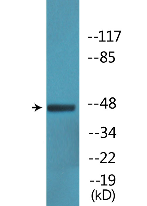
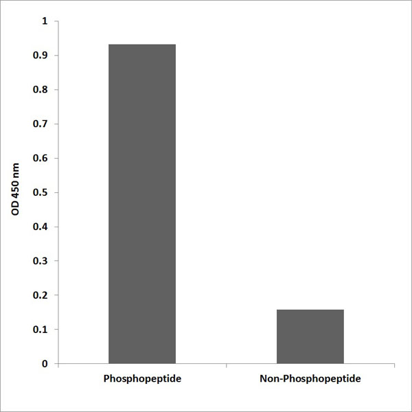
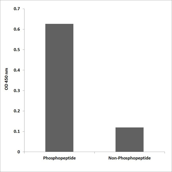
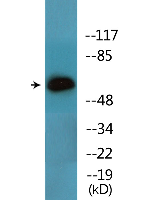
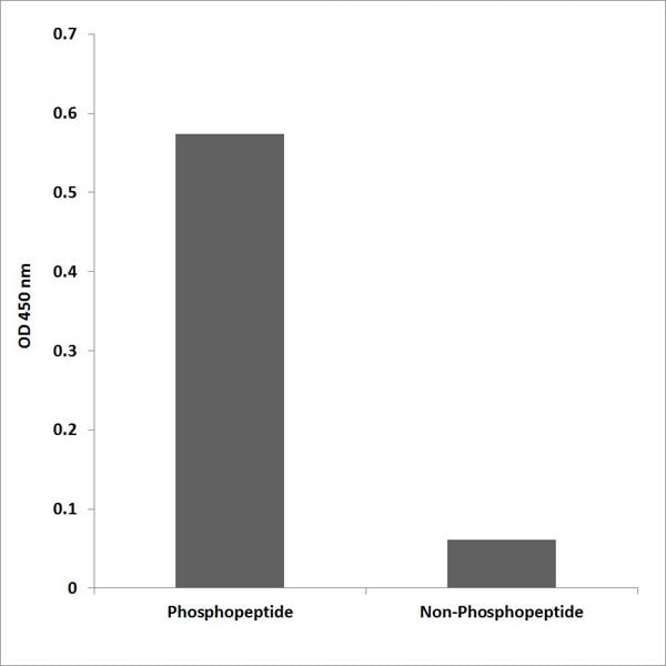
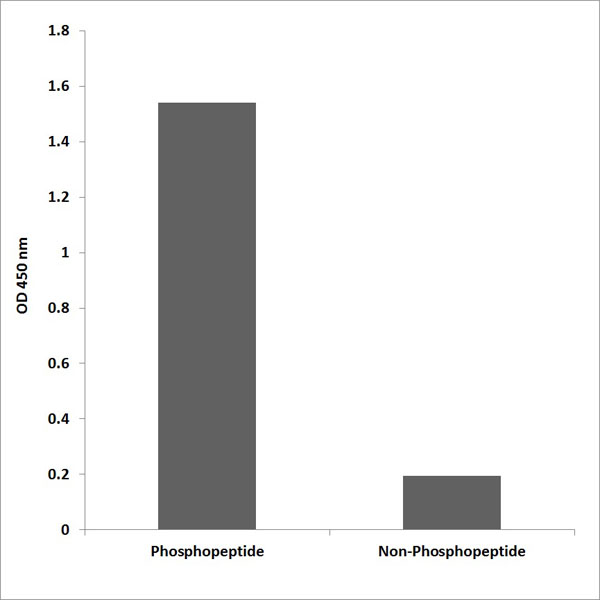
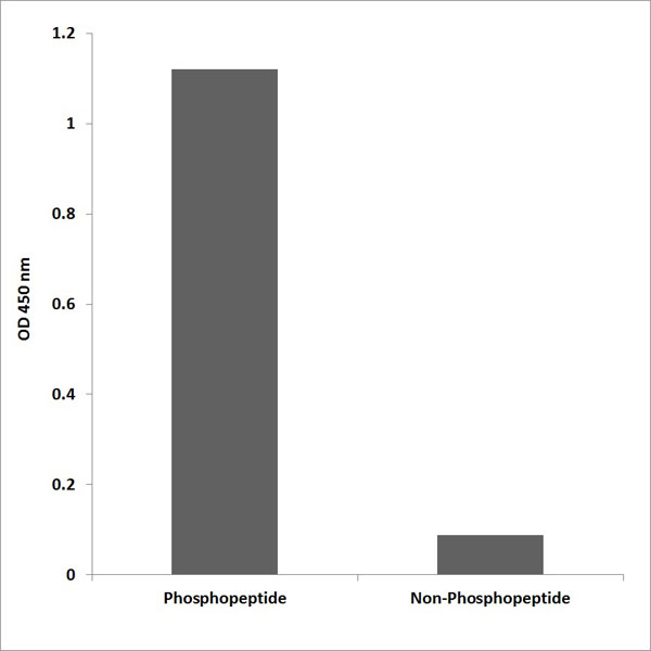
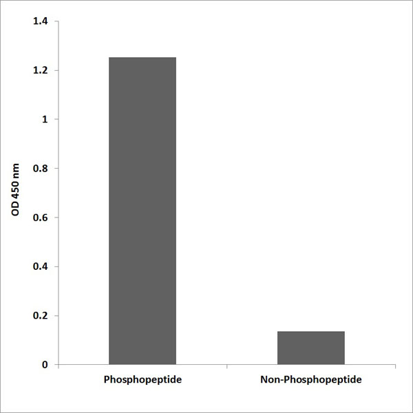
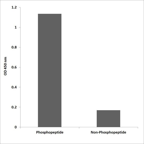
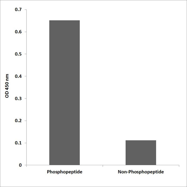
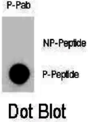
 DB (Dot Blot)
(Dot blot analysis of ULK1 Antibody (Phospho S317) Phospho-specific Pab on nitrocellulose membrane. 50ng of Phospho-peptide or Non Phospho-peptide per dot were adsorbed. Antibody working concentrations are 0.6ug per ml.)
DB (Dot Blot)
(Dot blot analysis of ULK1 Antibody (Phospho S317) Phospho-specific Pab on nitrocellulose membrane. 50ng of Phospho-peptide or Non Phospho-peptide per dot were adsorbed. Antibody working concentrations are 0.6ug per ml.)
Phospho-ULK1 (S317), Polyclonal Antibody (Cat# AAA289605)

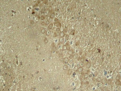
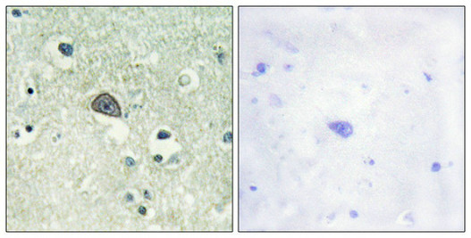
 IHC (Immunohiostchemistry)
(Immunohistochemistry analysis of paraffin-embedded human brain tissue using Trk A (Phospho-Tyr701) antibody. The picture on the right is treated with the synthesized peptide.)
IHC (Immunohiostchemistry)
(Immunohistochemistry analysis of paraffin-embedded human brain tissue using Trk A (Phospho-Tyr701) antibody. The picture on the right is treated with the synthesized peptide.)
Trk A, Polyclonal Antibody (Cat# AAA308022)
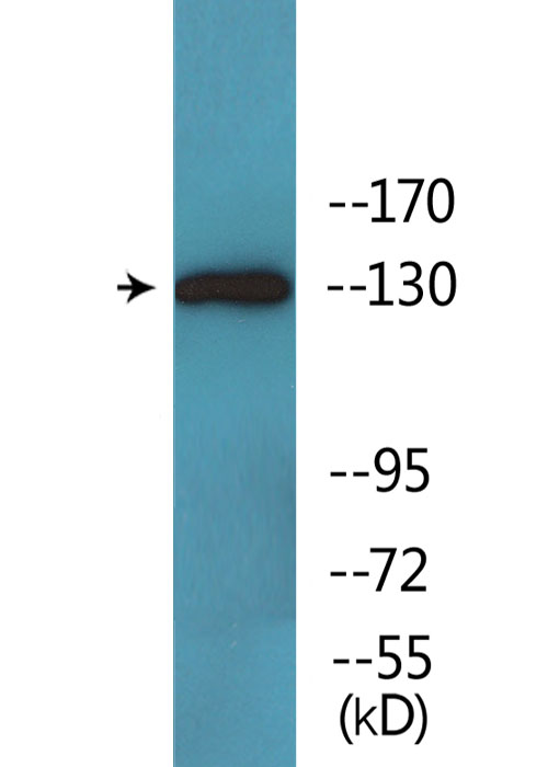
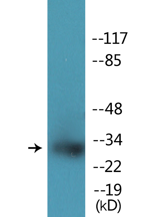
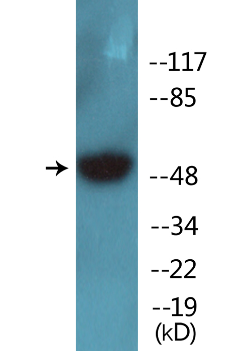
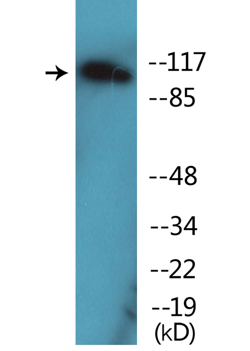
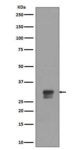
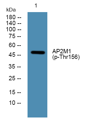 WB (Western Blot)
(Western blot analysis of lysates from K562 cells, primary antibody was diluted at 1:1000, 4 degree C over night)
WB (Western Blot)
(Western blot analysis of lysates from K562 cells, primary antibody was diluted at 1:1000, 4 degree C over night)
AP2M1, ELISA Kit (Cat# AAA319083)
What Are Phospho Antibodies?
Protein phosphorylation is a process where a phosphate group is added to certain amino acid residues of a protein – usually serine (S), threonine (T), or tyrosine (Y) - by enzymes called kinases. This process is integral in controlling cellular signaling, cellular growth, and other biological functions.
Our catalog includes a wide range of phospho-specific antibodies that can accurately detect this important marker. They perform strongly in widely-used laboratory applications such as Western blot, flow cytometry, immunohistochemistry, and immunofluorescence microscopy. We value your trust in us and are committed to providing top-quality products and services. All of our antibodies are guaranteed to work for the applications and species indicated on our website & associated product pages.
What Are The Key Applications of Phospho Antibodies?
1. Western Blotting
One of the first steps a researcher can take in utilizing these phospho-specific antibodies, is to check if the antibody works using a technique referred to as “Western blot”. For those unfamiliar, Western Blot aids in showing whether the protein that the antibody recognizes is appearing at the correct/expected size. These phospho-specific antibodies should also be able to detect changes in the target protein’s phosphorylation (on/off state) when cells are stimulated in certain ways.
2. Staining of Fixed Cells (Immunocytochemistry)
Another routine use of these phospho-specific antibodies, is to test if the antibody is able to demonstrate similar performance when used on fixed cells (intact cells that have been preserved) as it did in the Western blot tests. It is an important aspect in many cases to confirm that the antibody works in actual intact cell samples. Ideally, the method used for cellular fixation should be the same as what is used in pathology labs (like using 10% formalin). To check if the antibody works well in tissue sections (FFPE), researchers will often test it on fixed cells that are processed similar to tissue samples.
3. Specificity Tests Using Peptides
In order to make sure that the antibody is only binding to the right target:
- Laboratory technicians will mix the antibody with phospho-peptides (short segments of the protein containing the phosphate group modification).
- If the antibody signal disappears, it is confirmation that it is binding to the correct phosphorylated location.
- A more robust test is to use both the phosphorylated and non-phosphorylated (dephosphorylated) versions of the protein. The antibody should react only with the phosphorylated one.
- Another method sometimes utilized is to treat the sample with an enzyme, such as alkaline phosphatase, that specifically removes phosphate groups. If the antibody signal disappears after this, it also confirms specificity.
4. Genetic Confirmation
As a final step, scientists can genetically manipulate the nucleotide sequence and alter the target protein by removing the exact site where phosphorylation happens. If the antibody no longer appears to detect the modified protein, it is strong evidence supporting the antibody being specific for that phosphorylated site.
Why Buy Phospho Antibodies Through Us?
- The production laboratory adheres to strict and consistent protocols prior to releasing any of these phospho-specific antibodies:
- Standard methods and proper controls in all tests to ensure high quality.
- These antibodies are tested and validated in different cell types and species.
- High quality control criterion to ensure each batch is consistent, so you will obtain reliable results every time.
FAQ
1. What Are Phospho-Specific Antibodies?
Phospho-specific antibodies are made to detect proteins only when they have a phosphate group linked to a specific amino acid residue. This empowers scientists understand if a protein is "turned on" or active, based on its phosphorylation state.
2. How to Detect Phosphorylated Proteins in a Western Blot?
To find out if a protein is phosphorylated using Western blot:
- Use a phospho-specific antibody that binds only to the phosphorylated form of the protein.
- You can also use a “regular” antibody for the same amino acid sequence of the protein that the phospho-specific antibody is binding to (but in this case, this antibody will not bind if there is a phosphate group present) in order to compare how much of it is phosphorylated versus how much is non-phosphorylated (or “total” protein, if the “normal” antibody’s epitopes are non-phospho-site-specific).
3. How to Choose the Best Antibody?
Here are some simple tips to help you pick the right antibody:
- Know your target
- Match your sample characteristics
- Confirm the intended use is appropriate
- Check “host” and “type”
- Check the “quality” of the presented data/images
- Appraise whether the available validation meets your needs