Filters
▼Clonality
▼Type
▼Reactivity
▼Gene Name
▼Isotype
▼Host
▼Application
▼Clone
▼Phospho Antibodies
Phospho-specific antibodies’ typical purpose is to enable researchers to detect changes in proteins. They will exclusively bind to the amino acid sequence on a protein that has been phosphorylated (which is both a physical & chemical change) and do not bind to the same amino acid sequence on said protein if it lacks said phosphorylation. This aids in being able to clearly see and understand the data produced from this particular protein modification.
Viewing 3000-3050 of 5298 product results
 WB (Western Blot)
(Western blot analysis of lysates from COS7 cells treated with H2O2 100uM 30', using RAD52 (Phospho-Tyr104) Antibody. The lane on the right is blocked with the phospho peptide.)
WB (Western Blot)
(Western blot analysis of lysates from COS7 cells treated with H2O2 100uM 30', using RAD52 (Phospho-Tyr104) Antibody. The lane on the right is blocked with the phospho peptide.)
RAD52, Polyclonal Antibody (Cat# AAA316421)
 WB (Western Blot)
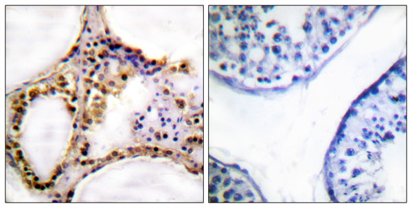
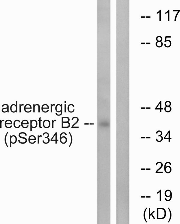
(Western blot analysis of lysates from HepG2 cells treated with nocodazole 1ug/ml 16h, using Adrenergic Receptor beta2 (Phospho-Ser346) Antibody. The lane on the right is blocked with the phospho peptide.)
WB (Western Blot)
(Western blot analysis of lysates from HepG2 cells treated with nocodazole 1ug/ml 16h, using Adrenergic Receptor beta2 (Phospho-Ser346) Antibody. The lane on the right is blocked with the phospho peptide.)
Adrenergic Receptor beta2, Polyclonal Antibody (Cat# AAA316424)
 Application Data
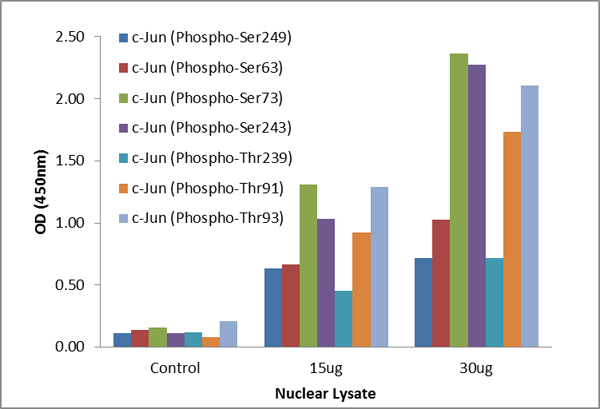
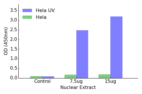
(The TFact c-Jun DNA-Binding ELISA kits detect active c-Jun in Hela Nuclear Extract. The Hela cells were grown 3 days in DMEM with 10% FBS and harvested for nuclear extract. The Hela cells were stimulated by UV (100J/M2) before harvest.)
Application Data
(The TFact c-Jun DNA-Binding ELISA kits detect active c-Jun in Hela Nuclear Extract. The Hela cells were grown 3 days in DMEM with 10% FBS and harvested for nuclear extract. The Hela cells were stimulated by UV (100J/M2) before harvest.)
c-Jun, ELISA Kit (Cat# AAA315851)
 Application Data
(The TFact ATF2 (Phospho-Thr71 or 53) DNA-Binding ELISA detects active ATF2 (Phospho-Thr71 or 53) in Hela Nuclear Extract. The Hela cells were grown 3 days in DMEM with 10% FBS and harvested for nuclear extract. The Hela cells were stimulated by UV (100J/M2) before harvest.)
Application Data
(The TFact ATF2 (Phospho-Thr71 or 53) DNA-Binding ELISA detects active ATF2 (Phospho-Thr71 or 53) in Hela Nuclear Extract. The Hela cells were grown 3 days in DMEM with 10% FBS and harvested for nuclear extract. The Hela cells were stimulated by UV (100J/M2) before harvest.)
ATF2, ELISA Kit (Cat# AAA315869)
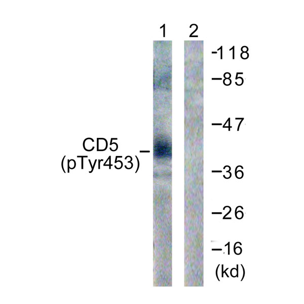 WB (Western Blot)
(Western blot analysis of lysates from 293 cells treated with PMA 125ng/ml 30', using CD5 (Phospho-Tyr453) Antibody. The lane on the right is blocked with the phospho peptide.)
WB (Western Blot)
(Western blot analysis of lysates from 293 cells treated with PMA 125ng/ml 30', using CD5 (Phospho-Tyr453) Antibody. The lane on the right is blocked with the phospho peptide.)
CD5, Polyclonal Antibody (Cat# AAA316330)
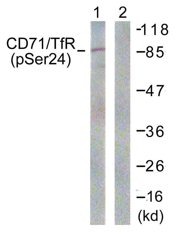 WB (Western Blot)
(Western blot analysis of lysates from 293 cells treated with PMA 125ng/ml 30', using CD71/TfR (Phospho-Ser24) Antibody. The lane on the right is blocked with the phospho peptide.)
WB (Western Blot)
(Western blot analysis of lysates from 293 cells treated with PMA 125ng/ml 30', using CD71/TfR (Phospho-Ser24) Antibody. The lane on the right is blocked with the phospho peptide.)
CD71/TfR, Polyclonal Antibody (Cat# AAA316331)
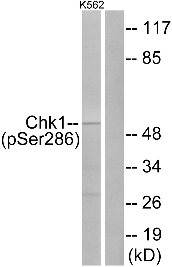 WB (Western Blot)
(Western blot analysis of lysates from K562 cells treated with Na3VO4 0.3uM 40', using Chk1 (Phospho-Ser286) Antibody. The lane on the right is blocked with the phospho peptide.)
WB (Western Blot)
(Western blot analysis of lysates from K562 cells treated with Na3VO4 0.3uM 40', using Chk1 (Phospho-Ser286) Antibody. The lane on the right is blocked with the phospho peptide.)
Chk1, Polyclonal Antibody (Cat# AAA316334)
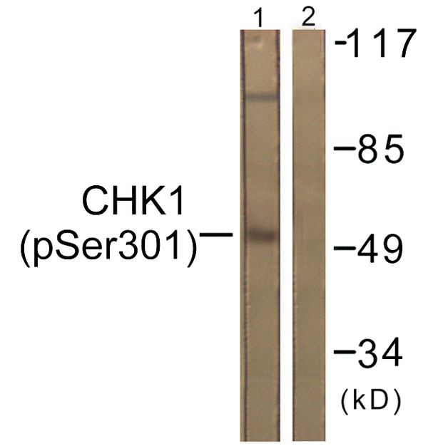 WB (Western Blot)
(Western blot analysis of lysates from 293 cells, using Chk1 (Phospho-Ser301) Antibody. The lane on the right is blocked with the phospho peptide.)
WB (Western Blot)
(Western blot analysis of lysates from 293 cells, using Chk1 (Phospho-Ser301) Antibody. The lane on the right is blocked with the phospho peptide.)
Chk1, Polyclonal Antibody (Cat# AAA316335)
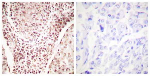
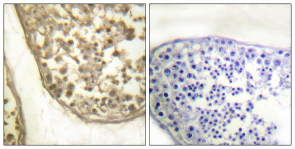 IHC (Immunohiostchemistry)
(Immunohistochemistry analysis of paraffin-embedded human testis, using c-Jun (Phospho-Thr231) Antibody. The picture on the right is blocked with the phospho peptide.)
IHC (Immunohiostchemistry)
(Immunohistochemistry analysis of paraffin-embedded human testis, using c-Jun (Phospho-Thr231) Antibody. The picture on the right is blocked with the phospho peptide.)
c-Jun, Polyclonal Antibody (Cat# AAA316337)
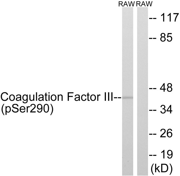 WB (Western Blot)
(Western blot analysis of lysates from RAW264.7 cells treated with TNF 20ng/ml 30', using Coagulation Factor III (Phospho-Ser290) Antibody. The lane on the right is blocked with the phospho peptide.)
WB (Western Blot)
(Western blot analysis of lysates from RAW264.7 cells treated with TNF 20ng/ml 30', using Coagulation Factor III (Phospho-Ser290) Antibody. The lane on the right is blocked with the phospho peptide.)
Coagulation Factor III, Polyclonal Antibody (Cat# AAA316340)
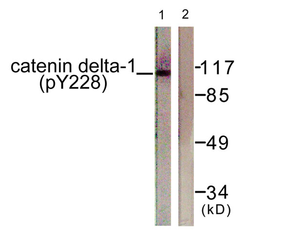 WB (Western Blot)
(Western blot analysis of lysates from HUVEC cells, using Catenin-delta1 (Phospho-Tyr228) Antibody. The lane on the right is blocked with the phospho peptide.)
WB (Western Blot)
(Western blot analysis of lysates from HUVEC cells, using Catenin-delta1 (Phospho-Tyr228) Antibody. The lane on the right is blocked with the phospho peptide.)
Catenin-delta1, Polyclonal Antibody (Cat# AAA316346)
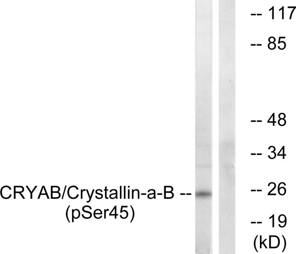 WB (Western Blot)
(Western blot analysis of lysates from COS7 cells treated with anisomycin 25ug/ml 30', using CRYAB (Phospho-Ser45) Antibody. The lane on the right is blocked with the phospho peptide.)
WB (Western Blot)
(Western blot analysis of lysates from COS7 cells treated with anisomycin 25ug/ml 30', using CRYAB (Phospho-Ser45) Antibody. The lane on the right is blocked with the phospho peptide.)
CRYAB, Polyclonal Antibody (Cat# AAA316348)
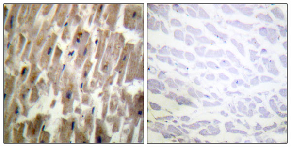
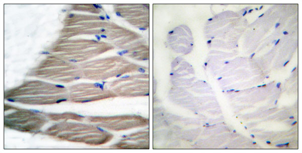
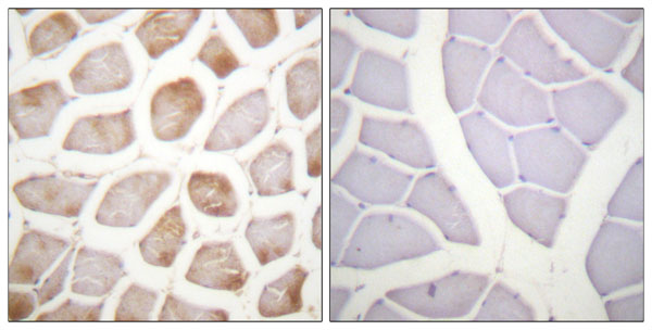 IHC (Immunohiostchemistry)
(Immunohistochemistry analysis of paraffin-embedded human skeletal muscle, using DAPK2 (Phospho-Ser318) Antibody. The picture on the right is blocked with the phospho peptide.)
IHC (Immunohiostchemistry)
(Immunohistochemistry analysis of paraffin-embedded human skeletal muscle, using DAPK2 (Phospho-Ser318) Antibody. The picture on the right is blocked with the phospho peptide.)
DAPK2, Polyclonal Antibody (Cat# AAA316349)
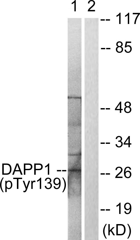 WB (Western Blot)
(Western blot analysis of lysates from 293 cells treated with Insulin 0.01U/ml 2', using DAPP1 (Phospho-Tyr139) Antibody. The lane on the right is blocked with the phospho peptide.)
WB (Western Blot)
(Western blot analysis of lysates from 293 cells treated with Insulin 0.01U/ml 2', using DAPP1 (Phospho-Tyr139) Antibody. The lane on the right is blocked with the phospho peptide.)
DAPP1, Polyclonal Antibody (Cat# AAA316351)
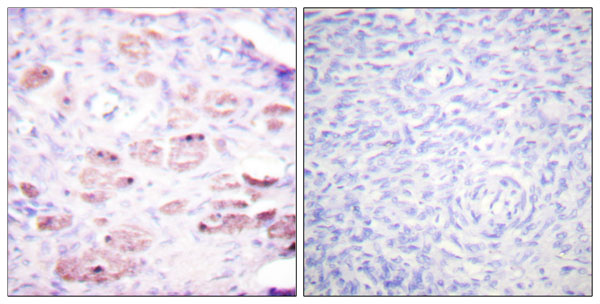
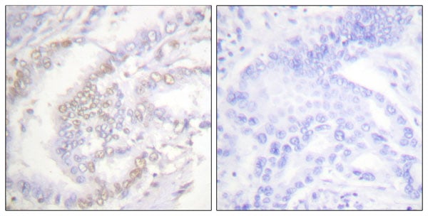
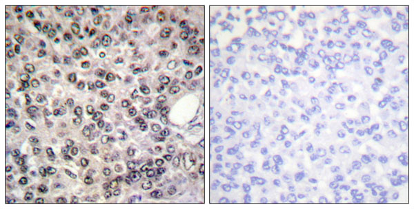 IHC (Immunohiostchemistry)
(Immunohistochemistry analysis of paraffin-embedded human breast carcinoma, using DDX5/DEAD-box Protein 5 (Phospho-Tyr593) Antibody. The picture on the right is blocked with the phospho peptide.)
IHC (Immunohiostchemistry)
(Immunohistochemistry analysis of paraffin-embedded human breast carcinoma, using DDX5/DEAD-box Protein 5 (Phospho-Tyr593) Antibody. The picture on the right is blocked with the phospho peptide.)
DDX5/DEAD-box Protein 5, Polyclonal Antibody (Cat# AAA316353)
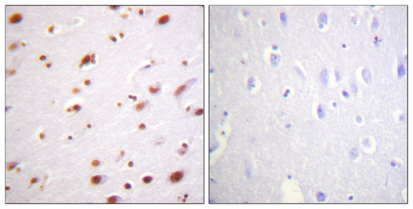
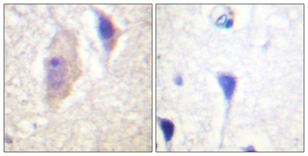
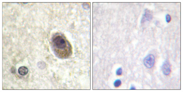
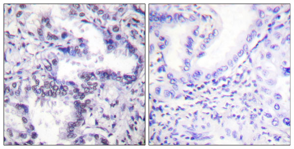
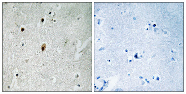 IHC (Immunohiostchemistry)
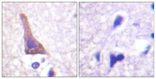
(Immunohistochemistry analysis of paraffin-embedded human brain, using DNA-PK (Phospho-Thr2647) Antibody. The picture on the right is blocked with the phospho peptide.)
IHC (Immunohiostchemistry)
(Immunohistochemistry analysis of paraffin-embedded human brain, using DNA-PK (Phospho-Thr2647) Antibody. The picture on the right is blocked with the phospho peptide.)
DNA-PK, Polyclonal Antibody (Cat# AAA316355)
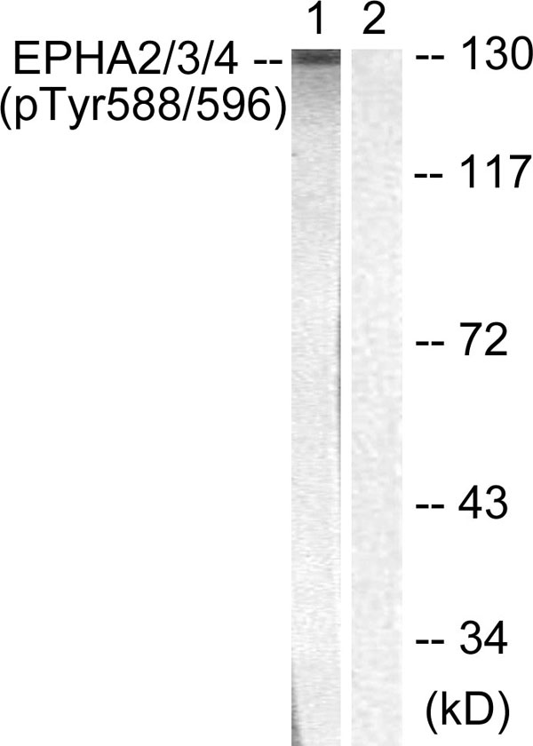 WB (Western Blot)
(Western blot analysis of lysates from HepG2 cells, using EPHA2/3 (Phospho-Tyr588/596) Antibody. The lane on the right is blocked with the phospho peptide.)
WB (Western Blot)
(Western blot analysis of lysates from HepG2 cells, using EPHA2/3 (Phospho-Tyr588/596) Antibody. The lane on the right is blocked with the phospho peptide.)
EPHA2/3, Polyclonal Antibody (Cat# AAA316356)
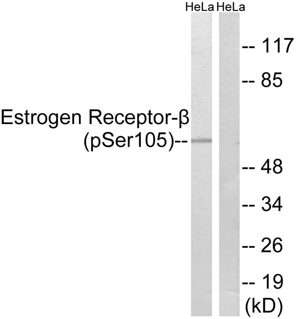 WB (Western Blot)
(Western blot analysis of lysates from HeLa cells, using Estrogen Receptor-beta (Phospho-Ser105) Antibody. The lane on the right is blocked with the phospho peptide.)
WB (Western Blot)
(Western blot analysis of lysates from HeLa cells, using Estrogen Receptor-beta (Phospho-Ser105) Antibody. The lane on the right is blocked with the phospho peptide.)
Estrogen Receptor-beta, Polyclonal Antibody (Cat# AAA316360)
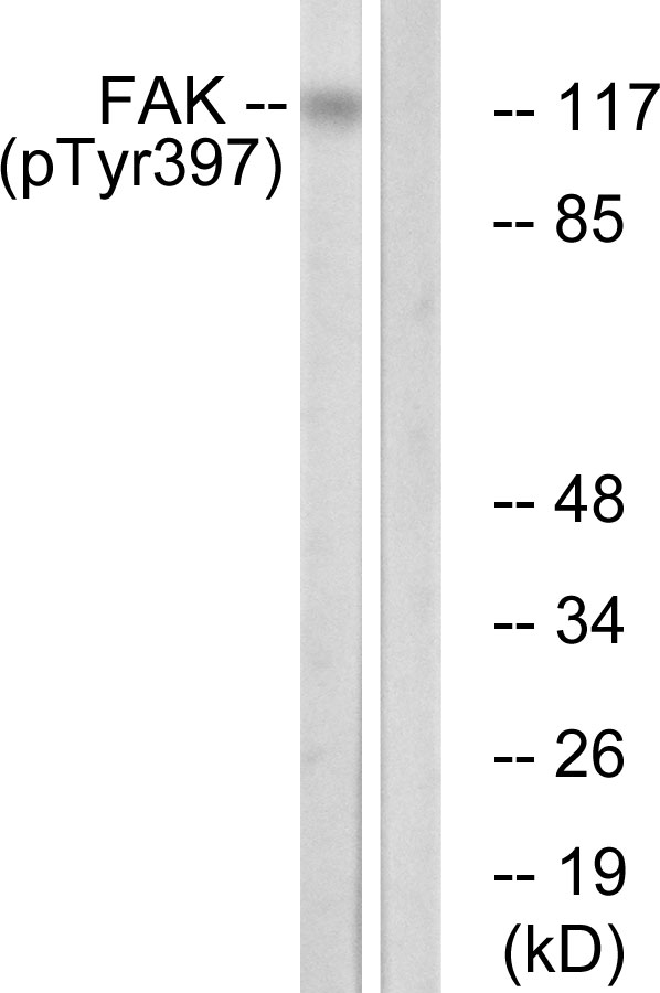 WB (Western Blot)
(Western blot analysis of lysates from Jurkat cells treated with Ca2+ 40nM 30', using FAK (Phospho-Tyr397) Antibody. The lane on the right is blocked with the phospho peptide.)
WB (Western Blot)
(Western blot analysis of lysates from Jurkat cells treated with Ca2+ 40nM 30', using FAK (Phospho-Tyr397) Antibody. The lane on the right is blocked with the phospho peptide.)
FAK, Polyclonal Antibody (Cat# AAA316363)
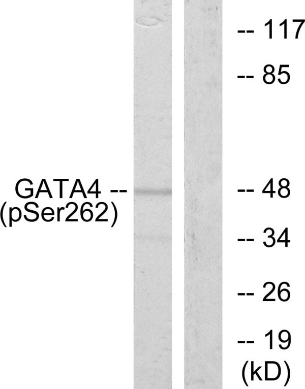 WB (Western Blot)
(Western blot analysis of lysates from 293 cells, using GATA4 (Phospho-Ser262) Antibody. The lane on the right is blocked with the phospho peptide.)
WB (Western Blot)
(Western blot analysis of lysates from 293 cells, using GATA4 (Phospho-Ser262) Antibody. The lane on the right is blocked with the phospho peptide.)
GATA4, Polyclonal Antibody (Cat# AAA316364)
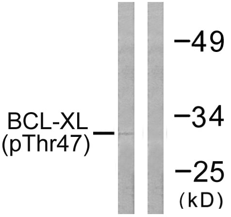 WB (Western Blot)
(Western blot analysis of lysates from 293 cells treated with UV 30', using BCL-XL (Phospho-Thr47) Antibody. The lane on the right is blocked with the phospho peptide.)
WB (Western Blot)
(Western blot analysis of lysates from 293 cells treated with UV 30', using BCL-XL (Phospho-Thr47) Antibody. The lane on the right is blocked with the phospho peptide.)
BCL-XL, Polyclonal Antibody (Cat# AAA316294)
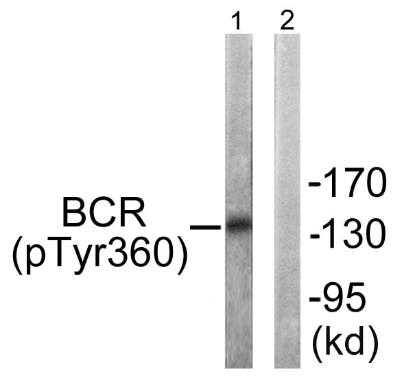 WB (Western Blot)
(Western blot analysis of lysates from COS7 cells, using Bcr (Phospho-Tyr360) Antibody. The lane on the right is blocked with the phospho peptide.)
WB (Western Blot)
(Western blot analysis of lysates from COS7 cells, using Bcr (Phospho-Tyr360) Antibody. The lane on the right is blocked with the phospho peptide.)
Bcr, Polyclonal Antibody (Cat# AAA316295)

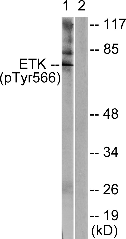 WB (Western Blot)
(Western blot analysis of lysates from HeLa cells treated with Serum 20% 15', using ETK (Phospho-Tyr566) Antibody. The lane on the right is blocked with the phospho peptide.)
WB (Western Blot)
(Western blot analysis of lysates from HeLa cells treated with Serum 20% 15', using ETK (Phospho-Tyr566) Antibody. The lane on the right is blocked with the phospho peptide.)
ETK, Polyclonal Antibody (Cat# AAA316300)
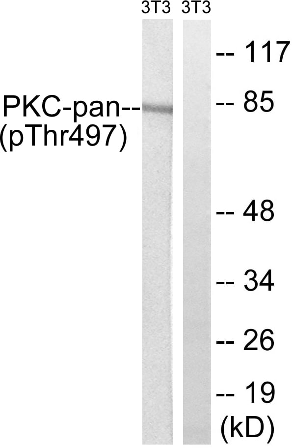 WB (Western Blot)
(Western blot analysis of lysates from NIH/3T3 cells treated with PMA 250ng/ml 15', using PKC-pan (Phospho-Thr497) Antibody. The lane on the right is blocked with the phospho peptide.)
WB (Western Blot)
(Western blot analysis of lysates from NIH/3T3 cells treated with PMA 250ng/ml 15', using PKC-pan (Phospho-Thr497) Antibody. The lane on the right is blocked with the phospho peptide.)
PKC-pan, Polyclonal Antibody (Cat# AAA316303)
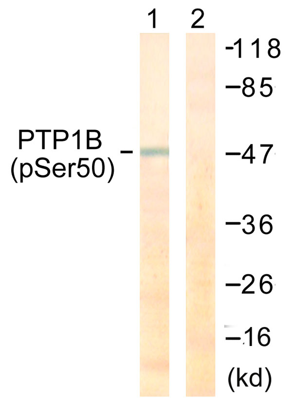 WB (Western Blot)
(Western blot analysis of lysates from COS7 cells treated with UV 30', using PTP1B (Phospho-Ser50) Antibody. The lane on the right is blocked with the phospho peptide.)
WB (Western Blot)
(Western blot analysis of lysates from COS7 cells treated with UV 30', using PTP1B (Phospho-Ser50) Antibody. The lane on the right is blocked with the phospho peptide.)
PTP1B, Polyclonal Antibody (Cat# AAA316308)
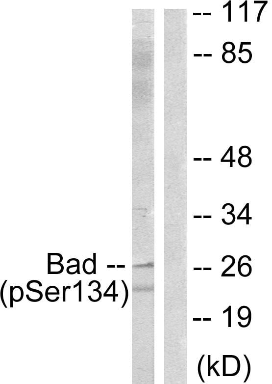 WB (Western Blot)
(Western blot analysis of lysates from mouse liver, using BAD (Phospho-Ser134) Antibody. The lane on the right is blocked with the phospho peptide.)
WB (Western Blot)
(Western blot analysis of lysates from mouse liver, using BAD (Phospho-Ser134) Antibody. The lane on the right is blocked with the phospho peptide.)
BAD, Polyclonal Antibody (Cat# AAA316315)
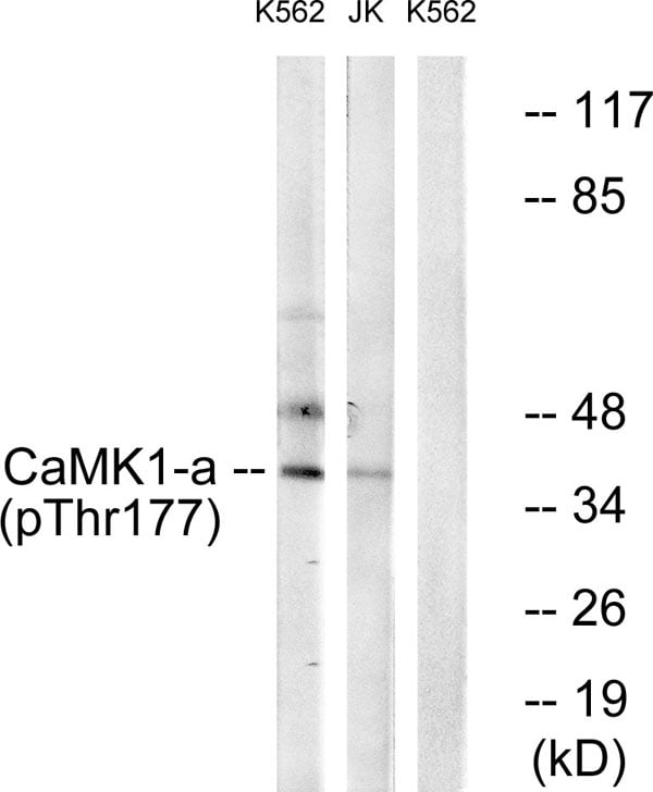 WB (Western Blot)
(Western blot analysis of lysates from K562 cells treated with insulin 0.01U/ml 15' and Jurkat cells treated with insulin 0.01U/ml 15', using CaMK1-alpha (Phospho-Thr177) Antibody. The lane on the right is blocked with the phospho peptide.)
WB (Western Blot)
(Western blot analysis of lysates from K562 cells treated with insulin 0.01U/ml 15' and Jurkat cells treated with insulin 0.01U/ml 15', using CaMK1-alpha (Phospho-Thr177) Antibody. The lane on the right is blocked with the phospho peptide.)
CaMK1-alpha, Polyclonal Antibody (Cat# AAA316320)
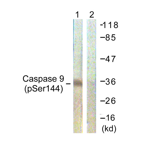 WB (Western Blot)
(Western blot analysis of lysates from K562 cells, using Caspase 9 (Phospho-Ser144) Antibody. The lane on the right is blocked with the phospho peptide.)
WB (Western Blot)
(Western blot analysis of lysates from K562 cells, using Caspase 9 (Phospho-Ser144) Antibody. The lane on the right is blocked with the phospho peptide.)
Caspase 9, Polyclonal Antibody (Cat# AAA316323)
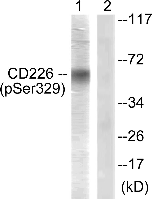 WB (Western Blot)
(Western blot analysis of lysates from COS7 cells, using CD226/DNAM-1 (Phospho-Ser329) Antibody. The lane on the right is blocked with the phospho peptide.)
WB (Western Blot)
(Western blot analysis of lysates from COS7 cells, using CD226/DNAM-1 (Phospho-Ser329) Antibody. The lane on the right is blocked with the phospho peptide.)
CD226/DNAM-1, Polyclonal Antibody (Cat# AAA316327)
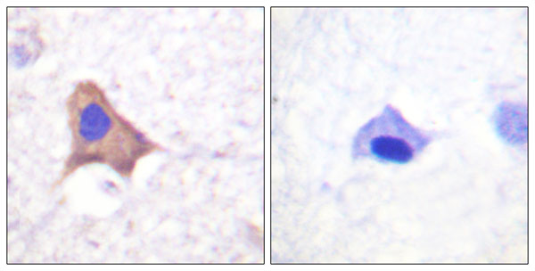
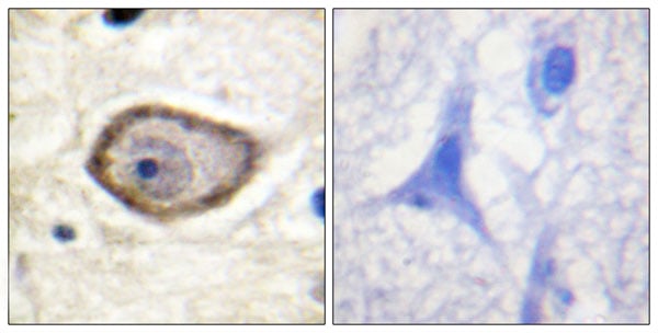
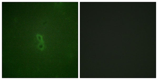
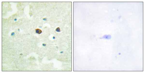
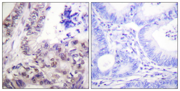
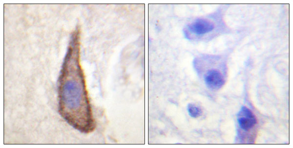
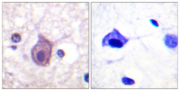 IHC (Immunohiostchemistry)
(Immunohistochemistry analysis of paraffin-embedded human brain, using CD4 (Phospho-Ser433) Antibody. The picture on the right is blocked with the phospho peptide.)
IHC (Immunohiostchemistry)
(Immunohistochemistry analysis of paraffin-embedded human brain, using CD4 (Phospho-Ser433) Antibody. The picture on the right is blocked with the phospho peptide.)
CD4, Polyclonal Antibody (Cat# AAA316328)
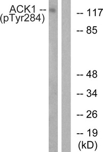
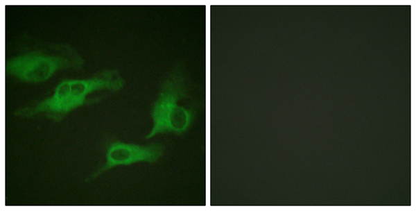
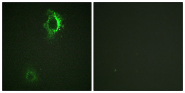
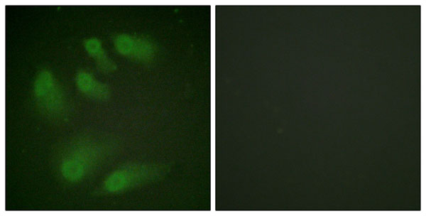
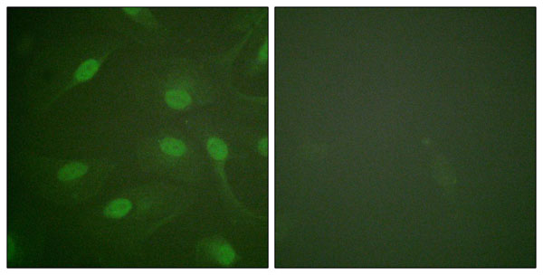
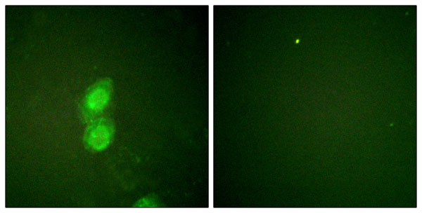
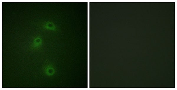
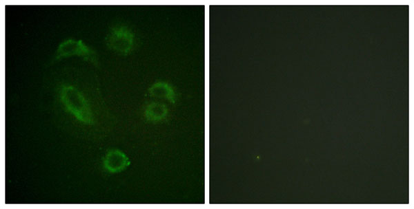
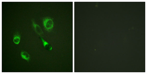
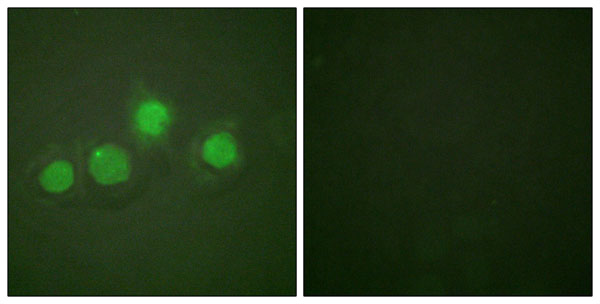
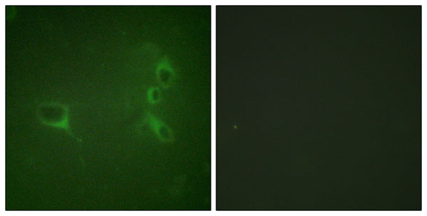
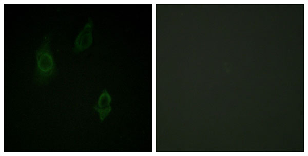
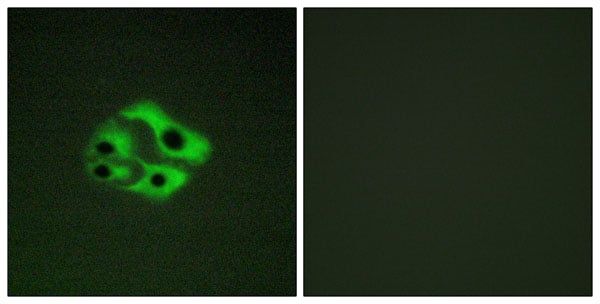
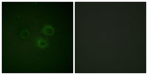
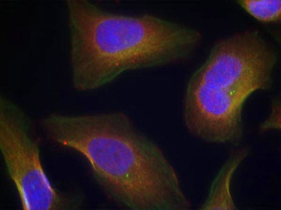
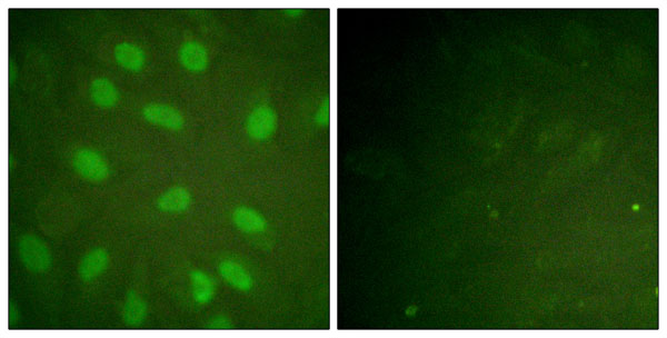
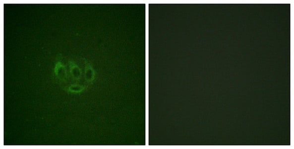 IF (Immunofluorescence)
(Immunofluorescence staining of methanol-fixed A549 cells using ACK1 (Phospho-Tyr284) Antibody.)
IF (Immunofluorescence)
(Immunofluorescence staining of methanol-fixed A549 cells using ACK1 (Phospho-Tyr284) Antibody.)
TNK2, Polyclonal Antibody (Cat# AAA243344)
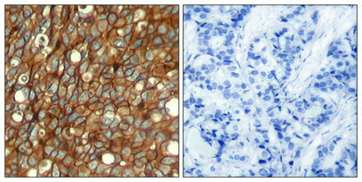
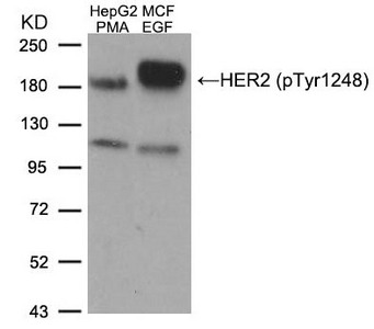
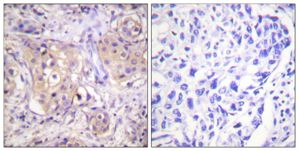
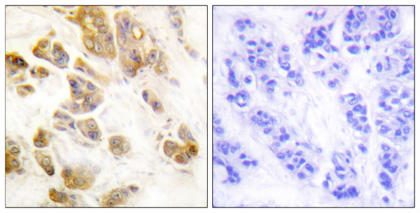
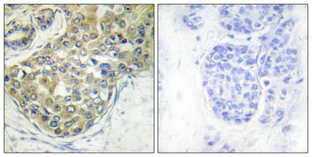
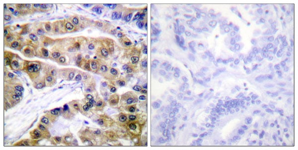
 IHC (Immunohistochemistry)
(Immunohistochemical analysis of paraffin-embedded human lung carcinoma tissue, using HER2 (Phospho-Tyr1248) Antibody.)
IHC (Immunohistochemistry)
(Immunohistochemical analysis of paraffin-embedded human lung carcinoma tissue, using HER2 (Phospho-Tyr1248) Antibody.)
ERBB2, Polyclonal Antibody (Cat# AAA243357)
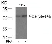
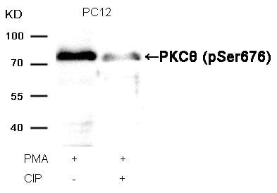 WB (Western Blot)
(Western blot analysis of extracts from PC12 cells, treated with PMA or calf intestinal phosphatase (CIP), using PKCtheta (Phospho-Ser676) Antibody.)
WB (Western Blot)
(Western blot analysis of extracts from PC12 cells, treated with PMA or calf intestinal phosphatase (CIP), using PKCtheta (Phospho-Ser676) Antibody.)
PRKCQ, Polyclonal Antibody (Cat# AAA243371)
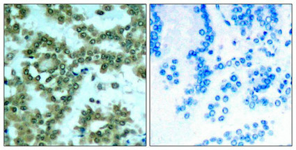
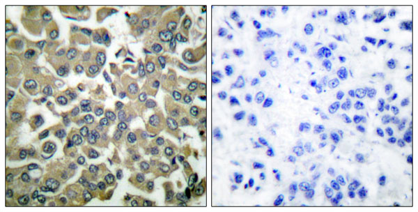
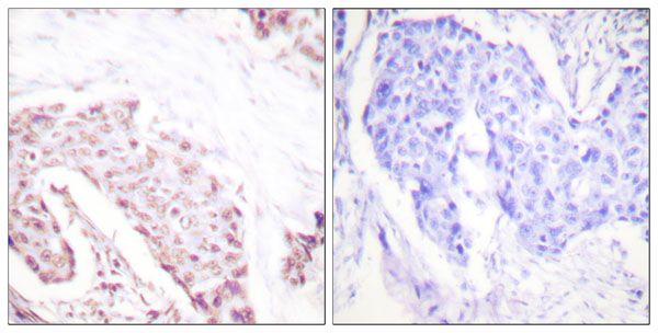
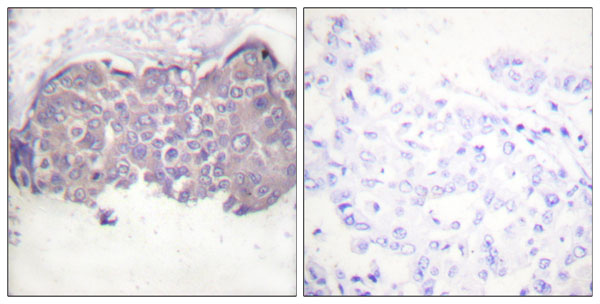
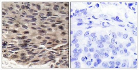 IHC (Immunohiostchemistry)
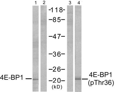
(Immunohistochemical analysis of paraffin-embedded human breast carcinoma, using 4E-BP1 (phospho-Thr36) antibody.)
IHC (Immunohiostchemistry)
(Immunohistochemical analysis of paraffin-embedded human breast carcinoma, using 4E-BP1 (phospho-Thr36) antibody.)
EIF4EBP1, Polyclonal Antibody (Cat# AAA243178)

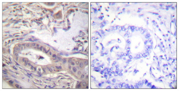
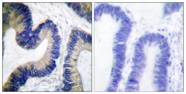
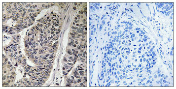 IHC (Immunohiostchemistry)
(Immunohistochemical analysis of paraffin-embedded human breast carcinoma tissue, using p130 Cas (Phospho-Tyr410) antibody (left)or the same antibody preincubated with blocking peptide (right).)
IHC (Immunohiostchemistry)
(Immunohistochemical analysis of paraffin-embedded human breast carcinoma tissue, using p130 Cas (Phospho-Tyr410) antibody (left)or the same antibody preincubated with blocking peptide (right).)
BCAR1, Polyclonal Antibody (Cat# AAA243185)

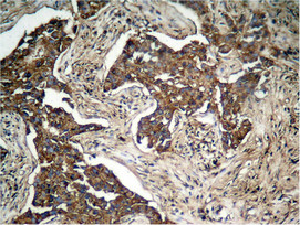
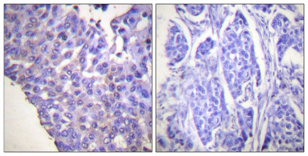
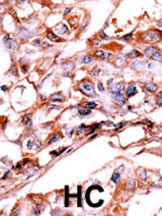 IHC (Immunohiostchemistry)
(Formalin-fixed and paraffin-embedded human cancer tissue reacted with the primary antibody, which was peroxidase-conjugated to the secondary antibody, followed by AEC staining. This data demonstrates the use of this antibody for immunohistochemistry; clinical relevance has not been evaluated. BC = breast carcinoma; HC = hepatocarcinoma.)
IHC (Immunohiostchemistry)
(Formalin-fixed and paraffin-embedded human cancer tissue reacted with the primary antibody, which was peroxidase-conjugated to the secondary antibody, followed by AEC staining. This data demonstrates the use of this antibody for immunohistochemistry; clinical relevance has not been evaluated. BC = breast carcinoma; HC = hepatocarcinoma.)
Phospho-HER4 (Y1188), Polyclonal Antibody (Cat# AAA284893)

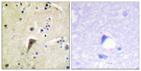
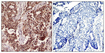
 IHC (Immunohiostchemistry)
(Immunohistochemical analysis of paraffin-embedded human brain tissue using PYK2 (Phospho-Tyr579) antibody (left)or the same antibody preincubated with blocking peptide (right).)
IHC (Immunohiostchemistry)
(Immunohistochemical analysis of paraffin-embedded human brain tissue using PYK2 (Phospho-Tyr579) antibody (left)or the same antibody preincubated with blocking peptide (right).)
PTK2B, Polyclonal Antibody (Cat# AAA243216)
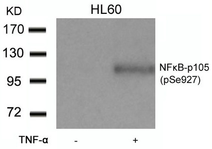
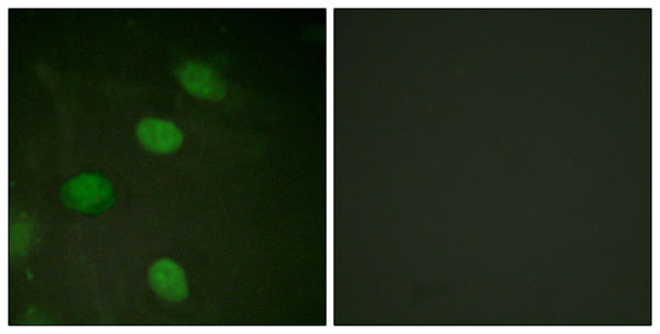
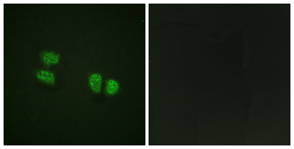
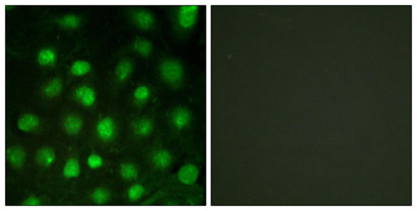
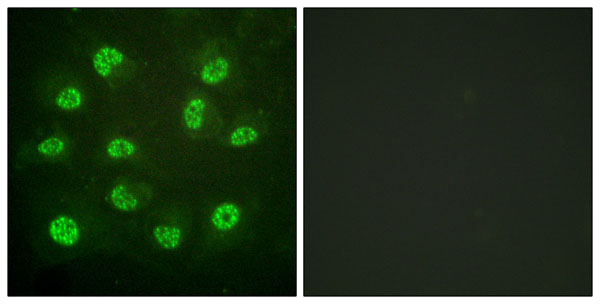
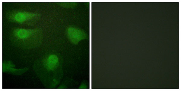
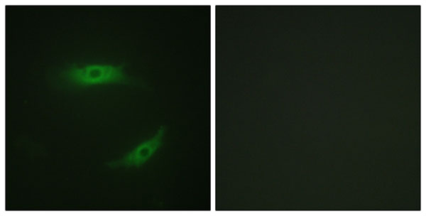
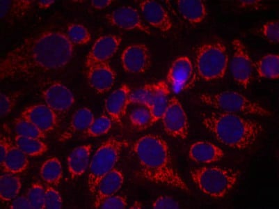
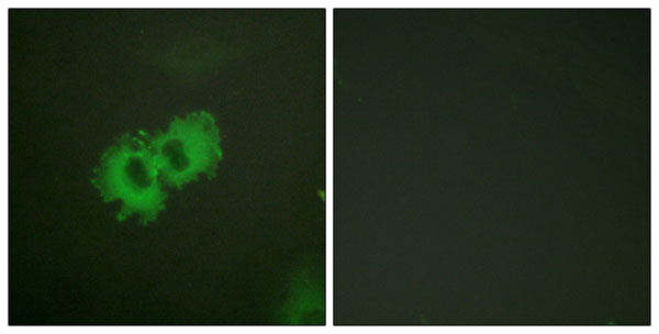
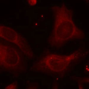 IF (Immunofluorescence)
(Immunofluorescence staining of methanol-fixed Hela cells using NFkB-p105(Phospho-Ser927) Antibody.)
IF (Immunofluorescence)
(Immunofluorescence staining of methanol-fixed Hela cells using NFkB-p105(Phospho-Ser927) Antibody.)
NFKB1, Polyclonal Antibody (Cat# AAA243319)
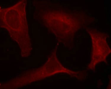

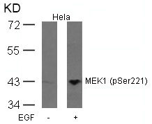
 WB (Western Blot)
(Western blot analysis of extracts from Hela cells, treated with calf intestinal phosphatase (CIP), using MEK1 (Phospho-Ser221) Antibody.)
WB (Western Blot)
(Western blot analysis of extracts from Hela cells, treated with calf intestinal phosphatase (CIP), using MEK1 (Phospho-Ser221) Antibody.)
MAP2K1, Polyclonal Antibody (Cat# AAA243363)
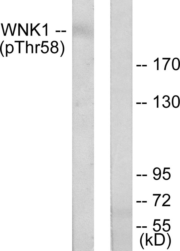 WB (Western Blot)
(Western blot analysis of lysates from 293 cells treated with EGF 200ng/ml 30', using WNK1 (Phospho-Thr58) Antibody. The lane on the right is blocked with the phospho peptide.)
WB (Western Blot)
(Western blot analysis of lysates from 293 cells treated with EGF 200ng/ml 30', using WNK1 (Phospho-Thr58) Antibody. The lane on the right is blocked with the phospho peptide.)
WNK1, Polyclonal Antibody (Cat# AAA316123)
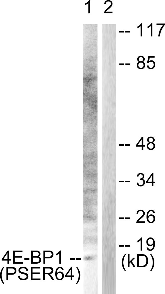 WB (Western Blot)
(Western blot analysis of lysates from Jurkat cells treated with Insulin 0.01U/ml 15', using 4E-BP1 (Phospho-Ser64) Antibody. The lane on the right is blocked with the phospho peptide.)
WB (Western Blot)
(Western blot analysis of lysates from Jurkat cells treated with Insulin 0.01U/ml 15', using 4E-BP1 (Phospho-Ser64) Antibody. The lane on the right is blocked with the phospho peptide.)
4E-BP1, Polyclonal Antibody (Cat# AAA316124)
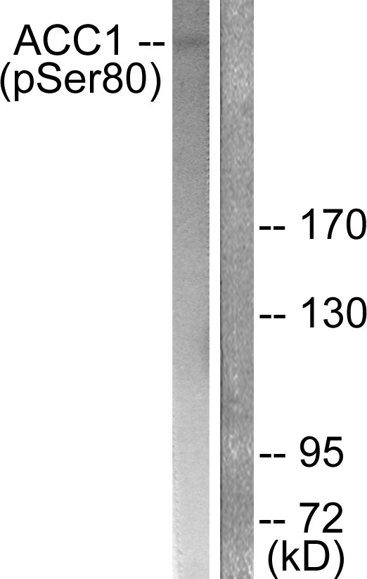 WB (Western Blot)
(Western blot analysis of lysates from K562 cells treated with Insulin 0.01U/ml 15', using ACC1 (Phospho-Ser80) Antibody. The lane on the right is blocked with the phospho peptide.)
WB (Western Blot)
(Western blot analysis of lysates from K562 cells treated with Insulin 0.01U/ml 15', using ACC1 (Phospho-Ser80) Antibody. The lane on the right is blocked with the phospho peptide.)
ACC1, Polyclonal Antibody (Cat# AAA316127)
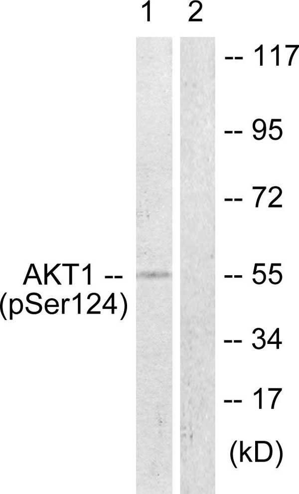 WB (Western Blot)
(Western blot analysis of lysates from mouse brain, using Akt (Phospho-Ser124) Antibody. The lane on the right is blocked with the phospho peptide.)
WB (Western Blot)
(Western blot analysis of lysates from mouse brain, using Akt (Phospho-Ser124) Antibody. The lane on the right is blocked with the phospho peptide.)
Akt, Polyclonal Antibody (Cat# AAA316129)
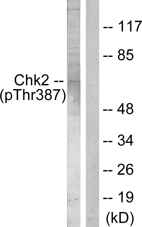 WB (Western Blot)
(Western blot analysis of lysates from Jurkat cells, using Chk2 (Phospho-Thr387) Antibody. The lane on the right is blocked with the phospho peptide.)
WB (Western Blot)
(Western blot analysis of lysates from Jurkat cells, using Chk2 (Phospho-Thr387) Antibody. The lane on the right is blocked with the phospho peptide.)
Chk2, Polyclonal Antibody (Cat# AAA316133)
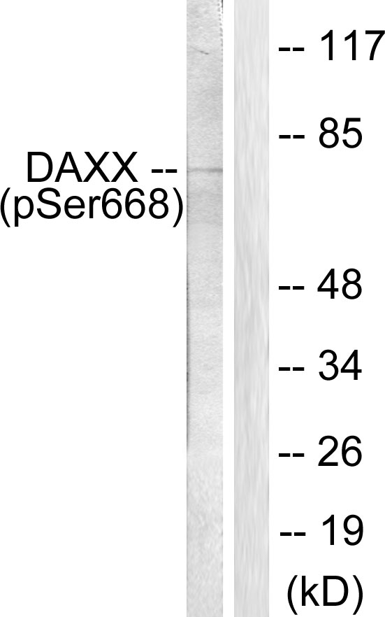 WB (Western Blot)
(Western blot analysis of lysates from 293 cells treated with PBS 60', using Daxx (Phospho-Ser668) Antibody. The lane on the right is blocked with the phospho peptide.)
WB (Western Blot)
(Western blot analysis of lysates from 293 cells treated with PBS 60', using Daxx (Phospho-Ser668) Antibody. The lane on the right is blocked with the phospho peptide.)
Daxx, Polyclonal Antibody (Cat# AAA316135)
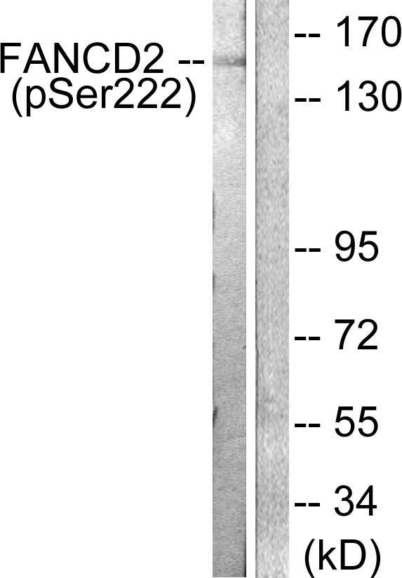 WB (Western Blot)
(Western blot analysis of lysates from HT29 cells treated with Calyculin A 50ng/ml 30', using FANCD2 (Phospho-Ser222) Antibody. The lane on the right is blocked with the phospho peptide.)
WB (Western Blot)
(Western blot analysis of lysates from HT29 cells treated with Calyculin A 50ng/ml 30', using FANCD2 (Phospho-Ser222) Antibody. The lane on the right is blocked with the phospho peptide.)
FANCD2, Polyclonal Antibody (Cat# AAA316140)
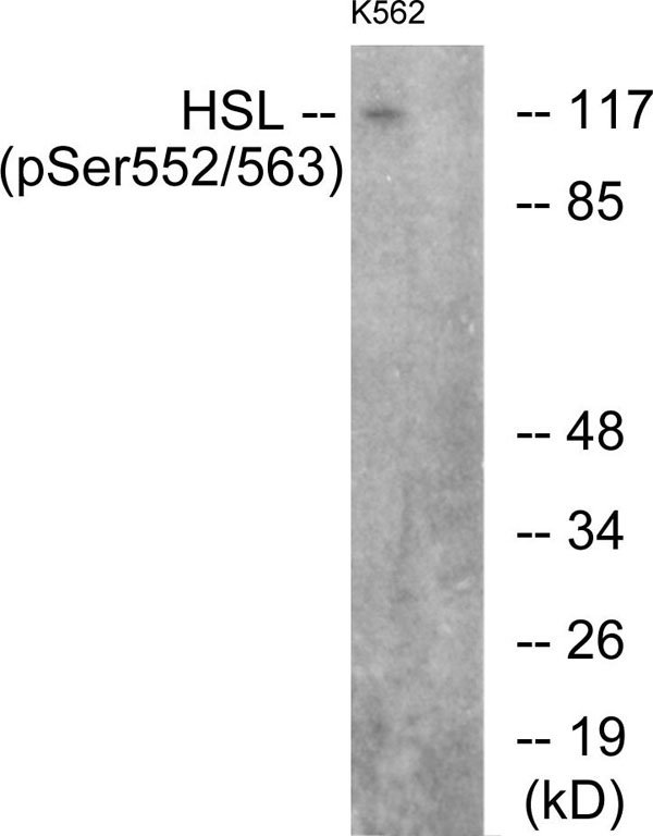 WB (Western Blot)
(Western blot analysis of lysates from K562 cells, using HSL (Phospho-Ser552) Antibody. The lane on the right is blocked with the phospho peptide.)
WB (Western Blot)
(Western blot analysis of lysates from K562 cells, using HSL (Phospho-Ser552) Antibody. The lane on the right is blocked with the phospho peptide.)
HSL, Polyclonal Antibody (Cat# AAA316147)
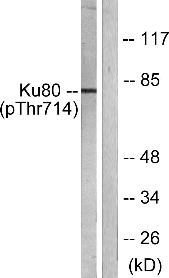 WB (Western Blot)
(Western blot analysis of lysates from COS7 cells, using Ku80 (Phospho-Thr714) Antibody. The lane on the right is blocked with the phospho peptide.)
WB (Western Blot)
(Western blot analysis of lysates from COS7 cells, using Ku80 (Phospho-Thr714) Antibody. The lane on the right is blocked with the phospho peptide.)
Ku80, Polyclonal Antibody (Cat# AAA316157)
 WB (Western Blot)
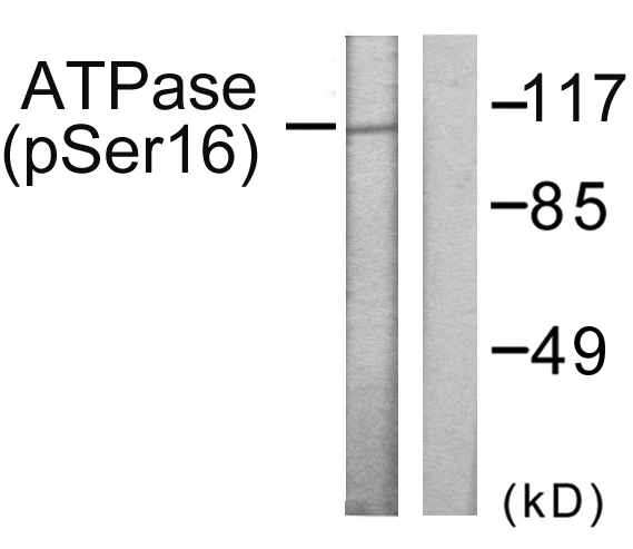
(Western blot analysis of lysates from 293 cells treated with PMA 125ng/ml 30', using ATPase (Phospho-Ser16) Antibody. The lane on the right is blocked with the phospho peptide.)
WB (Western Blot)
(Western blot analysis of lysates from 293 cells treated with PMA 125ng/ml 30', using ATPase (Phospho-Ser16) Antibody. The lane on the right is blocked with the phospho peptide.)
ATPase, Polyclonal Antibody (Cat# AAA316161)
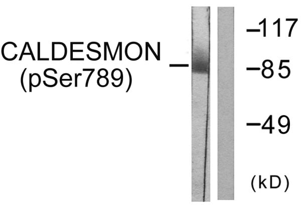 WB (Western Blot)
(Western blot analysis of lysates from HeLa cells treated with EGF 200ng/ml 30', using Caldesmon (Phospho-Ser789) Antibody. The lane on the right is blocked with the phospho peptide.)
WB (Western Blot)
(Western blot analysis of lysates from HeLa cells treated with EGF 200ng/ml 30', using Caldesmon (Phospho-Ser789) Antibody. The lane on the right is blocked with the phospho peptide.)
Caldesmon, Polyclonal Antibody (Cat# AAA316164)
What Are Phospho Antibodies?
Protein phosphorylation is a process where a phosphate group is added to certain amino acid residues of a protein – usually serine (S), threonine (T), or tyrosine (Y) - by enzymes called kinases. This process is integral in controlling cellular signaling, cellular growth, and other biological functions.
Our catalog includes a wide range of phospho-specific antibodies that can accurately detect this important marker. They perform strongly in widely-used laboratory applications such as Western blot, flow cytometry, immunohistochemistry, and immunofluorescence microscopy. We value your trust in us and are committed to providing top-quality products and services. All of our antibodies are guaranteed to work for the applications and species indicated on our website & associated product pages.
What Are The Key Applications of Phospho Antibodies?
1. Western Blotting
One of the first steps a researcher can take in utilizing these phospho-specific antibodies, is to check if the antibody works using a technique referred to as “Western blot”. For those unfamiliar, Western Blot aids in showing whether the protein that the antibody recognizes is appearing at the correct/expected size. These phospho-specific antibodies should also be able to detect changes in the target protein’s phosphorylation (on/off state) when cells are stimulated in certain ways.
2. Staining of Fixed Cells (Immunocytochemistry)
Another routine use of these phospho-specific antibodies, is to test if the antibody is able to demonstrate similar performance when used on fixed cells (intact cells that have been preserved) as it did in the Western blot tests. It is an important aspect in many cases to confirm that the antibody works in actual intact cell samples. Ideally, the method used for cellular fixation should be the same as what is used in pathology labs (like using 10% formalin). To check if the antibody works well in tissue sections (FFPE), researchers will often test it on fixed cells that are processed similar to tissue samples.
3. Specificity Tests Using Peptides
In order to make sure that the antibody is only binding to the right target:
- Laboratory technicians will mix the antibody with phospho-peptides (short segments of the protein containing the phosphate group modification).
- If the antibody signal disappears, it is confirmation that it is binding to the correct phosphorylated location.
- A more robust test is to use both the phosphorylated and non-phosphorylated (dephosphorylated) versions of the protein. The antibody should react only with the phosphorylated one.
- Another method sometimes utilized is to treat the sample with an enzyme, such as alkaline phosphatase, that specifically removes phosphate groups. If the antibody signal disappears after this, it also confirms specificity.
4. Genetic Confirmation
As a final step, scientists can genetically manipulate the nucleotide sequence and alter the target protein by removing the exact site where phosphorylation happens. If the antibody no longer appears to detect the modified protein, it is strong evidence supporting the antibody being specific for that phosphorylated site.
Why Buy Phospho Antibodies Through Us?
- The production laboratory adheres to strict and consistent protocols prior to releasing any of these phospho-specific antibodies:
- Standard methods and proper controls in all tests to ensure high quality.
- These antibodies are tested and validated in different cell types and species.
- High quality control criterion to ensure each batch is consistent, so you will obtain reliable results every time.
FAQ
1. What Are Phospho-Specific Antibodies?
Phospho-specific antibodies are made to detect proteins only when they have a phosphate group linked to a specific amino acid residue. This empowers scientists understand if a protein is "turned on" or active, based on its phosphorylation state.
2. How to Detect Phosphorylated Proteins in a Western Blot?
To find out if a protein is phosphorylated using Western blot:
- Use a phospho-specific antibody that binds only to the phosphorylated form of the protein.
- You can also use a “regular” antibody for the same amino acid sequence of the protein that the phospho-specific antibody is binding to (but in this case, this antibody will not bind if there is a phosphate group present) in order to compare how much of it is phosphorylated versus how much is non-phosphorylated (or “total” protein, if the “normal” antibody’s epitopes are non-phospho-site-specific).
3. How to Choose the Best Antibody?
Here are some simple tips to help you pick the right antibody:
- Know your target
- Match your sample characteristics
- Confirm the intended use is appropriate
- Check “host” and “type”
- Check the “quality” of the presented data/images
- Appraise whether the available validation meets your needs