Filters
▼Clonality
▼Type
▼Reactivity
▼Gene Name
▼Isotype
▼Host
▼Application
▼Clone
▼Monoclonal Antibodies
Get accurate results in your research with our Monoclonal Antibodies, which are specially made to target exactly what you require for your research, and will produce consistent, reliable performance in lab tests.
Viewing 3550-3600 of 27597 product results
Adenovirus, Monoclonal Antibody (Cat# AAA106396)
Adenovirus antibody was purified by Ion exchange chromatography.
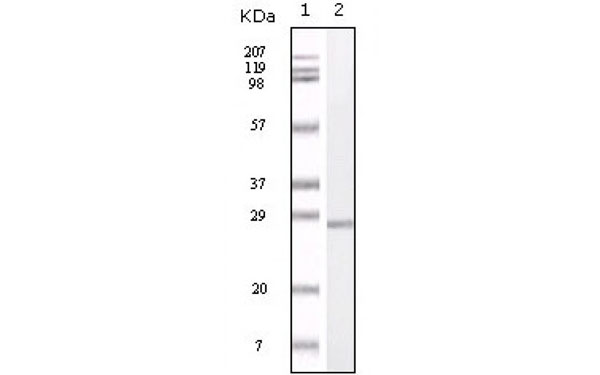 WB (Western Blot)
(Western Blot showing S100A antibody used against truncated S100A recombinant protein.)
WB (Western Blot)
(Western Blot showing S100A antibody used against truncated S100A recombinant protein.)
S100A1, Monoclonal Antibody (Cat# AAA106217)
OATP2, Monoclonal Antibody (Cat# AAA106224)
 IF (Immunofluorescence)
(Immunofluorescent staining of COS7 cells transiently transfected with recombinant DPP9 protein using DPP9 antibody)
IF (Immunofluorescence)
(Immunofluorescent staining of COS7 cells transiently transfected with recombinant DPP9 protein using DPP9 antibody)
DPP9, Monoclonal Antibody (Cat# AAA106253)
 IF (Immunofluorescence)
(Immunofluorescent staining of COS7 cells transiently transfected with recombinant APP protein using APP antibody)
IF (Immunofluorescence)
(Immunofluorescent staining of COS7 cells transiently transfected with recombinant APP protein using APP antibody)
APP, Monoclonal Antibody (Cat# AAA106431)
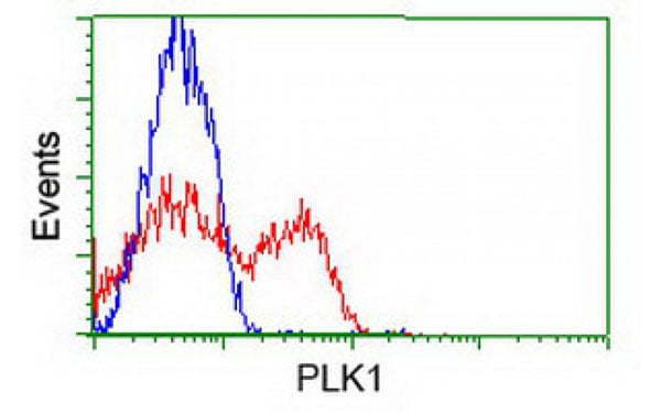
 Application Data
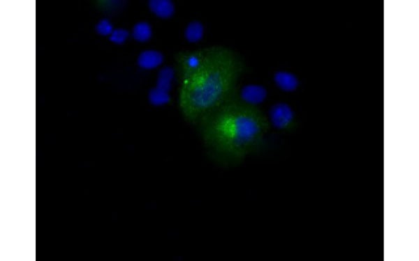
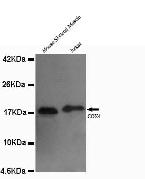
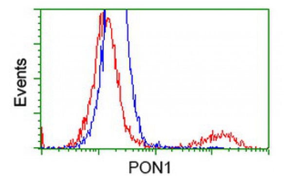
(Flow Cytometry analysis of K562 cells stained with COX4 (red, 1/100 dilution), followed by FITC-conjugated goat anti-mouse IgG. Blue line histogram represents the isotype control, normal mouse IgG.)
Application Data
(Flow Cytometry analysis of K562 cells stained with COX4 (red, 1/100 dilution), followed by FITC-conjugated goat anti-mouse IgG. Blue line histogram represents the isotype control, normal mouse IgG.)
COX4, Monoclonal Antibody (Cat# AAA111267)
 Application Data
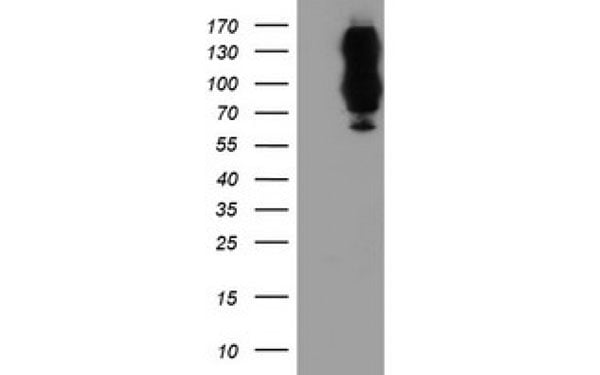
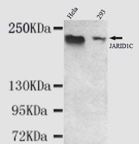
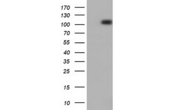
(Western blot detection of KDM5C(C-terminus) antibody in Hela&293 cell lysates using KDM5C(C-terminus) antibody (1:1000 diluted). Predicted band size: 176KDa Observed band size: 220KDa)
Application Data
(Western blot detection of KDM5C(C-terminus) antibody in Hela&293 cell lysates using KDM5C(C-terminus) antibody (1:1000 diluted). Predicted band size: 176KDa Observed band size: 220KDa)
KDM5C, Monoclonal Antibody (Cat# AAA111281)
 Application Data
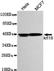
(Western blot detection of KRT18 in Hela&MCF7 cell lysates and using KRT18 antibody (1:1000 diluted). Predicted band size: 48KDa Observed band size: 48KDa)
Application Data
(Western blot detection of KRT18 in Hela&MCF7 cell lysates and using KRT18 antibody (1:1000 diluted). Predicted band size: 48KDa Observed band size: 48KDa)
KRT18, Monoclonal Antibody (Cat# AAA111286)
 Application Data
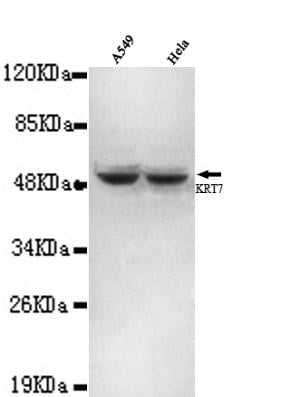
(Western blot detection of Keratin 7(N-terminus) antibody in A459&Hela cell lysates using Keratin 7(N-terminus) antibody (1:1000 diluted). Predicted band size: 50KDa Observed band size: 50KDa.)
Application Data
(Western blot detection of Keratin 7(N-terminus) antibody in A459&Hela cell lysates using Keratin 7(N-terminus) antibody (1:1000 diluted). Predicted band size: 50KDa Observed band size: 50KDa.)
Keratin 7, Monoclonal Antibody (Cat# AAA111287)
 Application Data
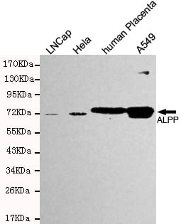
(Western blot detection of ALPP in Hela,LNCap,human Placenta &A549 cell lysates and using ALPP antibody (1:500 diluted). Predicted band size: 76KDa Observed band size:76KDa.)
Application Data
(Western blot detection of ALPP in Hela,LNCap,human Placenta &A549 cell lysates and using ALPP antibody (1:500 diluted). Predicted band size: 76KDa Observed band size:76KDa.)
ALPP, Monoclonal Antibody (Cat# AAA111289)
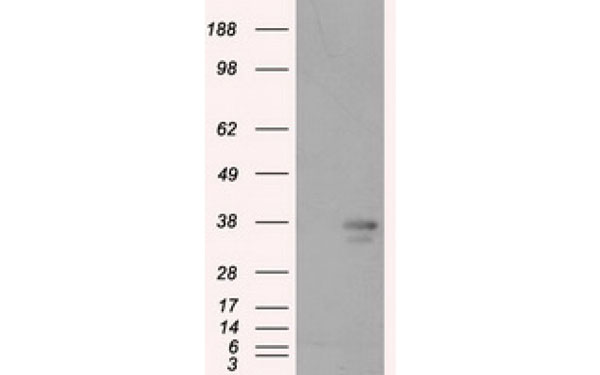
 Application Data
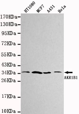
(Western blot detection of AKR1B1 in HT1080, MCF7, A431&Hela cell lysates using AKR1B1 antibody (1:1000 diluted). Predicted band size:36KDa. Observed band size:36KDa.)
Application Data
(Western blot detection of AKR1B1 in HT1080, MCF7, A431&Hela cell lysates using AKR1B1 antibody (1:1000 diluted). Predicted band size:36KDa. Observed band size:36KDa.)
PTK2B, Monoclonal Antibody (Cat# AAA111298)

 Application Data
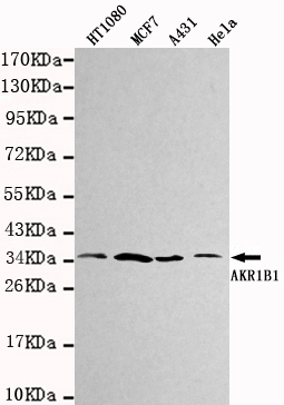
(Western blot detection of AKR1B1 in HT1080, MCF7, A431&Hela cell lysates using AKR1B1 antibody (1:1000 diluted). Predicted band size:36KDa. Observed band size:36KDa.)
Application Data
(Western blot detection of AKR1B1 in HT1080, MCF7, A431&Hela cell lysates using AKR1B1 antibody (1:1000 diluted). Predicted band size:36KDa. Observed band size:36KDa.)
AKR1B1, Monoclonal Antibody (Cat# AAA111299)
 Application Data
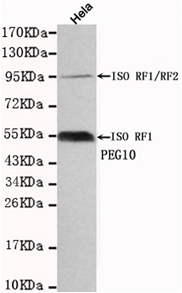
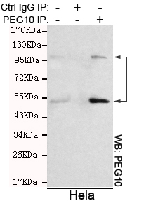
(Immunoprecipitation analysis of Hela cell lysates using PEG10 antibody.)
Application Data
(Immunoprecipitation analysis of Hela cell lysates using PEG10 antibody.)
PEG10, Monoclonal Antibody (Cat# AAA111303)
 Application Data
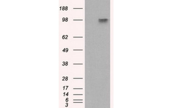
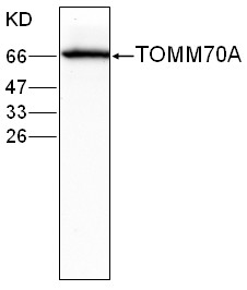
(WB(1:1000) analysis of TOMM70A expression in MCF-7 whole cell lysate with Anti-TOMM70A (AAA111305))
Application Data
(WB(1:1000) analysis of TOMM70A expression in MCF-7 whole cell lysate with Anti-TOMM70A (AAA111305))
TOMM70A, Monoclonal Antibody (Cat# AAA111305)
 Application Data
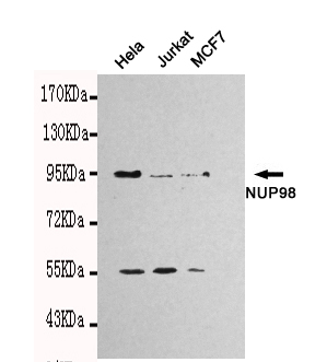
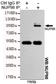
(Immunoprecipitation analysis of Hela cell lysates using NUP98 antibody.)
Application Data
(Immunoprecipitation analysis of Hela cell lysates using NUP98 antibody.)
NUP98, Monoclonal Antibody (Cat# AAA111310)
 Application Data
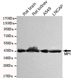
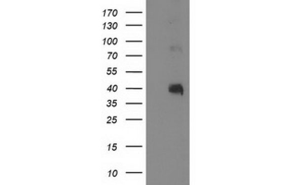
(Western blot detection of MPI in Rat kidney,Rat brain, A549&LNCAP cell lysates and using MPI antibody (1:1000 diluted). Predicted band size: 54KDa Observed band size: 45KDa.)
Application Data
(Western blot detection of MPI in Rat kidney,Rat brain, A549&LNCAP cell lysates and using MPI antibody (1:1000 diluted). Predicted band size: 54KDa Observed band size: 45KDa.)
MPI (Mannose Phosphate Isomerase), Monoclonal Antibody (Cat# AAA111318)
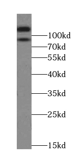
 WB (Western Blot)
(Western Blot analysis of Rat brain membrane lysate showing detection of SHANK1 protein using Mouse Anti-SHANK1 Monoclonal Antibody, Clone S22-21. Load: 15 ug. Block: 1.5% BSA for 30 minutes at RT. Primary Antibody: Mouse Anti-SHANK1 Monoclonal Antibody at 1:1000 for 2 hours at RT. Secondary Antibody: Sheep Anti-Mouse IgG: HRP for 1 hour at RT.)
WB (Western Blot)
(Western Blot analysis of Rat brain membrane lysate showing detection of SHANK1 protein using Mouse Anti-SHANK1 Monoclonal Antibody, Clone S22-21. Load: 15 ug. Block: 1.5% BSA for 30 minutes at RT. Primary Antibody: Mouse Anti-SHANK1 Monoclonal Antibody at 1:1000 for 2 hours at RT. Secondary Antibody: Sheep Anti-Mouse IgG: HRP for 1 hour at RT.)
Shank1, Monoclonal Antibody (Cat# AAA102947)
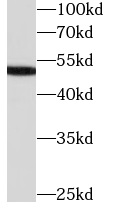
 WB (Western Blot)
(COLO 320 cells were subjected to SDS PAGE followed by western blot with AAA102787 (LIMA1 antibody) at dilution of 1:1000)
WB (Western Blot)
(COLO 320 cells were subjected to SDS PAGE followed by western blot with AAA102787 (LIMA1 antibody) at dilution of 1:1000)
EPLIN, Monoclonal Antibody (Cat# AAA102787)
Protein A+G purification
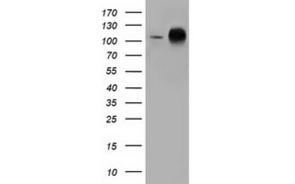
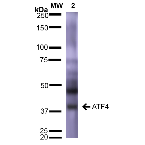 WB (Western Blot)
(Western Blot analysis of Rat Brain showing detection of ~39 kDa (isoform 2) ATF4 protein using Mouse Anti-ATF4 Monoclonal Antibody, Clone S360A-24 . Lane 1: Molecular Weight Ladder (MW). Lane 2: Rat Brain. Load: 15 ug. Block: 5% Skim Milk in 1X TBST. Primary Antibody: Mouse Anti-ATF4 Monoclonal Antibody at 1:1000 for 2 hours at RT. Secondary Antibody: Goat Anti-Mouse IgG: HRP at 1:2000 for 60 min at RT. Color Development: ECL solution for 5 min at RT. Predicted/Observed Size: ~39 kDa (isoform 2).)
WB (Western Blot)
(Western Blot analysis of Rat Brain showing detection of ~39 kDa (isoform 2) ATF4 protein using Mouse Anti-ATF4 Monoclonal Antibody, Clone S360A-24 . Lane 1: Molecular Weight Ladder (MW). Lane 2: Rat Brain. Load: 15 ug. Block: 5% Skim Milk in 1X TBST. Primary Antibody: Mouse Anti-ATF4 Monoclonal Antibody at 1:1000 for 2 hours at RT. Secondary Antibody: Goat Anti-Mouse IgG: HRP at 1:2000 for 60 min at RT. Color Development: ECL solution for 5 min at RT. Predicted/Observed Size: ~39 kDa (isoform 2).)
ATF4, Monoclonal Antibody (Cat# AAA103483)
 WB (Western Blot)
(L02 cells were subjected to SDS PAGE followed by western blot with AAA102693 (SERPINC1 Antibody) at dilution of 1:1000)
WB (Western Blot)
(L02 cells were subjected to SDS PAGE followed by western blot with AAA102693 (SERPINC1 Antibody) at dilution of 1:1000)
Antithrombin III, Monoclonal Antibody (Cat# AAA102693)
Purification: Protein A+G purification
 WB (Western Blot)
(human plasma tissue were subjected to SDS PAGE followed by western blot with AAA102717 (C4 (gamma chain) antibody at dilution of 1:8000)
WB (Western Blot)
(human plasma tissue were subjected to SDS PAGE followed by western blot with AAA102717 (C4 (gamma chain) antibody at dilution of 1:8000)
C4 (gamma chain), Monoclonal Antibody (Cat# AAA102717)
Protein A+G purification
 WB (Western Blot)
(K-562 cells were subjected to SDS PAGE followed by western blot with AAA102742 (CDK2 Antibody) at dilution of 1:1000)
WB (Western Blot)
(K-562 cells were subjected to SDS PAGE followed by western blot with AAA102742 (CDK2 Antibody) at dilution of 1:1000)
CDK2, Monoclonal Antibody (Cat# AAA102742)
Protein A+G purification
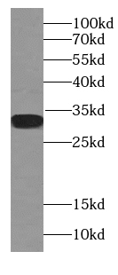
 WB (Western Blot)
(WB (1:2000) analysis of His-tagged (N-terminal) fusion protein with Anti-His Tag.)
WB (Western Blot)
(WB (1:2000) analysis of His-tagged (N-terminal) fusion protein with Anti-His Tag.)
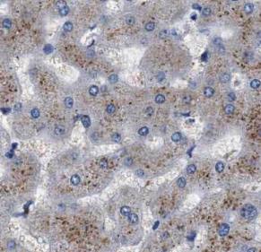
 IHC (Immunohiostchemistry)
(IHC (1:10) to human liver tissue.)
IHC (Immunohiostchemistry)
(IHC (1:10) to human liver tissue.)
IL-6, Monoclonal Antibody (Cat# AAA110028)
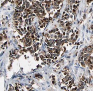
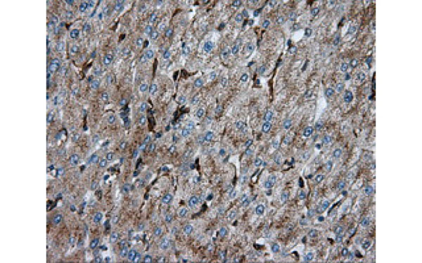
 IHC (Immunohiostchemistry)
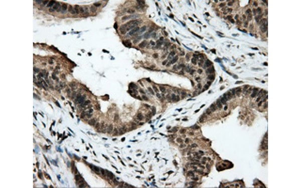
(IHC (1:100 200x) analysis of GOT2 expression in liver Cancer with Anti-GOT2.)
IHC (Immunohiostchemistry)
(IHC (1:100 200x) analysis of GOT2 expression in liver Cancer with Anti-GOT2.)
GOT2, Monoclonal Antibody (Cat# AAA110070)
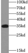
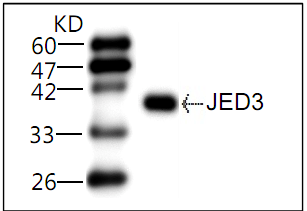 Application Data
(WB (1:1000) analysis of recombinant protein JEVE-D3 with Anti-JEV E-D3.)
Application Data
(WB (1:1000) analysis of recombinant protein JEVE-D3 with Anti-JEV E-D3.)
JEV E-D3, Monoclonal Antibody (Cat# AAA110348)
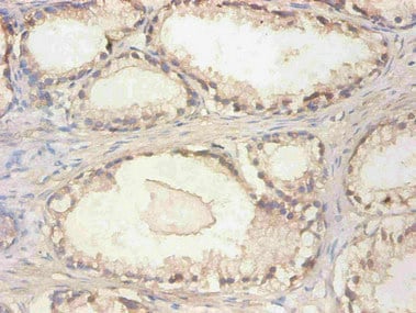
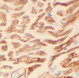
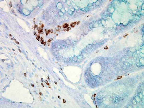
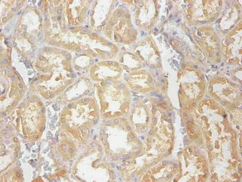 IHC (Immunohiostchemistry)
(Immunohistochemical of paraffin-embedded human kidney tissue using AAA114274 at dilution of 1:200)
IHC (Immunohiostchemistry)
(Immunohistochemical of paraffin-embedded human kidney tissue using AAA114274 at dilution of 1:200)
Cystatin C, Monoclonal Antibody (Cat# AAA114274)
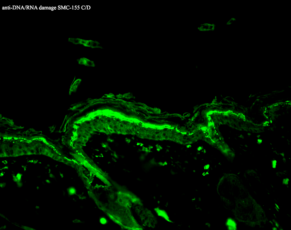
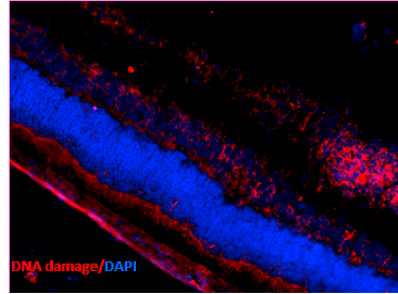
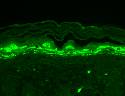
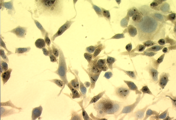
 IHC (Immunohistochemistry)
(Immunohistochemistry analysis using Mouse Anti-DNA Damage Monoclonal Antibody, Clone 15A3. Tissue: Ischemic fresh brain tissue. Species: Rat. Primary Antibody: Mouse Anti-DNA Damage Monoclonal Antibody at 1:1000 for 16 hours at RT. Secondary Antibody: Alexa Fluor 546 Goat Anti-mouse (Red) at 1:500 for 1 hour at RT. Localization: Cerebral Cortex. Courtesy of: Dr. Yi Yang, U. New Mexico.)
IHC (Immunohistochemistry)
(Immunohistochemistry analysis using Mouse Anti-DNA Damage Monoclonal Antibody, Clone 15A3. Tissue: Ischemic fresh brain tissue. Species: Rat. Primary Antibody: Mouse Anti-DNA Damage Monoclonal Antibody at 1:1000 for 16 hours at RT. Secondary Antibody: Alexa Fluor 546 Goat Anti-mouse (Red) at 1:500 for 1 hour at RT. Localization: Cerebral Cortex. Courtesy of: Dr. Yi Yang, U. New Mexico.)
DNA/RNA Damage, Monoclonal Antibody (Cat# AAA103193)
 IHC (Immunohistochemisry)
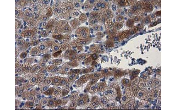
(Immunohistochemical analysis of PON1 protein in paraffin embedded Human liver tissue using PON1 antibody)
IHC (Immunohistochemisry)
(Immunohistochemical analysis of PON1 protein in paraffin embedded Human liver tissue using PON1 antibody)
PON1, Monoclonal Antibody (Cat# AAA108090)
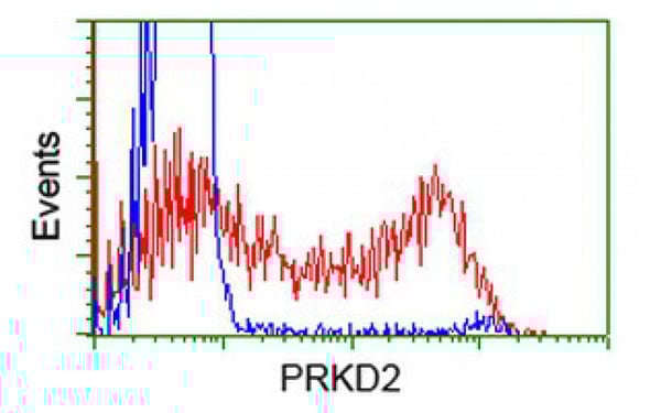
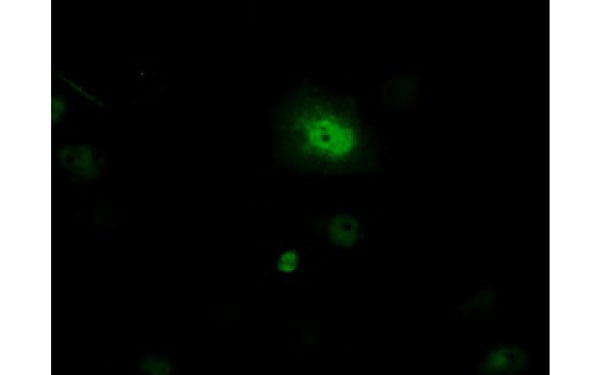
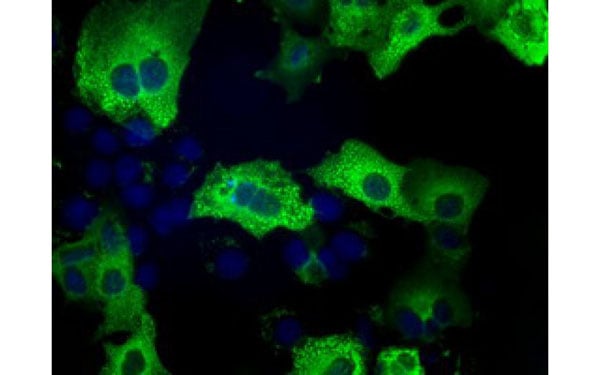
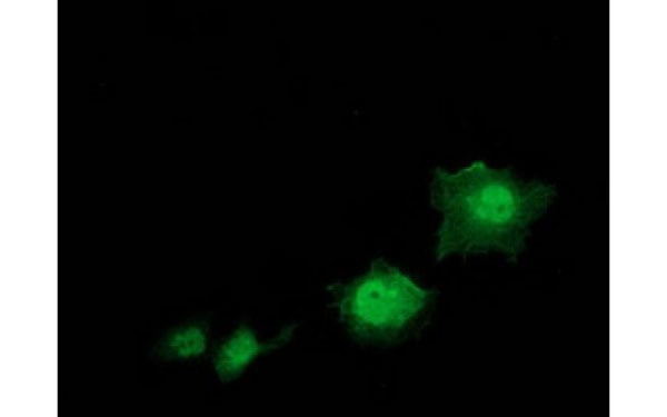
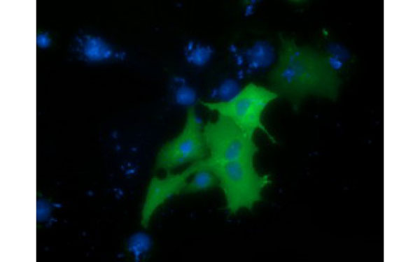 IF (Immunofluorescence)
(Immunofluorescent staining of COS7 cells transiently transfected with recombinant PRKD2 protein using PRKD2 antibody)
IF (Immunofluorescence)
(Immunofluorescent staining of COS7 cells transiently transfected with recombinant PRKD2 protein using PRKD2 antibody)
PRKD2, Monoclonal Antibody (Cat# AAA108101)
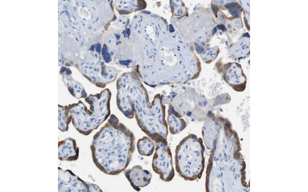
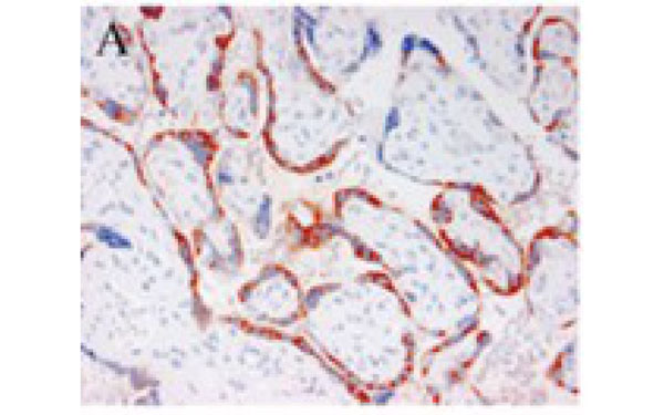 IHC (Immunohiostchemistry)
(Immunohistochemical staining of a formalin-fixed, paraffin-embedded placental tissue with PAPPA antibody)
IHC (Immunohiostchemistry)
(Immunohistochemical staining of a formalin-fixed, paraffin-embedded placental tissue with PAPPA antibody)
PAPPA, Monoclonal Antibody (Cat# AAA108118)
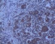
 IHC (Immunohistochemisry)
(Immunohistochemical analysis of PIM2 protein in paraffin embedded Human liver tissue using PIM2 antibody)
IHC (Immunohistochemisry)
(Immunohistochemical analysis of PIM2 protein in paraffin embedded Human liver tissue using PIM2 antibody)
PIM2, Monoclonal Antibody (Cat# AAA108137)
 IF (Immunofluorescence)
(Immunofluorescent staining of COS7 cells transiently transfected with recombinant TACC3 protein using TACC3 antibody)
IF (Immunofluorescence)
(Immunofluorescent staining of COS7 cells transiently transfected with recombinant TACC3 protein using TACC3 antibody)
TACC3, Monoclonal Antibody (Cat# AAA108157)
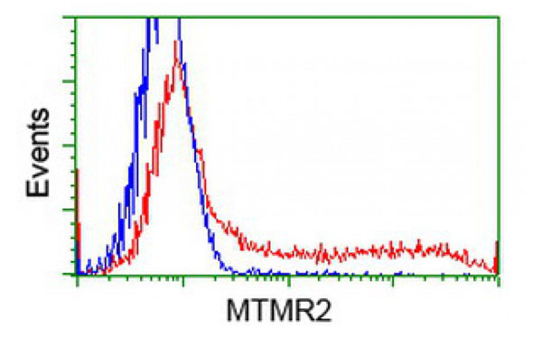
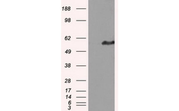
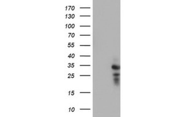
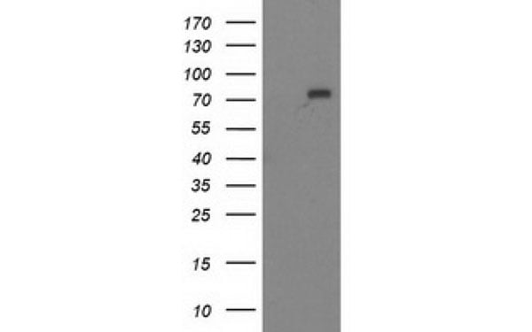 WB (Western Blot)
(Western Blot analysis of HEK293T cell lysates (5 ug) transfected with either recombinant MTMR2 protein (Right) or empty vector (Left) detected with MTMR2 antibody)
WB (Western Blot)
(Western Blot analysis of HEK293T cell lysates (5 ug) transfected with either recombinant MTMR2 protein (Right) or empty vector (Left) detected with MTMR2 antibody)
MTMR2, Monoclonal Antibody (Cat# AAA108252)
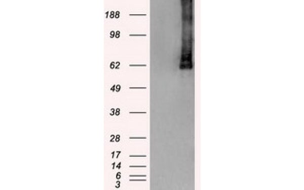
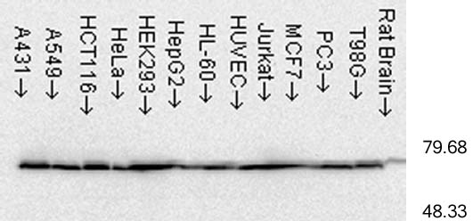
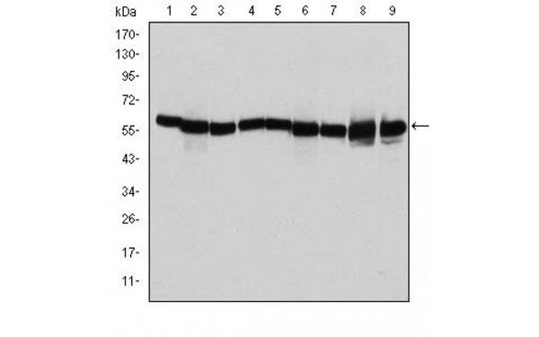 WB (Western Blot)
(Western blot analysis using HSP60 antibody against T47D (1), Hela (2), HepG2 (3), A549 (4), Jurkat (5), HEK293 (6), NIH/3T3 (7), PC-12 (8) and Cos7 (9) cell lysate.)
WB (Western Blot)
(Western blot analysis using HSP60 antibody against T47D (1), Hela (2), HepG2 (3), A549 (4), Jurkat (5), HEK293 (6), NIH/3T3 (7), PC-12 (8) and Cos7 (9) cell lysate.)
SHP60, Monoclonal Antibody (Cat# AAA108178)
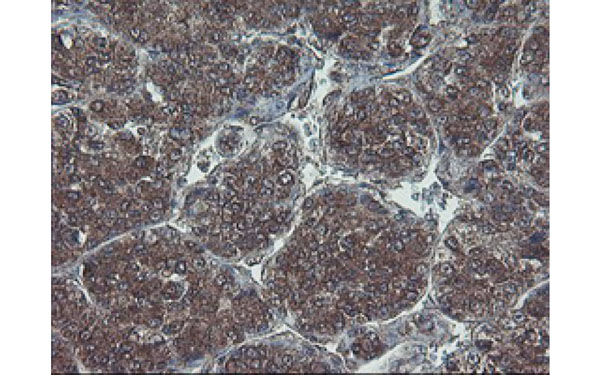
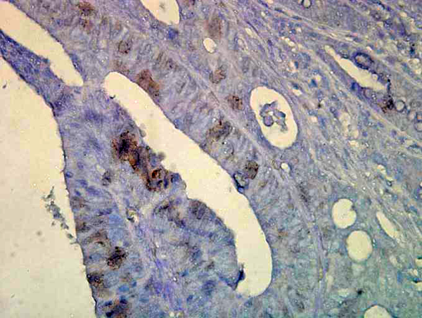
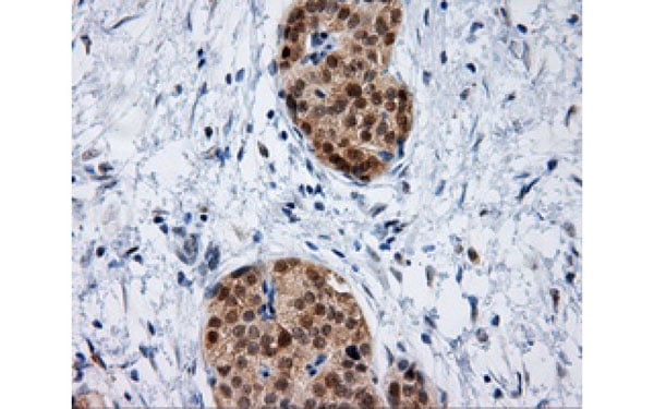 IHC (Immunohiostchemistry)
(Immunohistochemical analysis of IFT57 protein in paraffin embedded Carcinoma of Human pancreas tissue using IFT57 antibody)
IHC (Immunohiostchemistry)
(Immunohistochemical analysis of IFT57 protein in paraffin embedded Carcinoma of Human pancreas tissue using IFT57 antibody)
IFT57, Monoclonal Antibody (Cat# AAA108042)
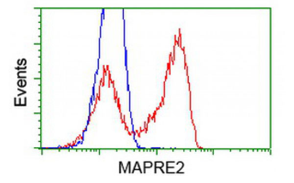
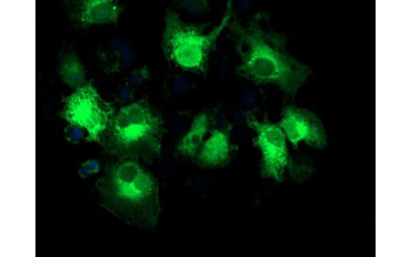 IF (Immunofluorescence)
(Immunofluorescent staining of COS7 cells transiently transfected with recombinant MAPRE2 protein using MAPRE2 antibody)
IF (Immunofluorescence)
(Immunofluorescent staining of COS7 cells transiently transfected with recombinant MAPRE2 protein using MAPRE2 antibody)
MAPRE2, Monoclonal Antibody (Cat# AAA108080)
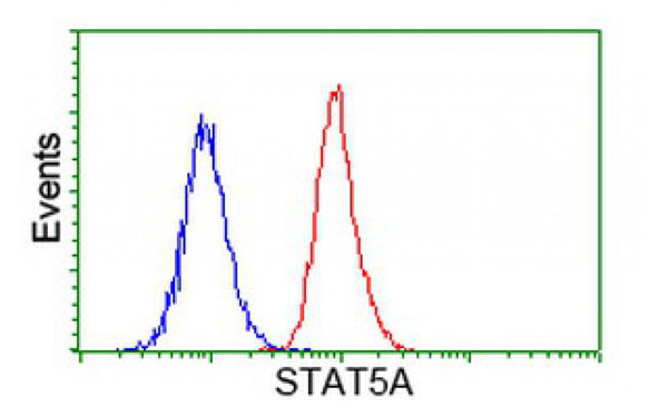
 IF (Immunofluorescence)
(Immunofluorescent staining of COS7 cells transiently transfected with recombinant STAT5A protein using STAT5A antibody)
IF (Immunofluorescence)
(Immunofluorescent staining of COS7 cells transiently transfected with recombinant STAT5A protein using STAT5A antibody)
STAT5A, Monoclonal Antibody (Cat# AAA107974)

 IHC (Immunohistochemisry)
(Immunohistochemical analysis of HDHD1 protein in paraffin embedded Carcinoma of Human liver tissue using HDHD1 antibody)
IHC (Immunohistochemisry)
(Immunohistochemical analysis of HDHD1 protein in paraffin embedded Carcinoma of Human liver tissue using HDHD1 antibody)
HDHD1, Monoclonal Antibody (Cat# AAA107975)
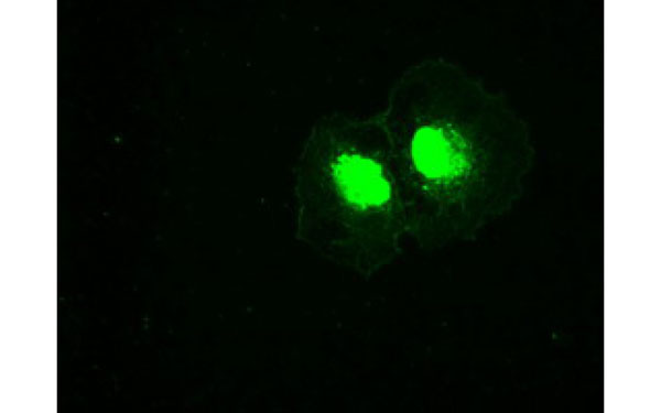 IF (Immunofluorescence)
(Immunofluorescent staining of COS7 cells transiently transfected with recombinant PLK1 protein using PLK1 antibody)
IF (Immunofluorescence)
(Immunofluorescent staining of COS7 cells transiently transfected with recombinant PLK1 protein using PLK1 antibody)
PLK1, Monoclonal Antibody (Cat# AAA107997)

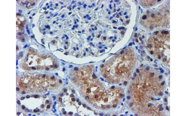 IHC (Immunohiostchemistry)
(Immunohistochemical analysis of ALOX15 protein in paraffin embedded Human Kidney tissue using ALOX15 antibody)
IHC (Immunohiostchemistry)
(Immunohistochemical analysis of ALOX15 protein in paraffin embedded Human Kidney tissue using ALOX15 antibody)
ALOX15, Monoclonal Antibody (Cat# AAA108026)
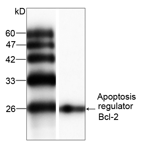
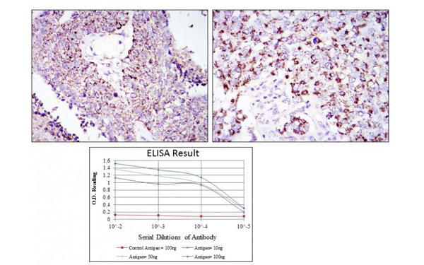
 Application Data
BCL2, Monoclonal Antibody (Cat# AAA108918)
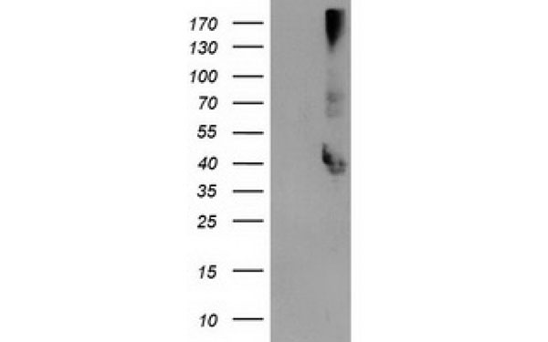
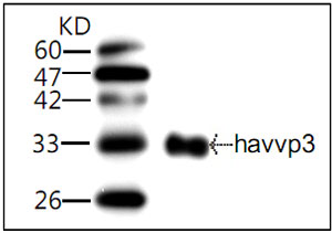 WB (Western Blot)
(WB (1:1000) analysis of recombinant protein HAV VP3 with Anti-HAV VP3 (AAA108986))
WB (Western Blot)
(WB (1:1000) analysis of recombinant protein HAV VP3 with Anti-HAV VP3 (AAA108986))
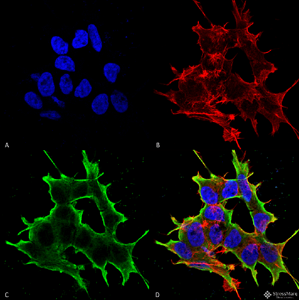
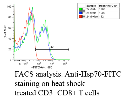 IF (Immunofluorescence)
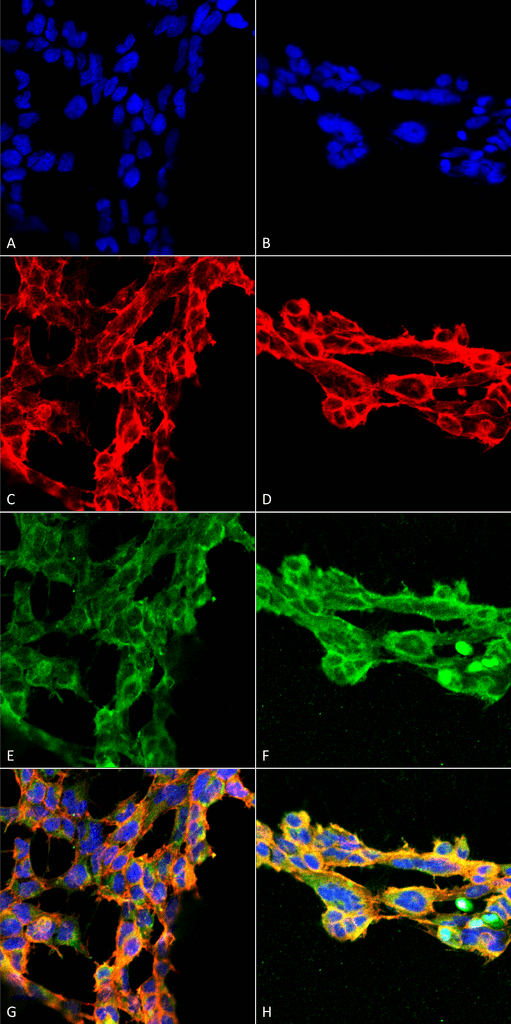
(Fluorescence Activated Cell Sorting analysis using Mouse Anti-Hsp70: FITC Monoclonal Antibody, Clone C92. Tissue: Heat Shocked CD3+ CD8+ T cells . Species: Mouse. Primary Antibody: Mouse Anti-Hsp70: FITC Monoclonal Antibody at 1:1000. Courtesy of: Cheryl Cameron, Vaccine and Gene Therapy Instit. Florida.)
IF (Immunofluorescence)
(Fluorescence Activated Cell Sorting analysis using Mouse Anti-Hsp70: FITC Monoclonal Antibody, Clone C92. Tissue: Heat Shocked CD3+ CD8+ T cells . Species: Mouse. Primary Antibody: Mouse Anti-Hsp70: FITC Monoclonal Antibody at 1:1000. Courtesy of: Cheryl Cameron, Vaccine and Gene Therapy Instit. Florida.)
HSP70, Monoclonal Antibody (Cat# AAA103782)
HSP70, Monoclonal Antibody (Cat# AAA103789)

 WB (Western Blot)
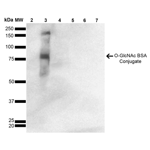
(Western Blot analysis of GlcNAc-BSA Conjugate showing detection of 67 kDa O-GlcNAc protein using Mouse Anti-O-GlcNAc Monoclonal Antibody, Clone 9H6. Lane 1: Molecular Weight Ladder (MW). Lane 2: BSA. Lane 3: GlcNAc-BSA. Lane 4: GalNAc-BSA. Lane 5: Galactose-BSA. Lane 6: Glucose-BSA. Lane 7: Citrulline-BSA. Load: 2.0 ug. Block: 5% Skim Milk in TBST. Primary Antibody: Mouse Anti-O-GlcNAc Monoclonal Antibody at 1:1000 for 2 hours at RT. Secondary Antibody: Goat Anti-Mouse IgG: HRP at 1:2000 for 60 min at RT. Color Development: ECL solution for 5 min in RT. Predicted/Observed Size: 67 kDa.)
WB (Western Blot)
(Western Blot analysis of GlcNAc-BSA Conjugate showing detection of 67 kDa O-GlcNAc protein using Mouse Anti-O-GlcNAc Monoclonal Antibody, Clone 9H6. Lane 1: Molecular Weight Ladder (MW). Lane 2: BSA. Lane 3: GlcNAc-BSA. Lane 4: GalNAc-BSA. Lane 5: Galactose-BSA. Lane 6: Glucose-BSA. Lane 7: Citrulline-BSA. Load: 2.0 ug. Block: 5% Skim Milk in TBST. Primary Antibody: Mouse Anti-O-GlcNAc Monoclonal Antibody at 1:1000 for 2 hours at RT. Secondary Antibody: Goat Anti-Mouse IgG: HRP at 1:2000 for 60 min at RT. Color Development: ECL solution for 5 min in RT. Predicted/Observed Size: 67 kDa.)
O-GlcNAc, Monoclonal Antibody (Cat# AAA103977)
 WB (Western Blot)
(Western Blot analysis of GlcNAc-BSA Conjugate showing detection of 67 kDa O-GlcNAc protein using Mouse Anti-O-GlcNAc Monoclonal Antibody, Clone 9H6. Lane 1: Molecular Weight Ladder (MW). Lane 2: BSA. Lane 3: GlcNAc-BSA. Lane 4: GalNAc-BSA. Lane 5: Galactose-BSA. Lane 6: Glucose-BSA. Lane 7: Citrulline-BSA. Load: 2.0 ug. Block: 5% Skim Milk in TBST. Primary Antibody: Mouse Anti-O-GlcNAc Monoclonal Antibody at 1:1000 for 2 hours at RT. Secondary Antibody: Goat Anti-Mouse IgG: HRP at 1:2000 for 60 min at RT. Color Development: ECL solution for 5 min in RT. Predicted/Observed Size: 67 kDa.)
WB (Western Blot)
(Western Blot analysis of GlcNAc-BSA Conjugate showing detection of 67 kDa O-GlcNAc protein using Mouse Anti-O-GlcNAc Monoclonal Antibody, Clone 9H6. Lane 1: Molecular Weight Ladder (MW). Lane 2: BSA. Lane 3: GlcNAc-BSA. Lane 4: GalNAc-BSA. Lane 5: Galactose-BSA. Lane 6: Glucose-BSA. Lane 7: Citrulline-BSA. Load: 2.0 ug. Block: 5% Skim Milk in TBST. Primary Antibody: Mouse Anti-O-GlcNAc Monoclonal Antibody at 1:1000 for 2 hours at RT. Secondary Antibody: Goat Anti-Mouse IgG: HRP at 1:2000 for 60 min at RT. Color Development: ECL solution for 5 min in RT. Predicted/Observed Size: 67 kDa.)
O-GlcNAc, Monoclonal Antibody (Cat# AAA103978)
 WB (Western Blot)
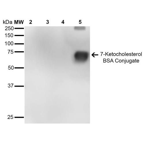
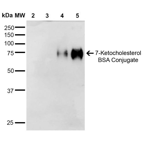
(Western Blot analysis of 7-Ketocholesterol-BSA Conjugate showing detection of 67 kDa 7-Ketocholesterol protein using Mouse Anti-7-Ketocholesterol Monoclonal Antibody, Clone 7E1. Lane 1: Molecular Weight Ladder (MW). Lane 2: BSA (0.5 ug). Lane 3: BSA (2.0 ug). Lane 4: 7-ketocholesterol-BSA (0.5 ug). Lane 5: 7-ketocholesterol-BSA (2.0 ug). Block: 5% Skim Milk in TBST. Primary Antibody: Mouse Anti-7-Ketocholesterol Monoclonal Antibody at 1:1000 for 2 hours at RT. Secondary Antibody: Goat Anti-Mouse IgG: HRP at 1:2000 for 60 min at RT. Color Development: ECL solution for 5 min in RT. Predicted/Observed Size: 67 kDa.)
WB (Western Blot)
(Western Blot analysis of 7-Ketocholesterol-BSA Conjugate showing detection of 67 kDa 7-Ketocholesterol protein using Mouse Anti-7-Ketocholesterol Monoclonal Antibody, Clone 7E1. Lane 1: Molecular Weight Ladder (MW). Lane 2: BSA (0.5 ug). Lane 3: BSA (2.0 ug). Lane 4: 7-ketocholesterol-BSA (0.5 ug). Lane 5: 7-ketocholesterol-BSA (2.0 ug). Block: 5% Skim Milk in TBST. Primary Antibody: Mouse Anti-7-Ketocholesterol Monoclonal Antibody at 1:1000 for 2 hours at RT. Secondary Antibody: Goat Anti-Mouse IgG: HRP at 1:2000 for 60 min at RT. Color Development: ECL solution for 5 min in RT. Predicted/Observed Size: 67 kDa.)
7-Ketocholesterol, Monoclonal Antibody (Cat# AAA104051)

 WB (Western Blot)
(Western Blot analysis of 7-Ketocholesterol-BSA Conjugate showing detection of 67 kDa 7-Ketocholesterol protein using Mouse Anti-7-Ketocholesterol Monoclonal Antibody, Clone 7E1. Lane 1: Molecular Weight Ladder (MW). Lane 2: BSA (0.5 ug). Lane 3: BSA (2.0 ug). Lane 4: 7-ketocholesterol-BSA (0.5 ug). Lane 5: 7-ketocholesterol-BSA (2.0 ug). Block: 5% Skim Milk in TBST. Primary Antibody: Mouse Anti-7-Ketocholesterol Monoclonal Antibody at 1:1000 for 2 hours at RT. Secondary Antibody: Goat Anti-Mouse IgG: HRP at 1:2000 for 60 min at RT. Color Development: ECL solution for 5 min in RT. Predicted/Observed Size: 67 kDa.)
WB (Western Blot)
(Western Blot analysis of 7-Ketocholesterol-BSA Conjugate showing detection of 67 kDa 7-Ketocholesterol protein using Mouse Anti-7-Ketocholesterol Monoclonal Antibody, Clone 7E1. Lane 1: Molecular Weight Ladder (MW). Lane 2: BSA (0.5 ug). Lane 3: BSA (2.0 ug). Lane 4: 7-ketocholesterol-BSA (0.5 ug). Lane 5: 7-ketocholesterol-BSA (2.0 ug). Block: 5% Skim Milk in TBST. Primary Antibody: Mouse Anti-7-Ketocholesterol Monoclonal Antibody at 1:1000 for 2 hours at RT. Secondary Antibody: Goat Anti-Mouse IgG: HRP at 1:2000 for 60 min at RT. Color Development: ECL solution for 5 min in RT. Predicted/Observed Size: 67 kDa.)
7-Ketocholesterol, Monoclonal Antibody (Cat# AAA104053)
 WB (Western Blot)
(Western Blot analysis of 7-Ketocholesterol-BSA Conjugate showing detection of 67 kDa 7-Ketocholesterol protein using Mouse Anti-7-Ketocholesterol Monoclonal Antibody, Clone 3F7. Lane 1: Molecular Weight Ladder (MW). Lane 2: BSA (0.5 ug). Lane 3: BSA (2.0 ug). Lane 4: 7-ketocholesterol-BSA (0.5 ug). Lane 5: 7-ketocholesterol-BSA (2.0 ug). Block: 5% Skim Milk in TBST. Primary Antibody: Mouse Anti-7-Ketocholesterol Monoclonal Antibody at 1:1000 for 2 hours at RT. Secondary Antibody: Goat Anti-Mouse IgG: HRP at 1:2000 for 60 min at RT. Color Development: ECL solution for 5 min in RT. Predicted/Observed Size: 67 kDa.)
WB (Western Blot)
(Western Blot analysis of 7-Ketocholesterol-BSA Conjugate showing detection of 67 kDa 7-Ketocholesterol protein using Mouse Anti-7-Ketocholesterol Monoclonal Antibody, Clone 3F7. Lane 1: Molecular Weight Ladder (MW). Lane 2: BSA (0.5 ug). Lane 3: BSA (2.0 ug). Lane 4: 7-ketocholesterol-BSA (0.5 ug). Lane 5: 7-ketocholesterol-BSA (2.0 ug). Block: 5% Skim Milk in TBST. Primary Antibody: Mouse Anti-7-Ketocholesterol Monoclonal Antibody at 1:1000 for 2 hours at RT. Secondary Antibody: Goat Anti-Mouse IgG: HRP at 1:2000 for 60 min at RT. Color Development: ECL solution for 5 min in RT. Predicted/Observed Size: 67 kDa.)
7-Ketocholesterol, Monoclonal Antibody (Cat# AAA104063)
What are Monoclonal Antibodies?
Monoclonal antibodies are specialized laboratory-produced proteins developed for binding to specific biological antigens or other molecular targets. Since they come from a single cell (or clone), they are especially consistent and accurate in the data they are involved in producing.
This type of antibody material has been shown to be a powerful tool in finding and subsequently destroying harmful cells in an organism, such as those found in cancers or various autoimmune diseases. This makes them excellent aids in medical testing and research, which is why they are so widely used.
AAA Biotech offers a comprehensive range of high-quality monoclonal antibodies that perform effectively in various laboratory tests, including (amongst others) ELISA, western blotting, immunohistochemistry, and flow cytometry. All of the products in our catalog are thoroughly quality tested to make sure that they are reliable and will consistently perform well in your research.
What Are The Uses of Monoclonal Antibodies
Monoclonal antibodies are used in many lab tests, including (amongst others) ELISA, western blotting, immunohistochemistry, and flow cytometry.
ELISA is a test that helps detect a specific substance/analyte in a sample. It uses antibodies (often monoclonal) bound to a solid surface (such as the well of a microplate) to “capture” the substance/analyte in the sample and immobilize it so that the detection antibody component can then bind to it and produce a signal, which can then be measured.
Western blotting identifies specific proteins in a sample. The sample is first separated on a gel, and then antibodies are applied that will typically bind to the target, which will all be localized to a single band in a lane.
Immunohistochemistry helps locate specific proteins in cells or tissue samples using antibodies.
Flow cytometry looks at and sorts cells. It uses antibodies that are conjugated to reporter molecules called “fluorophores”, which, under special lights, emit light themselves, which can then be measured by a detector instrument.
How Monoclonal Antibodies Are Used as Medicine?
Please note that all of the products listed in AAA Biotech’s also known as AAA Bio or AAABio catalog are strictly for research-use only (RUO).
Monoclonal antibodies can also be used as therapeutic/medical treatments, particularly in the context of cancers. They are designed to find and bind to specific cells or proteins, helping the immune system recognize and attack the cancer. These treatments work in different ways, such as:
- Radioimmunotherapy attaches a small amount of radioactive molecule to the antibody, so it delivers the radiation directly to the cancer cells that the antibody is specifically binding to.
- Antibody-directed enzyme prodrug therapy uses antibodies that are specifically bound to special enzymes. These enzymes activate a harmless drug in the body and turn it into a cancer-killing drug only near the cancer cells—this helps avoid harming healthy cells.
- Immunoliposomes are tiny “bubbles” filled with medicine/drug and coated with antibodies. They carry the drug straight to the cancer cells.
Why Buy Monoclonal Antibodies From Us?
At AAA Biotech, we provide high-performance monoclonal antibodies designed to support a wide range of research needs.
1. Validated for Versatile Applications
The antibodies in our catalog are extensively validated and compatible with multiple techniques, including (but not limited to) ELISA, flow cytometry (FC), immunocytochemistry (ICC), immunofluorescence (IF), immunohistochemistry (IHC), immunoprecipitation (IP), and western blotting (WB).
2. Wide Selection & Specialized Options
We offer antibodies for common and rare species, that are available in various conjugated forms, and also in recombinant formats. Essentially, there is almost anything one might need to meet their experimental model’s requirements.
3. High-Quality Proteins
Our proteins meet high purity standards—90% or more as confirmed by SDS-PAGE. Many are available with tags like His, Flag, GST, or MBP, and we also supply native and biologically active proteins for functional studies.
Frequently Asked Questions
1. Are your monoclonal antibodies validated for specific applications?
Yes, our antibodies are tested and validated for use in methods such as ELISA, western blot, IHC, flow cytometry, and more. Refer to specific product pages or datasheets for individual product information.
2. How do I choose the right monoclonal antibody for my application?
Review the product details directly for application validation, species reactivity, and target information. You may also contact our support team at any time for help.
3. How quickly can I receive my order?
Most orders are processed and shipped within 1–3 business days, depending on product availability and your shipping location.