Filters
▼Clonality
▼Type
▼Reactivity
▼Gene Name
▼Isotype
▼Host
▼Application
▼Clone
▼Monoclonal Antibodies
Get accurate results in your research with our Monoclonal Antibodies, which are specially made to target exactly what you require for your research, and will produce consistent, reliable performance in lab tests.
Viewing 3500-3550 of 27597 product results
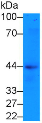
 WB (Western Blot)
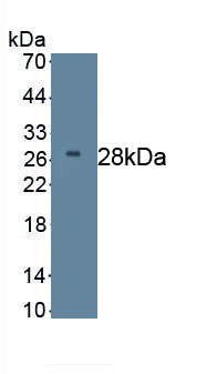
(Figure. Western Blot: Sample: Recombinant DKK2, Human.)
WB (Western Blot)
(Figure. Western Blot: Sample: Recombinant DKK2, Human.)
Dickkopf Related Protein 2 (DKK2), Monoclonal Antibody (Cat# AAA149444)
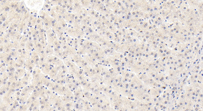
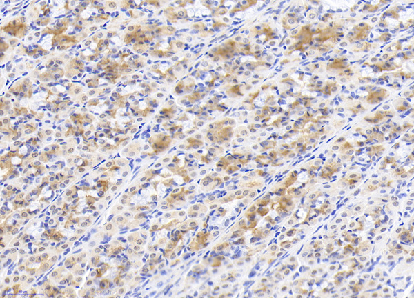
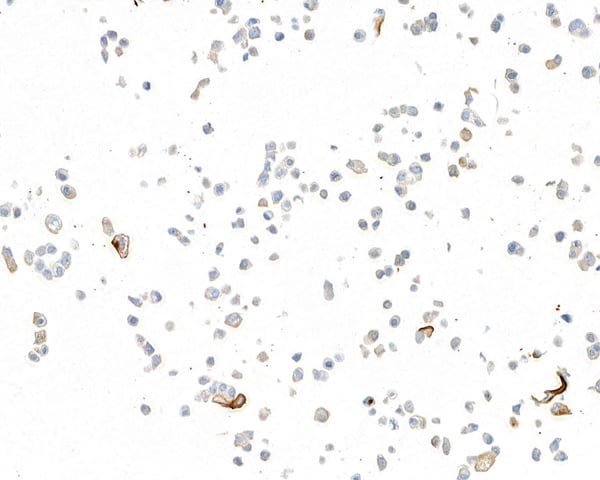
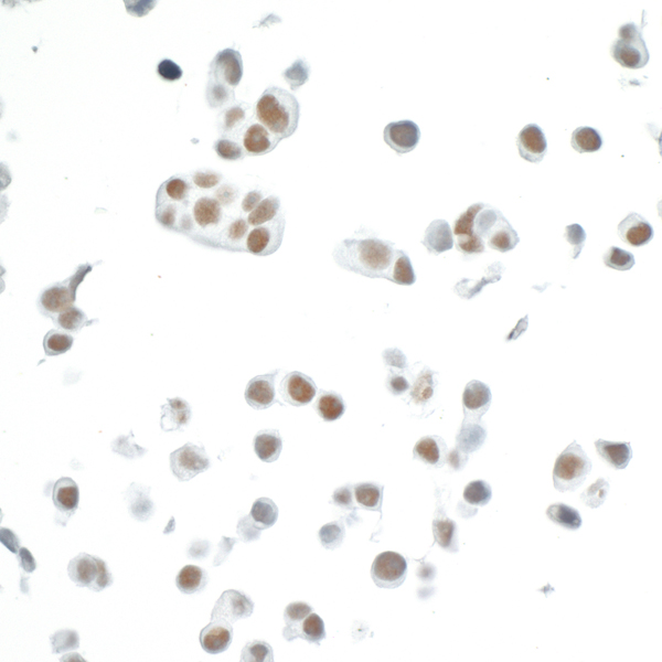
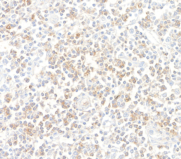
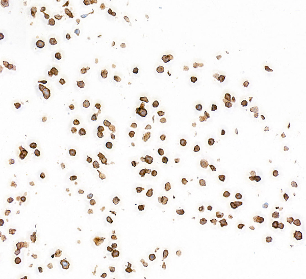
 IHC (Immunohistochemisry)
(DAB staining on IHC-P;Samples: Human Liver Tissue;Primary Ab: 30ug/ml Mouse Anti-Human AST AntibodySecond Ab: 2ug/mL HRP-Linked Caprine Anti-Mouse IgG Polyclonal Antibody)
IHC (Immunohistochemisry)
(DAB staining on IHC-P;Samples: Human Liver Tissue;Primary Ab: 30ug/ml Mouse Anti-Human AST AntibodySecond Ab: 2ug/mL HRP-Linked Caprine Anti-Mouse IgG Polyclonal Antibody)
Aspartate Aminotransferase (AST), Monoclonal Antibody (Cat# AAA149446)
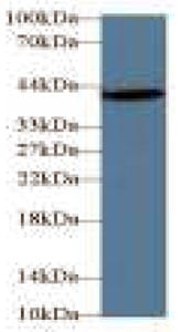
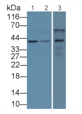
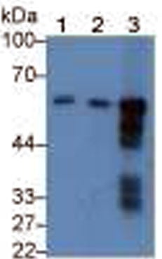 WB (Western Blot)
(Western Blot;Sample:Lane 1: Human Saliva;Lane 2: Rat Pancreas lysate;Lane 3: Mouse Pancreas lysatePrimary Ab: 0.3ug/ml Mouse Anti-Human AMY1 AntibodySecond Ab: 0.2ug/mL HRPLinked Caprine Anti-Mouse IgG Polyclonal Antibody)
WB (Western Blot)
(Western Blot;Sample:Lane 1: Human Saliva;Lane 2: Rat Pancreas lysate;Lane 3: Mouse Pancreas lysatePrimary Ab: 0.3ug/ml Mouse Anti-Human AMY1 AntibodySecond Ab: 0.2ug/mL HRPLinked Caprine Anti-Mouse IgG Polyclonal Antibody)
Amylase Alpha 1 (AMY1), Monoclonal Antibody (Cat# AAA149449)
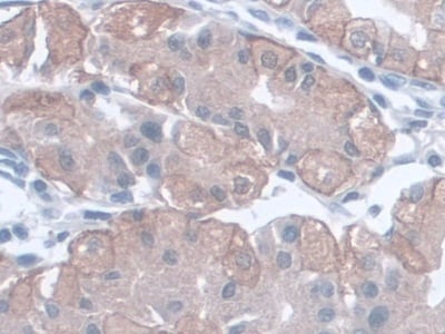
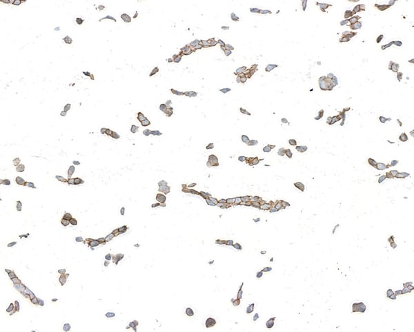
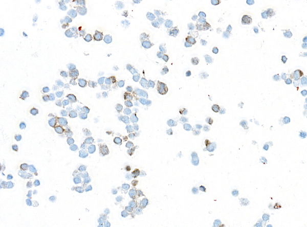
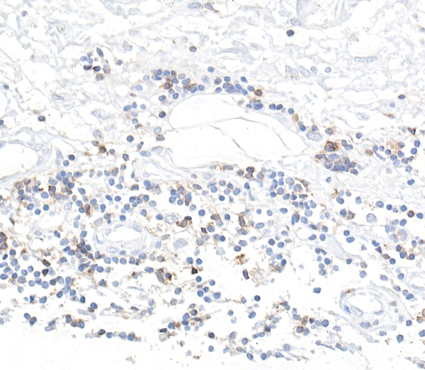
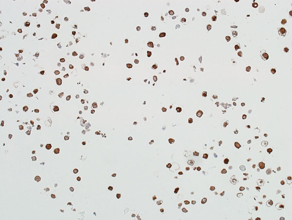
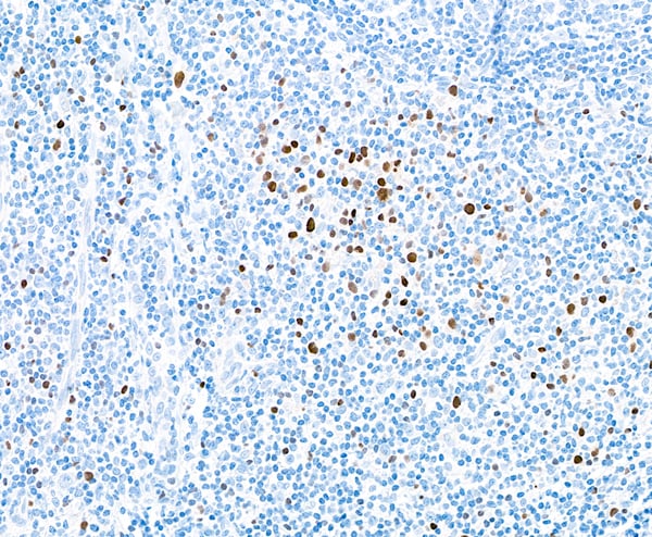
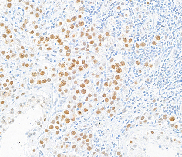
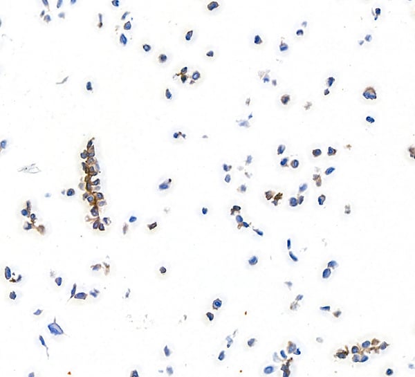
 IHC (Immunohiostchemistry)
(DAB staining on IHC-P;Samples: Human Cerebrum Tissue;Primary Ab: 30ug/ml Mouse Anti-Human SIGLEC8 AntibodySecond Ab: 2ug/mL HRP-Linked Caprine Anti-Mouse IgG Polyclonal Antibody (Catalog: ))
IHC (Immunohiostchemistry)
(DAB staining on IHC-P;Samples: Human Cerebrum Tissue;Primary Ab: 30ug/ml Mouse Anti-Human SIGLEC8 AntibodySecond Ab: 2ug/mL HRP-Linked Caprine Anti-Mouse IgG Polyclonal Antibody (Catalog: ))
Sialic Acid Binding Ig Like Lectin 8 (SIGLEC8), Monoclonal Antibody (Cat# AAA149452)
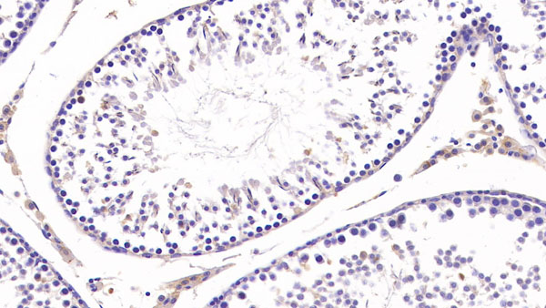 IHC (Immunohiostchemistry)
(DAB staining on IHC-P; Samples: Rat Testis Tissue)
IHC (Immunohiostchemistry)
(DAB staining on IHC-P; Samples: Rat Testis Tissue)
Collagen Type III Alpha 1 (COL3a1), Monoclonal Antibody (Cat# AAA149456)
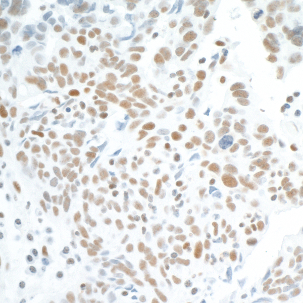
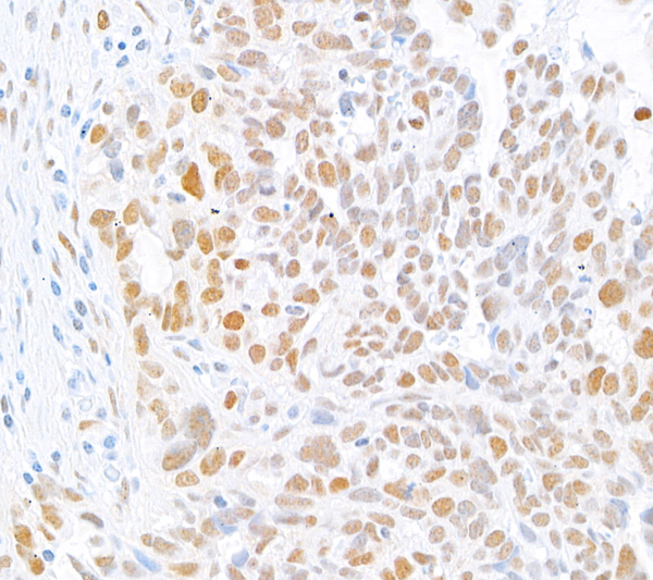
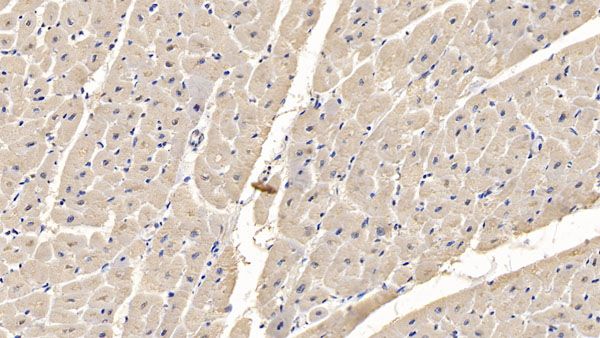 IHC (Immunohiostchemistry)
(DAB staining on IHC-P; Samples: Human Cardiac Muscle Tissue)
IHC (Immunohiostchemistry)
(DAB staining on IHC-P; Samples: Human Cardiac Muscle Tissue)
Myosin Heavy Chain 4 (MYH4), Monoclonal Antibody (Cat# AAA149457)
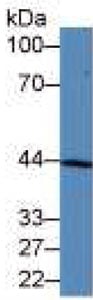
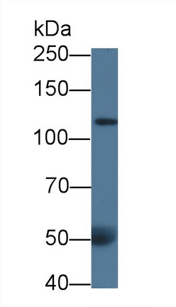
 WB (Western Blot)
(Western Blot: Sample: Human Serum)
WB (Western Blot)
(Western Blot: Sample: Human Serum)
Phospholipase A2 Receptor 1 (PLA2R1), Monoclonal Antibody (Cat# AAA149459)
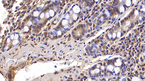 IHC (Immunohiostchemistry)
(DAB staining on IHC-P; Samples: Human Small intestine Tissue)
IHC (Immunohiostchemistry)
(DAB staining on IHC-P; Samples: Human Small intestine Tissue)
Fibulin 3 (FBLN3), Monoclonal Antibody (Cat# AAA149461)
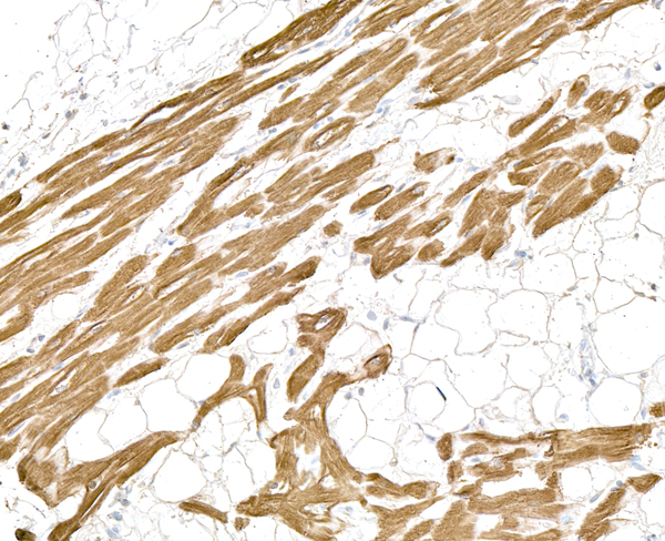
 IHC (Immunohiostchemistry)
(DAB staining on IHC-P; Samples: Human Skeletal muscle Tissue)
IHC (Immunohiostchemistry)
(DAB staining on IHC-P; Samples: Human Skeletal muscle Tissue)
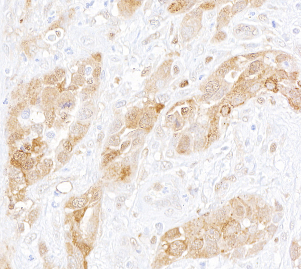
Mesothelin (MSLN), Monoclonal Antibody (Cat# AAA149463)
Myelin Oligodendrocyte Glycoprotein (MOG), Monoclonal Antibody (Cat# AAA149338)
Apolipoprotein A1 (APOA1), Monoclonal Antibody (Cat# AAA149342)
Myelin Basic Protein (MBP), Monoclonal Antibody (Cat# AAA149347)
Ghrelin (GHRL), Monoclonal Antibody (Cat# AAA149369)
Surfactant Associated Protein C (SPC), Monoclonal Antibody (Cat# AAA149375)
Regenerating Islet Derived Protein 3 Gamma (REG3g), Monoclonal Antibody (Cat# AAA149399)
G Protein Coupled Receptor 35 (GPR35), Monoclonal Antibody (Cat# AAA149404)
Myxovirus Resistance 1 (MX1), Monoclonal Antibody (Cat# AAA149407)
 IHC (Immunohiostchemistry)
(DAB staining on IHC-P;Sample: Human Liver Tissue;Primary Ab: 30ug/ml Mouse Anti-Human IGFBP3 AntibodySecond Ab: 2ug/mL HRP-Linked Caprine Anti-Mouse IgG Polyclonal Antibody)
IHC (Immunohiostchemistry)
(DAB staining on IHC-P;Sample: Human Liver Tissue;Primary Ab: 30ug/ml Mouse Anti-Human IGFBP3 AntibodySecond Ab: 2ug/mL HRP-Linked Caprine Anti-Mouse IgG Polyclonal Antibody)
Insulin Like Growth Factor Binding Protein 3 (IGFBP3), Monoclonal Antibody (Cat# AAA149417)
Interferon Gamma (IFNg), Monoclonal Antibody (Cat# AAA145991)
Interferon Gamma (IFNg), Monoclonal Antibody (Cat# AAA145992)
Interleukin 17 (IL17), Monoclonal Antibody (Cat# AAA145999)
Plasminogen Activator, Urokinase Receptor (uPAR), Monoclonal Antibody (Cat# AAA149854)
Procollagen III N-Terminal Propeptide (PIIINP), Monoclonal Antibody (Cat# AAA149784)
Toll Like Receptor 4 (TLR4), Monoclonal Antibody (Cat# AAA149819)
Mucin 5 Subtype AC (MUC5AC), Monoclonal Antibody (Cat# AAA149820)
Procollagen I N-Terminal Propeptide (PINP), Monoclonal Antibody (Cat# AAA149821)
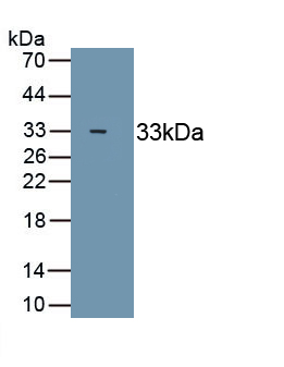
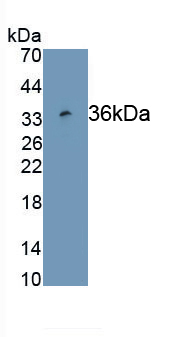
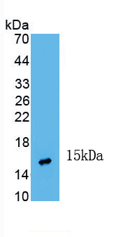 WB (Western Blot)
(Figure. Western Blot; Sample: Recombinant INHbA, Human.)
WB (Western Blot)
(Figure. Western Blot; Sample: Recombinant INHbA, Human.)
Inhibin Beta A (INHbA), Monoclonal Antibody (Cat# AAA150802)

 WB (Western Blot)
(Figure. Western Blot; Sample: Recombinant INHbA, Human.)
WB (Western Blot)
(Figure. Western Blot; Sample: Recombinant INHbA, Human.)
Inhibin Beta A (INHbA), Monoclonal Antibody (Cat# AAA150807)
 WB (Western Blot)
(Western Blot; Sample: Lane1: Human Skeletal muscle lysate; Lane2: Human MCF7 cell lysate; Primary Ab)
WB (Western Blot)
(Western Blot; Sample: Lane1: Human Skeletal muscle lysate; Lane2: Human MCF7 cell lysate; Primary Ab)
Insulin Like Growth Factor 1 Receptor (IGF1R), Monoclonal Antibody (Cat# AAA150814)
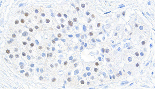
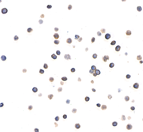
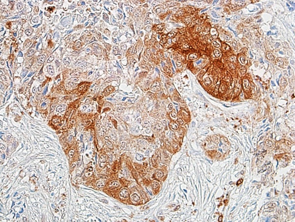
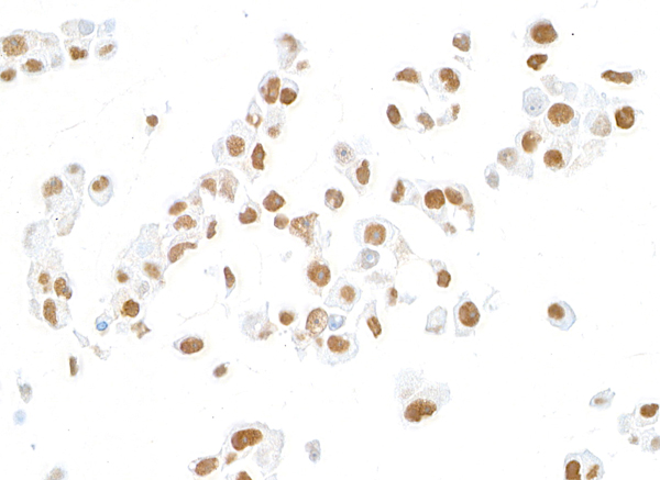
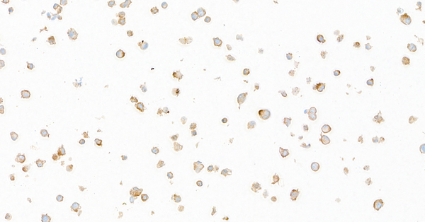
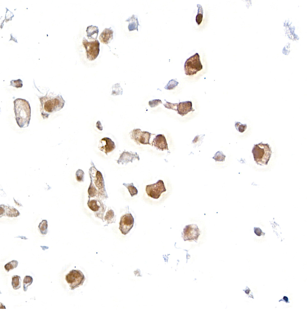
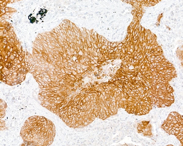 IHC (Immunohiostchemistry)
(Detection of human Desmoglein 3 by immunohistochemistry. Sample: FFPE section of lung squamous cell carcinoma. Antibody: Mouse anti-Desmoglein 3 monoclonal antibody [BC11] (AAA213519-1). Detection: Biocare Medical MACH 4 mouse probe/HRP Polymer.)
IHC (Immunohiostchemistry)
(Detection of human Desmoglein 3 by immunohistochemistry. Sample: FFPE section of lung squamous cell carcinoma. Antibody: Mouse anti-Desmoglein 3 monoclonal antibody [BC11] (AAA213519-1). Detection: Biocare Medical MACH 4 mouse probe/HRP Polymer.)
Desmoglein 3, Monoclonal Antibody (Cat# AAA213519)
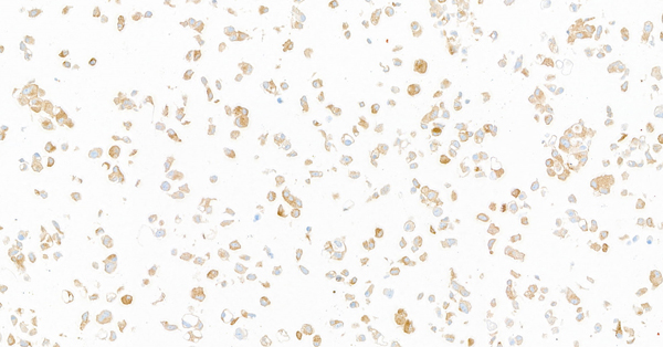
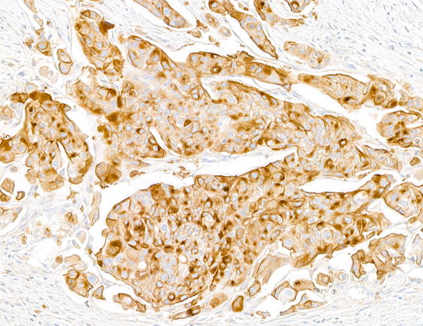 IHC (Immunohiostchemistry)
(Detection of human Uroplakin III by immunohistochemistry. Sample: FFPE section of bladder carcinoma. Antibody: Mouse anti-Uroplakin III monoclonal antibody [BC12] (AAA213520-1). Secondary: HRP-conjugated goat anti-mouse IgG .)
IHC (Immunohiostchemistry)
(Detection of human Uroplakin III by immunohistochemistry. Sample: FFPE section of bladder carcinoma. Antibody: Mouse anti-Uroplakin III monoclonal antibody [BC12] (AAA213520-1). Secondary: HRP-conjugated goat anti-mouse IgG .)
Uroplakin III, Monoclonal Antibody (Cat# AAA213520)
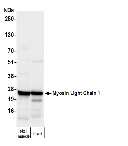

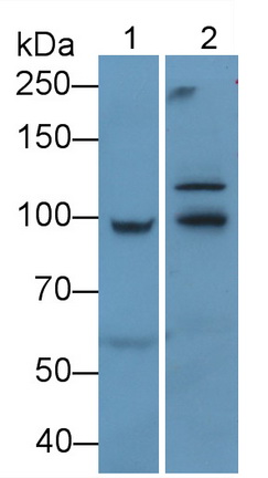
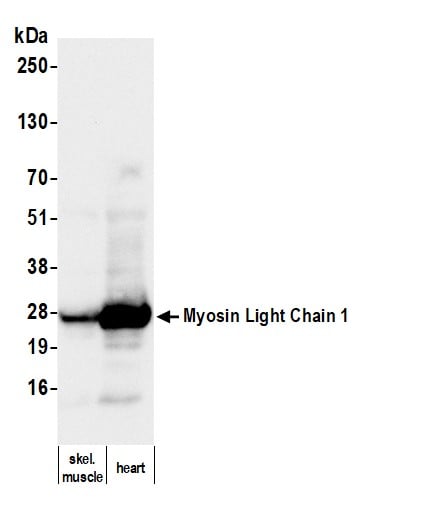 WB (Western Blot)
(Detection of mouse Myosin Light Chain 1 by western blot. Samples: Tissue lysate (50 ug) from mouse skeletal muscle and heart. Antibody: Mouse anti-Myosin Light Chain 1 monoclonal antibody [39-15] (AAA213526 lot 1) used at 1:1000. Secondary: HRP-conjugated goat anti-mouse IgG . Detection: Chemiluminescence with an exposure time of 3 seconds.)
WB (Western Blot)
(Detection of mouse Myosin Light Chain 1 by western blot. Samples: Tissue lysate (50 ug) from mouse skeletal muscle and heart. Antibody: Mouse anti-Myosin Light Chain 1 monoclonal antibody [39-15] (AAA213526 lot 1) used at 1:1000. Secondary: HRP-conjugated goat anti-mouse IgG . Detection: Chemiluminescence with an exposure time of 3 seconds.)
Myosin Light Chain 1, Monoclonal Antibody (Cat# AAA213526)
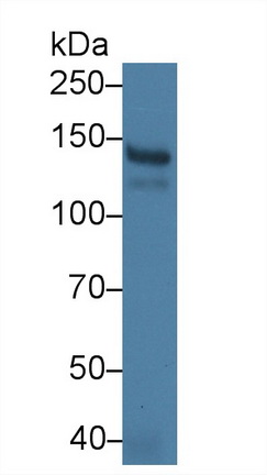
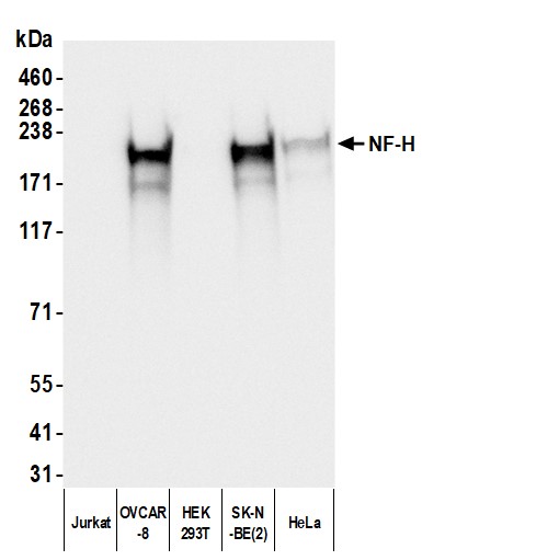 WB (Western Blot)
(Detection of human NF-H using mouse anti-Chicken IgY secondary antibody by western blot. Samples: Whole cell lysate (50 ug) from Jurkat, OVCAR-8, HEK293T, SK-N-BE(2), and HeLa cells prepared using NETN lysis buffer. Primary: Chicken anti-NF-H antibody. Secondary: mouse anti-Chicken IgY Light Chain monoclonal antibody [1Y-263] (AAA213527 lot 1) used at 1:1000. Tertiary: HRP-conjugated goat anti-mouse IgG . Detection: Chemiluminescence with an exposure time of 1 second.)
WB (Western Blot)
(Detection of human NF-H using mouse anti-Chicken IgY secondary antibody by western blot. Samples: Whole cell lysate (50 ug) from Jurkat, OVCAR-8, HEK293T, SK-N-BE(2), and HeLa cells prepared using NETN lysis buffer. Primary: Chicken anti-NF-H antibody. Secondary: mouse anti-Chicken IgY Light Chain monoclonal antibody [1Y-263] (AAA213527 lot 1) used at 1:1000. Tertiary: HRP-conjugated goat anti-mouse IgG . Detection: Chemiluminescence with an exposure time of 1 second.)
IgY Light Chain, Monoclonal Secondary Antibody (Cat# AAA213527)

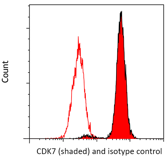
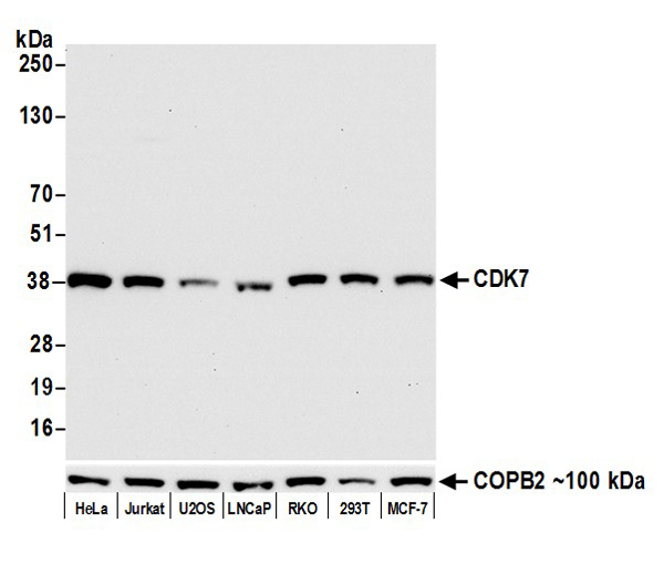 WB (Western Blot)
(Detection of human CDK7 by western blot. Samples: Whole cell lysate (15 ug) from HeLa, Jurkat, U2OS, LNCaP, RKO, HEK293T, and MCF-7 cells prepared using NETN lysis buffer. Antibody: Rabbit anti-CDK7 recombinant monoclonal antibody [BL-80-5D4] (AAA213531 lot 2) used at 1:1000. Secondary: HRP-conjugated goat anti-rabbit IgG . Detection: Chemiluminescence with an exposure time of 10 seconds. Lower Panel: Rabbit anti-COPB2 .)
WB (Western Blot)
(Detection of human CDK7 by western blot. Samples: Whole cell lysate (15 ug) from HeLa, Jurkat, U2OS, LNCaP, RKO, HEK293T, and MCF-7 cells prepared using NETN lysis buffer. Antibody: Rabbit anti-CDK7 recombinant monoclonal antibody [BL-80-5D4] (AAA213531 lot 2) used at 1:1000. Secondary: HRP-conjugated goat anti-rabbit IgG . Detection: Chemiluminescence with an exposure time of 10 seconds. Lower Panel: Rabbit anti-COPB2 .)
CDK7, Monoclonal Recombinant Antibody (Cat# AAA213531)
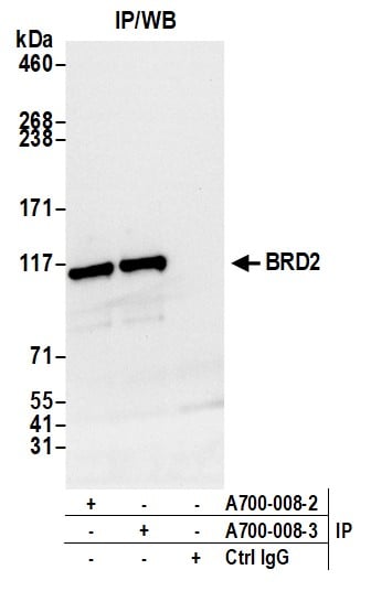
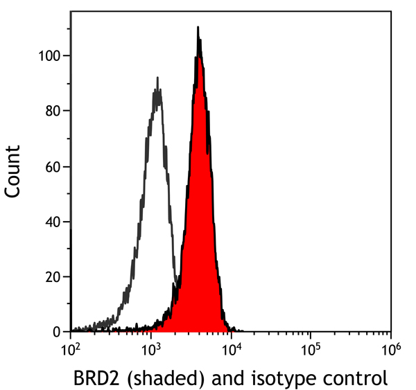
 WB (Western Blot)
(Detection of human BRD2 by western blot. Samples: Whole cell lysate (50 ug) from HeLa, HEK293T, MCF-7, Hep-G2, A-549, SW620, SK-MEL-28, and Jurkat cells prepared using NETN lysis buffer. Antibody: Rabbit anti-BRD2 recombinant monoclonal antibody [BL-167-2A2] (AAA213532 lot 3) used at 1:1000. Secondary: HRP-conjugated goat anti-rabbit IgG . Detection: Chemiluminescence with an exposure time of 10 seconds.)
WB (Western Blot)
(Detection of human BRD2 by western blot. Samples: Whole cell lysate (50 ug) from HeLa, HEK293T, MCF-7, Hep-G2, A-549, SW620, SK-MEL-28, and Jurkat cells prepared using NETN lysis buffer. Antibody: Rabbit anti-BRD2 recombinant monoclonal antibody [BL-167-2A2] (AAA213532 lot 3) used at 1:1000. Secondary: HRP-conjugated goat anti-rabbit IgG . Detection: Chemiluminescence with an exposure time of 10 seconds.)
BRD2, Monoclonal Recombinant Antibody (Cat# AAA213532)
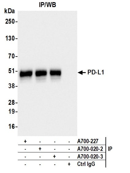
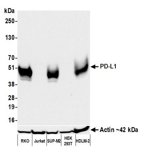 WB (Western Blot)
(Detection of human PD-L1 by western blot. Samples: Whole cell lysate (50 ug) from RKO, Jurkat, SUP-M2, HEK293T, and HDLM-2 cells prepared using NETN lysis buffer. Antibody: Rabbit anti-PD-L1 recombinant monoclonal antibody (AAA213536 lot 3) used at 1:1000. Secondary: HRP-conjugated goat anti-rabbit IgG . Detection: Chemiluminescence with an exposure time of 30 seconds. Lower Panel: Rabbit anti-Actin recombinant monoclonal antibody .)
WB (Western Blot)
(Detection of human PD-L1 by western blot. Samples: Whole cell lysate (50 ug) from RKO, Jurkat, SUP-M2, HEK293T, and HDLM-2 cells prepared using NETN lysis buffer. Antibody: Rabbit anti-PD-L1 recombinant monoclonal antibody (AAA213536 lot 3) used at 1:1000. Secondary: HRP-conjugated goat anti-rabbit IgG . Detection: Chemiluminescence with an exposure time of 30 seconds. Lower Panel: Rabbit anti-Actin recombinant monoclonal antibody .)
PD-L1, Monoclonal Recombinant Antibody (Cat# AAA213536)
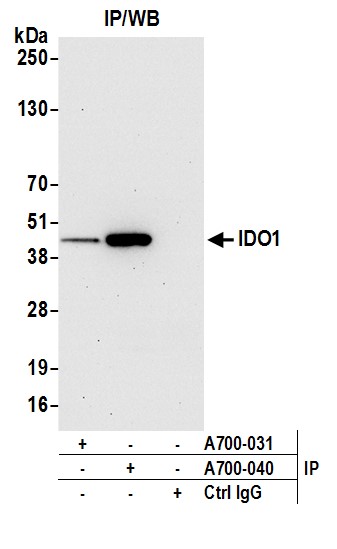
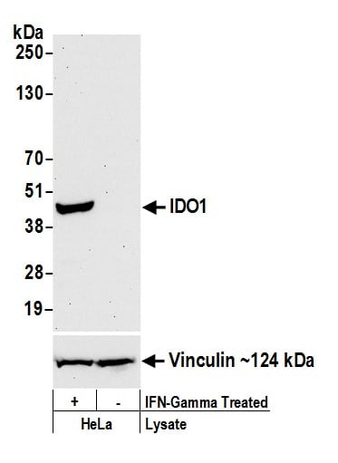 WB (Western Blot)
(Detection of human IDO1 by western blot. Samples: Whole cell lysate (50 ug) from HeLa cells treated with IFN-gamma (+) or mock treated (-). Antibody: Rabbit anti-IDO1 recombinant monoclonal antibody (AAA213543 lot 1) used at 1:1000. Secondary: HRP-conjugated goat anti-rabbit IgG . Detection: Chemiluminescence with an exposure time of 30 seconds. Lower Panel: Rabbit anti-Vinculin .)
WB (Western Blot)
(Detection of human IDO1 by western blot. Samples: Whole cell lysate (50 ug) from HeLa cells treated with IFN-gamma (+) or mock treated (-). Antibody: Rabbit anti-IDO1 recombinant monoclonal antibody (AAA213543 lot 1) used at 1:1000. Secondary: HRP-conjugated goat anti-rabbit IgG . Detection: Chemiluminescence with an exposure time of 30 seconds. Lower Panel: Rabbit anti-Vinculin .)
IDO1, Monoclonal Recombinant Antibody (Cat# AAA213543)
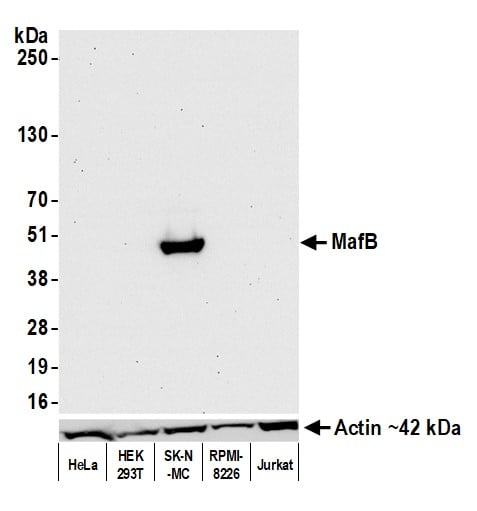
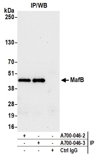
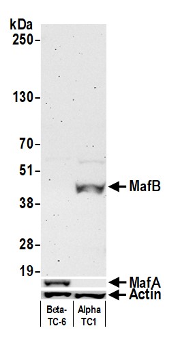 WB (Western Blot)
(Detection of mouse MafB by western blot. Samples: Whole cell lysate (50 ug) from Beta-TC-6 and AlphaTC1 Clone 9 cells prepared using NETN lysis buffer. Antibody: Rabbit anti-MafB recombinant monoclonal antibody (AAA213545 lot 3) used at 1:1000. Secondary: HRP-conjugated goat anti-rabbit IgG . Detection: Chemiluminescence with an exposure time of 3 minutes. Lower Panels: Rabbit anti-MafA recombinant monoclonal antibody and rabbit anti-Actin recombinant monoclonal antibody (AAA213545).)
WB (Western Blot)
(Detection of mouse MafB by western blot. Samples: Whole cell lysate (50 ug) from Beta-TC-6 and AlphaTC1 Clone 9 cells prepared using NETN lysis buffer. Antibody: Rabbit anti-MafB recombinant monoclonal antibody (AAA213545 lot 3) used at 1:1000. Secondary: HRP-conjugated goat anti-rabbit IgG . Detection: Chemiluminescence with an exposure time of 3 minutes. Lower Panels: Rabbit anti-MafA recombinant monoclonal antibody and rabbit anti-Actin recombinant monoclonal antibody (AAA213545).)
MafB, Monoclonal Recombinant Antibody (Cat# AAA213545)
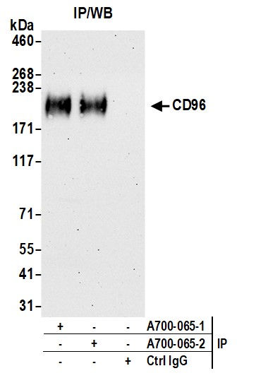
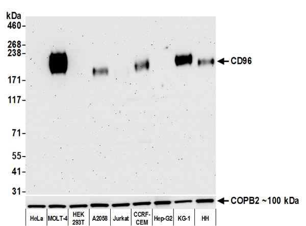 WB (Western Blot)
(Detection of human CD96 by western blot. Samples: Whole cell lysate (10 ug) from HeLa, MOLT-4, HEK293T, A2058, Jurkat, CCRF-CEM, Hep-G2, KG-1, and HH cells prepared using NETN lysis buffer. Antibody: Rabbit anti-CD96 recombinant monoclonal antibody [855-3C7] (AAA213558 lot 2) used at 1:1000. Secondary: HRP-conjugated goat anti-rabbit IgG . Detection: Chemiluminescence with an exposure time of 3 minutes.)
WB (Western Blot)
(Detection of human CD96 by western blot. Samples: Whole cell lysate (10 ug) from HeLa, MOLT-4, HEK293T, A2058, Jurkat, CCRF-CEM, Hep-G2, KG-1, and HH cells prepared using NETN lysis buffer. Antibody: Rabbit anti-CD96 recombinant monoclonal antibody [855-3C7] (AAA213558 lot 2) used at 1:1000. Secondary: HRP-conjugated goat anti-rabbit IgG . Detection: Chemiluminescence with an exposure time of 3 minutes.)
CD96, Monoclonal Recombinant Antibody (Cat# AAA213558)
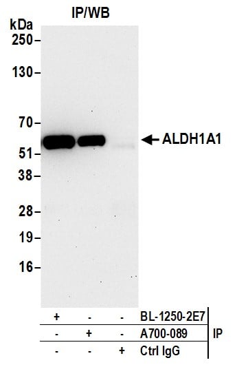

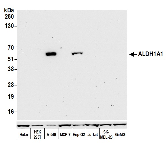 WB (Western Blot)
(Detection of human ALDH1A1 by western blot. Samples: Whole cell lysate (50 ug) from HeLa, HEK293T, MCF-7, Hep-G2, Jurkat, SK-MEL-28, GaMG and (10 ug) A-549 cells prepared using NETN lysis buffer. Antibody: Rabbit anti-ALDH1A1 recombinant monoclonal antibody (AAA213569 lot 1) used at 1:1000. Secondary: HRP-conjugated goat anti-rabbit IgG . Detection: Chemiluminescence with an exposure time of 30 seconds. Lower Panel: Rabbit anti-Cytoskeletal Actin recombinant monoclonal antibody .)
WB (Western Blot)
(Detection of human ALDH1A1 by western blot. Samples: Whole cell lysate (50 ug) from HeLa, HEK293T, MCF-7, Hep-G2, Jurkat, SK-MEL-28, GaMG and (10 ug) A-549 cells prepared using NETN lysis buffer. Antibody: Rabbit anti-ALDH1A1 recombinant monoclonal antibody (AAA213569 lot 1) used at 1:1000. Secondary: HRP-conjugated goat anti-rabbit IgG . Detection: Chemiluminescence with an exposure time of 30 seconds. Lower Panel: Rabbit anti-Cytoskeletal Actin recombinant monoclonal antibody .)
ALDH1A1, Monoclonal Recombinant Antibody (Cat# AAA213569)
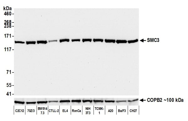
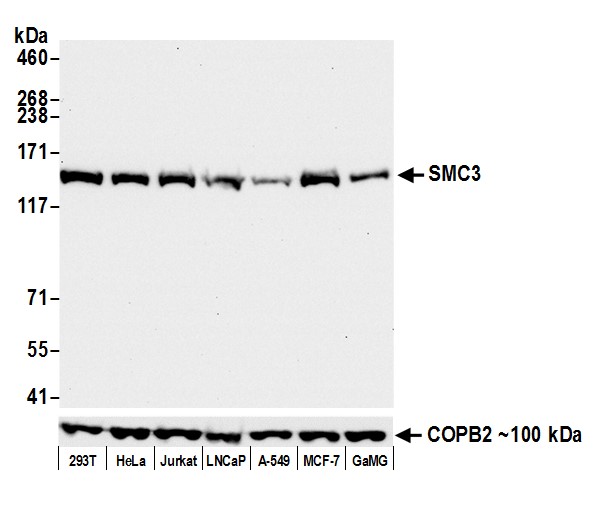 WB (Western Blot)
(Detection of human SMC3 by western blot. Samples: Whole cell lysate (50 ug) from HEK293T, HeLa, Jurkat, LNCaP, A-549, MCF-7, and GaMG cells prepared using NETN lysis buffer. Antibody: Rabbit anti-SMC3 recombinant monoclonal antibody (AAA213575 lot 1) used at 1:1000. Secondary: HRP-conjugated goat anti-rabbit IgG . Detection: Chemiluminescence with an exposure time of 10 seconds. Lower Panel: Rabbit anti-COPB2 antibody .)
WB (Western Blot)
(Detection of human SMC3 by western blot. Samples: Whole cell lysate (50 ug) from HEK293T, HeLa, Jurkat, LNCaP, A-549, MCF-7, and GaMG cells prepared using NETN lysis buffer. Antibody: Rabbit anti-SMC3 recombinant monoclonal antibody (AAA213575 lot 1) used at 1:1000. Secondary: HRP-conjugated goat anti-rabbit IgG . Detection: Chemiluminescence with an exposure time of 10 seconds. Lower Panel: Rabbit anti-COPB2 antibody .)
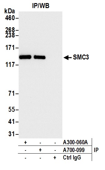
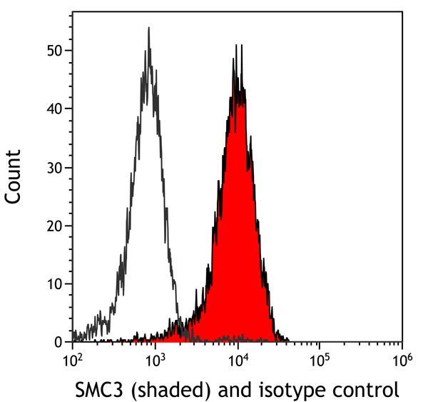
SMC3, Monoclonal Recombinant Antibody (Cat# AAA213575)
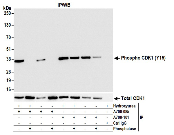
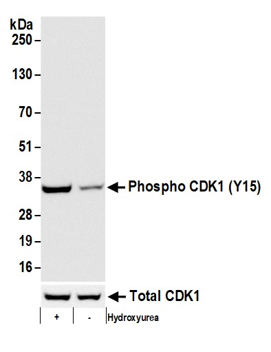 WB (Western Blot)
(Detection of human Phospho CDK1 (Y15) by western blot. Samples: Whole cell lysate (50 ug) from HeLa cells treated (+) with Hydroxyurea or mock treated (-) prepared using NETN lysis buffer. Antibody: Rabbit anti-Phospho CDK1 (Y15) recombinant monoclonal antibody (AAA213576 lot 1) used at 1:1000. Secondary: HRP-conjugated goat anti-rabbit IgG . Detection: Chemiluminescence with an exposure time of 10 seconds. Lower panel shows WB for total CDK1 using rabbit anti-CDK1 recombinant monoclonal .)
WB (Western Blot)
(Detection of human Phospho CDK1 (Y15) by western blot. Samples: Whole cell lysate (50 ug) from HeLa cells treated (+) with Hydroxyurea or mock treated (-) prepared using NETN lysis buffer. Antibody: Rabbit anti-Phospho CDK1 (Y15) recombinant monoclonal antibody (AAA213576 lot 1) used at 1:1000. Secondary: HRP-conjugated goat anti-rabbit IgG . Detection: Chemiluminescence with an exposure time of 10 seconds. Lower panel shows WB for total CDK1 using rabbit anti-CDK1 recombinant monoclonal .)
CDK1, Monoclonal Recombinant Antibody (Cat# AAA213576)
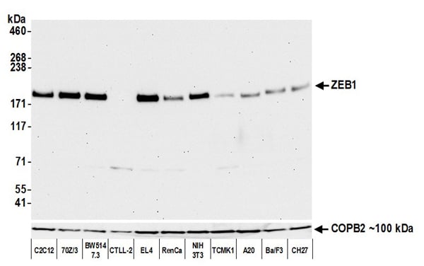
 WB (Western Blot)
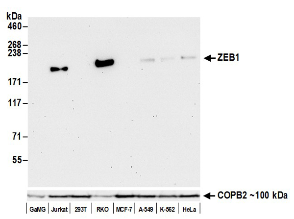
(Detection of human ZEB1 by western blot. Samples: Whole cell lysate (50 ug) from GaMG, Jurkat, HEK293T, RKO, MCF-7, A-549, K-562, and HeLa cells prepared using NETN lysis buffer. Antibody: Rabbit anti-ZEB1 recombinant monoclonal antibody (AAA213577 lot 1) used at 1:1000. Secondary: HRP-conjugated goat anti-rabbit IgG . Detection: Chemiluminescence with an exposure time of 30 seconds. Lower Panel: Rabbit anti-COPB2 antibody .)
WB (Western Blot)
(Detection of human ZEB1 by western blot. Samples: Whole cell lysate (50 ug) from GaMG, Jurkat, HEK293T, RKO, MCF-7, A-549, K-562, and HeLa cells prepared using NETN lysis buffer. Antibody: Rabbit anti-ZEB1 recombinant monoclonal antibody (AAA213577 lot 1) used at 1:1000. Secondary: HRP-conjugated goat anti-rabbit IgG . Detection: Chemiluminescence with an exposure time of 30 seconds. Lower Panel: Rabbit anti-COPB2 antibody .)
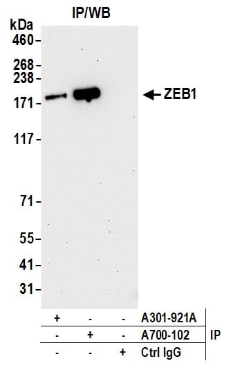
ZEB1, Monoclonal Recombinant Antibody (Cat# AAA213577)

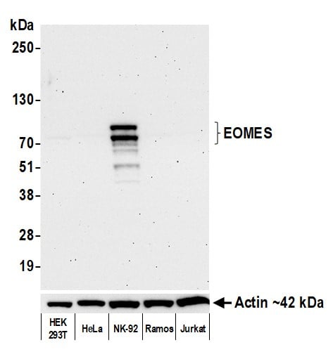 WB (Western Blot)
(Detection of human EOMES by western blot. Samples: Whole cell lysate (10 ug) from HEK293T, HeLa, NK-92, Ramos, and Jurkat cells prepared using NETN lysis buffer. Antibody: Rabbit anti-EOMES recombinant monoclonal antibody (AAA213579 lot 2) used at 1:1000. Secondary: HRP-conjugated goat anti-rabbit IgG . Detection: Chemiluminescence with an exposure time of 75 seconds. Lower Panel: Rabbit anti-Actin recombinant monoclonal antibody .)
WB (Western Blot)
(Detection of human EOMES by western blot. Samples: Whole cell lysate (10 ug) from HEK293T, HeLa, NK-92, Ramos, and Jurkat cells prepared using NETN lysis buffer. Antibody: Rabbit anti-EOMES recombinant monoclonal antibody (AAA213579 lot 2) used at 1:1000. Secondary: HRP-conjugated goat anti-rabbit IgG . Detection: Chemiluminescence with an exposure time of 75 seconds. Lower Panel: Rabbit anti-Actin recombinant monoclonal antibody .)
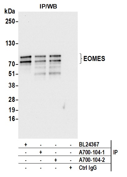
EOMES, Monoclonal Recombinant Antibody (Cat# AAA213579)
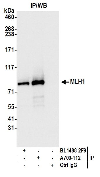
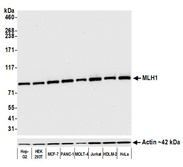 WB (Western Blot)
(Detection of human MLH1 by western blot. Samples: Whole cell lysate (5 ug) from Hep-G2, HEK293T, MCF-7, PANC-1, MOLT-4, Jurkat, HDLM-2, and HeLa cells prepared using NETN lysis buffer. Antibody: Rabbit anti-MLH1 recombinant monoclonal antibody (AAA213583 lot 1) used at 1:1000. Secondary: HRP-conjugated goat anti-rabbit IgG . Detection: Chemiluminescence with an exposure time of 10 seconds. Lower Panel: Rabbit anti-Actin recombinant monoclonal .)
WB (Western Blot)
(Detection of human MLH1 by western blot. Samples: Whole cell lysate (5 ug) from Hep-G2, HEK293T, MCF-7, PANC-1, MOLT-4, Jurkat, HDLM-2, and HeLa cells prepared using NETN lysis buffer. Antibody: Rabbit anti-MLH1 recombinant monoclonal antibody (AAA213583 lot 1) used at 1:1000. Secondary: HRP-conjugated goat anti-rabbit IgG . Detection: Chemiluminescence with an exposure time of 10 seconds. Lower Panel: Rabbit anti-Actin recombinant monoclonal .)
MLH1, Monoclonal Recombinant Antibody (Cat# AAA213583)

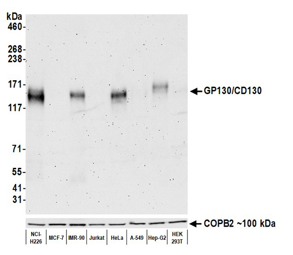 WB (Western Blot)
(Detection of human GP130/CD130 by western blot. Samples: Whole cell lysate (50 ug) from NCI-H226, MCF-7, IMR-90, Jurkat, HeLa, A-549, Hep-G2, and HEK293T cells prepared using NETN lysis buffer. Antibody: Rabbit anti-GP130/CD130 recombinant monoclonal antibody (AAA213585 Lot 1) used at 1:1000. Secondary: HRP-conjugated goat anti-rabbit IgG . Detection: Chemiluminescence with an exposure time of 30 seconds. Lower Panel: Rabbit anti-COPB2 .)
WB (Western Blot)
(Detection of human GP130/CD130 by western blot. Samples: Whole cell lysate (50 ug) from NCI-H226, MCF-7, IMR-90, Jurkat, HeLa, A-549, Hep-G2, and HEK293T cells prepared using NETN lysis buffer. Antibody: Rabbit anti-GP130/CD130 recombinant monoclonal antibody (AAA213585 Lot 1) used at 1:1000. Secondary: HRP-conjugated goat anti-rabbit IgG . Detection: Chemiluminescence with an exposure time of 30 seconds. Lower Panel: Rabbit anti-COPB2 .)
GP130/CD130, Monoclonal Recombinant Antibody (Cat# AAA213585)
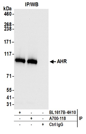
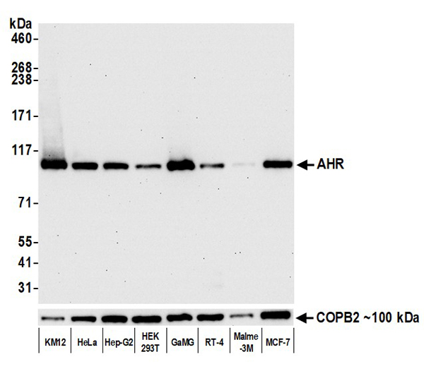 WB (Western Blot)
(Detection of human AHR by western blot. Samples: Whole cell lysate (50 ug) from KM12, HeLa, Hep-G2, HEK293T, GaMG, RT-4, Malme-3M, and MCF-7 cells prepared using NETN lysis buffer. Antibody: Rabbit anti-AHR recombinant monoclonal antibody (AAA213589 Lot 1) used at 1:1000. Secondary: HRP-conjugated goat anti-rabbit IgG . Detection: Chemiluminescence with an exposure time of 30 seconds. Lower Panel: Rabbit anti-COPB2 antibody .)
WB (Western Blot)
(Detection of human AHR by western blot. Samples: Whole cell lysate (50 ug) from KM12, HeLa, Hep-G2, HEK293T, GaMG, RT-4, Malme-3M, and MCF-7 cells prepared using NETN lysis buffer. Antibody: Rabbit anti-AHR recombinant monoclonal antibody (AAA213589 Lot 1) used at 1:1000. Secondary: HRP-conjugated goat anti-rabbit IgG . Detection: Chemiluminescence with an exposure time of 30 seconds. Lower Panel: Rabbit anti-COPB2 antibody .)
AHR, Monoclonal Recombinant Antibody (Cat# AAA213589)
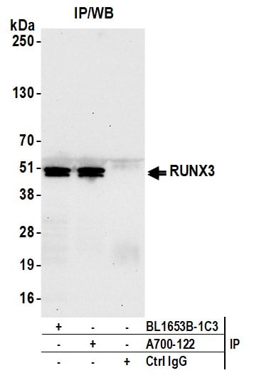
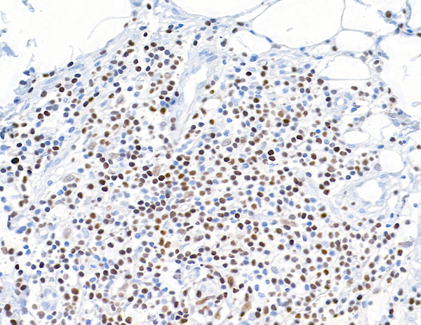
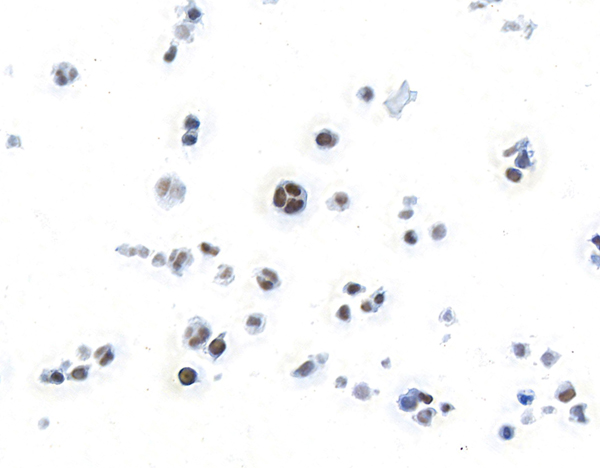
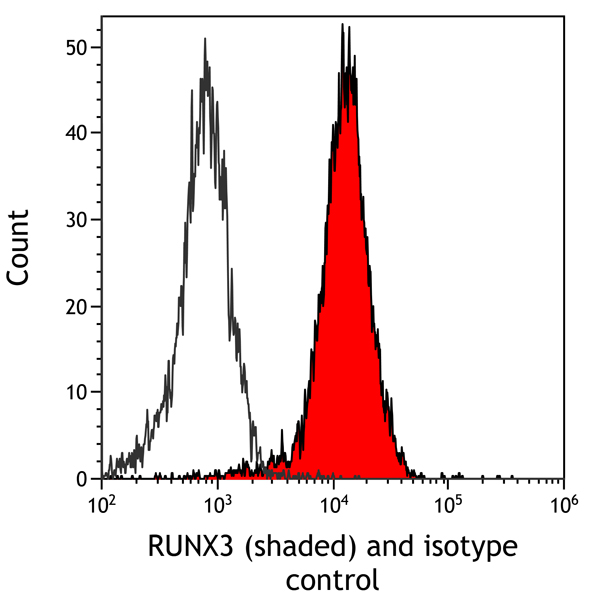
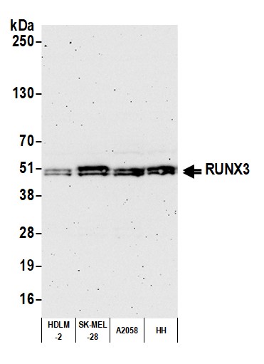 WB (Western Blot)
(Detection of human RUNX3 by western blot. Samples: Whole cell lysate (25 ug) from HDLM-2, SK-MEL-28, A2058, and HH cells prepared using NETN lysis buffer. Antibody: Rabbit anti-RUNX3 recombinant monoclonal antibody (AAA213590 lot 1) used at 1:1000. Secondary: HRP-conjugated goat anti-rabbit IgG . Detection: Chemiluminescence with an exposure time of 3 minutes.)
WB (Western Blot)
(Detection of human RUNX3 by western blot. Samples: Whole cell lysate (25 ug) from HDLM-2, SK-MEL-28, A2058, and HH cells prepared using NETN lysis buffer. Antibody: Rabbit anti-RUNX3 recombinant monoclonal antibody (AAA213590 lot 1) used at 1:1000. Secondary: HRP-conjugated goat anti-rabbit IgG . Detection: Chemiluminescence with an exposure time of 3 minutes.)
RUNX3, Monoclonal Recombinant Antibody (Cat# AAA213590)
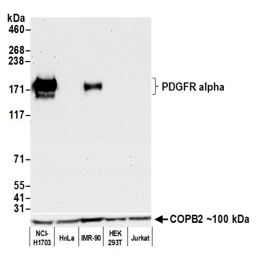
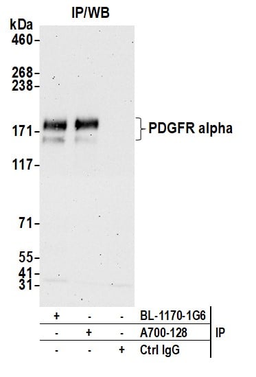
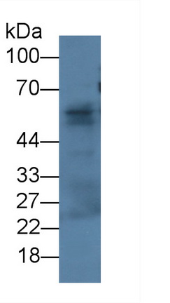
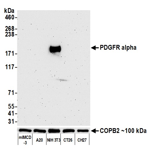 WB (Western Blot)
(Detection of mouse PDGFR alpha by western blot. Samples: Whole cell lysate (50 ug) from mIMCD-3, A20, NIH 3T3, CT26, and CH27 cells prepared using NETN lysis buffer. Antibody: Rabbit anti-PDGFR alpha recombinant monoclonal antibody (AAA213594 Lot 1) used at 1:1000. Secondary: HRP-conjugated goat anti-rabbit IgG . Detection: Chemiluminescence with an exposure time of 3 minutes. Lower Panel: Rabbit anti-COPB2 antibody .)
WB (Western Blot)
(Detection of mouse PDGFR alpha by western blot. Samples: Whole cell lysate (50 ug) from mIMCD-3, A20, NIH 3T3, CT26, and CH27 cells prepared using NETN lysis buffer. Antibody: Rabbit anti-PDGFR alpha recombinant monoclonal antibody (AAA213594 Lot 1) used at 1:1000. Secondary: HRP-conjugated goat anti-rabbit IgG . Detection: Chemiluminescence with an exposure time of 3 minutes. Lower Panel: Rabbit anti-COPB2 antibody .)
PDGFR alpha, Monoclonal Recombinant Antibody (Cat# AAA213594)

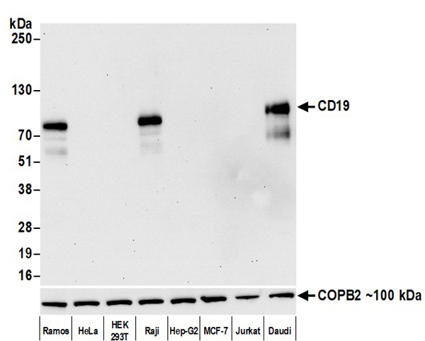 WB (Western Blot)
(Detection of human CD19 by western blot. Samples: Whole cell lysate (10 ug) from Ramos, HeLa, HEK293T, Raji, Hep-G2, MCF-7, Jurkat, and Daudi cells prepared using NETN lysis buffer. Antibody: Rabbit anti-CD19 recombinant monoclonal antibody (AAA213598 Lot 1) used at 1:1000. Secondary: HRP-conjugated goat anti-rabbit IgG . Detection: Chemiluminescence with an exposure time of 30 seconds. Lower Panel: Rabbit anti-COPB2 antibody .)
WB (Western Blot)
(Detection of human CD19 by western blot. Samples: Whole cell lysate (10 ug) from Ramos, HeLa, HEK293T, Raji, Hep-G2, MCF-7, Jurkat, and Daudi cells prepared using NETN lysis buffer. Antibody: Rabbit anti-CD19 recombinant monoclonal antibody (AAA213598 Lot 1) used at 1:1000. Secondary: HRP-conjugated goat anti-rabbit IgG . Detection: Chemiluminescence with an exposure time of 30 seconds. Lower Panel: Rabbit anti-COPB2 antibody .)
CD19, Monoclonal Recombinant Antibody (Cat# AAA213598)
What are Monoclonal Antibodies?
Monoclonal antibodies are specialized laboratory-produced proteins developed for binding to specific biological antigens or other molecular targets. Since they come from a single cell (or clone), they are especially consistent and accurate in the data they are involved in producing.
This type of antibody material has been shown to be a powerful tool in finding and subsequently destroying harmful cells in an organism, such as those found in cancers or various autoimmune diseases. This makes them excellent aids in medical testing and research, which is why they are so widely used.
AAA Biotech offers a comprehensive range of high-quality monoclonal antibodies that perform effectively in various laboratory tests, including (amongst others) ELISA, western blotting, immunohistochemistry, and flow cytometry. All of the products in our catalog are thoroughly quality tested to make sure that they are reliable and will consistently perform well in your research.
What Are The Uses of Monoclonal Antibodies
Monoclonal antibodies are used in many lab tests, including (amongst others) ELISA, western blotting, immunohistochemistry, and flow cytometry.
ELISA is a test that helps detect a specific substance/analyte in a sample. It uses antibodies (often monoclonal) bound to a solid surface (such as the well of a microplate) to “capture” the substance/analyte in the sample and immobilize it so that the detection antibody component can then bind to it and produce a signal, which can then be measured.
Western blotting identifies specific proteins in a sample. The sample is first separated on a gel, and then antibodies are applied that will typically bind to the target, which will all be localized to a single band in a lane.
Immunohistochemistry helps locate specific proteins in cells or tissue samples using antibodies.
Flow cytometry looks at and sorts cells. It uses antibodies that are conjugated to reporter molecules called “fluorophores”, which, under special lights, emit light themselves, which can then be measured by a detector instrument.
How Monoclonal Antibodies Are Used as Medicine?
Please note that all of the products listed in AAA Biotech’s also known as AAA Bio or AAABio catalog are strictly for research-use only (RUO).
Monoclonal antibodies can also be used as therapeutic/medical treatments, particularly in the context of cancers. They are designed to find and bind to specific cells or proteins, helping the immune system recognize and attack the cancer. These treatments work in different ways, such as:
- Radioimmunotherapy attaches a small amount of radioactive molecule to the antibody, so it delivers the radiation directly to the cancer cells that the antibody is specifically binding to.
- Antibody-directed enzyme prodrug therapy uses antibodies that are specifically bound to special enzymes. These enzymes activate a harmless drug in the body and turn it into a cancer-killing drug only near the cancer cells—this helps avoid harming healthy cells.
- Immunoliposomes are tiny “bubbles” filled with medicine/drug and coated with antibodies. They carry the drug straight to the cancer cells.
Why Buy Monoclonal Antibodies From Us?
At AAA Biotech, we provide high-performance monoclonal antibodies designed to support a wide range of research needs.
1. Validated for Versatile Applications
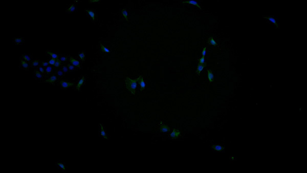
The antibodies in our catalog are extensively validated and compatible with multiple techniques, including (but not limited to) ELISA, flow cytometry (FC), immunocytochemistry (ICC), immunofluorescence (IF), immunohistochemistry (IHC), immunoprecipitation (IP), and western blotting (WB).
2. Wide Selection & Specialized Options
We offer antibodies for common and rare species, that are available in various conjugated forms, and also in recombinant formats. Essentially, there is almost anything one might need to meet their experimental model’s requirements.
3. High-Quality Proteins
Our proteins meet high purity standards—90% or more as confirmed by SDS-PAGE. Many are available with tags like His, Flag, GST, or MBP, and we also supply native and biologically active proteins for functional studies.
Frequently Asked Questions
1. Are your monoclonal antibodies validated for specific applications?
Yes, our antibodies are tested and validated for use in methods such as ELISA, western blot, IHC, flow cytometry, and more. Refer to specific product pages or datasheets for individual product information.
2. How do I choose the right monoclonal antibody for my application?
Review the product details directly for application validation, species reactivity, and target information. You may also contact our support team at any time for help.
3. How quickly can I receive my order?
Most orders are processed and shipped within 1–3 business days, depending on product availability and your shipping location.