Filters
▼Clonality
▼Type
▼Reactivity
▼Gene Name
▼Isotype
▼Host
▼Application
▼Clone
▼Monoclonal Antibodies
Get accurate results in your research with our Monoclonal Antibodies, which are specially made to target exactly what you require for your research, and will produce consistent, reliable performance in lab tests.
Viewing 3050-3100 of 27597 product results
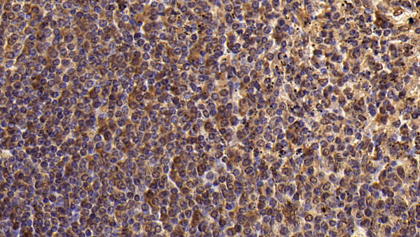
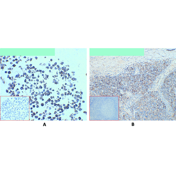
 IHC (Immunohiostchemistry)
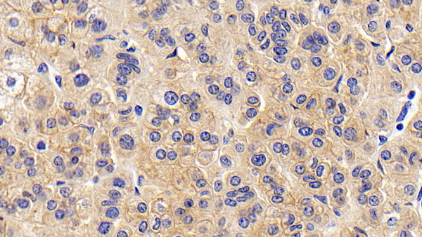
(DAB staining on IHCP;Sample: Human Lung cancer Tissue;Primary Ab: 30ug/ml Mouse AntiHuman CD147 AntibodySecond Ab: 2ug/mL HRPLinked Caprine AntiMouse IgG Polyclonal Antibody(Catalog: SAA544Mu19))
IHC (Immunohiostchemistry)
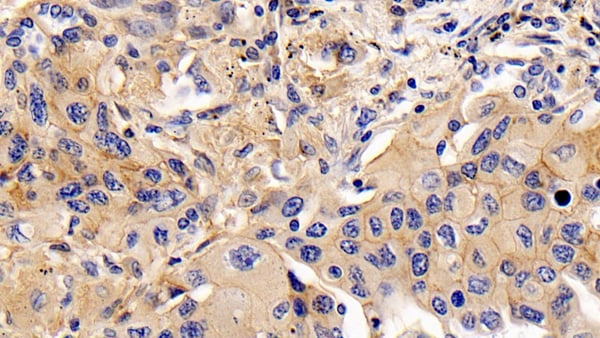
(DAB staining on IHCP;Sample: Human Lung cancer Tissue;Primary Ab: 30ug/ml Mouse AntiHuman CD147 AntibodySecond Ab: 2ug/mL HRPLinked Caprine AntiMouse IgG Polyclonal Antibody(Catalog: SAA544Mu19))
Cluster Of Differentiation 147 (CD147), Monoclonal Antibody (Cat# AAA151723)
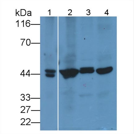
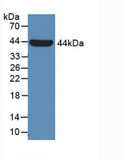
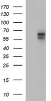
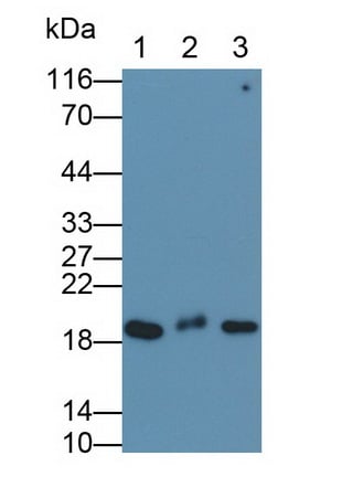 WB (Western Blot)
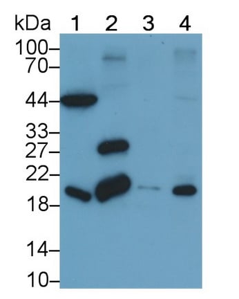
(Western Blot; Sample: Lane1: A549 cell lysate; Lane2: HepG2 cell lysate; Lane3: MCF7 cell lysatePrimary Ab: 0.3ug/ml Mouse AntiHuman CFL1 AntibodySecond Ab: 0.2ug/mL HRPLinked Caprine AntiMouse IgG Polyclonal Antibody(Catalog: SAA544Mu19))
WB (Western Blot)
(Western Blot; Sample: Lane1: A549 cell lysate; Lane2: HepG2 cell lysate; Lane3: MCF7 cell lysatePrimary Ab: 0.3ug/ml Mouse AntiHuman CFL1 AntibodySecond Ab: 0.2ug/mL HRPLinked Caprine AntiMouse IgG Polyclonal Antibody(Catalog: SAA544Mu19))
Cofilin 1 (CFL1), Monoclonal Antibody (Cat# AAA151725)
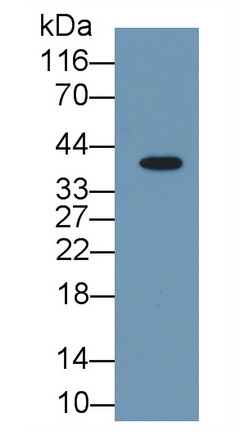
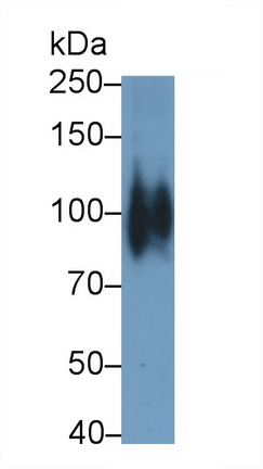
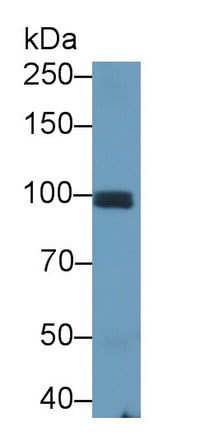
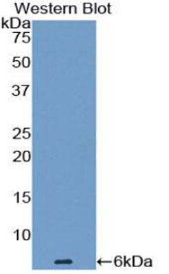
 WB (Western Blot)
(Western Blot; Sample: Rat Spleen lysate; Primary Ab: 3ug/ml Mouse AntiHuman KIR2DS4 AntibodySecond Ab: 0.2ug/mL HRPLinked Caprine AntiMouse IgG Polyclonal Antibody(Catalog: SAA544Mu19))
WB (Western Blot)
(Western Blot; Sample: Rat Spleen lysate; Primary Ab: 3ug/ml Mouse AntiHuman KIR2DS4 AntibodySecond Ab: 0.2ug/mL HRPLinked Caprine AntiMouse IgG Polyclonal Antibody(Catalog: SAA544Mu19))
Killer Cell Immunoglobulin Like Receptor 2DS4 (KIR2DS4), Monoclonal Antibody (Cat# AAA151728)
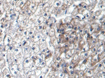
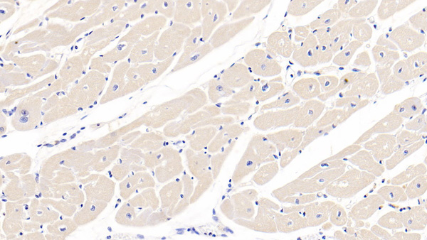 IHC (Immunohistochemistry)
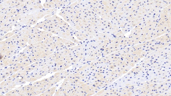
(DAB staining on IHCP;Sample: Porcine Cardiac Muscle Tissue; Primary Ab: 30ug/ml Mouse AntiHuman GPC3 AntibodySecond Ab: 2ug/mL HRPLinked Caprine AntiMouse IgG Polyclonal Antibody(Catalog: SAA544Mu19))
IHC (Immunohistochemistry)
(DAB staining on IHCP;Sample: Porcine Cardiac Muscle Tissue; Primary Ab: 30ug/ml Mouse AntiHuman GPC3 AntibodySecond Ab: 2ug/mL HRPLinked Caprine AntiMouse IgG Polyclonal Antibody(Catalog: SAA544Mu19))
Glypican 3 (GPC3), Monoclonal Antibody (Cat# AAA151665)

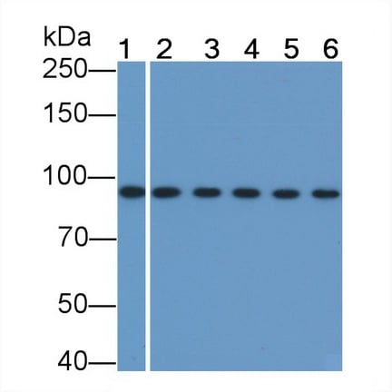 WB (Western Blot)
(Western Blot; Sample: Lane1: Mouse Cerebrum lysate; Lane2: Rat Cerebrum lysate; Lane3: Canine Heart lysate; Lane4: Canine Cerebrum lysate; Lane5: Gallus Heart lysate; Lane6: Gallus Cerebrum lysatePrimary Ab: 2ug/ml Mouse AntiHuman betacatenin AntibodySecond Ab: 0.2ug/mL HRPLinked Caprine AntiMouse IgG Polyclonal Antibody(Catalog: SAA544Mu19))
WB (Western Blot)
(Western Blot; Sample: Lane1: Mouse Cerebrum lysate; Lane2: Rat Cerebrum lysate; Lane3: Canine Heart lysate; Lane4: Canine Cerebrum lysate; Lane5: Gallus Heart lysate; Lane6: Gallus Cerebrum lysatePrimary Ab: 2ug/ml Mouse AntiHuman betacatenin AntibodySecond Ab: 0.2ug/mL HRPLinked Caprine AntiMouse IgG Polyclonal Antibody(Catalog: SAA544Mu19))
Beta Catenin (betacatenin), Monoclonal Antibody (Cat# AAA151669)
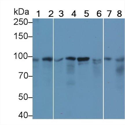
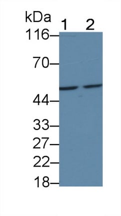
 WB (Western Blot)
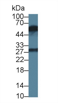
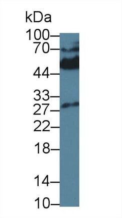
(Western Blot; Sample: Lane1: Porcine Liver lysate; Lane2: HepG2 cell lysatePrimary Ab: 0.2ug/ml Mouse AntiHuman Arg AntibodySecond Ab: 0.2ug/mL HRPLinked Caprine AntiMouse IgG Polyclonal Antibody(Catalog: SAA544Mu19))
WB (Western Blot)
(Western Blot; Sample: Lane1: Porcine Liver lysate; Lane2: HepG2 cell lysatePrimary Ab: 0.2ug/ml Mouse AntiHuman Arg AntibodySecond Ab: 0.2ug/mL HRPLinked Caprine AntiMouse IgG Polyclonal Antibody(Catalog: SAA544Mu19))
Arginase (ARG), Monoclonal Antibody (Cat# AAA151680)
 WB (Western Blot)
Cadherin, Osteoblast (CDHOB), Monoclonal Antibody (Cat# AAA151682)

 WB (Western Blot)
(Western Blot; Sample: Lane1: Rat Thymus lysate; Lane2: Rat Spleen lysate; Lane3: Raji cell lysate; Lane4: K562 cell lysate Primary Ab: 3ug/ml Mouse AntiHuman CD4 Antibody Second Ab: 0.2ug/mL HRPLinked Caprine AntiMouse IgG Polyclonal Antibody (Catalog: SAA544Mu19))
WB (Western Blot)
(Western Blot; Sample: Lane1: Rat Thymus lysate; Lane2: Rat Spleen lysate; Lane3: Raji cell lysate; Lane4: K562 cell lysate Primary Ab: 3ug/ml Mouse AntiHuman CD4 Antibody Second Ab: 0.2ug/mL HRPLinked Caprine AntiMouse IgG Polyclonal Antibody (Catalog: SAA544Mu19))
Cluster Of Differentiation 4 (CD4), Monoclonal Antibody (Cat# AAA151686)
 WB (Western Blot)
(Western Blot; Sample: Lane1: Raji cell lysate; Lane2: THP1 cell lysate Primary Ab: 0.1ug/ml Mouse AntiHuman CD4 Antibody Second Ab: 0.2ug/mL HRPLinked Caprine AntiMouse IgG Polyclonal Antibody (Catalog: SAA544Mu19))
WB (Western Blot)
(Western Blot; Sample: Lane1: Raji cell lysate; Lane2: THP1 cell lysate Primary Ab: 0.1ug/ml Mouse AntiHuman CD4 Antibody Second Ab: 0.2ug/mL HRPLinked Caprine AntiMouse IgG Polyclonal Antibody (Catalog: SAA544Mu19))
Cluster Of Differentiation 4 (CD4), Monoclonal Antibody (Cat# AAA151687)
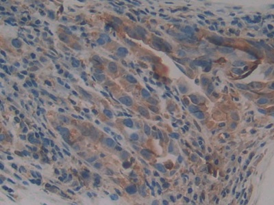
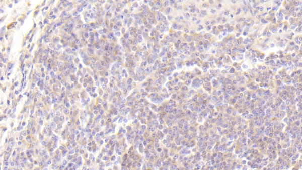 IHC (Immunohistochemistry)
(DAB staining on IHC-P;Sample: human Spleen Tissue; Primary Ab: 20ug/ml Mouse Anti-human TGFa AntibodySecond Ab: 2ug/mL HRP-Linked Caprine Anti-Mouse IgG Polyclonal Antibody)
IHC (Immunohistochemistry)
(DAB staining on IHC-P;Sample: human Spleen Tissue; Primary Ab: 20ug/ml Mouse Anti-human TGFa AntibodySecond Ab: 2ug/mL HRP-Linked Caprine Anti-Mouse IgG Polyclonal Antibody)
Transforming Growth Factor Alpha (TGFa), Monoclonal Antibody (Cat# AAA152708)
 IHC (Immunohistochemisry)
(DAB staining on IHC-P;Samples: Human Stomach Tissue;Primary Ab: 20ug/ml Mouse Anti-Human EDN1 AntibodySecond Ab: 2ug/mL HRP-Linked CaprineAnti-Mouse IgG Polyclonal Antibody)
IHC (Immunohistochemisry)
(DAB staining on IHC-P;Samples: Human Stomach Tissue;Primary Ab: 20ug/ml Mouse Anti-Human EDN1 AntibodySecond Ab: 2ug/mL HRP-Linked CaprineAnti-Mouse IgG Polyclonal Antibody)
Endothelin 1 (EDN1), Monoclonal Antibody (Cat# AAA130615)
 IHC (Immunohistochemisry)
(DAB staining on IHC-P;Samples: Human Stomach Tissue;Primary Ab: 40ug/ml Mouse Anti-Human GLa AntibodySecond Ab: 2ug/mL HRP-Linked Caprine Anti-Mouse IgG Polyclonal Antibody)
IHC (Immunohistochemisry)
(DAB staining on IHC-P;Samples: Human Stomach Tissue;Primary Ab: 40ug/ml Mouse Anti-Human GLa AntibodySecond Ab: 2ug/mL HRP-Linked Caprine Anti-Mouse IgG Polyclonal Antibody)
Galactosidase Alpha (GLa), Monoclonal Antibody (Cat# AAA130640)
C Reactive Protein (CRP), Monoclonal Antibody (Cat# AAA133838)
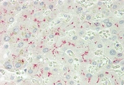
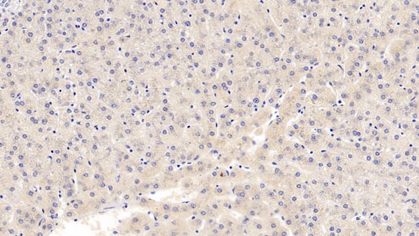
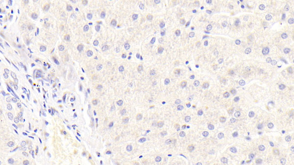
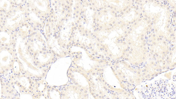
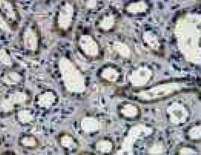
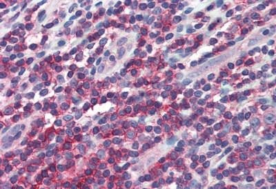
 IHC (Immunohiostchemistry)
(ANPEP/CD13 Antibody-Human Kidney: Formalin-Fixed, Paraffin-Embedded (FFPE))
IHC (Immunohiostchemistry)
(ANPEP/CD13 Antibody-Human Kidney: Formalin-Fixed, Paraffin-Embedded (FFPE))
ANPEP/CD13, Monoclonal Antibody (Cat# AAA162699)
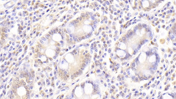
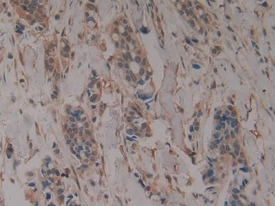
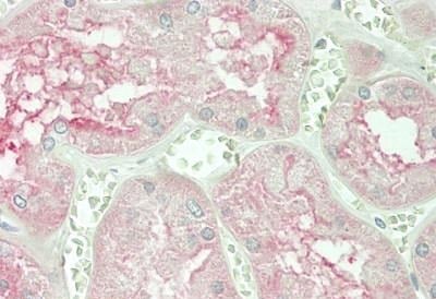
 IHC (Immunohiostchemistry)
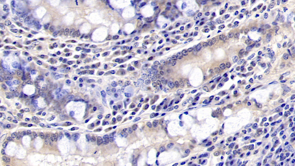
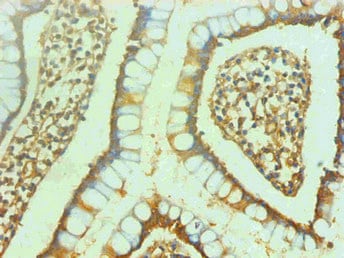
(IL10RA Antibody-Human Small Intestine, MALT: Formalin-Fixed, Paraffin-Embedded (FFPE), at a concentration of 10 ug/ml.)
IHC (Immunohiostchemistry)
(IL10RA Antibody-Human Small Intestine, MALT: Formalin-Fixed, Paraffin-Embedded (FFPE), at a concentration of 10 ug/ml.)
IL10RA, Monoclonal Antibody (Cat# AAA162853)
TCR gamma/delta, Monoclonal Antibody (Cat# AAA129125)
CD184, Monoclonal Antibody (Cat# AAA129129)
Ly6G, Monoclonal Antibody (Cat# AAA129137)
MHC Class II, Monoclonal Antibody (Cat# AAA129138)
CD235a, Monoclonal Antibody (Cat# AAA129139)
CD52, Monoclonal Antibody (Cat# AAA129148)
CD184, Monoclonal Antibody (Cat# AAA129150)
CD52, Monoclonal Antibody (Cat# AAA129161)
CD44, Monoclonal Antibody (Cat# AAA129166)
CD184, Monoclonal Antibody (Cat# AAA129169)
Ig Kappa Light Chain, Monoclonal Antibody (Cat# AAA129171)
CD235a, Monoclonal Antibody (Cat# AAA129173)
CD61r, Monoclonal Antibody (Cat# AAA129176)
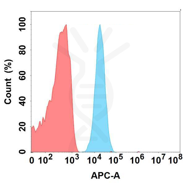
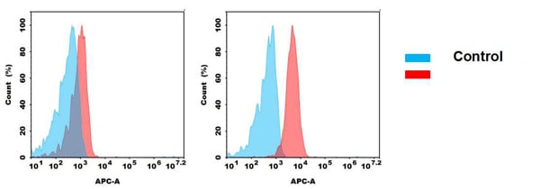
 FCM/FACS (Flow Cytometry)
(Figure 2. Flow cytometry analysis of antigen binding of anti-human CD93 mAb(AAA129453).)
FCM/FACS (Flow Cytometry)
(Figure 2. Flow cytometry analysis of antigen binding of anti-human CD93 mAb(AAA129453).)
CD93, Monoclonal Antibody (Cat# AAA129453)
 FCM/FACS (Flow Cytometry)
(Figure 2. Flow cytometry analysis of antigen binding of anti-human MICA mAb(AAA129456).)
FCM/FACS (Flow Cytometry)
(Figure 2. Flow cytometry analysis of antigen binding of anti-human MICA mAb(AAA129456).)
MICA, Monoclonal Antibody (Cat# AAA129456)
 FCM/FACS (Flow Cytometry)
(Figure 2. Flow cytometry analysis of antigen binding of anti-human CD45 mAb(AAA129458).)
FCM/FACS (Flow Cytometry)
(Figure 2. Flow cytometry analysis of antigen binding of anti-human CD45 mAb(AAA129458).)
CD45, Monoclonal Antibody (Cat# AAA129458)
TENM4, Monoclonal Antibody (Cat# AAA129413)
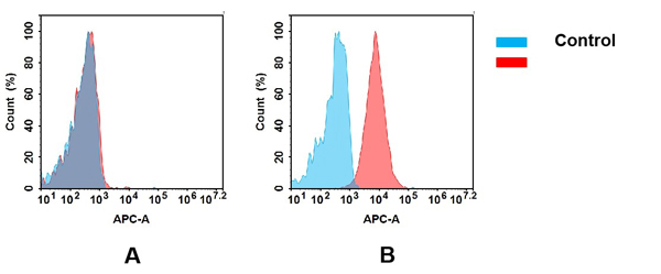
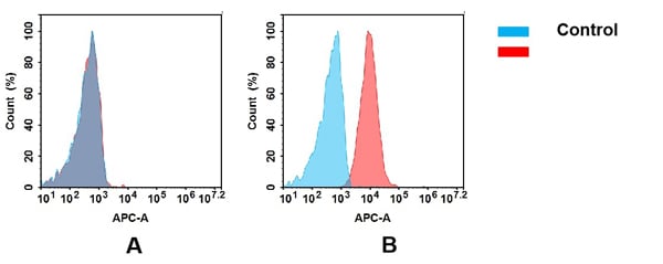
 FCM/FACS (Flow Cytometry)
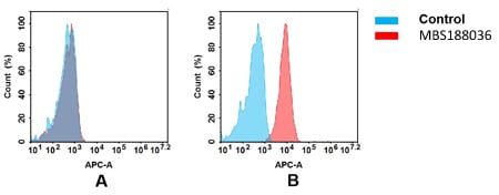
(Figure 2. Flow cytometry analysis of antigen binding of anti-human LIV-1 mAb(AAA129414).(A) AAA129414 does not bind to CHO-S cells that do not express LIV-1.(B) A clear peak shift of AAA129414 was seen compared to the control when incubated with LIV-1-expressing Raji cells, indicating strong binding of AAA129414 to LIV-1. Antibodies were incubated at 5 ug/mL.)
FCM/FACS (Flow Cytometry)
(Figure 2. Flow cytometry analysis of antigen binding of anti-human LIV-1 mAb(AAA129414).(A) AAA129414 does not bind to CHO-S cells that do not express LIV-1.(B) A clear peak shift of AAA129414 was seen compared to the control when incubated with LIV-1-expressing Raji cells, indicating strong binding of AAA129414 to LIV-1. Antibodies were incubated at 5 ug/mL.)
LIV-1, Monoclonal Antibody (Cat# AAA129414)
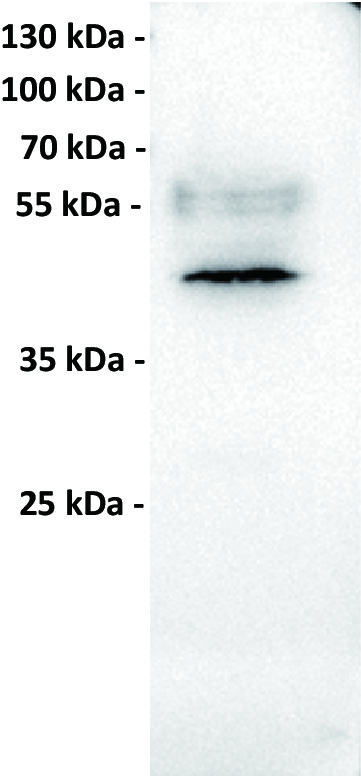
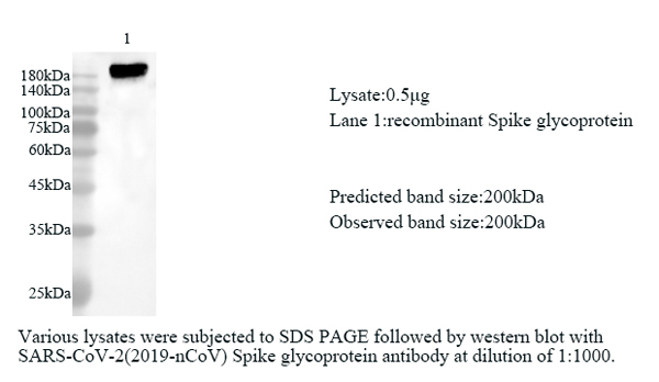 WB (Western Blot)
COVID 19 Spike glycoprotein Coronavirus, Monoclonal Antibody (Cat# AAA119905)
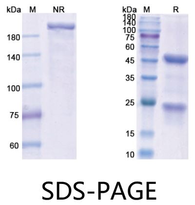 SDS-PAGE
(SDS PAGE for NeuGcGM3)
SDS-PAGE
(SDS PAGE for NeuGcGM3)
NeuGcGM3 (14F7), Monoclonal Recombinant Antibody (Cat# AAA119950)
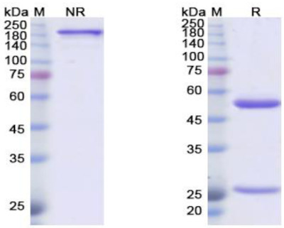 SDS-PAGE
Sotigalimab, Monoclonal Antibody (Cat# AAA119954)
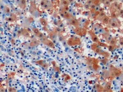
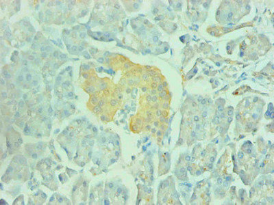 IHC (Immunohistochemisry)
(Immunohistochemical of paraffin-embedded human pancreas using AAA119807 at dilution of 1:200)
IHC (Immunohistochemisry)
(Immunohistochemical of paraffin-embedded human pancreas using AAA119807 at dilution of 1:200)
Gamma-enolase, Monoclonal Antibody (Cat# AAA119807)
CD42b/GP1BA, Monoclonal Antibody (Cat# AAA120162)
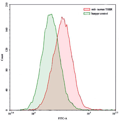
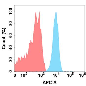
 FCM/FACS (Flow Cytometry)
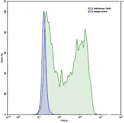
(Flow-cytometry using anti-human TSHR antibody. TSHR Transfected CHO cells were stained with an irrelevant antibody (Blue Histogram) or an anti-human TSHR antibody monoclonal antibody (Catalog # AAA120166, Green Histogram) at a concentration of 5 ug/ml for 30 mins at RT. After washing, bound antibody was detected using a FITC conjugated goat anti-human antibody (Catalog # Please inquire) and cells analysed on a NovoCyte Flow Cytometer.)
FCM/FACS (Flow Cytometry)
(Flow-cytometry using anti-human TSHR antibody. TSHR Transfected CHO cells were stained with an irrelevant antibody (Blue Histogram) or an anti-human TSHR antibody monoclonal antibody (Catalog # AAA120166, Green Histogram) at a concentration of 5 ug/ml for 30 mins at RT. After washing, bound antibody was detected using a FITC conjugated goat anti-human antibody (Catalog # Please inquire) and cells analysed on a NovoCyte Flow Cytometer.)
TSHR/LGR3, Monoclonal Antibody (Cat# AAA120166)
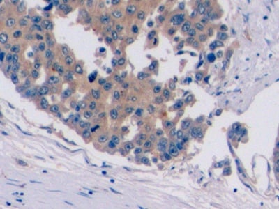
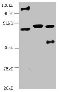
 IHC (Immunohiostchemistry)
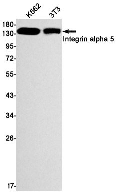
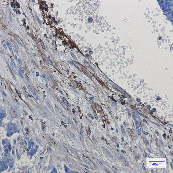
(Immunohistochemistry of Integrin alpha 5 in paraffin-embedded Human breast cancer tissue using Integrin alpha 5 Rabbit mAb at dilution 1:50)
IHC (Immunohiostchemistry)
(Immunohistochemistry of Integrin alpha 5 in paraffin-embedded Human breast cancer tissue using Integrin alpha 5 Rabbit mAb at dilution 1:50)
Integrin alpha 5, Monoclonal Antibody (Cat# AAA178859)

 IHC (Immunohiostchemistry)
(Immunohistochemistry of p16 ARC in paraffin-embedded Human Cholangiocarcinoma using p16 ARC Rabbit mAb at dilution 1/20)
IHC (Immunohiostchemistry)
(Immunohistochemistry of p16 ARC in paraffin-embedded Human Cholangiocarcinoma using p16 ARC Rabbit mAb at dilution 1/20)
p16 ARC, Monoclonal Antibody (Cat# AAA178870)
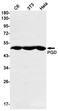
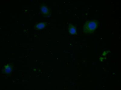
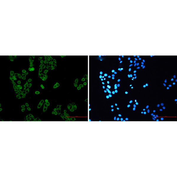
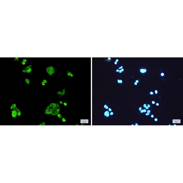 IF (Immunofluorescence)
(Immunofluorescence of PGD(green) in Hela cells using PGD Rabbit mAb at dilution 1:200, and DAPI(blue))
IF (Immunofluorescence)
(Immunofluorescence of PGD(green) in Hela cells using PGD Rabbit mAb at dilution 1:200, and DAPI(blue))
PGD, Monoclonal Antibody (Cat# AAA178873)
 IHC (Immunohistochemisry)
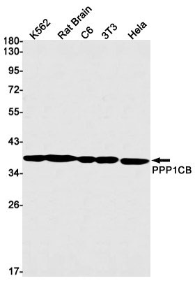
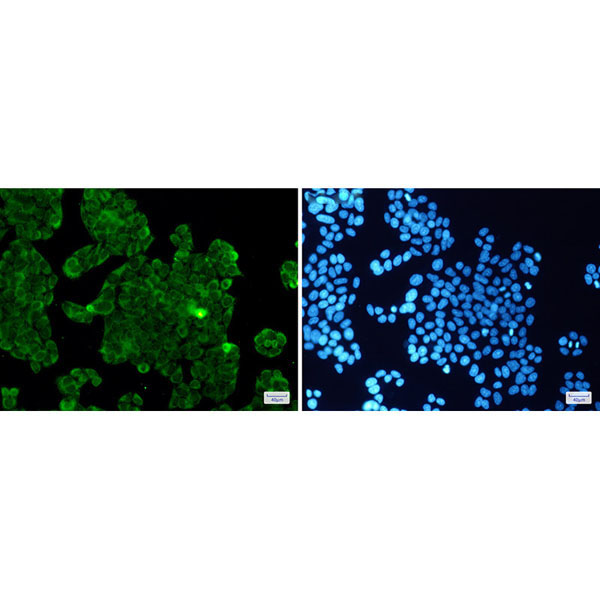
(Immunohistochemistry of PPP1CB in paraffin-embedded Human colon cancer tissue using PPP1CB Rabbit mAb at dilution 1:100)
IHC (Immunohistochemisry)
(Immunohistochemistry of PPP1CB in paraffin-embedded Human colon cancer tissue using PPP1CB Rabbit mAb at dilution 1:100)
PP1C beta, Monoclonal Antibody (Cat# AAA178874)
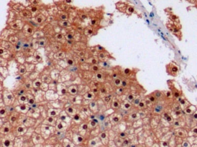
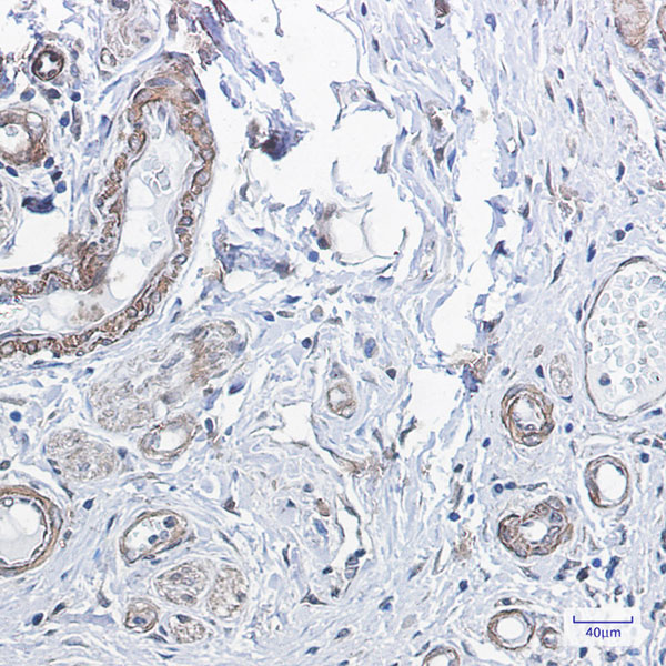
 IHC (Immunohiostchemistry)
(Immunohistochemistry of Prohibitin in paraffin-embedded Human Cholangiocarcinoma using Prohibitin Rabbit mAb at dilution 1:50)
IHC (Immunohiostchemistry)
(Immunohistochemistry of Prohibitin in paraffin-embedded Human Cholangiocarcinoma using Prohibitin Rabbit mAb at dilution 1:50)
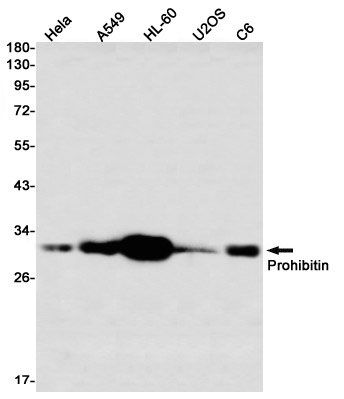
Prohibitin, Monoclonal Antibody (Cat# AAA178875)
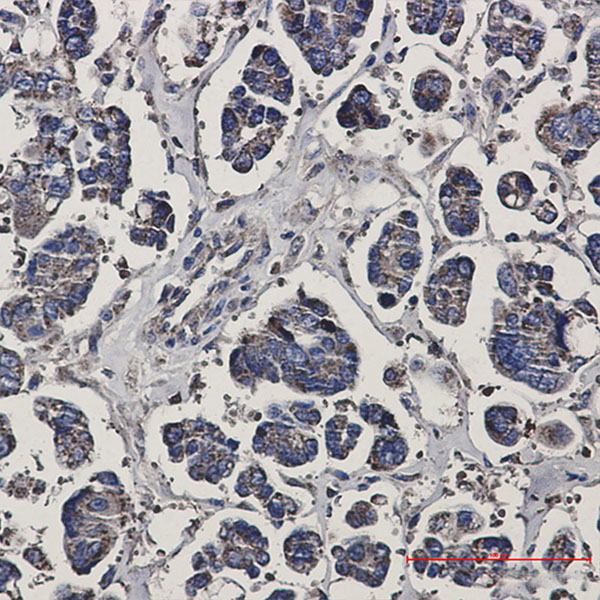
 IHC (Immunohistochemisry)
(Immunohistochemistry of RENT1 in paraffin-embedded Human Cholangiocarcinoma using RENT1 Rabbit mAb at dilution 1:50)
IHC (Immunohistochemisry)
(Immunohistochemistry of RENT1 in paraffin-embedded Human Cholangiocarcinoma using RENT1 Rabbit mAb at dilution 1:50)
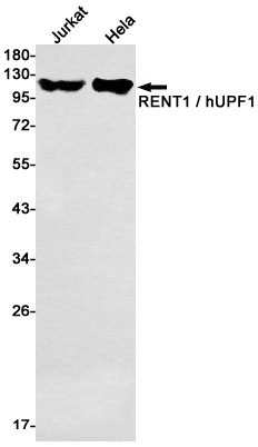
RENT1, Monoclonal Antibody (Cat# AAA178877)
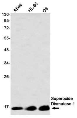
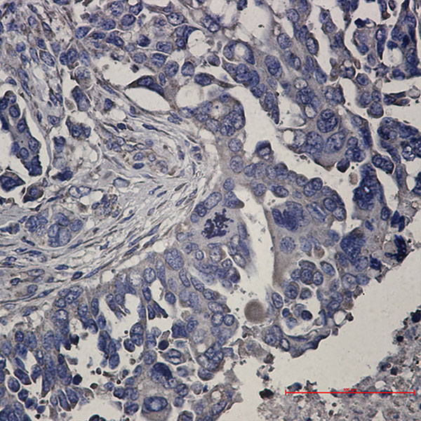
 IHC (Immunohiostchemistry)
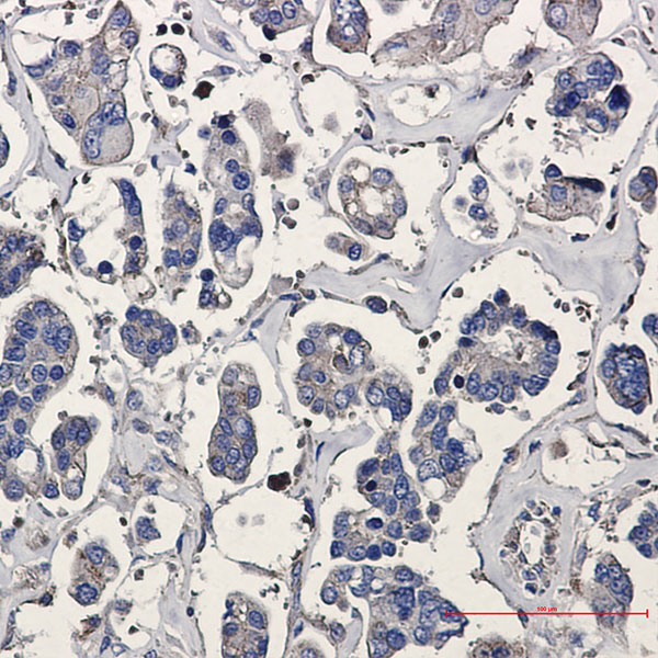
(Immunohistochemical of Superoxide Dismutase 1 in Human breast cancer tissue using Superoxide Dismutase 1 antibody at dilution 1:50)
IHC (Immunohiostchemistry)
(Immunohistochemical of Superoxide Dismutase 1 in Human breast cancer tissue using Superoxide Dismutase 1 antibody at dilution 1:50)
Superoxide Dismutase 1, Monoclonal Antibody (Cat# AAA178881)
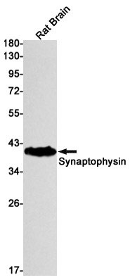
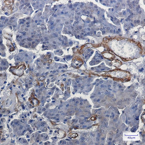
 IHC (Immunohiostchemistry)
(Immunohistochemistry of Synaptophysin in paraffin-embedded Human Brain using Synaptophysin Rabbit mAb at dilution 1:20)
IHC (Immunohiostchemistry)
(Immunohistochemistry of Synaptophysin in paraffin-embedded Human Brain using Synaptophysin Rabbit mAb at dilution 1:20)
Synaptophysin, Monoclonal Antibody (Cat# AAA178883)
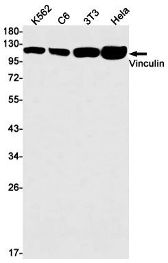
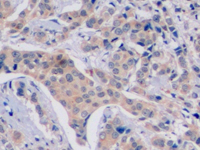
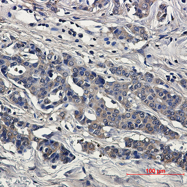
 IHC (Immunohiostchemistry)
(Immunohistochemistry of Vinculin in paraffin-embedded Human breast cancer tissue using Vinculin Rabbit mAb at dilution 1:50)
IHC (Immunohiostchemistry)
(Immunohistochemistry of Vinculin in paraffin-embedded Human breast cancer tissue using Vinculin Rabbit mAb at dilution 1:50)
Vinculin, Monoclonal Antibody (Cat# AAA178888)
Corticosterone, Monoclonal Antibody (Cat# AAA117776)
What are Monoclonal Antibodies?
Monoclonal antibodies are specialized laboratory-produced proteins developed for binding to specific biological antigens or other molecular targets. Since they come from a single cell (or clone), they are especially consistent and accurate in the data they are involved in producing.
This type of antibody material has been shown to be a powerful tool in finding and subsequently destroying harmful cells in an organism, such as those found in cancers or various autoimmune diseases. This makes them excellent aids in medical testing and research, which is why they are so widely used.
AAA Biotech offers a comprehensive range of high-quality monoclonal antibodies that perform effectively in various laboratory tests, including (amongst others) ELISA, western blotting, immunohistochemistry, and flow cytometry. All of the products in our catalog are thoroughly quality tested to make sure that they are reliable and will consistently perform well in your research.
What Are The Uses of Monoclonal Antibodies
Monoclonal antibodies are used in many lab tests, including (amongst others) ELISA, western blotting, immunohistochemistry, and flow cytometry.
ELISA is a test that helps detect a specific substance/analyte in a sample. It uses antibodies (often monoclonal) bound to a solid surface (such as the well of a microplate) to “capture” the substance/analyte in the sample and immobilize it so that the detection antibody component can then bind to it and produce a signal, which can then be measured.
Western blotting identifies specific proteins in a sample. The sample is first separated on a gel, and then antibodies are applied that will typically bind to the target, which will all be localized to a single band in a lane.
Immunohistochemistry helps locate specific proteins in cells or tissue samples using antibodies.
Flow cytometry looks at and sorts cells. It uses antibodies that are conjugated to reporter molecules called “fluorophores”, which, under special lights, emit light themselves, which can then be measured by a detector instrument.
How Monoclonal Antibodies Are Used as Medicine?
Please note that all of the products listed in AAA Biotech’s also known as AAA Bio or AAABio catalog are strictly for research-use only (RUO).
Monoclonal antibodies can also be used as therapeutic/medical treatments, particularly in the context of cancers. They are designed to find and bind to specific cells or proteins, helping the immune system recognize and attack the cancer. These treatments work in different ways, such as:
- Radioimmunotherapy attaches a small amount of radioactive molecule to the antibody, so it delivers the radiation directly to the cancer cells that the antibody is specifically binding to.
- Antibody-directed enzyme prodrug therapy uses antibodies that are specifically bound to special enzymes. These enzymes activate a harmless drug in the body and turn it into a cancer-killing drug only near the cancer cells—this helps avoid harming healthy cells.
- Immunoliposomes are tiny “bubbles” filled with medicine/drug and coated with antibodies. They carry the drug straight to the cancer cells.
Why Buy Monoclonal Antibodies From Us?
At AAA Biotech, we provide high-performance monoclonal antibodies designed to support a wide range of research needs.
1. Validated for Versatile Applications
The antibodies in our catalog are extensively validated and compatible with multiple techniques, including (but not limited to) ELISA, flow cytometry (FC), immunocytochemistry (ICC), immunofluorescence (IF), immunohistochemistry (IHC), immunoprecipitation (IP), and western blotting (WB).
2. Wide Selection & Specialized Options
We offer antibodies for common and rare species, that are available in various conjugated forms, and also in recombinant formats. Essentially, there is almost anything one might need to meet their experimental model’s requirements.
3. High-Quality Proteins
Our proteins meet high purity standards—90% or more as confirmed by SDS-PAGE. Many are available with tags like His, Flag, GST, or MBP, and we also supply native and biologically active proteins for functional studies.
Frequently Asked Questions
1. Are your monoclonal antibodies validated for specific applications?
Yes, our antibodies are tested and validated for use in methods such as ELISA, western blot, IHC, flow cytometry, and more. Refer to specific product pages or datasheets for individual product information.
2. How do I choose the right monoclonal antibody for my application?
Review the product details directly for application validation, species reactivity, and target information. You may also contact our support team at any time for help.
3. How quickly can I receive my order?
Most orders are processed and shipped within 1–3 business days, depending on product availability and your shipping location.