Filters
▼Clonality
▼Type
▼Reactivity
▼Gene Name
▼Isotype
▼Host
▼Application
▼Clone
▼Active Proteins
AAA Biotech also known as AAA Bio or AAABio provides a variety of high-quality recombinant and natural/native proteins that are proven to work in a wide range of experiments. Explore our products to find the active protein that best fits your needs or experimental model.
Viewing 350-400 of 2567 product results
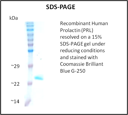
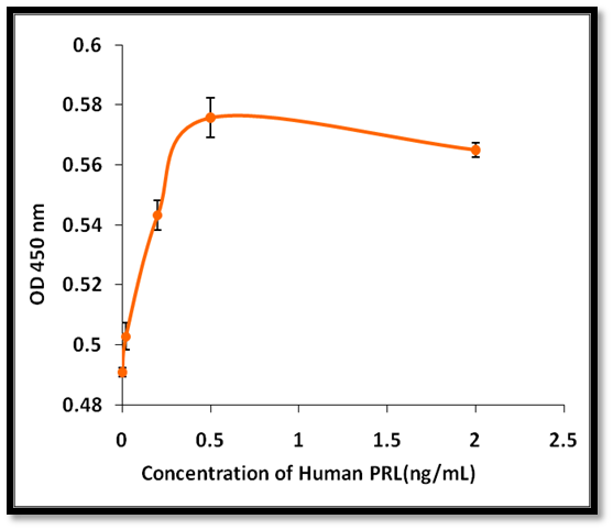 Application Data
Prolactin (PRL), Active Protein (Cat# AAA214290)
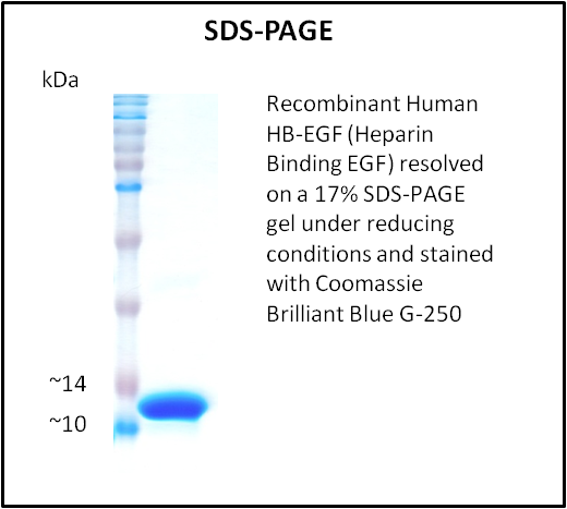
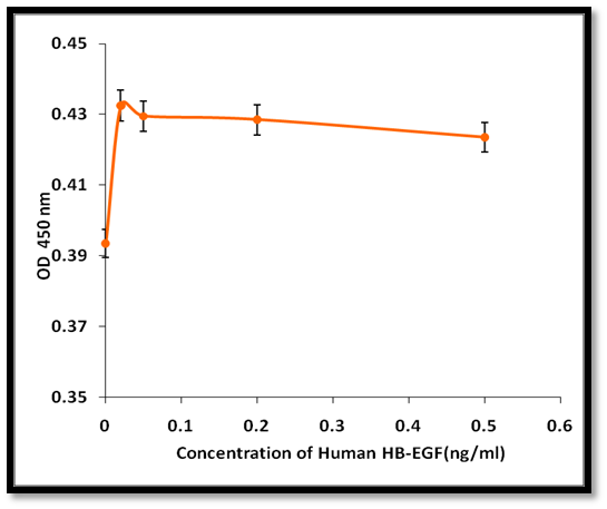 Application Data
HB-EGF (Heparin Binding EGF), Active Protein (Cat# AAA214291)
 Application Data
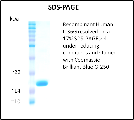
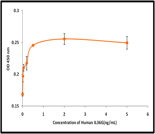
IL36G, Active Protein (Cat# AAA214293)
 Application Data
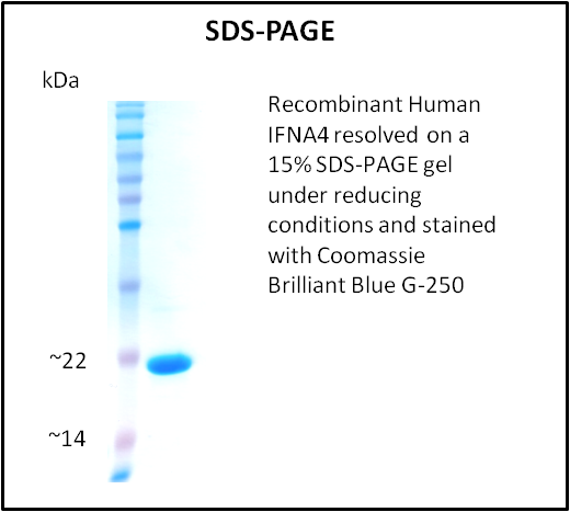
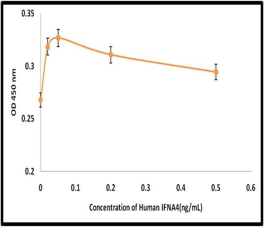
IFNA4, Active Protein (Cat# AAA214301)
 Application Data
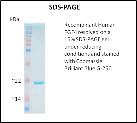
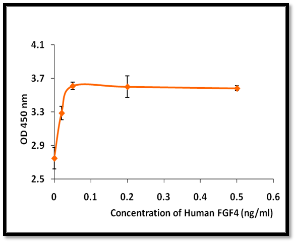
FGF4, Active Protein (Cat# AAA214307)
 Application Data
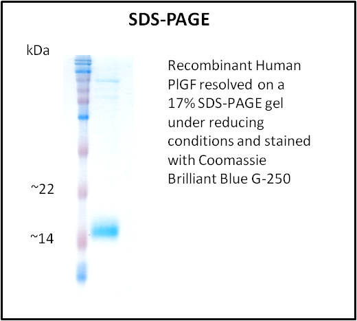
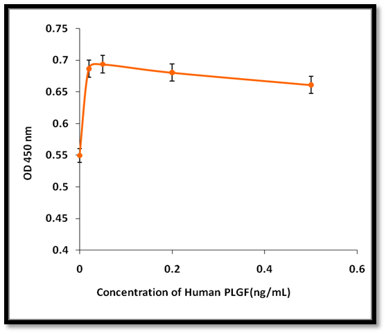
PlGF, Active Protein (Cat# AAA214308)
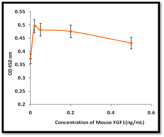 Application Data
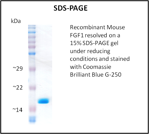
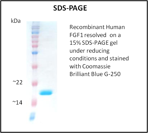
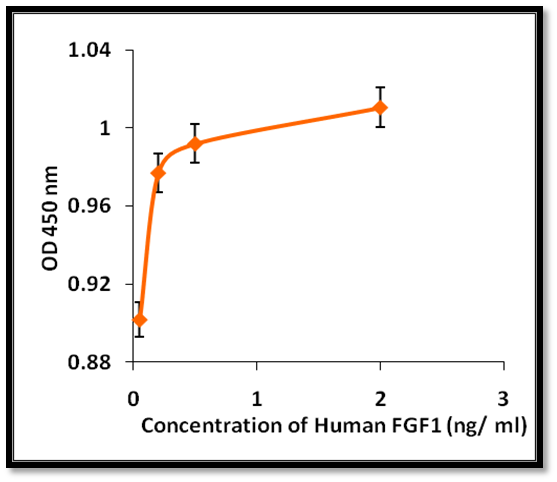
FGF1, Active Protein (Cat# AAA214309)
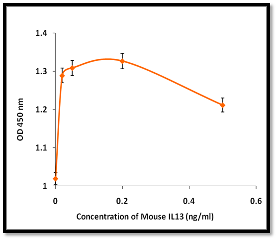 Application Data
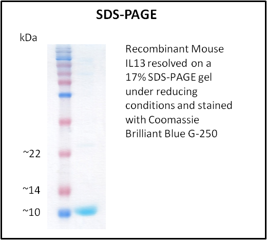
IL13, Active Protein (Cat# AAA214322)
Beta-Defensin 2 (DEFB2), Active Protein (Cat# AAA214326)
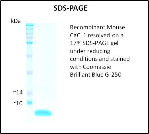
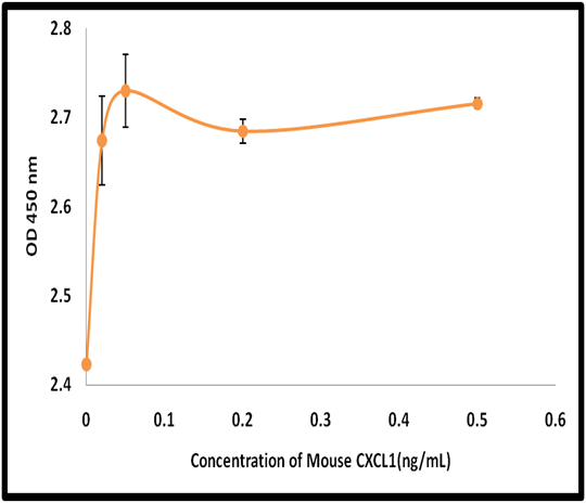 Application Data
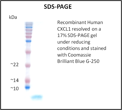
CXCL1, Active Protein (Cat# AAA214328)
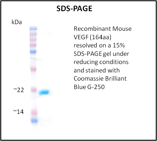
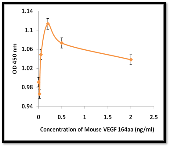 Application Data
VEGF, Active Protein (Cat# AAA214333)
 Application Data
FGF1, Active Protein (Cat# AAA214210)
 Application Data
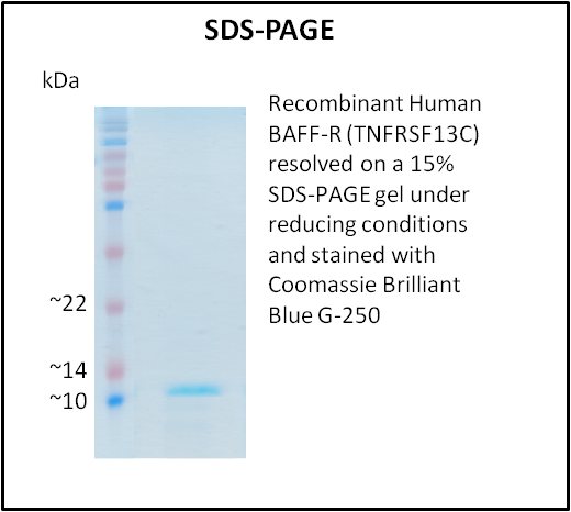
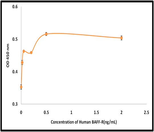
BAFF-R (TNFRSF13C), Active Protein (Cat# AAA214211)
 Application Data
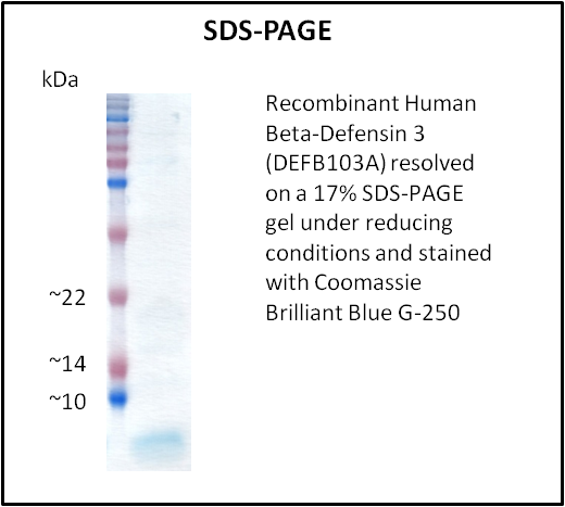
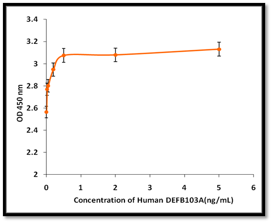
Beta-Defensin 3 (DEFB103A), Active Protein (Cat# AAA214214)
 Application Data
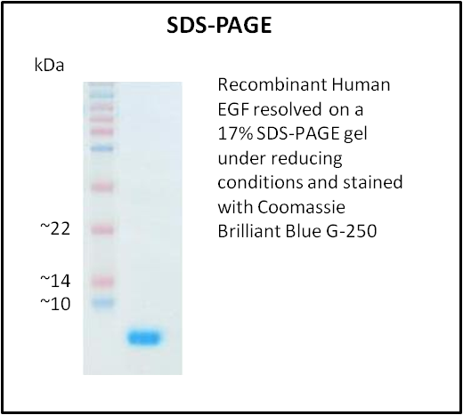
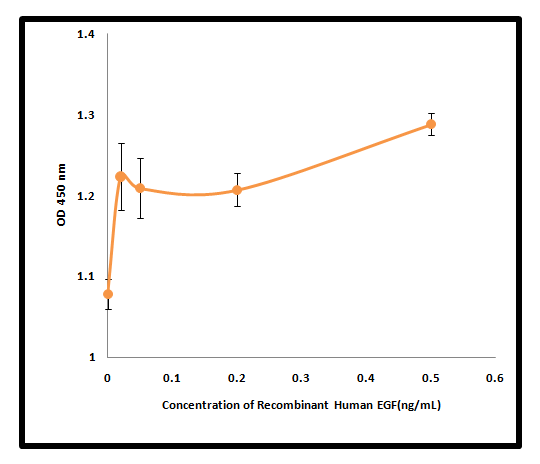
EGF, Active Protein (Cat# AAA214216)

 Application Data
Eotaxin (CCL11), Active Protein (Cat# AAA214217)
 Application Data
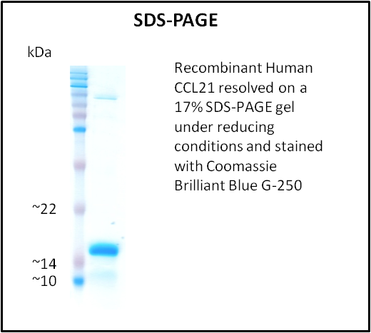
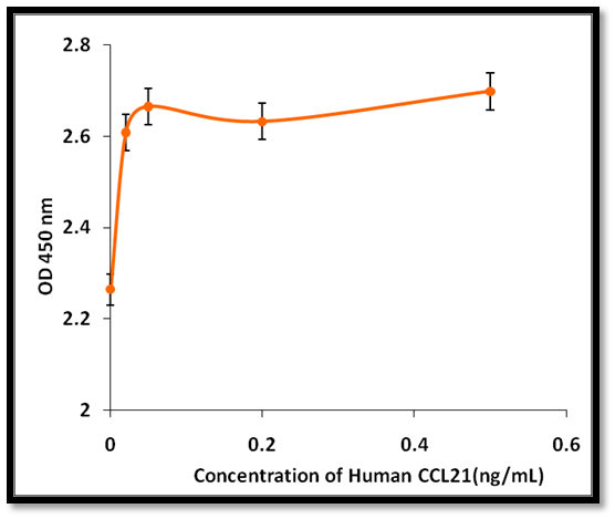
CCL21, Active Protein (Cat# AAA214218)
 Application Data
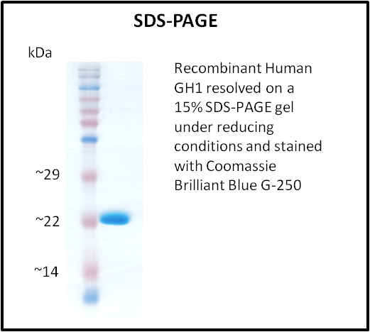
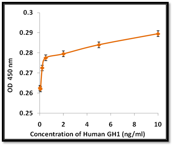
GH1, Active Protein (Cat# AAA214221)
 Application Data
CXCL1, Active Protein (Cat# AAA214223)

 Application Data
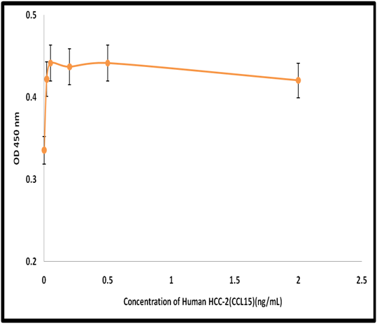
HCC-2 (CCL15), Active Protein (Cat# AAA214226)
 Application Data
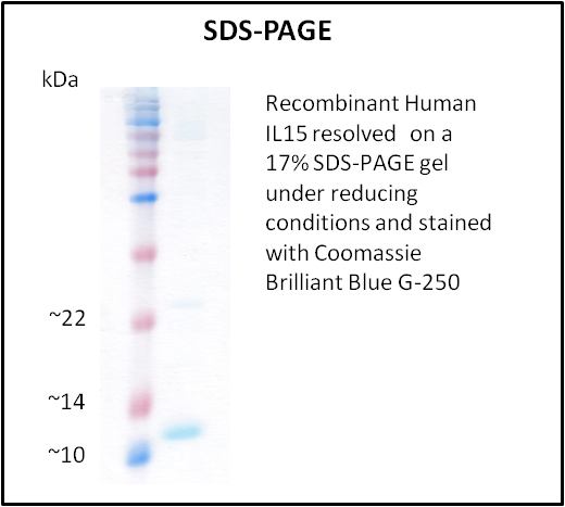
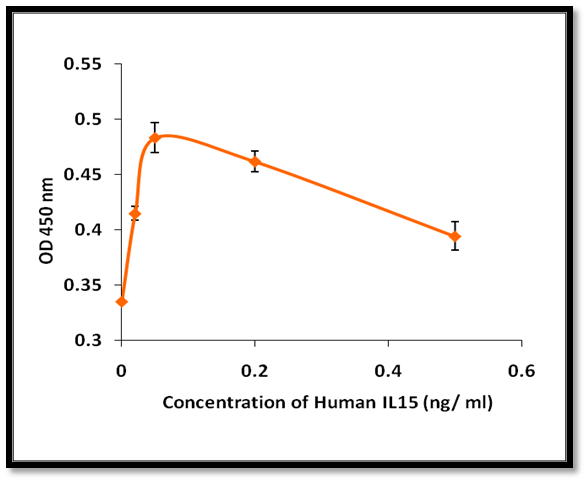
IL15, Active Protein (Cat# AAA214232)
 Application Data
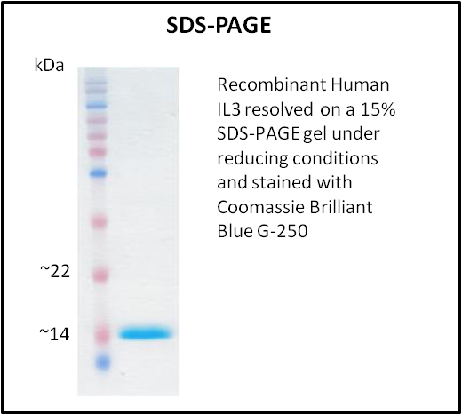
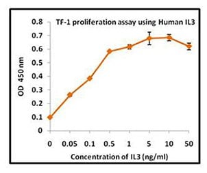
IL3, Active Protein (Cat# AAA214236)
 Application Data
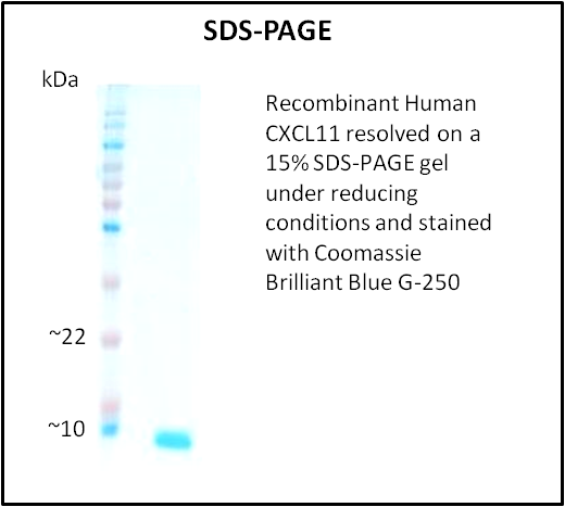
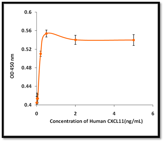
CXCL11, Active Protein (Cat# AAA214238)
 Application Data
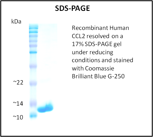
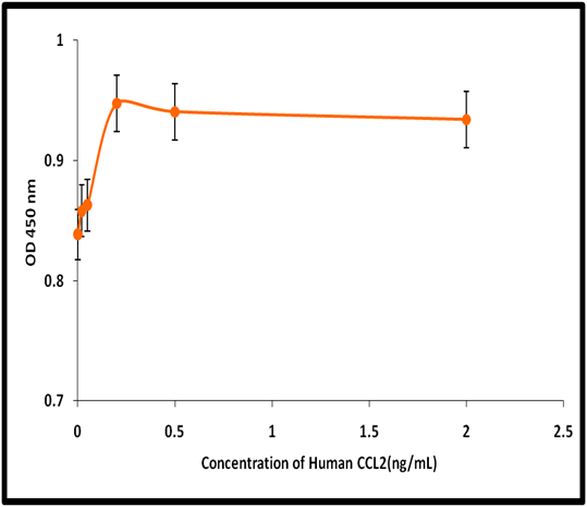
CCL2, Active Protein (Cat# AAA214242)
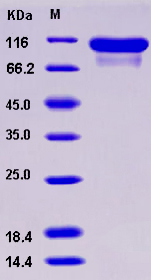
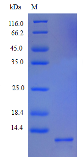
 SDS-PAGE
CD32a / FCGR2A, Active Protein (Cat# AAA173506)
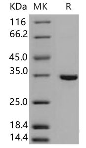
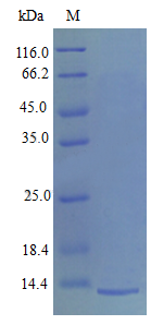
 SDS-PAGE
EGFR / HER1 / ErbB1, Active Protein (Cat# AAA173510)
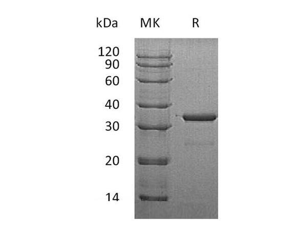
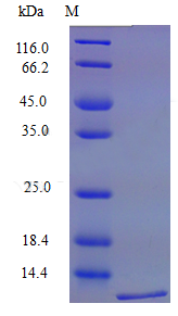
 SDS-PAGE
IL22BP / IL22RA2, Active Protein (Cat# AAA173554)
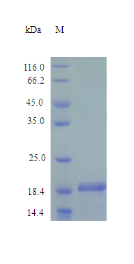
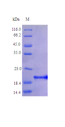
 SDS-PAGE
HER3 / ErbB3, Active Protein (Cat# AAA173572)
 SDS-PAGE
Cytosolic Sulfotransferase Family 1B Member 1, Active Protein (Cat# AAA174591)
 SDS-PAGE
Cytosolic Sulfotransferase Family 1A Member 3, Active Protein (Cat# AAA174592)
 SDS-PAGE
Growth hormone receptor (GHR), Active Protein (Cat# AAA113773)
Glucagon receptor (GCGR), Active Protein (Cat# AAA114042)
 SDS-PAGE
Desert hedgehog protein (DHH), Active Protein (Cat# AAA114231)
 SDS-PAGE
C-X-C motif chemokine 5 (CXCL5), Active Protein (Cat# AAA114232)
 SDS-PAGE
Platelet factor 4 protein (Pf4), Active Protein (Cat# AAA114234)
 SDS-PAGE
C-C motif chemokine 19 protein (Ccl19), Active Protein (Cat# AAA114235)
 SDS-PAGE
Interleukin-6 protein (IL6), Active Protein (Cat# AAA114237)
 SDS-PAGE
Leukemia inhibitory factor protein (LIF), Active Protein (Cat# AAA114239)
 SDS-PAGE
Tumor necrosis factor receptor superfamily member 10B protein (TNFRSF10B), Active Protein (Cat# AAA114240)
 SDS-PAGE
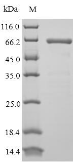
Interferon omega-1 protein (IFNW1), Active Protein (Cat# AAA114245)
Bone morphogenetic protein 2 protein (BMP2), Active Protein (Cat# AAA114247)
 SDS-PAGE
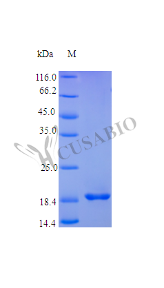
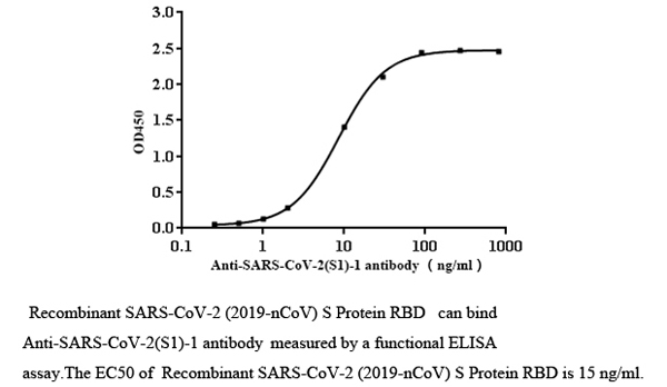
COVID 19 Spike S RBD Coronavirus, Active Protein (Cat# AAA119896)
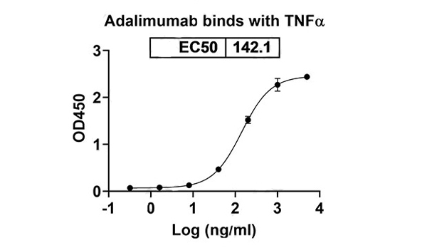 Bioactivity
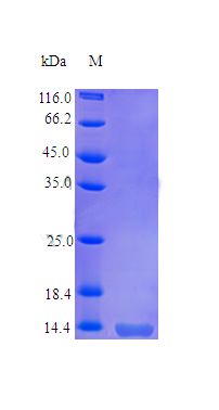
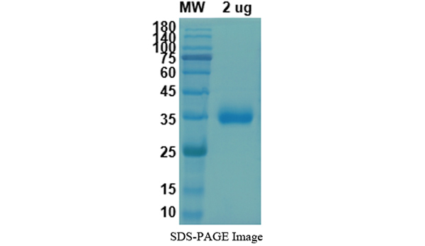
Tnf, Active Protein (Cat# AAA119943)
 SDS-PAGE
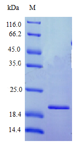
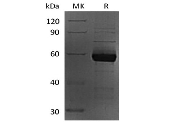
Delta-like protein 3 (DLL3), Active Protein (Cat# AAA117499)
IL-27 EBI3, Active Protein (Cat# AAA178005)
 Bioactivity
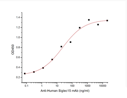
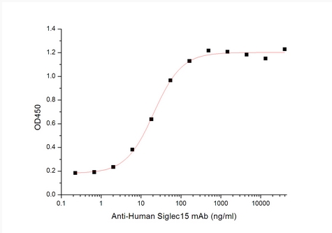
Sialic acid-binding Ig-like lectin 15/Siglec-15/CD33L3, Active Protein (Cat# AAA177934)
 Bioactivity
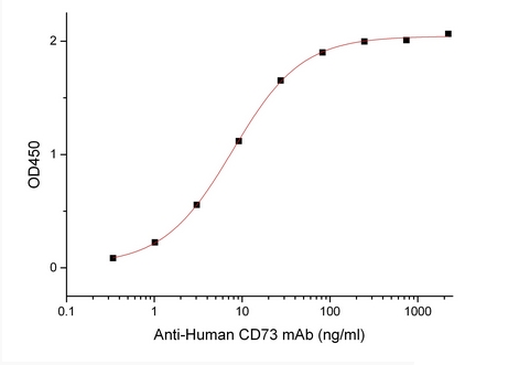
5'-Nucleotidase/5'-NT/CD73, Active Protein (Cat# AAA177936)
 Bioactivity
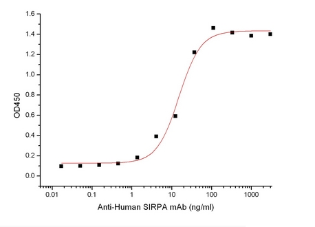
Signal-Regulatory Protein alpha-1/SIRPA/CD172a, Active Protein (Cat# AAA177940)
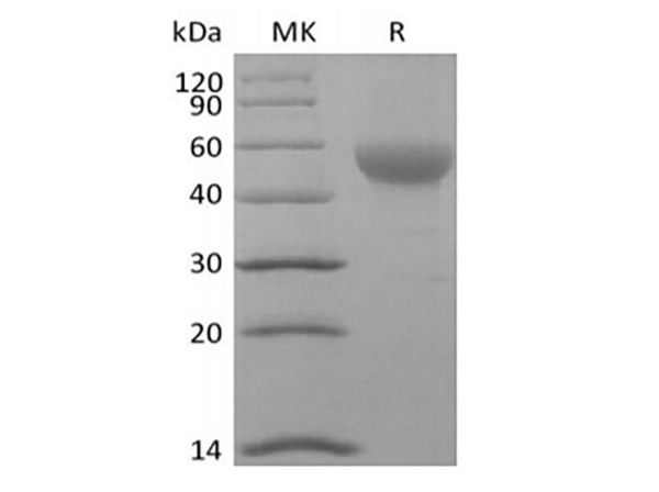
 Bioactivity
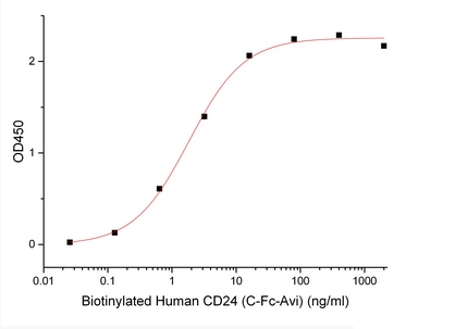
Signal Transducer CD24/CD24 Biotinylated, Active Protein (Cat# AAA177962)
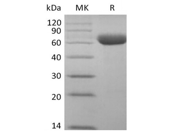
 Bioactivity
Sialic acid-binding Ig-like lectin 15/Siglec-15/CD33L3, Active Protein (Cat# AAA177977)
What Are Active Proteins?
Proteins are large molecules made up of long chains of amino acids.
They will typically fold into a very particular 3-dimensional shape/conformation, that is sometimes referred to as their “native” form, which allows them to work properly in the body. For the purposes of product categorization, AAA Biotech will typically refer to proteins purified from their original animal host as being “native” proteins (this is to signify their difference compared to their “recombinant” or “synthetic” protein counterparts).
If a protein successfully folds into the correct shape, it is will typically display high fidelity characteristics to its original protein in its original animal host, and be classified as an active protein, as it will be able to function “normally” in most enzymatic or binding capacities. If it loses this shape, due to factors such as heat or strong chemicals (such as detergents), it becomes inactive and is no longer able to perform its basic functions. All of the proteins in this category are made under strict quality control, and they are active, pure, low in contaminants, and stable.
Most are stored as freeze-dried powders and come without extra tags, so they’re very close to the actual natural/native form.
Key Applications of Active Proteins
1. Scientific Research
- Aid in the study of how proteins function in the body
- Aid in understanding various disease processes
2. Drug Development
- Powerful tools to investigate how potential drugs interact with specific proteins
- Ideal for identifying drug targets
3. Cell Culture
- Are routinely utilized to support cell growth and function (e.g., using exogenous growth factors)
- Can be used to promote cellular development into specific types (differentiation)
4. Diagnostics
- Regularly utilized in tests to detect diseases or infections (e.g., COVID-19, cancer)
- Note: All products are strictly for research-use only (RUO).
5. Therapeutics
- Some active proteins are used directly as treatments (e.g., insulin, enzymes)
- Note: All products are strictly for research-use only (RUO).
6. Vaccine Development
- Used to create or test vaccines by mimicking parts of viruses or bacteria
7. Biochemical Assays
- They can facilitate the characterization of enzyme activity, binding strength, or protein interactions in lab tests
Why Buy Active Proteins from AAA Biotech?
- High biological activity – Verified to perform as expected or indicated on datasheet
- Strict quality control – We are confident in our active proteins’ reliability and consistency
- High purity & low endotoxin – Ideal for applications involving sensitive or precious samples/components
- Freeze-dried for stability – Long shelf life and straightforward storage
- Mostly tag-free – Closer to natural/native protein form
FAQ
1. What are active proteins used for in research?
Active proteins are used primarily in the study of how proteins function, in characterizing/discovering drug interactions, supporting cell growth, running biochemical assays, and in development of diagnostics or therapeutics.
2. How are AAA Biotech's active proteins validated?
AAA Biotech’s active proteins are validated through strict quality control and functional assays to ensure they are properly folded and active. “Active”, though, can be an ambiguous term, so if a specific “activity” or “binding” capability of a protein is of crucial interest to you, please inquire with us prior to purchase, and we will provide further details on how the “Active” modifier was determined to be applicable.
3. Are these proteins tested for biological activity?
Yes, all active proteins from AAA Biotech are tested to confirm they have the expected biological activity before being offered for use. Though, said “biological activity” can be either “enzymatic”, “binding”, or both.