Filters
▼Clonality
▼Type
▼Reactivity
▼Gene Name
▼Isotype
▼Host
▼Application
▼Clone
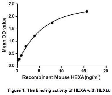
▼Active Proteins
AAA Biotech also known as AAA Bio or AAABio provides a variety of high-quality recombinant and natural/native proteins that are proven to work in a wide range of experiments. Explore our products to find the active protein that best fits your needs or experimental model.
Viewing 300-350 of 2567 product results
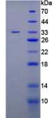
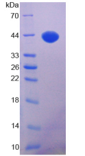
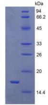
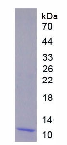 SDS-PAGE
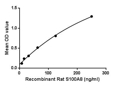
S100 Calcium Binding Protein A8 (S100A8), Active Protein (Cat# AAA149251)
 Bioactivity
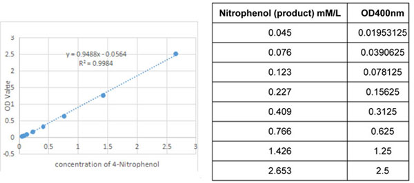
(The standard curve of 4-Nitrophenol.Carboxylesterase 1 (CES1) also known as Liver carboxylesterase 1 is a serine esterase and member of a large multigene carboxylesterase family. The protein Involved in the detoxification of xenobiotics and in the activation of ester and amide prodrugs. Hydrolyzes aromatic and aliphatic esters, but has no catalytic activity toward amides or a fatty acyl-CoA ester. Hydrolyzes the methyl ester group of cocaine to form benzoylecgonine. Thus, the recombinant human CES1 activity was measured by its ability to hydrolyze 4-Nitrophenyl acetate (4-NPA) to 4-Nitrophenol. The reaction was performed in 50mM Tris, pH 7.5 (Assay Buffer), initiated by addition 50uL of various concentrations of CES1 (dilute by assay buffer) to 50uL of 2mM Substrate 4-NPA (100mM stock in Acetone, dilute by deionized water). Incubated at 37 degree C for 10min, then read at a wavelength of 400nm.One unit of enzyme activity is defined as the 1ug of enzyme required to convert 1pmol of 4-Nitrophenyl acetate to 4-Nitrophenol in 1min at 37°C. The specific activity of recombinant human CES1 is 1396 pmol/min/ug.Specific Activity (pmol/min/ug) = ΔOD*F/T*NΔOD=Adjusted for Substrate BlankF=Conversion Factor(convert from standard curve of 4-Nitrophenol)T=TimeN=Amount of enzyme)
Bioactivity
(The standard curve of 4-Nitrophenol.Carboxylesterase 1 (CES1) also known as Liver carboxylesterase 1 is a serine esterase and member of a large multigene carboxylesterase family. The protein Involved in the detoxification of xenobiotics and in the activation of ester and amide prodrugs. Hydrolyzes aromatic and aliphatic esters, but has no catalytic activity toward amides or a fatty acyl-CoA ester. Hydrolyzes the methyl ester group of cocaine to form benzoylecgonine. Thus, the recombinant human CES1 activity was measured by its ability to hydrolyze 4-Nitrophenyl acetate (4-NPA) to 4-Nitrophenol. The reaction was performed in 50mM Tris, pH 7.5 (Assay Buffer), initiated by addition 50uL of various concentrations of CES1 (dilute by assay buffer) to 50uL of 2mM Substrate 4-NPA (100mM stock in Acetone, dilute by deionized water). Incubated at 37 degree C for 10min, then read at a wavelength of 400nm.One unit of enzyme activity is defined as the 1ug of enzyme required to convert 1pmol of 4-Nitrophenyl acetate to 4-Nitrophenol in 1min at 37°C. The specific activity of recombinant human CES1 is 1396 pmol/min/ug.Specific Activity (pmol/min/ug) = ΔOD*F/T*NΔOD=Adjusted for Substrate BlankF=Conversion Factor(convert from standard curve of 4-Nitrophenol)T=TimeN=Amount of enzyme)
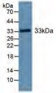
Carboxylesterase 1 (CES1), Active Protein (Cat# AAA149260)
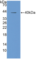
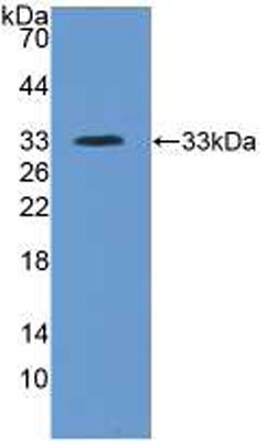 WB (Western Blot)
(Sample: Recombinant ITIH4, Human;Antibody: Rabbit Anti-Human ITIH4 Ab)
WB (Western Blot)
(Sample: Recombinant ITIH4, Human;Antibody: Rabbit Anti-Human ITIH4 Ab)
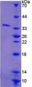
Inter Alpha-Globulin Inhibitor H4 (ITIH4), Active Protein (Cat# AAA149264)


 Bioactivity
(Figure. The binding activity of IFN2 with IFNa/bR2.)
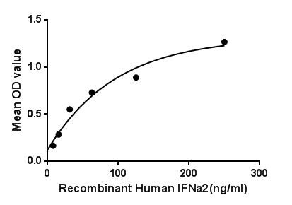
Bioactivity
(Figure. The binding activity of IFN2 with IFNa/bR2.)
Interferon Alpha 2, Active Protein (Cat# AAA150081)

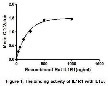
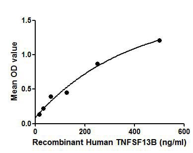 Bioactivity
(Figure. The binding activity of TNFSF13B with ITGb1.Tumor necrosis factor ligand superfamily member 13B protein (TNFSF13B) also known as B-cell activating factor (BAFF) is a cytokine that belongs to the tumor necrosis factor (TNF) ligand family. This cytokine is a ligand for receptors TNFRSF13B/TACI, TNFRSF17/BCMA, and TNFRSF13C/BAFF-R. This cytokine is expressed in B cell lineage cells, and acts as a potent B cell activator. It has been also shown to play an important role in the proliferation and differentiation of B cells. Besides, Integrin Beta 1 (ITGb1) has been identified as an interactor of TNFSF13B, thus a binding ELISA assay was conducted to detect the interaction of recombinant human TNFSF13B and recombinant human ITGb1. Briefly, TNFSF13B were diluted serially in PBS, with 0.01% BSA (pH 7.4). Duplicate samples of 100uL were then transferred to ITGb1-coated microtiter wells and incubated for 2h at 37. Wells were washed with PBST and incubated for 1h with anti-TNFSF13B pAb, then aspirated and washed 3 times. After incubation with HRP labelled secondary antibody, wells were aspirated and washed 3 times. With the addition of substrate solution, wells were incubated 15-25 minutes at 37. Finally, add 50uL stop solution to the wells and read at 450nm immediately. The binding activity of TNFSF13B and ITGb1 was shown in Figure 1, and this effect was in a dose dependent manner.)
Bioactivity
(Figure. The binding activity of TNFSF13B with ITGb1.Tumor necrosis factor ligand superfamily member 13B protein (TNFSF13B) also known as B-cell activating factor (BAFF) is a cytokine that belongs to the tumor necrosis factor (TNF) ligand family. This cytokine is a ligand for receptors TNFRSF13B/TACI, TNFRSF17/BCMA, and TNFRSF13C/BAFF-R. This cytokine is expressed in B cell lineage cells, and acts as a potent B cell activator. It has been also shown to play an important role in the proliferation and differentiation of B cells. Besides, Integrin Beta 1 (ITGb1) has been identified as an interactor of TNFSF13B, thus a binding ELISA assay was conducted to detect the interaction of recombinant human TNFSF13B and recombinant human ITGb1. Briefly, TNFSF13B were diluted serially in PBS, with 0.01% BSA (pH 7.4). Duplicate samples of 100uL were then transferred to ITGb1-coated microtiter wells and incubated for 2h at 37. Wells were washed with PBST and incubated for 1h with anti-TNFSF13B pAb, then aspirated and washed 3 times. After incubation with HRP labelled secondary antibody, wells were aspirated and washed 3 times. With the addition of substrate solution, wells were incubated 15-25 minutes at 37. Finally, add 50uL stop solution to the wells and read at 450nm immediately. The binding activity of TNFSF13B and ITGb1 was shown in Figure 1, and this effect was in a dose dependent manner.)
B-Cell Activating Factor, Active Protein (Cat# AAA150113)
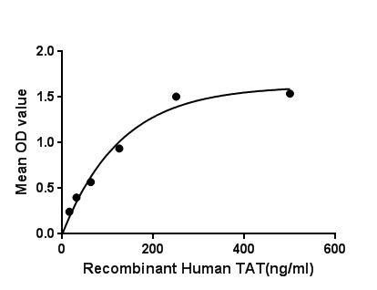 Bioactivity
(Figure. The binding activity of TAT with HSPA8)
Bioactivity
(Figure. The binding activity of TAT with HSPA8)
Tyrosine Aminotransferase, Active Protein (Cat# AAA150132)
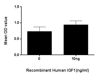 Bioactivity
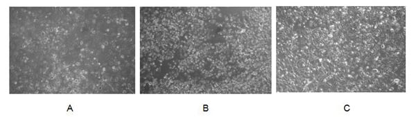
(Figure. Cell proliferation of MCF-7 cells after stimulated with IGF1.)
Bioactivity
(Figure. Cell proliferation of MCF-7 cells after stimulated with IGF1.)
Insulin Like Growth Factor 1, Active Protein (Cat# AAA150053)
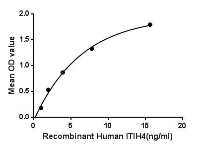
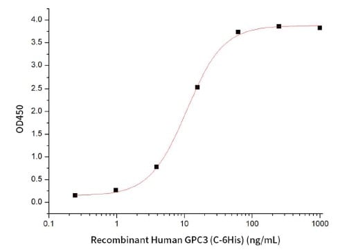 Bioactivity
Glypican 3 (GPC3), Active Protein (Cat# AAA190412)
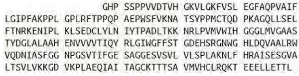
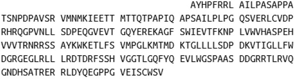
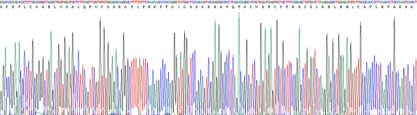
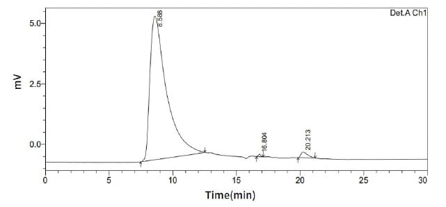
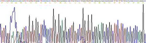
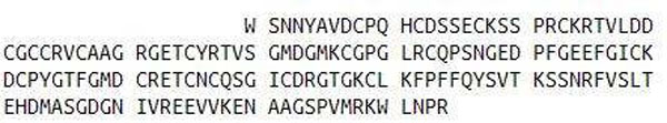
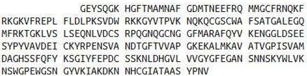
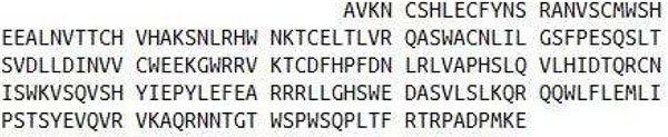
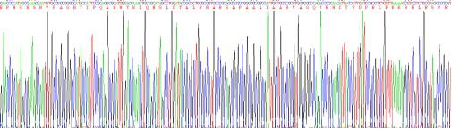
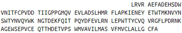
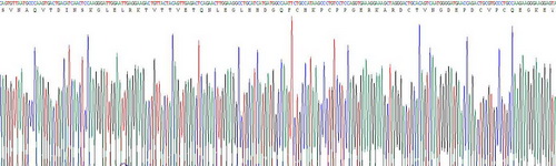
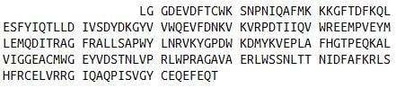 Sequence
(Figure 4. Sequence)
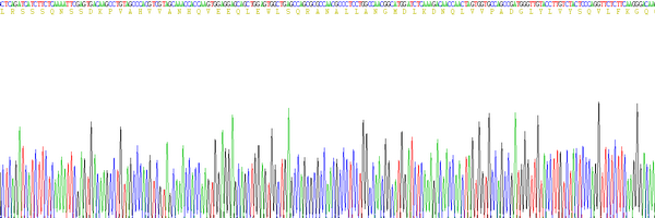
Sequence
(Figure 4. Sequence)
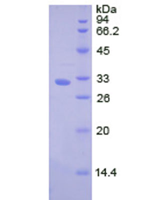
Hexosaminidase A Alpha (HEXa), Active Protein (Cat# AAA148166)

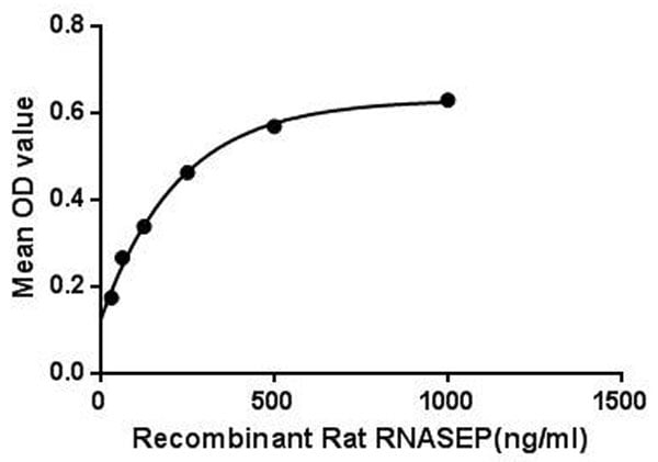

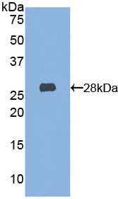
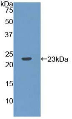 WB (Western Blot)
(Sample: Recombinant RNASEP, Rat;Antibody: Rabbit Anti-Rat RNASEP Ab)
WB (Western Blot)
(Sample: Recombinant RNASEP, Rat;Antibody: Rabbit Anti-Rat RNASEP Ab)
Ribonuclease P (RNASEP), Active Protein (Cat# AAA148167)
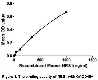
 Application Data
Nesfatin 1 (NES1), Active Protein (Cat# AAA148169)
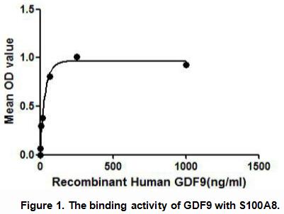
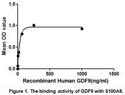 Application Data
Growth Differentiation Factor 9 (GDF9), Active Protein (Cat# AAA148170)
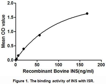

 Application Data
Insulin (INS), Active Protein (Cat# AAA148171)
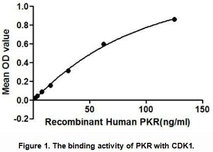

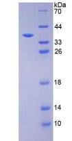
 Application Data
Protein Kinase R (PKR), Active Protein (Cat# AAA148174)
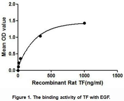
 Application Data
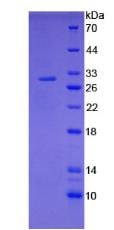
Tissue Factor (TF), Active Protein (Cat# AAA148175)
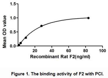
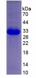
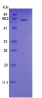
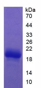 SDS-PAGE
(Sample: Active recombinant F2, Human)
SDS-PAGE
(Sample: Active recombinant F2, Human)
Coagulation Factor II (F2), Active Protein (Cat# AAA148183)
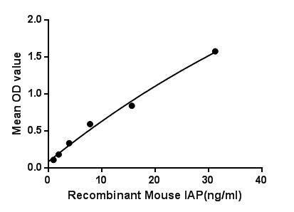
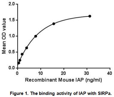 Application Data
Integrin Associated Protein (IAP), Active Protein (Cat# AAA148184)

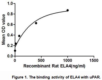 Application Data
Elastase 4 (ELA4), Active Protein (Cat# AAA148191)
 Application Data
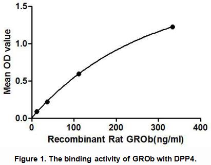
Growth Regulated Oncogene Beta (GROb), Active Protein (Cat# AAA148193)
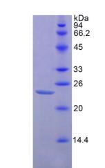
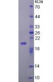
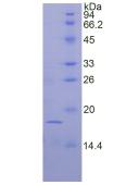
 SDS_PAGE
(Sample: Active recombinant ESM1, Human)
SDS_PAGE
(Sample: Active recombinant ESM1, Human)
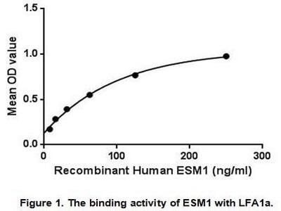
Endothelial Cell Specific Molecule 1 (ESM1), Active Protein (Cat# AAA148202)
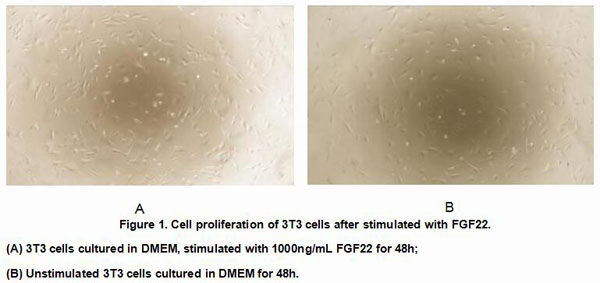

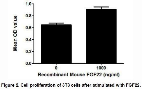 Application Data
Fibroblast Growth Factor 22 (FGF22), Active Protein (Cat# AAA148204)

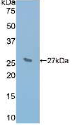
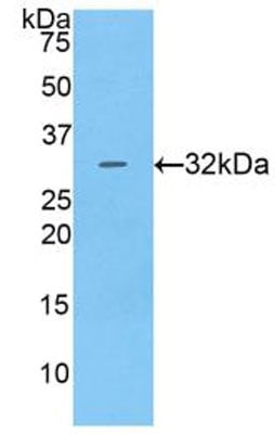 WB (Western Blot)
(Figure 3. Western BlotSample: Recombinant CTSV, Human;Antibody: Rabbit Anti-Human CTSV Ab)
WB (Western Blot)
(Figure 3. Western BlotSample: Recombinant CTSV, Human;Antibody: Rabbit Anti-Human CTSV Ab)
Cathepsin V (CTSV), Active Protein (Cat# AAA148206)
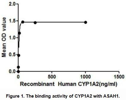

 Application Data
Cytochrome P450 1A2 (CYP1A2), Active Protein (Cat# AAA148207)
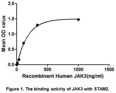

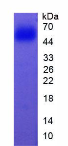
 Application Data
Janus Kinase 3 (JAK3), Active Protein (Cat# AAA148213)
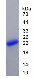
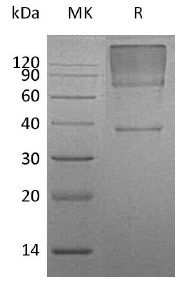
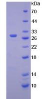
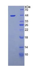
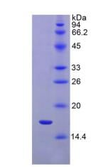
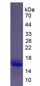
 SDS-PAGE
Interleukin 1 Alpha (IL1a), Active Protein (Cat# AAA148220)
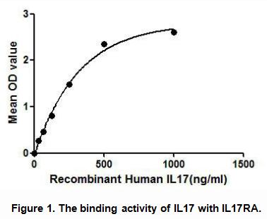
 SDS-PAGE
Interleukin 17 (IL17), Active Protein (Cat# AAA148224)
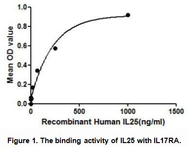
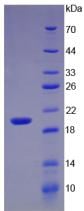
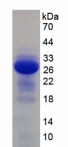
 SDS-PAGE
Interleukin 25 (IL25), Active Protein (Cat# AAA148228)
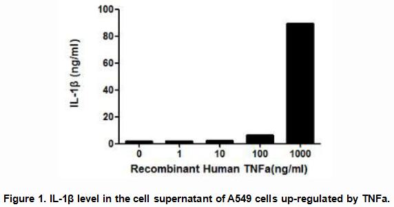
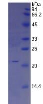
 SDS-PAGE
Tumor Necrosis Factor Alpha (TNFa), Active Protein (Cat# AAA148231)
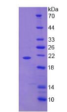
 SDS-PAGE
Interleukin 1 Receptor Type I (IL1R1), Active Protein (Cat# AAA148236)
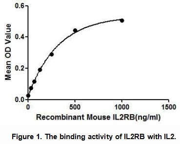
 WB (Western Blot)
(Sample: Recombinant IL2Rb, Mouse;Antibody: Rabbit Anti-Mouse IL2Rb Ab)
WB (Western Blot)
(Sample: Recombinant IL2Rb, Mouse;Antibody: Rabbit Anti-Mouse IL2Rb Ab)
Interleukin 2 Receptor Beta (IL2Rb), Active Protein (Cat# AAA148239)
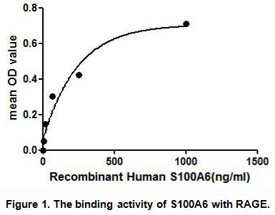
 SDS-PAGE
S100 Calcium Binding Protein A6 (S100A6), Active Protein (Cat# AAA148243)
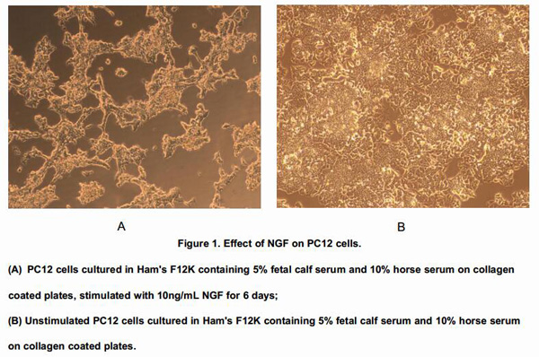

 Application Data
Nerve Growth Factor (NGF), Active Protein (Cat# AAA146613)
 Application Data
Tumor Necrosis Factor Alpha (TNFa), Active Protein (Cat# AAA146614)
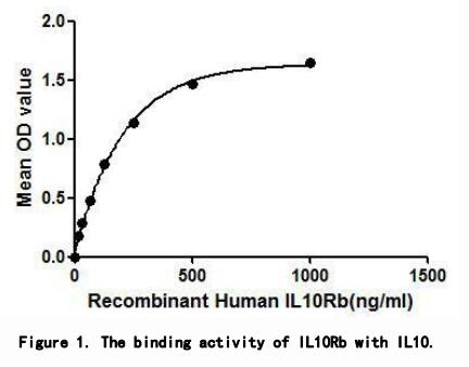
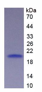
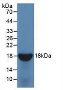 WB (Western Blot)
(Sample: Recombinant IL10Rb, Human;Antibody: Rabbit Anti-Human IL10Rb Ab)
WB (Western Blot)
(Sample: Recombinant IL10Rb, Human;Antibody: Rabbit Anti-Human IL10Rb Ab)
Interleukin 10 Receptor Beta (IL10Rb), Active Protein (Cat# AAA148125)

 Application Data
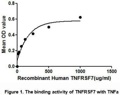
Tumor Necrosis Factor Receptor Superfamily, Member 7 (TNFRSF7), Active Protein (Cat# AAA148126)

 Application Data
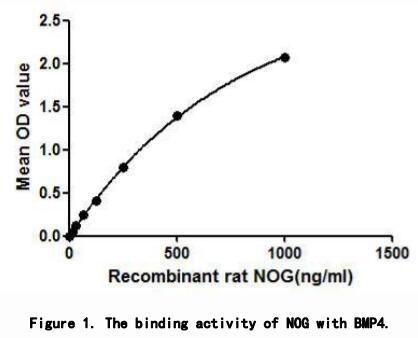
Noggin (NOG), Active Protein (Cat# AAA148132)

 Application Data
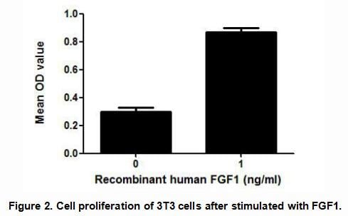
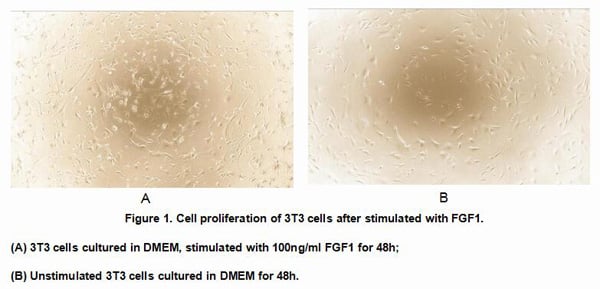
Fibroblast Growth Factor 1, Acidic (FGF1), Active Protein (Cat# AAA148139)
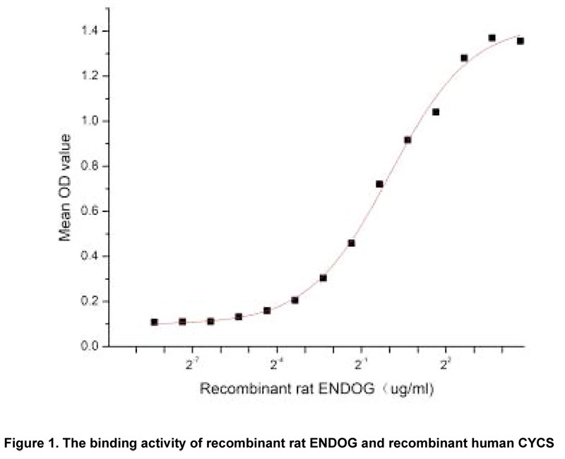
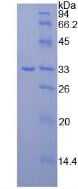
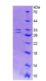
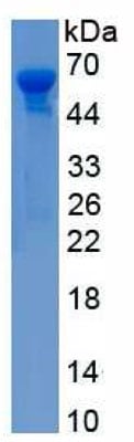 SDS_PAGE
(Sample: Active recombinant ENDOG, Rat)
SDS_PAGE
(Sample: Active recombinant ENDOG, Rat)
Endonuclease G, Mitochondrial (ENDOG), Active Protein (Cat# AAA148142)
 Application Data
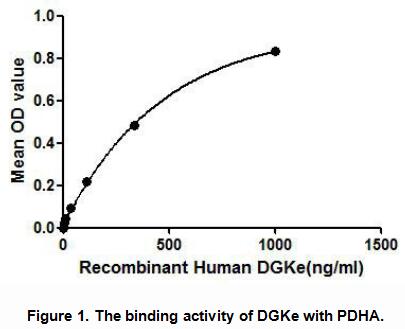
Diacylglycerol Kinase Epsilon (DGKe), Active Protein (Cat# AAA148145)
 Application Data
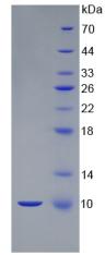
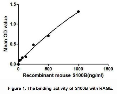
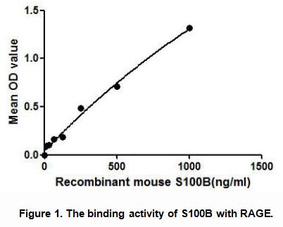
S100 Calcium Binding Protein B (S100B), Active Protein (Cat# AAA148149)

 Application Data
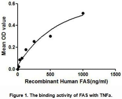
Factor Related Apoptosis (FAS), Active Protein (Cat# AAA148153)

 Application Data
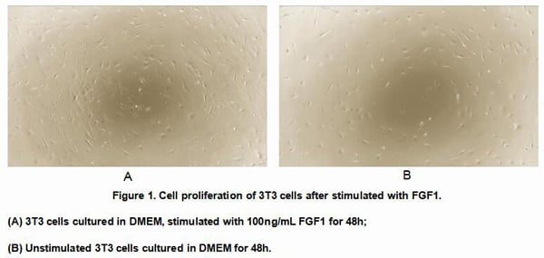
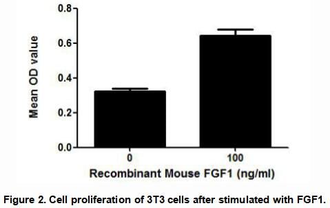
Fibroblast Growth Factor 1, Acidic (FGF1), Active Protein (Cat# AAA148154)
 Application Data
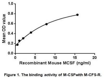
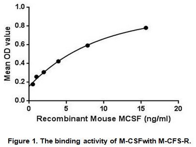
Colony Stimulating Factor 1, Macrophage (MCSF), Active Protein (Cat# AAA148162)
 Application Data
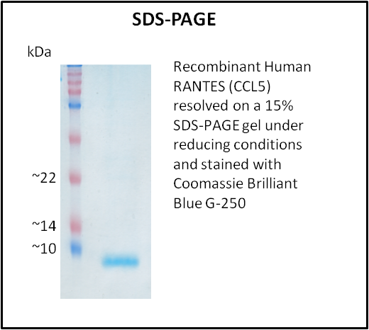
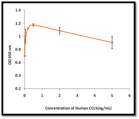
RANTES (CCL5), Active Protein (Cat# AAA214251)
 Application Data
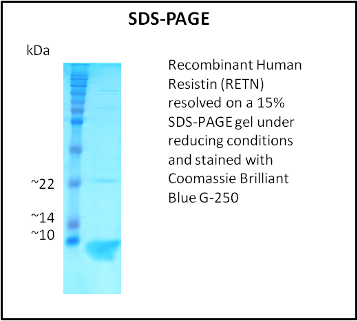
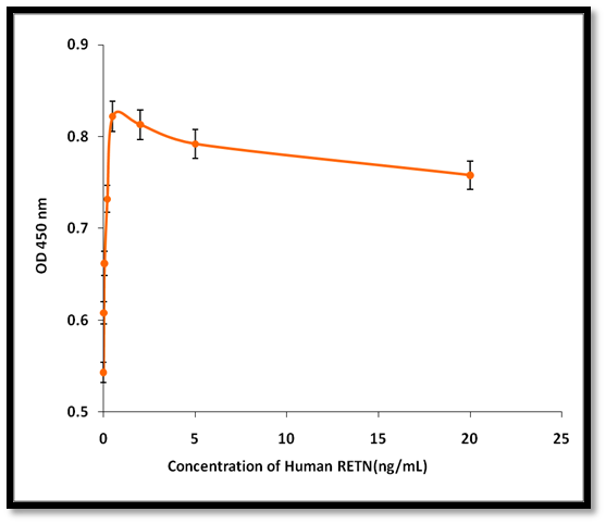
Resistin (RETN), Active Protein (Cat# AAA214252)
 Application Data
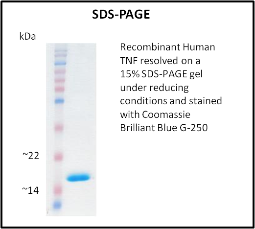
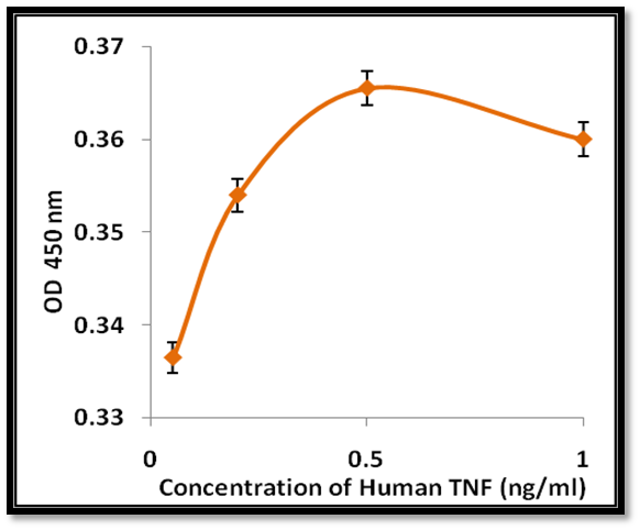
TNF, Active Protein (Cat# AAA214253)
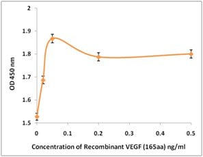 Application Data
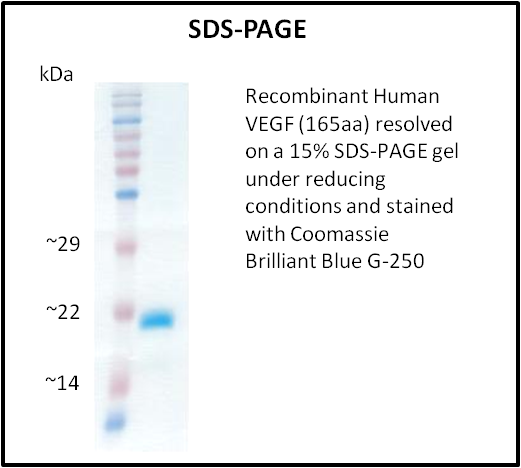
VEGF, Active Protein (Cat# AAA214255)
 Application Data
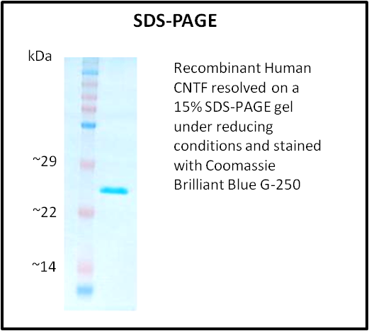
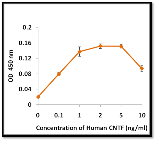
CNTF, Active Protein (Cat# AAA214258)
 Application Data
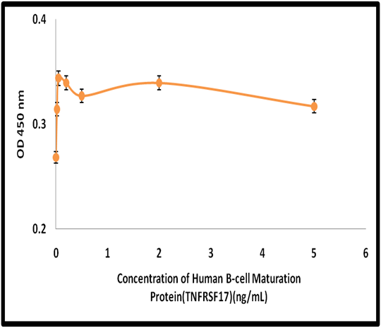
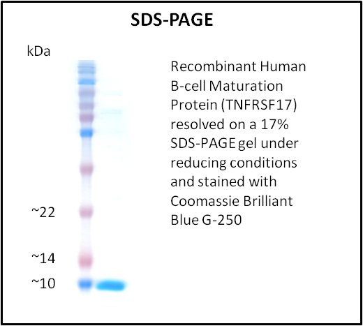
B-Cell Maturation (TNFRSF17), Active Protein (Cat# AAA214266)
 Application Data
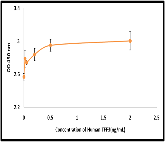
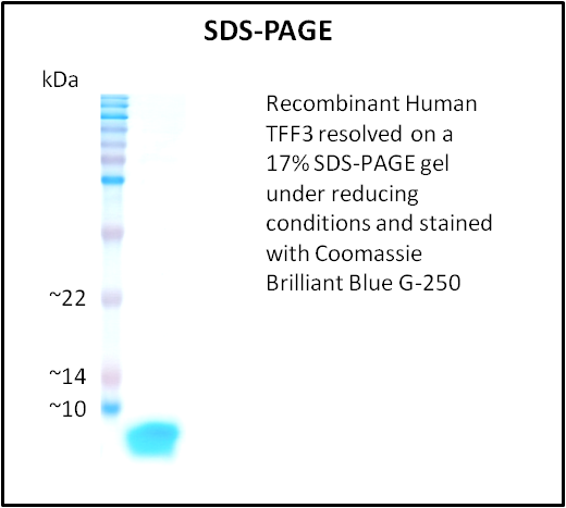
TFF3, Active Protein (Cat# AAA214283)
What Are Active Proteins?
Proteins are large molecules made up of long chains of amino acids.
They will typically fold into a very particular 3-dimensional shape/conformation, that is sometimes referred to as their “native” form, which allows them to work properly in the body. For the purposes of product categorization, AAA Biotech will typically refer to proteins purified from their original animal host as being “native” proteins (this is to signify their difference compared to their “recombinant” or “synthetic” protein counterparts).
If a protein successfully folds into the correct shape, it is will typically display high fidelity characteristics to its original protein in its original animal host, and be classified as an active protein, as it will be able to function “normally” in most enzymatic or binding capacities. If it loses this shape, due to factors such as heat or strong chemicals (such as detergents), it becomes inactive and is no longer able to perform its basic functions. All of the proteins in this category are made under strict quality control, and they are active, pure, low in contaminants, and stable.
Most are stored as freeze-dried powders and come without extra tags, so they’re very close to the actual natural/native form.
Key Applications of Active Proteins
1. Scientific Research
- Aid in the study of how proteins function in the body
- Aid in understanding various disease processes
2. Drug Development
- Powerful tools to investigate how potential drugs interact with specific proteins
- Ideal for identifying drug targets
3. Cell Culture
- Are routinely utilized to support cell growth and function (e.g., using exogenous growth factors)
- Can be used to promote cellular development into specific types (differentiation)
4. Diagnostics
- Regularly utilized in tests to detect diseases or infections (e.g., COVID-19, cancer)
- Note: All products are strictly for research-use only (RUO).
5. Therapeutics
- Some active proteins are used directly as treatments (e.g., insulin, enzymes)
- Note: All products are strictly for research-use only (RUO).
6. Vaccine Development
- Used to create or test vaccines by mimicking parts of viruses or bacteria
7. Biochemical Assays
- They can facilitate the characterization of enzyme activity, binding strength, or protein interactions in lab tests
Why Buy Active Proteins from AAA Biotech?
- High biological activity – Verified to perform as expected or indicated on datasheet
- Strict quality control – We are confident in our active proteins’ reliability and consistency
- High purity & low endotoxin – Ideal for applications involving sensitive or precious samples/components
- Freeze-dried for stability – Long shelf life and straightforward storage
- Mostly tag-free – Closer to natural/native protein form
FAQ
1. What are active proteins used for in research?
Active proteins are used primarily in the study of how proteins function, in characterizing/discovering drug interactions, supporting cell growth, running biochemical assays, and in development of diagnostics or therapeutics.
2. How are AAA Biotech's active proteins validated?
AAA Biotech’s active proteins are validated through strict quality control and functional assays to ensure they are properly folded and active. “Active”, though, can be an ambiguous term, so if a specific “activity” or “binding” capability of a protein is of crucial interest to you, please inquire with us prior to purchase, and we will provide further details on how the “Active” modifier was determined to be applicable.
3. Are these proteins tested for biological activity?
Yes, all active proteins from AAA Biotech are tested to confirm they have the expected biological activity before being offered for use. Though, said “biological activity” can be either “enzymatic”, “binding”, or both.