Filters
▼Clonality
▼Type
▼Reactivity
▼Gene Name
▼Isotype
▼Host
▼Application
▼Clone
▼Active Proteins
AAA Biotech also known as AAA Bio or AAABio provides a variety of high-quality recombinant and natural/native proteins that are proven to work in a wide range of experiments. Explore our products to find the active protein that best fits your needs or experimental model.
Viewing 500-550 of 2567 product results
 WB (Western Blot)
(Sample: Recombinant TNFRSF12A, Human; Antibody: Rabbit Anti-Human TNFRSF12A Ab)
WB (Western Blot)
(Sample: Recombinant TNFRSF12A, Human; Antibody: Rabbit Anti-Human TNFRSF12A Ab)
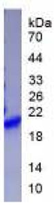
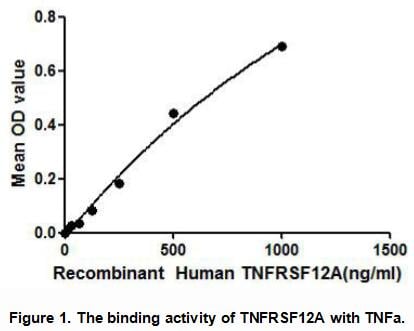
Tumor Necrosis Factor Receptor Superfamily, Member 12A (TNFRSF12A), Active Protein (Cat# AAA148180)
 Application Data
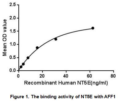
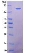
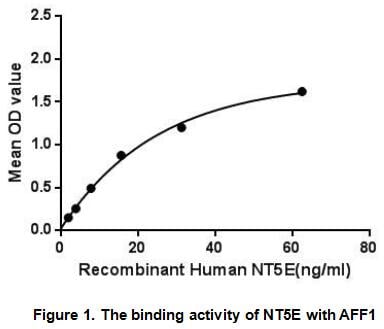
5'-Nucleotidase, Ecto (NT5E), Active Protein (Cat# AAA148190)
 Application Data
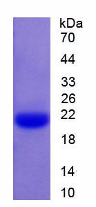
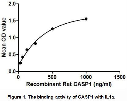
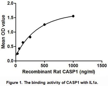
Caspase 1 (CASP1), Active Protein (Cat# AAA148192)
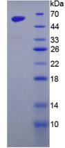
 Application Data
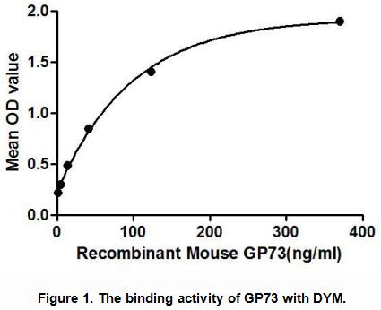
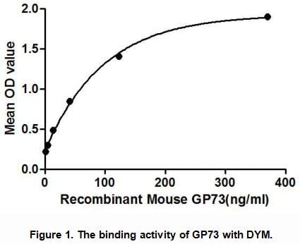
Golgi Protein 73 (GP73), Active Protein (Cat# AAA148195)

 Application Data
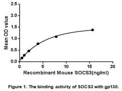
Suppressors Of Cytokine Signaling 3 (SOCS3), Active Protein (Cat# AAA148196)
 Application Data
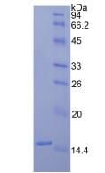
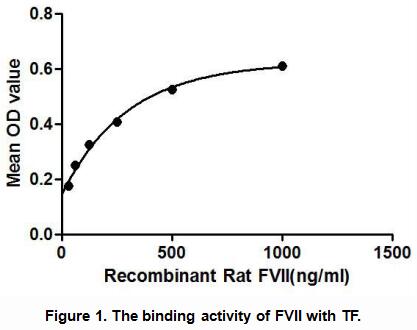
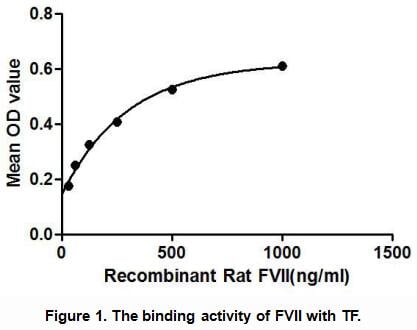
Coagulation Factor VII (F7), Active Protein (Cat# AAA148198)
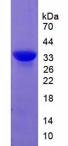
 Application Data
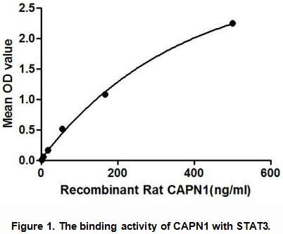
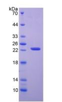
Calpain 1, Large Subunit (CAPN1), Active Protein (Cat# AAA148200)

 Application Data
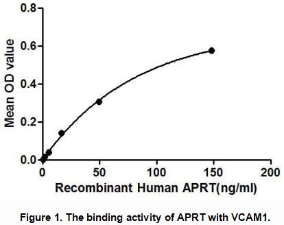
Adenine Phosphoribosyltransferase (APRT), Active Protein (Cat# AAA148201)
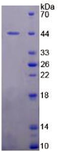
 WB (Western Blot)
(Western BlotSample: Recombinant MDM2, Human;Antibody: Rabbit Anti-Human MDM2 Ab)
WB (Western Blot)
(Western BlotSample: Recombinant MDM2, Human;Antibody: Rabbit Anti-Human MDM2 Ab)
Mdm2 p53 Binding Protein Homolog (MDM2), Active Protein (Cat# AAA148216)


 Application Data
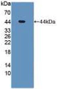
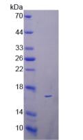
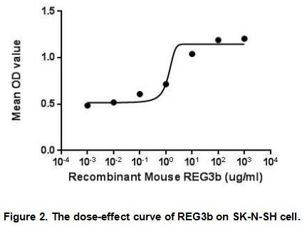
Regenerating Islet Derived Protein 3 Beta (REG3b), Active Protein (Cat# AAA148217)
 WB (Western Blot)
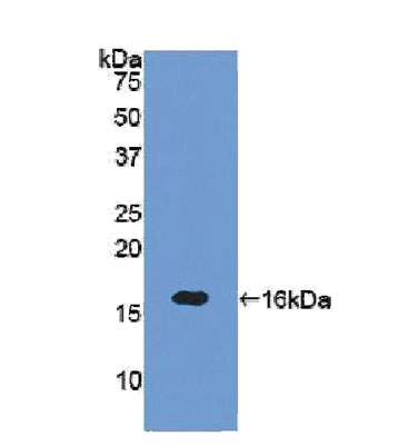
(Figure 4. Western BlotSample: Recombinant SEMA3A, Human;Antibody: Rabbit Anti-Human SEMA3A Ab)
WB (Western Blot)
(Figure 4. Western BlotSample: Recombinant SEMA3A, Human;Antibody: Rabbit Anti-Human SEMA3A Ab)
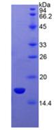
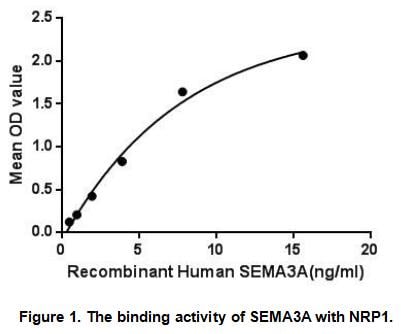
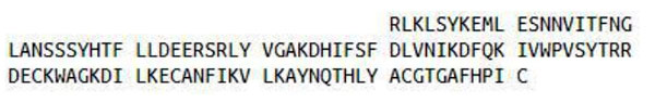
Semaphorin 3A (SEMA3A), Active Protein (Cat# AAA148218)

 Application Data
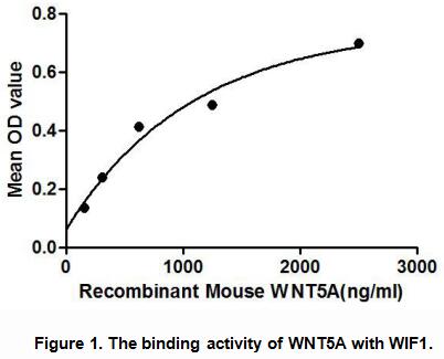
Wingless Type MMTV Integration Site Family, Member 5A (WNT5A), Active Protein (Cat# AAA148219)
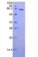
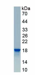
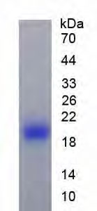
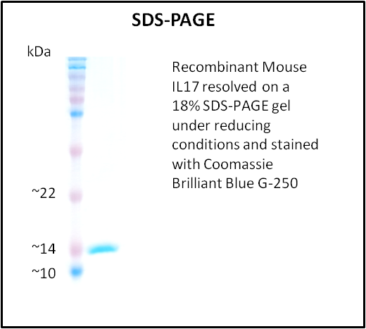
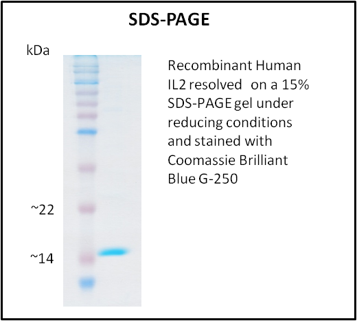
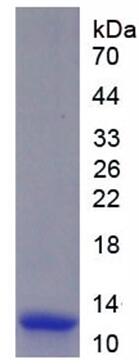
 SDS-PAGE
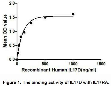
Interleukin 17D (IL17D), Active Protein (Cat# AAA148227)
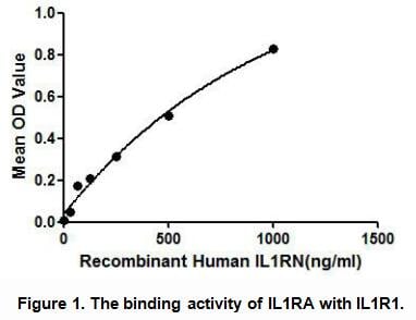
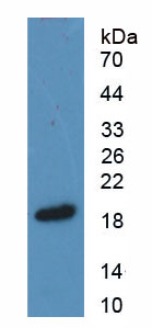
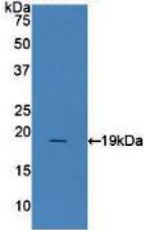 WB (Western Blot)
(Sample: Recombinant IL1RA, Human;Antibody: Rabbit Anti-Human IL1RA Ab)
WB (Western Blot)
(Sample: Recombinant IL1RA, Human;Antibody: Rabbit Anti-Human IL1RA Ab)
Interleukin 1 Receptor Antagonist (IL1RA), Active Protein (Cat# AAA148229)
 WB (Western Blot)
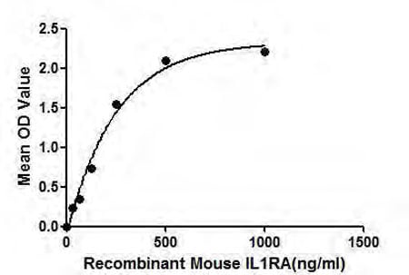
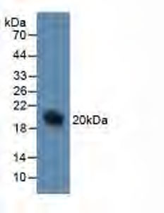
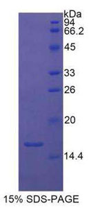
(Figure 4. Western BlotSample: Recombinant IL1RA, Mouse; Antibody: Rabbit Anti-Mouse IL1RA Ab)
WB (Western Blot)
(Figure 4. Western BlotSample: Recombinant IL1RA, Mouse; Antibody: Rabbit Anti-Mouse IL1RA Ab)
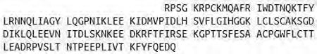
Interleukin 1 Receptor Antagonist (IL1RA), Active Protein (Cat# AAA148230)
 WB (Western Blot)
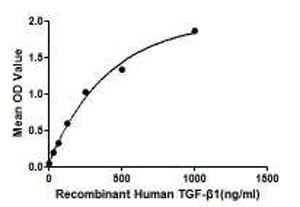
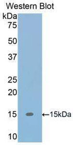
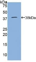
(Figure 4. Western Blot Sample: Recombinant TGF-Beta1, Human; Antibody: Rabbit Anti8Human TGF-Beta1 Ab)
WB (Western Blot)
(Figure 4. Western Blot Sample: Recombinant TGF-Beta1, Human; Antibody: Rabbit Anti8Human TGF-Beta1 Ab)
Transforming Growth Factor Beta 1 (TGFb1), Active Protein (Cat# AAA148233)
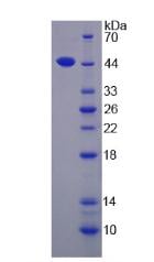
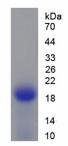
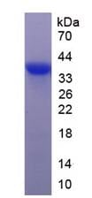
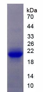 SDS-PAGE
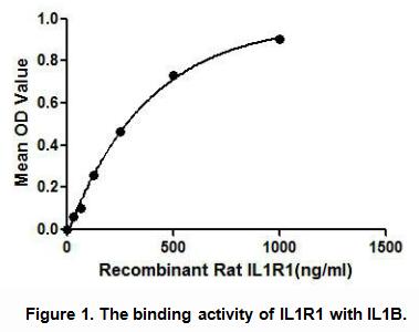
Interleukin 1 Receptor Type I (IL1R1), Active Protein (Cat# AAA148237)

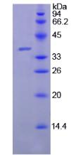
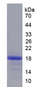 SDS-PAGE
Epidermal Growth Factor (EGF), Active Protein (Cat# AAA148240)
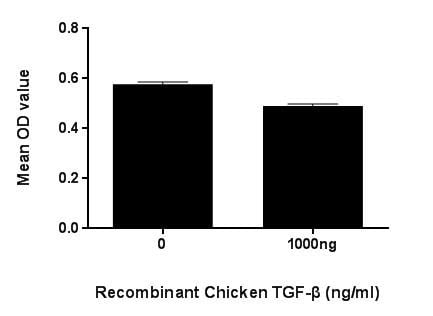 Application Data
(Figure. TGF- inhibit cell proliferation of HepG2 cells.)
Application Data
(Figure. TGF- inhibit cell proliferation of HepG2 cells.)
Transforming Growth Factor Beta 2, Active Protein (Cat# AAA150083)
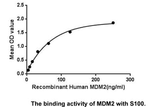
 Bioactivity
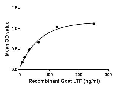
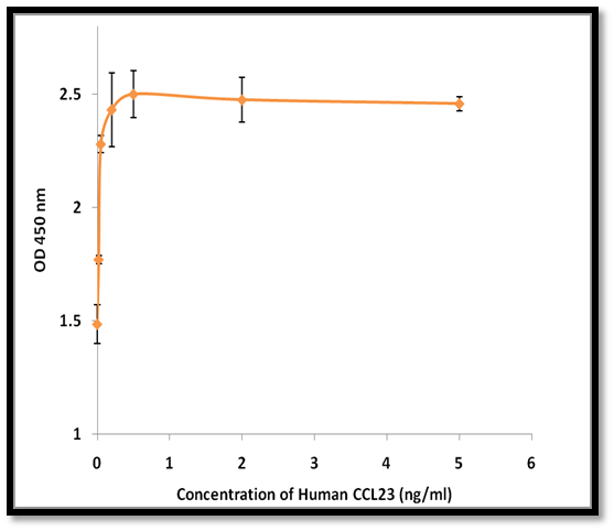
(Figure. The binding activity of LTF with CLU.Lactotransferrin (LTF), also known as actoferrin (LF), is a multifunctional protein of the transferrin family. Lactoferrin belongs to the innate immune system. Apart from its main biological function, namely binding and transport of iron ions, lactoferrin also has antibacterial, antiviral, antiparasitic, catalytic, anti-cancer, and anti-allergic functions and properties. LTF is widely represented in various secretory fluids, such as milk, saliva, tears, and nasal secretions. It also present in secondary granules of PMN and is secreted by some acinar cells. Besides, Clusterin (CLU) has been identified as an interactor of LTF, thus a binding ELISA assay was conducted to detect the interaction of recombinant goat LTF and recombinant goat CLU. Briefly, LTF were diluted serially in PBS, with 0.01% BSA (pH 7.4). Duplicate samples of 100uL were then transferred to CLU-coated microtiter wells and incubated for 2h at 37. Wells were washed with PBST and incubated for 1h with anti-LTF pAb, then aspirated and washed 3 times. After incubation with HRP labelled secondary antibody, wells were aspirated and washed 3 times. With the addition of substrate solution, wells were incubated 15-25 minutes at 37. Finally, add 50uL stop solution to the wells and read at 450nm immediately. The binding activity of LTF and CLU was shown in Figure 1, and this effect was in a dose dependent manner.)
Bioactivity
(Figure. The binding activity of LTF with CLU.Lactotransferrin (LTF), also known as actoferrin (LF), is a multifunctional protein of the transferrin family. Lactoferrin belongs to the innate immune system. Apart from its main biological function, namely binding and transport of iron ions, lactoferrin also has antibacterial, antiviral, antiparasitic, catalytic, anti-cancer, and anti-allergic functions and properties. LTF is widely represented in various secretory fluids, such as milk, saliva, tears, and nasal secretions. It also present in secondary granules of PMN and is secreted by some acinar cells. Besides, Clusterin (CLU) has been identified as an interactor of LTF, thus a binding ELISA assay was conducted to detect the interaction of recombinant goat LTF and recombinant goat CLU. Briefly, LTF were diluted serially in PBS, with 0.01% BSA (pH 7.4). Duplicate samples of 100uL were then transferred to CLU-coated microtiter wells and incubated for 2h at 37. Wells were washed with PBST and incubated for 1h with anti-LTF pAb, then aspirated and washed 3 times. After incubation with HRP labelled secondary antibody, wells were aspirated and washed 3 times. With the addition of substrate solution, wells were incubated 15-25 minutes at 37. Finally, add 50uL stop solution to the wells and read at 450nm immediately. The binding activity of LTF and CLU was shown in Figure 1, and this effect was in a dose dependent manner.)
Lactoferrin, Active Protein (Cat# AAA150100)
 Application Data
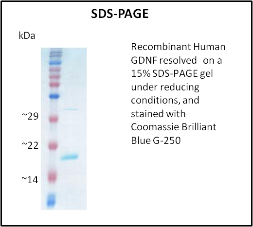
GDNF, Active Protein (Cat# AAA214260)
 Application Data
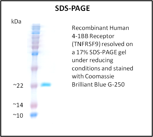
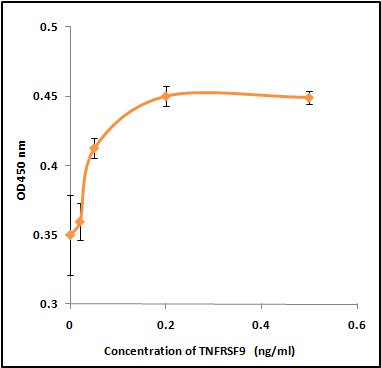
4-1BB Receptor (TNFRSF9), Active Protein (Cat# AAA214261)
IL24, Active Protein (Cat# AAA214264)
 Application Data
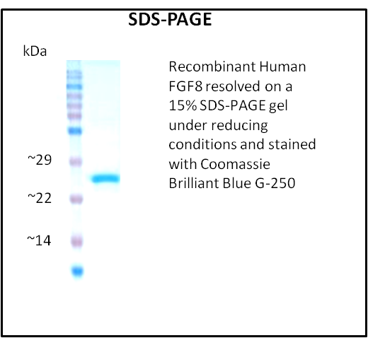
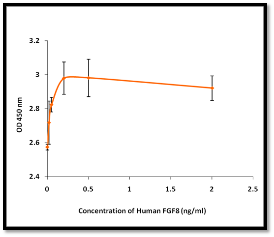
FGF8, Active Protein (Cat# AAA214272)

 Application Data
Galectin-1 (LGALS1), Active Protein (Cat# AAA214275)
 Application Data
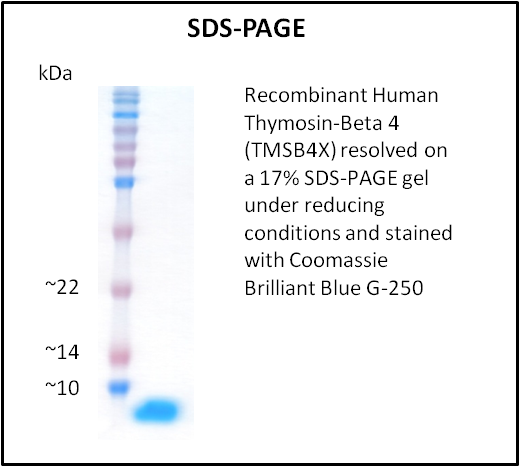
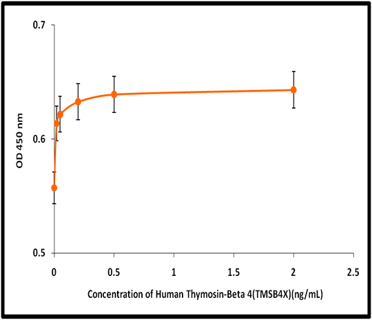
Thymosin-Beta 4 (TMSB4X), Active Protein (Cat# AAA214284)
 Application Data
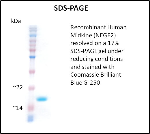
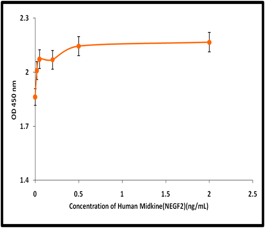
Midkine (NEGF2), Active Protein (Cat# AAA214292)
 Application Data
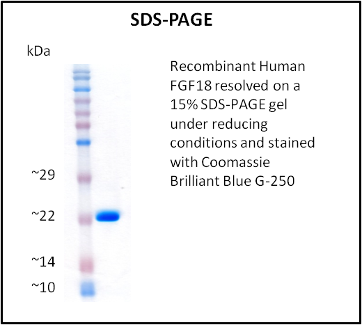
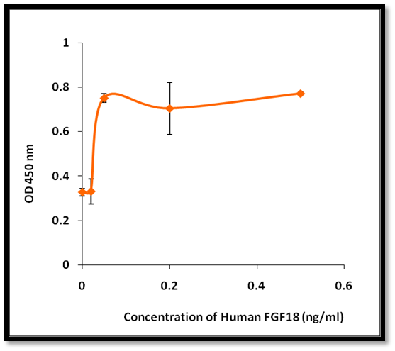
FGF18, Active Protein (Cat# AAA214296)
 Application Data
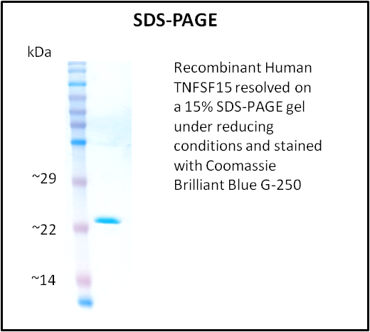
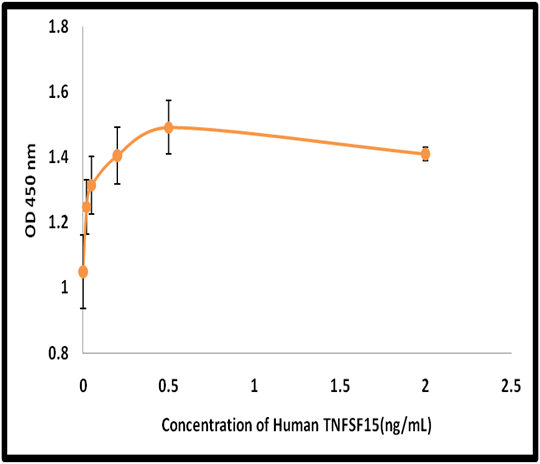
TNFSF15, Active Protein (Cat# AAA214297)
 Application Data
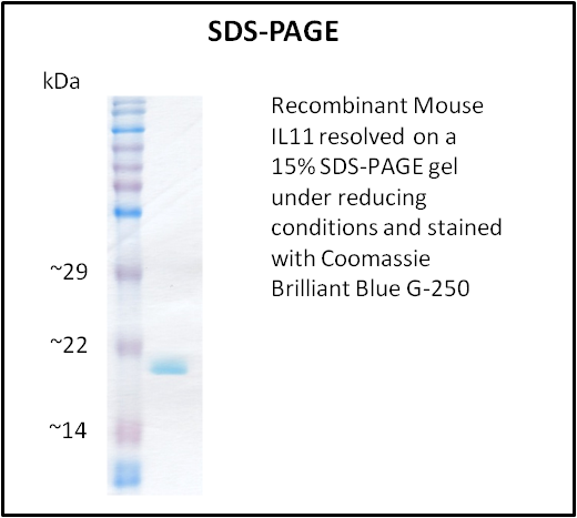
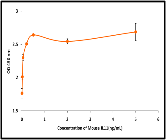
IL11, Active Protein (Cat# AAA214315)
 Application Data
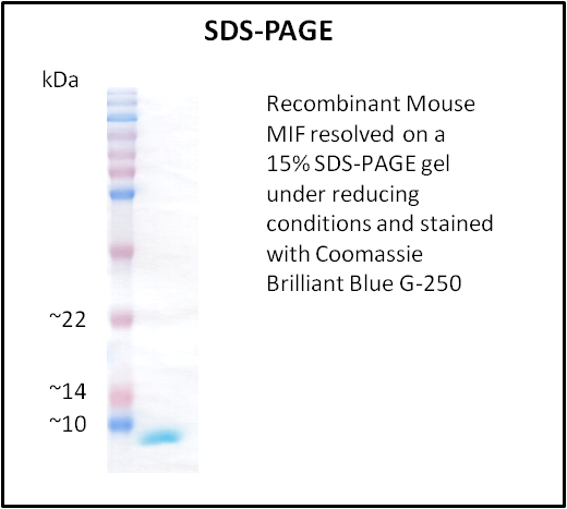
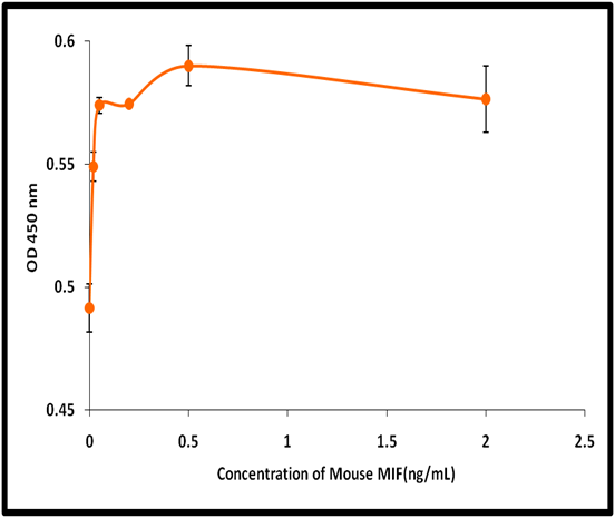
MIF, Active Protein (Cat# AAA214327)
 Application Data
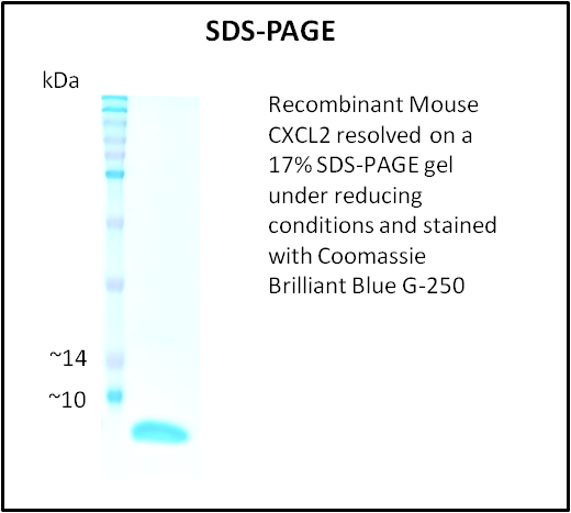
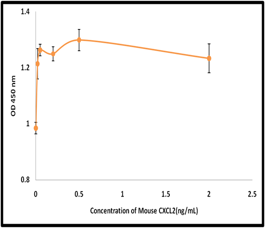
CXCL2, Active Protein (Cat# AAA214329)
 Application Data
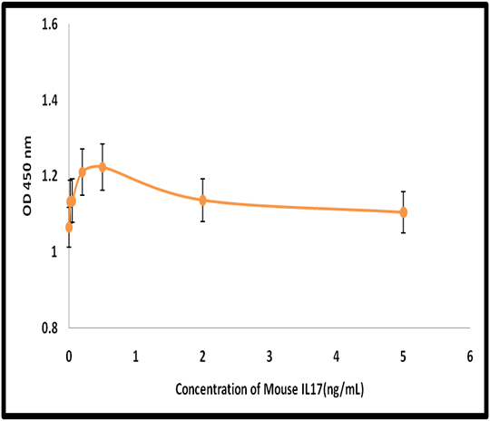
IL17, Active Protein (Cat# AAA214331)
 Application Data
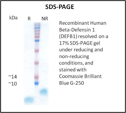
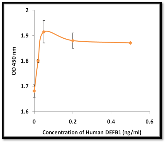
Beta-Defensin 1 (DEFB1), Active Protein (Cat# AAA214212)
 Application Data
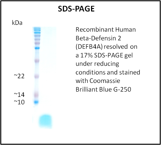
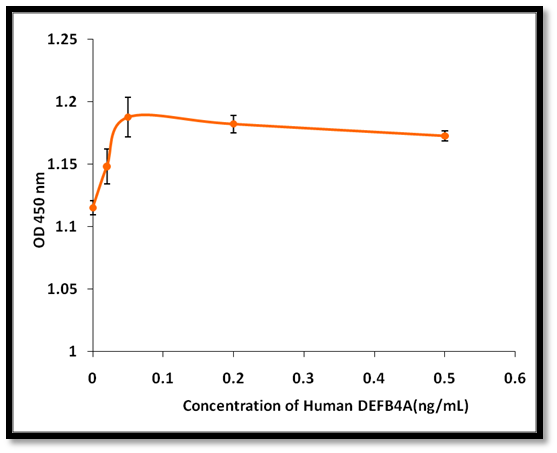
Beta-Defensin 2 (DEFB4A), Active Protein (Cat# AAA214213)
 Application Data
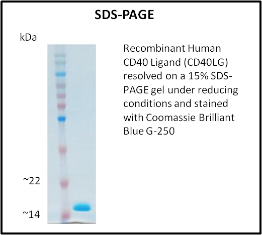
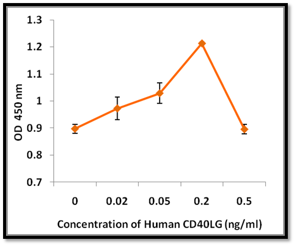
CD40 Ligand (CD40LG), Active Protein (Cat# AAA214215)
 Application Data
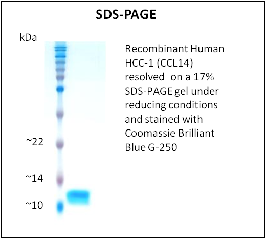
HCC-1 (CCL14), Active Protein (Cat# AAA214225)
 Application Data
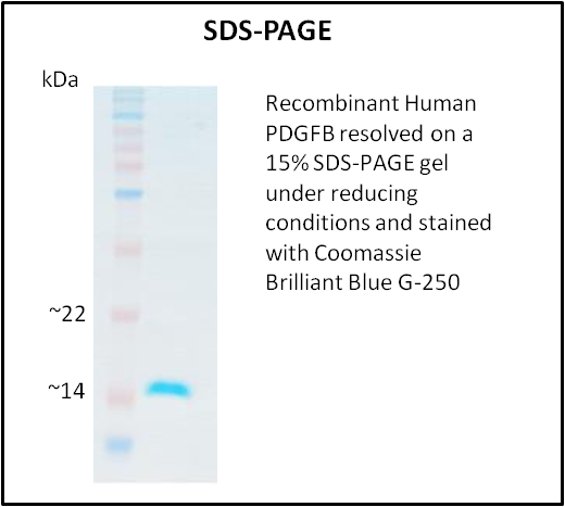
PDGFB, Active Protein (Cat# AAA214227)
 Application Data
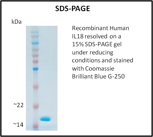
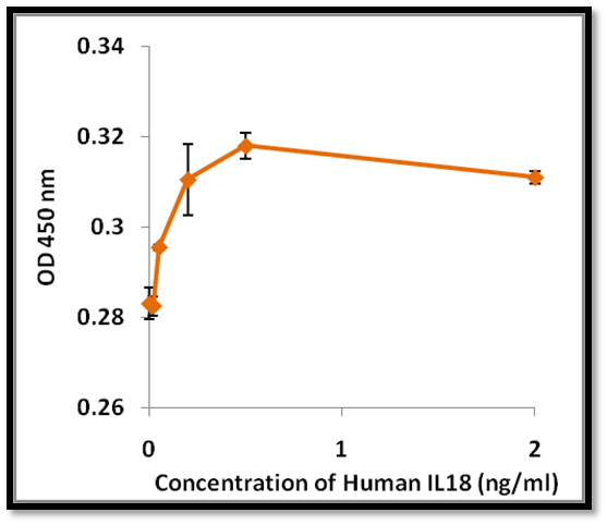
IL18, Active Protein (Cat# AAA214234)
 Application Data
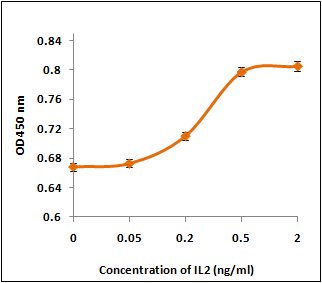
IL2, Active Protein (Cat# AAA214235)
 Application Data
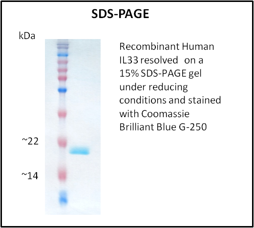
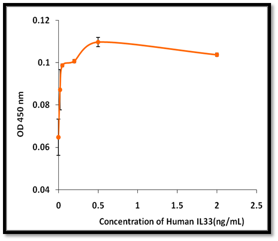
IL33, Active Protein (Cat# AAA214237)
 Application Data
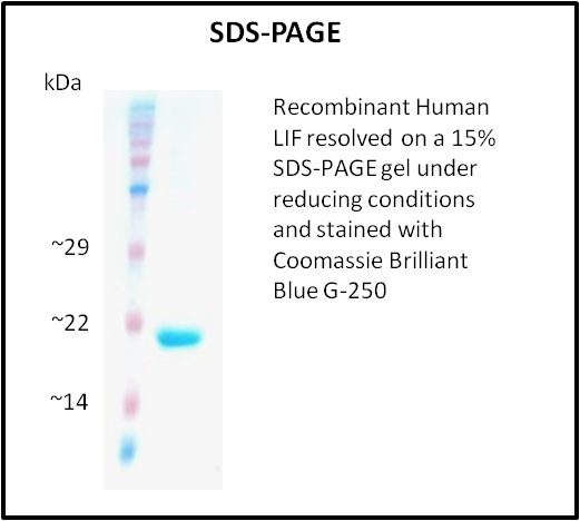
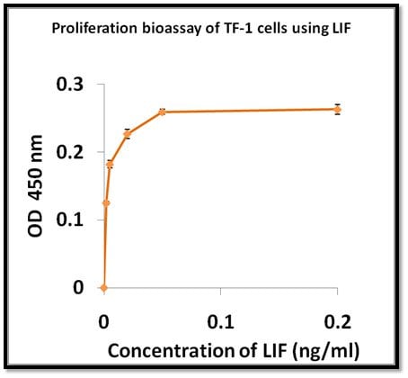
LIF, Active Protein (Cat# AAA214240)

 Application Data
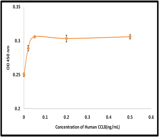
MCP-2 (CCL8), Active Protein (Cat# AAA214243)
 Application Data
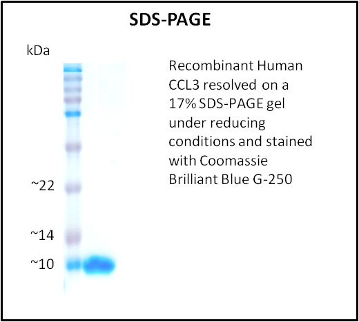
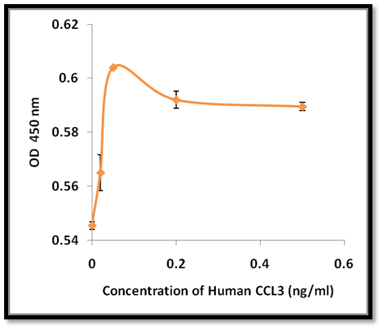
CCL3, Active Protein (Cat# AAA214246)
 Application Data
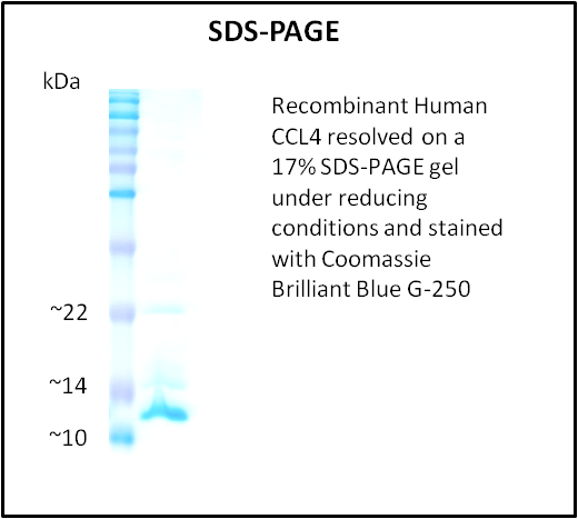
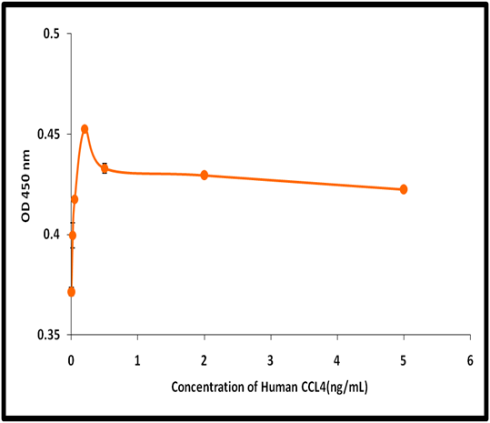
CCL4, Active Protein (Cat# AAA214247)
 Application Data
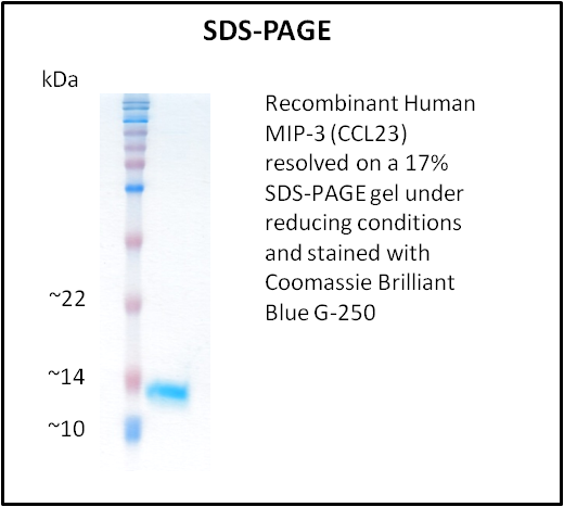
MIP-3 (CCL23), Active Protein (Cat# AAA214248)
 Bioactivity
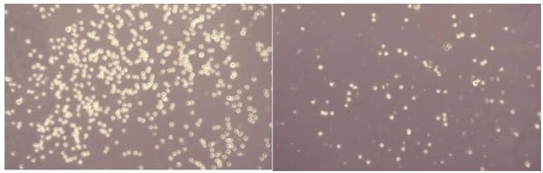
(Neutrophil Activating Protein 3 (NAP3),also known as chemokine (C-X-C motif) ligand 1 (CXCL1) is a small cytokine belonging to the CXC chemokine family. NAP3 plays a role in spinal cord development by inhibiting the migration of oligodendrocyte precursors and is involved in the processes of angiogenesis, arteriogenesis, inflammation, wound healing, and tumorigenesis. Thus, chemotaxis assay used 24-well microchemotaxis system was undertaken to detect the chemotactic effect of NAP3 on the human monocytic cell line THP-1. Briefly, THP-1 cells were seeded into the upper chambers (100uL cell suspension, 5×10⁵ cells/ml in RPMI 1640 with FBS free) and recombinant human NAP3 (0.01ng/mL, 0.1ng/mL, 1ng/mL, 10ng/mL, 100ng/mL, 1000ng/mL diluted separately in serum free RPMI 1640) was added in lower chamber with a polycarbonate filter (8um pore size) used to separate the two compartments. After incubation at 37 degree C with 5% CO2 for 1h, the filter was removed, then cells in low chamber were observed by inverted microscope at low magnification (×100) and the number of migrated cells were counted at high magnification (×200) randomly (five fields for each filter). Result shows recombinant human NAP3 is able to induce migration of THP-1 cells. The migrated THP-1 cells in low chamber at low magnification(×100) were shown in Figure 1. Five fields of each chamber were randomly chosen, and the migrated cells were counted at high magnification(×200). Statistical results were shown in Figure 2. The optimum chemotaxis of NAP3 occurs at 0.1~1ng/mL.)
Bioactivity
(Neutrophil Activating Protein 3 (NAP3),also known as chemokine (C-X-C motif) ligand 1 (CXCL1) is a small cytokine belonging to the CXC chemokine family. NAP3 plays a role in spinal cord development by inhibiting the migration of oligodendrocyte precursors and is involved in the processes of angiogenesis, arteriogenesis, inflammation, wound healing, and tumorigenesis. Thus, chemotaxis assay used 24-well microchemotaxis system was undertaken to detect the chemotactic effect of NAP3 on the human monocytic cell line THP-1. Briefly, THP-1 cells were seeded into the upper chambers (100uL cell suspension, 5×10⁵ cells/ml in RPMI 1640 with FBS free) and recombinant human NAP3 (0.01ng/mL, 0.1ng/mL, 1ng/mL, 10ng/mL, 100ng/mL, 1000ng/mL diluted separately in serum free RPMI 1640) was added in lower chamber with a polycarbonate filter (8um pore size) used to separate the two compartments. After incubation at 37 degree C with 5% CO2 for 1h, the filter was removed, then cells in low chamber were observed by inverted microscope at low magnification (×100) and the number of migrated cells were counted at high magnification (×200) randomly (five fields for each filter). Result shows recombinant human NAP3 is able to induce migration of THP-1 cells. The migrated THP-1 cells in low chamber at low magnification(×100) were shown in Figure 1. Five fields of each chamber were randomly chosen, and the migrated cells were counted at high magnification(×200). Statistical results were shown in Figure 2. The optimum chemotaxis of NAP3 occurs at 0.1~1ng/mL.)
Chemokine (C-X-C Motif) Ligand 1 (CXCL1), Active Protein (Cat# AAA152237)
 Bioactivity
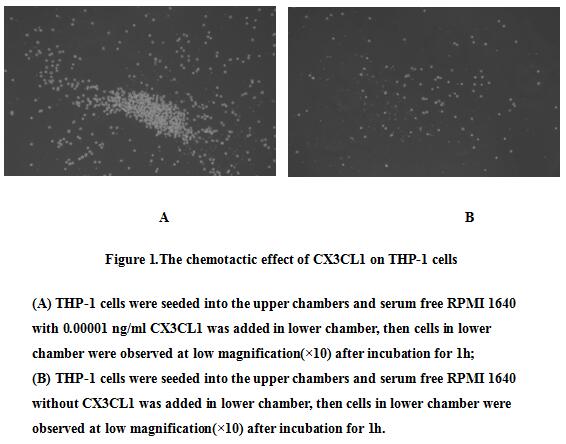
(Chemokine C-X3-C-Motif Ligand 1 (CX3CL1) also known as fractalkine is a large cytokine protein of 373 amino acids, it contains multiple domains and is the only known member of the CX3C chemokine family. Soluble CX3CL1 potently chemoattracts T cells and mo)
Bioactivity
(Chemokine C-X3-C-Motif Ligand 1 (CX3CL1) also known as fractalkine is a large cytokine protein of 373 amino acids, it contains multiple domains and is the only known member of the CX3C chemokine family. Soluble CX3CL1 potently chemoattracts T cells and mo)
Chemokine C-X3-C-Motif Ligand 1 (CX3CL1), Active Protein (Cat# AAA152958)
 Bioactivity
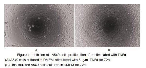
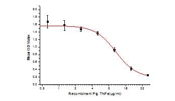
(Figure 2. Inhibition of A549 cells proliferation after stimulated with TNFalpha.)
Bioactivity
(Figure 2. Inhibition of A549 cells proliferation after stimulated with TNFalpha.)
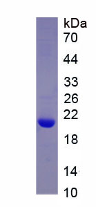
Tumor Necrosis Factor Alpha (TNFa), Active Protein (Cat# AAA152990)
 Bioactivity
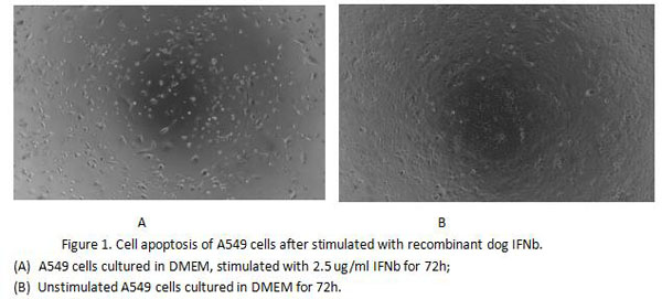
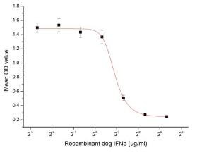
(Figure 2. Cell apoptosis of A549 cells after stimulated with recombinant dog IFNb.)
Bioactivity
(Figure 2. Cell apoptosis of A549 cells after stimulated with recombinant dog IFNb.)
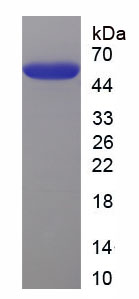
Interferon Beta (IFNb), Active Protein (Cat# AAA153001)
What Are Active Proteins?
Proteins are large molecules made up of long chains of amino acids.
They will typically fold into a very particular 3-dimensional shape/conformation, that is sometimes referred to as their “native” form, which allows them to work properly in the body. For the purposes of product categorization, AAA Biotech will typically refer to proteins purified from their original animal host as being “native” proteins (this is to signify their difference compared to their “recombinant” or “synthetic” protein counterparts).
If a protein successfully folds into the correct shape, it is will typically display high fidelity characteristics to its original protein in its original animal host, and be classified as an active protein, as it will be able to function “normally” in most enzymatic or binding capacities. If it loses this shape, due to factors such as heat or strong chemicals (such as detergents), it becomes inactive and is no longer able to perform its basic functions. All of the proteins in this category are made under strict quality control, and they are active, pure, low in contaminants, and stable.
Most are stored as freeze-dried powders and come without extra tags, so they’re very close to the actual natural/native form.
Key Applications of Active Proteins
1. Scientific Research
- Aid in the study of how proteins function in the body
- Aid in understanding various disease processes
2. Drug Development
- Powerful tools to investigate how potential drugs interact with specific proteins
- Ideal for identifying drug targets
3. Cell Culture
- Are routinely utilized to support cell growth and function (e.g., using exogenous growth factors)
- Can be used to promote cellular development into specific types (differentiation)
4. Diagnostics
- Regularly utilized in tests to detect diseases or infections (e.g., COVID-19, cancer)
- Note: All products are strictly for research-use only (RUO).
5. Therapeutics
- Some active proteins are used directly as treatments (e.g., insulin, enzymes)
- Note: All products are strictly for research-use only (RUO).
6. Vaccine Development
- Used to create or test vaccines by mimicking parts of viruses or bacteria
7. Biochemical Assays
- They can facilitate the characterization of enzyme activity, binding strength, or protein interactions in lab tests
Why Buy Active Proteins from AAA Biotech?
- High biological activity – Verified to perform as expected or indicated on datasheet
- Strict quality control – We are confident in our active proteins’ reliability and consistency
- High purity & low endotoxin – Ideal for applications involving sensitive or precious samples/components
- Freeze-dried for stability – Long shelf life and straightforward storage
- Mostly tag-free – Closer to natural/native protein form
FAQ
1. What are active proteins used for in research?
Active proteins are used primarily in the study of how proteins function, in characterizing/discovering drug interactions, supporting cell growth, running biochemical assays, and in development of diagnostics or therapeutics.
2. How are AAA Biotech's active proteins validated?
AAA Biotech’s active proteins are validated through strict quality control and functional assays to ensure they are properly folded and active. “Active”, though, can be an ambiguous term, so if a specific “activity” or “binding” capability of a protein is of crucial interest to you, please inquire with us prior to purchase, and we will provide further details on how the “Active” modifier was determined to be applicable.
3. Are these proteins tested for biological activity?
Yes, all active proteins from AAA Biotech are tested to confirm they have the expected biological activity before being offered for use. Though, said “biological activity” can be either “enzymatic”, “binding”, or both.