Filters
▼Clonality
▼Type
▼Reactivity
▼Gene Name
▼Isotype
▼Host
▼Application
▼Clone
▼Polyclonal Antibodies
At AAA Biotech also known as AAA Bio or AAABio, we provide a broad range of purified polyclonal antibodies (pAbs) that are able to all be browsed online through our website. Due to their high specificity and strong binding affinity, these antibodies are ideal for wide swathes of research and experimental applications.
Our polyclonal antibodies can easily support your work, whether you use them for Western Blotting, Immunocytochemistry (with or without Immunofluorescence used in conjunction), Immunohistochemistry, Immunoprecipitation, and ELISA tests. We highly encourage you to browse our range of pAbs and choose the one that best suits your experimental model.
Viewing 7150-7200 of 96805 product results
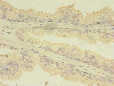
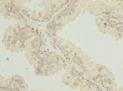
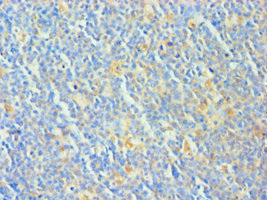
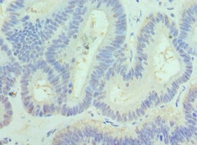
 IHC (Immunohiostchemistry)
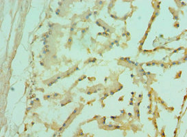
(Immunohistochemistry of paraffin-embedded human pancreatic tissue using AAA119531 at dilution of 1:100)
IHC (Immunohiostchemistry)
(Immunohistochemistry of paraffin-embedded human pancreatic tissue using AAA119531 at dilution of 1:100)
TBCA, Polyclonal Antibody (Cat# AAA119531)
Peptide deformylase, Polyclonal Antibody (Cat# AAA119535)
Mycoplasma hyopneumoniae 46 kDa surface antigen, Polyclonal Antibody (Cat# AAA119537)
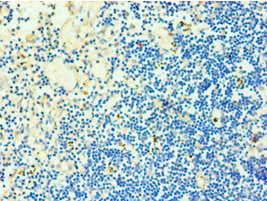
 IHC (Immunohistochemistry)
(Immunohistochemistry of paraffin-embedded human thymus using AAA119539 at dilution 1:100)
IHC (Immunohistochemistry)
(Immunohistochemistry of paraffin-embedded human thymus using AAA119539 at dilution 1:100)
PBK, Polyclonal Antibody (Cat# AAA119539)
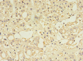
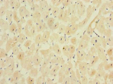
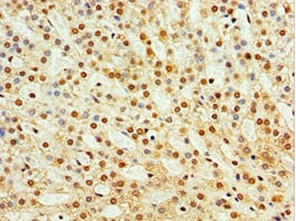
 IHC (Immunohiostchemistry)
(Immunohistochemistry of paraffin-embedded human liver tissue using AAA119541 at dilution of 1:100)
IHC (Immunohiostchemistry)
(Immunohistochemistry of paraffin-embedded human liver tissue using AAA119541 at dilution of 1:100)
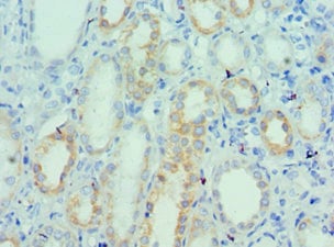
B3GALT2, Polyclonal Antibody (Cat# AAA119541)
 IHC (Immunohiostchemistry)
(Immunohistochemistry of paraffin-embedded human tonsil using AAA119542 at dilution 1:100)
IHC (Immunohiostchemistry)
(Immunohistochemistry of paraffin-embedded human tonsil using AAA119542 at dilution 1:100)
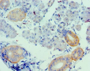
EIF2AK4, Polyclonal Antibody (Cat# AAA119542)
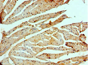
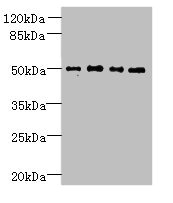
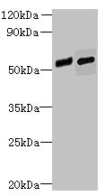
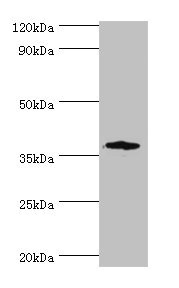
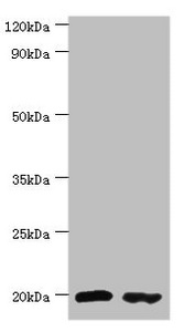
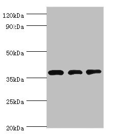
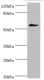
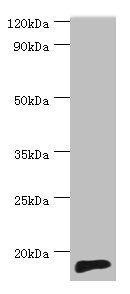 WB (Western Blot)
(Western blotAll lanes: TNNC2 antibody at 10ug/ml+mouse skeletal muscle tissueSecondaryGoat polyclonal to rabbit at 1/10000 dilutionPredicted band size: 18kDaObserved band size: 18kDa)
WB (Western Blot)
(Western blotAll lanes: TNNC2 antibody at 10ug/ml+mouse skeletal muscle tissueSecondaryGoat polyclonal to rabbit at 1/10000 dilutionPredicted band size: 18kDaObserved band size: 18kDa)
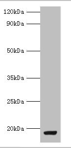
TNNC2, Polyclonal Antibody (Cat# AAA119543)
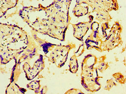
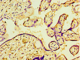
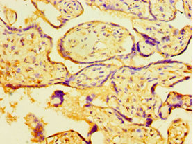
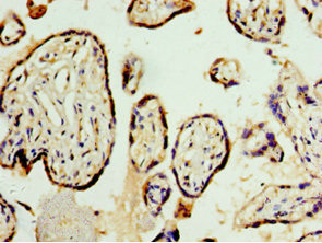
 IHC (Immunohiostchemistry)
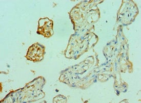
(Immunohistochemistry of paraffin-embedded human placenta tissue using AAA119547 at dilution 1:100)
IHC (Immunohiostchemistry)
(Immunohistochemistry of paraffin-embedded human placenta tissue using AAA119547 at dilution 1:100)
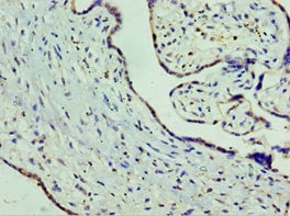
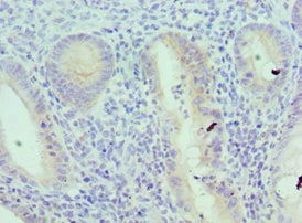
SLC25A15, Polyclonal Antibody (Cat# AAA119547)
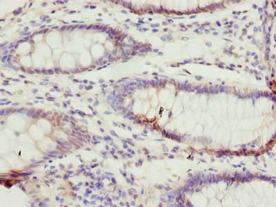
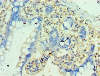
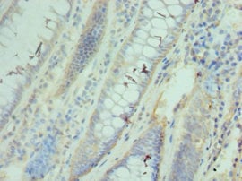
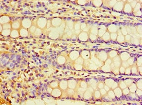
 IHC (Immunohistochemisry)
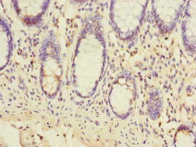
(Immunohistochemistry of paraffin-embedded human small intestine tissue using AAA119549 at dilution of 1:100)
IHC (Immunohistochemisry)
(Immunohistochemistry of paraffin-embedded human small intestine tissue using AAA119549 at dilution of 1:100)
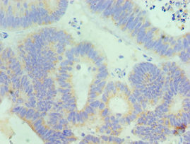
RAB6B, Polyclonal Antibody (Cat# AAA119549)
 IF (Immunofluorescence)
(Immunofluorescent analysis of Hela cells using AAA119552 at a dilution of 1:100 and Alexa Fluor 488-congugated AffiniPure Goat Anti-Rabbit IgG(H+L))
IF (Immunofluorescence)
(Immunofluorescent analysis of Hela cells using AAA119552 at a dilution of 1:100 and Alexa Fluor 488-congugated AffiniPure Goat Anti-Rabbit IgG(H+L))
NKG7, Polyclonal Antibody (Cat# AAA119552)
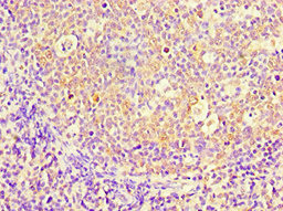
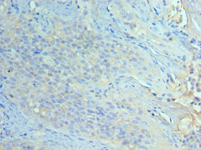
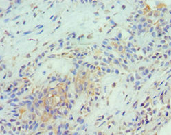
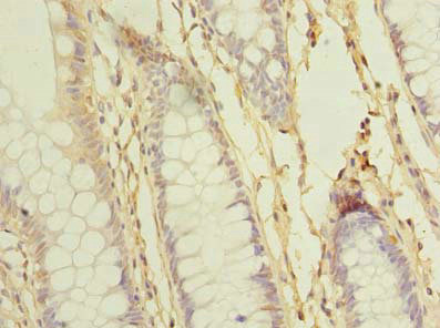
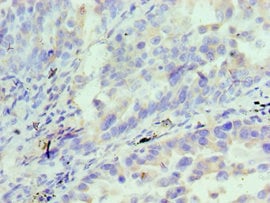
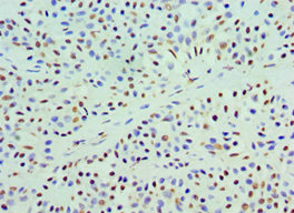
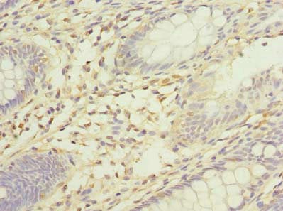
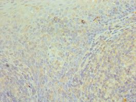
 IHC (Immunohistochemisry)
(Immunohistochemistry of paraffin-embedded human colon cancer using AAA119723 at dilution of 1:100)
IHC (Immunohistochemisry)
(Immunohistochemistry of paraffin-embedded human colon cancer using AAA119723 at dilution of 1:100)
TTC38, Polyclonal Antibody (Cat# AAA119723)
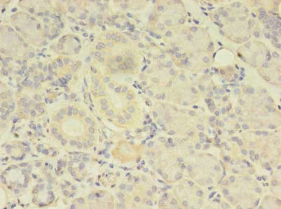
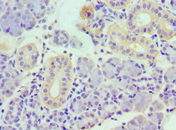
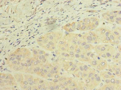
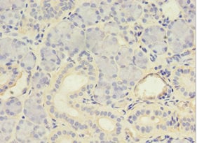 IHC (Immunohiostchemistry)
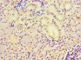
(Immunohistochemistry of paraffin-embedded human pancreas tissue using AAA119725 at dilution 1:100)
IHC (Immunohiostchemistry)
(Immunohistochemistry of paraffin-embedded human pancreas tissue using AAA119725 at dilution 1:100)
C-Myc-binding, Polyclonal Antibody (Cat# AAA119725)
 IHC (Immunohistochemisry)
(Immunohistochemistry of paraffin-embedded human testis using AAA119729 at dilution 1:100)
IHC (Immunohistochemisry)
(Immunohistochemistry of paraffin-embedded human testis using AAA119729 at dilution 1:100)
Diacylglycerol kinase epsilon, Polyclonal Antibody (Cat# AAA119729)
 IF (Immunofluorescence)
(Immunofluorescent analysis of MCF-7 cells using AAA119737 at a dilution of 1:100 and Alexa Fluor 488-congugated AffiniPure Goat Anti-Rabbit IgG(H+L))
IF (Immunofluorescence)
(Immunofluorescent analysis of MCF-7 cells using AAA119737 at a dilution of 1:100 and Alexa Fluor 488-congugated AffiniPure Goat Anti-Rabbit IgG(H+L))
GPM6A, Polyclonal Antibody (Cat# AAA119737)
 IHC (Immunohiostchemistry)
(Immunohistochemistry of paraffin-embedded human small intestine cancer using AAA119745 at dilution 1:100)
IHC (Immunohiostchemistry)
(Immunohistochemistry of paraffin-embedded human small intestine cancer using AAA119745 at dilution 1:100)
Angiopoietin-related protein 2, Polyclonal Antibody (Cat# AAA119745)
 IHC (Immunohiostchemistry)
(Immunohistochemistry of paraffin-embedded human lung using AAA119746 at dilution of 1:100)
IHC (Immunohiostchemistry)
(Immunohistochemistry of paraffin-embedded human lung using AAA119746 at dilution of 1:100)
FAM124B, Polyclonal Antibody (Cat# AAA119746)
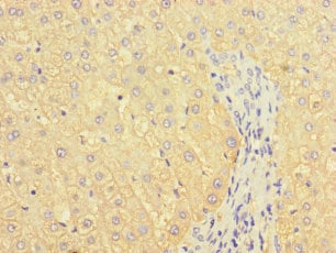
 IHC (Immunohiostchemistry)
(Immunohistochemistry of paraffin-embedded human liver tissue using AAA119749 at dilution of 1:100)
IHC (Immunohiostchemistry)
(Immunohistochemistry of paraffin-embedded human liver tissue using AAA119749 at dilution of 1:100)
BEND7, Polyclonal Antibody (Cat# AAA119749)
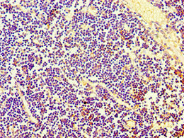
 IHC (Immunohiostchemistry)
(Immunohistochemistry of paraffin-embedded human thymus using AAA119752 at dilution 1:100)
IHC (Immunohiostchemistry)
(Immunohistochemistry of paraffin-embedded human thymus using AAA119752 at dilution 1:100)
Treacle protein, Polyclonal Antibody (Cat# AAA119752)
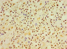
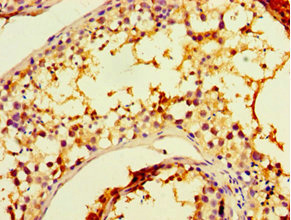
 IHC (Immunohiostchemistry)
(Immunohistochemistry of paraffin-embedded human pancreas using AAA119753 at dilution 1:100)
IHC (Immunohiostchemistry)
(Immunohistochemistry of paraffin-embedded human pancreas using AAA119753 at dilution 1:100)
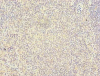
Tumor necrosis factor ligand superfamily member 13, Polyclonal Antibody (Cat# AAA119753)
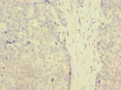
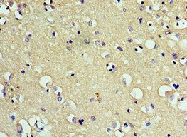 IHC (Immunohiostchemistry)
(Immunohistochemistry of paraffin-embedded human brain tissue using AAA119754 at dilution 1:100)
IHC (Immunohiostchemistry)
(Immunohistochemistry of paraffin-embedded human brain tissue using AAA119754 at dilution 1:100)
CD200R1, Polyclonal Antibody (Cat# AAA119754)
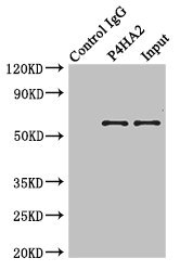 IP (Immunoprecipitation)
(Immunoprecipitating P4HA2 in Hela whole cell lysateLane 1: Rabbit monoclonal IgG(1ug)instead of AAA119756 in Hela whole cell lysate. For western blotting, a HRP-conjugated anti-rabbit IgG, specific to the non-reduced form of IgG was used as the Secondary antibody (1/50000)Lane 2: AAA119756(4ug)+ Hela whole cell lysate(500ug)Lane 3: Hela whole cell lysate (20ug))
IP (Immunoprecipitation)
(Immunoprecipitating P4HA2 in Hela whole cell lysateLane 1: Rabbit monoclonal IgG(1ug)instead of AAA119756 in Hela whole cell lysate. For western blotting, a HRP-conjugated anti-rabbit IgG, specific to the non-reduced form of IgG was used as the Secondary antibody (1/50000)Lane 2: AAA119756(4ug)+ Hela whole cell lysate(500ug)Lane 3: Hela whole cell lysate (20ug))
P4HA2, Polyclonal Antibody (Cat# AAA119756)
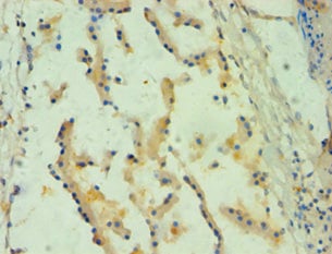
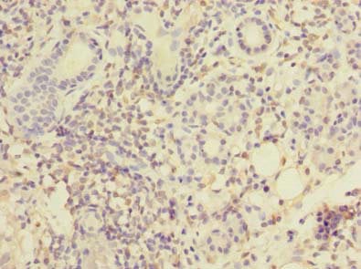
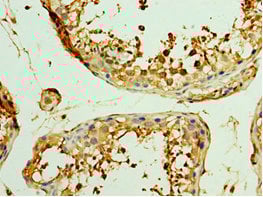
 IHC (Immunohiostchemistry)
(Immunohistochemistry of paraffin-embedded human liver cancer using AAA119759 at dilution of 1:100)
IHC (Immunohiostchemistry)
(Immunohistochemistry of paraffin-embedded human liver cancer using AAA119759 at dilution of 1:100)
SCFD2, Polyclonal Antibody (Cat# AAA119759)
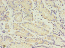
 IHC (Immunohistochemisry)
(Immunohistochemistry of paraffin-embedded human pancreas tissueAAA119762 at dilution 1:100)
IHC (Immunohistochemisry)
(Immunohistochemistry of paraffin-embedded human pancreas tissueAAA119762 at dilution 1:100)
MYDGF, Polyclonal Antibody (Cat# AAA119762)
 IHC (Immunohiostchemistry)
(Immunohistochemistry of paraffin-embedded human lung tissue using AAA119763 at dilution of 1:100)
IHC (Immunohiostchemistry)
(Immunohistochemistry of paraffin-embedded human lung tissue using AAA119763 at dilution of 1:100)
Zinc finger protein 212, Polyclonal Antibody (Cat# AAA119763)
 IHC (Immunohistochemisry)
(Immunohistochemistry of paraffin-embedded human epityphlon using AAA119765at dilution 1:100)
IHC (Immunohistochemisry)
(Immunohistochemistry of paraffin-embedded human epityphlon using AAA119765at dilution 1:100)
Interleukin-18 receptor accessory protein, Polyclonal Antibody (Cat# AAA119765)
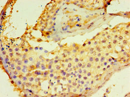
 IHC (Immunohiostchemistry)
(Immunohistochemistry of paraffin-embedded human pancreas using AAA119767 at dilution 1:100)
IHC (Immunohiostchemistry)
(Immunohistochemistry of paraffin-embedded human pancreas using AAA119767 at dilution 1:100)
Urotensin-2, Polyclonal Antibody (Cat# AAA119767)
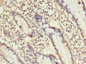
 IHC (Immunohiostchemistry)
(Immunohistochemistry of paraffin-embedded human small intestine using AAA119774 at dilution 1:100)
IHC (Immunohiostchemistry)
(Immunohistochemistry of paraffin-embedded human small intestine using AAA119774 at dilution 1:100)
Peptidyl-tRNA hydrolase 2, Polyclonal Antibody (Cat# AAA119774)
Arg2, Polyclonal Antibody (Cat# AAA119775)
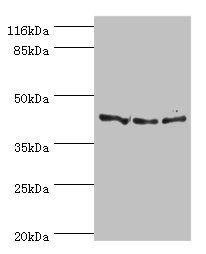
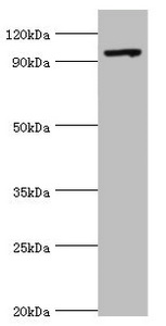
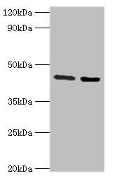
 WB (Western Blot)
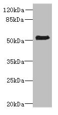
(Western blotAll lanes: MAVS antibody at 2ug/mlLane 1:mouse heart tissueLane 2:jurkat whole cell lysateLane 3:293T whole cell lysateSecondaryGoat polyclonal to rabbit at 1/10000 dilutionPredicted band size: 57,16,18,41,15 kDaObserved band size: 57 kDa)
WB (Western Blot)
(Western blotAll lanes: MAVS antibody at 2ug/mlLane 1:mouse heart tissueLane 2:jurkat whole cell lysateLane 3:293T whole cell lysateSecondaryGoat polyclonal to rabbit at 1/10000 dilutionPredicted band size: 57,16,18,41,15 kDaObserved band size: 57 kDa)
MAVS, Polyclonal Antibody (Cat# AAA119781)
TMEM100, Polyclonal Antibody (Cat# AAA119782)
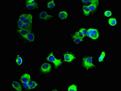
 IF (Immunofluorescence)
(Immunofluorescent analysis of 293T cells using AAA119783 at a dilution of 1:100 and Alexa Fluor 488-congugated AffiniPure Goat Anti-Rabbit IgG(H+L))
IF (Immunofluorescence)
(Immunofluorescent analysis of 293T cells using AAA119783 at a dilution of 1:100 and Alexa Fluor 488-congugated AffiniPure Goat Anti-Rabbit IgG(H+L))
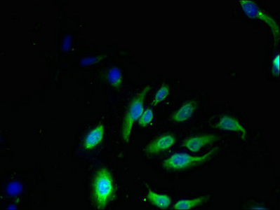
Glucagon-like peptide 1 receptor, Polyclonal Antibody (Cat# AAA119783)

 IHC (Immunohiostchemistry)
(Immunohistochemistry of paraffin-embedded human prostate using AAA119784 at dilution 1:100)
IHC (Immunohiostchemistry)
(Immunohistochemistry of paraffin-embedded human prostate using AAA119784 at dilution 1:100)
Kunitz-type protease inhibitor 1, Polyclonal Antibody (Cat# AAA119784)
 IHC (Immunohiostchemistry)
(Immunohistochemistry of paraffin-embedded human breast cancer using AAA119788 at dilution 1:100)
IHC (Immunohiostchemistry)
(Immunohistochemistry of paraffin-embedded human breast cancer using AAA119788 at dilution 1:100)
Annexin A4, Polyclonal Antibody (Cat# AAA119788)
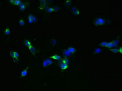
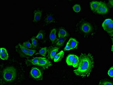
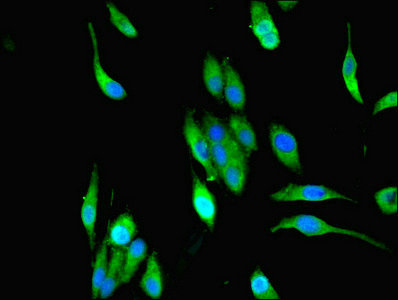 IHC (Immunohistochemisry)
(Immunofluorescent analysis of Hela cells using AAA119792 at a dilution of 1:100 and Alexa Fluor 488-congugated AffiniPure Goat Anti-Rabbit IgG(H+L))
IHC (Immunohistochemisry)
(Immunofluorescent analysis of Hela cells using AAA119792 at a dilution of 1:100 and Alexa Fluor 488-congugated AffiniPure Goat Anti-Rabbit IgG(H+L))
MPPED1, Polyclonal Antibody (Cat# AAA119792)
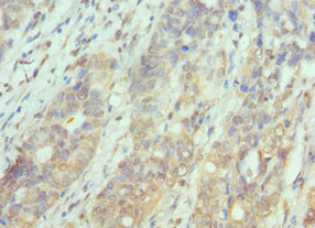
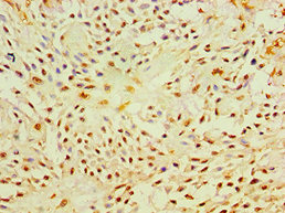
 IHC (Immunohiostchemistry)
(Immunohistochemistry of paraffin-embedded human colon cancer using AAA119795 at dilution 1:100)
IHC (Immunohiostchemistry)
(Immunohistochemistry of paraffin-embedded human colon cancer using AAA119795 at dilution 1:100)
Serine/threonine-protein kinase PAK 1, Polyclonal Antibody (Cat# AAA119795)
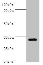
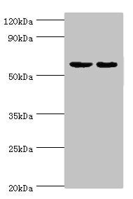
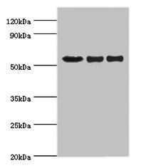
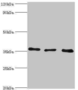
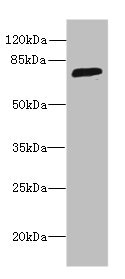
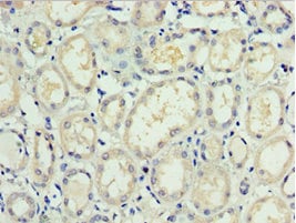
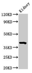 WB (Western Blot)
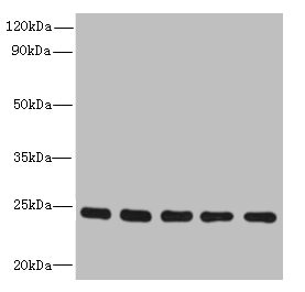
(Western BlotPositive WB detected in: Mouse kidney tissueAll lanes: Wnt3 antibody at 3.2ug/mlSecondaryGoat polyclonal to rabbit IgG at 1/50000 dilutionPredicted band size: 40 KDaObserved band size: 40 KDa)
WB (Western Blot)
(Western BlotPositive WB detected in: Mouse kidney tissueAll lanes: Wnt3 antibody at 3.2ug/mlSecondaryGoat polyclonal to rabbit IgG at 1/50000 dilutionPredicted band size: 40 KDaObserved band size: 40 KDa)
Wnt3, Polyclonal Antibody (Cat# AAA119801)
Fcgr3, Polyclonal Antibody (Cat# AAA119805)
 IHC (Immunohistochemisry)
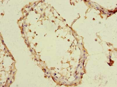
(Immunohistochemistry of paraffin-embedded human placenta tissue using AAA119806 at dilution 1:100)
IHC (Immunohistochemisry)
(Immunohistochemistry of paraffin-embedded human placenta tissue using AAA119806 at dilution 1:100)
STRAP, Polyclonal Antibody (Cat# AAA119806)
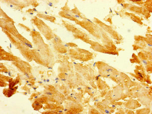
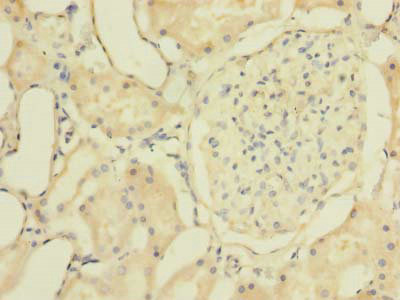
 IHC (Immunohiostchemistry)
(Immunohistochemistry of paraffin-embedded human skeletal muscle using AAA119557 at dilution 1:100)
IHC (Immunohiostchemistry)
(Immunohistochemistry of paraffin-embedded human skeletal muscle using AAA119557 at dilution 1:100)
Ubiquinone biosynthesis protein COQ7 homolog, Polyclonal Antibody (Cat# AAA119557)
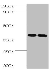
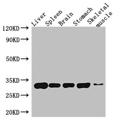
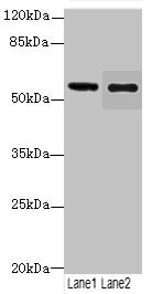
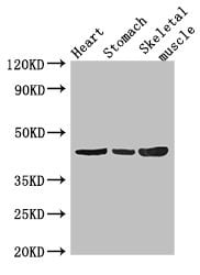 WB (Western Blot)
(Western BlotPositive WB detected in:Mouse heart tissue,Mouse stomach tissue,Mouse skeletal muscle tissueAll lanes: ZNF707 antibody at 3.4ug/mlSecondaryGoat polyclonal to rabbit IgG at 1/50000 dilutionPredicted band size: 44 kDaObserved band size: 44 kDa)
WB (Western Blot)
(Western BlotPositive WB detected in:Mouse heart tissue,Mouse stomach tissue,Mouse skeletal muscle tissueAll lanes: ZNF707 antibody at 3.4ug/mlSecondaryGoat polyclonal to rabbit IgG at 1/50000 dilutionPredicted band size: 44 kDaObserved band size: 44 kDa)
ZNF707, Polyclonal Antibody (Cat# AAA119565)
Enterobacteria phage H19B Shiga-like toxin 1 subunit B, Polyclonal Antibody (Cat# AAA119569)
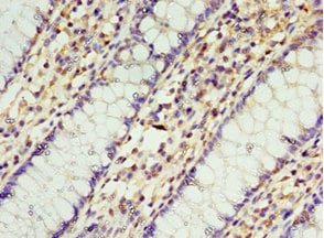
 IHC (Immunohistochemisry)
(Immunohistochemistry of paraffin-embedded human colon cancer using AAA119572 at dilution of 1:100)
IHC (Immunohistochemisry)
(Immunohistochemistry of paraffin-embedded human colon cancer using AAA119572 at dilution of 1:100)
SORBS3, Polyclonal Antibody (Cat# AAA119572)
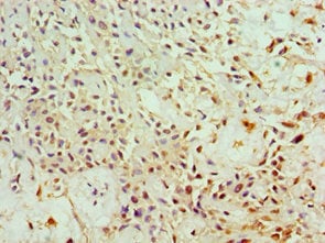
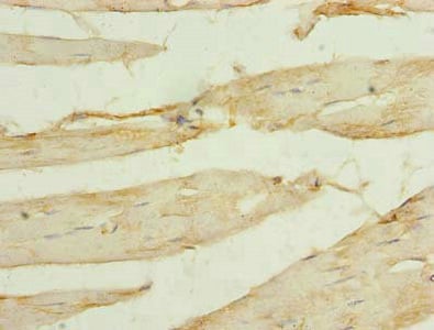
 IHC (Immunohiostchemistry)
(Immunohistochemistry of paraffin-embedded human skeletal muscle using AAA119575 at dilution 1:100)
IHC (Immunohiostchemistry)
(Immunohistochemistry of paraffin-embedded human skeletal muscle using AAA119575 at dilution 1:100)
WNT1-inducible-signaling pathway protein 2, Polyclonal Antibody (Cat# AAA119575)
 IHC (Immunohistochemisry)
(Immunohistochemistry of paraffin-embedded human adrenal gland using AAA119579 at dilution 1:100)
IHC (Immunohistochemisry)
(Immunohistochemistry of paraffin-embedded human adrenal gland using AAA119579 at dilution 1:100)
TAF1C, Polyclonal Antibody (Cat# AAA119579)
 IHC (Immunohistochemisry)
(Immunohistochemistry of paraffin-embedded human colon cancer using AAA119580 at dilution 1:100)
IHC (Immunohistochemisry)
(Immunohistochemistry of paraffin-embedded human colon cancer using AAA119580 at dilution 1:100)
T-complex protein 1 subunit gamma, Polyclonal Antibody (Cat# AAA119580)
cytomegalovirus Envelope glycoprotein L, Polyclonal Antibody (Cat# AAA119586)
 IHC (Immunohiostchemistry)
(Immunohistochemistry of paraffin-embedded human placenta using AAA119587 at dilution 1:100)
IHC (Immunohiostchemistry)
(Immunohistochemistry of paraffin-embedded human placenta using AAA119587 at dilution 1:100)
Forkhead box protein P2, Polyclonal Antibody (Cat# AAA119587)
Interferon tau-1, Polyclonal Antibody (Cat# AAA119594)
 IHC (Immunohistochemisry)
(Immunofluorescent analysis of MCF-7 cells using AAA119596 at a dilution of 1:100 and Alexa Fluor 488-congugated AffiniPure Goat Anti-Rabbit IgG(H+L))
IHC (Immunohistochemisry)
(Immunofluorescent analysis of MCF-7 cells using AAA119596 at a dilution of 1:100 and Alexa Fluor 488-congugated AffiniPure Goat Anti-Rabbit IgG(H+L))
EMC6, Polyclonal Antibody (Cat# AAA119596)
 IHC (Immunohistochemisry)
(Immunohistochemistry of paraffin-embedded human testis tissue using AAA119598 at dilution 1:100)
IHC (Immunohistochemisry)
(Immunohistochemistry of paraffin-embedded human testis tissue using AAA119598 at dilution 1:100)
CHRFAM7A, Polyclonal Antibody (Cat# AAA119598)
What are Polyclonal Antibodies?
Polyclonal antibodies are antibodies that come from multiple B cell clones of a host animal. The typical hosts used for the majority of polyclonal antibody production are rabbits, goats, sheep, and donkeys. These polyclonal antibodies, once having identified their target, will bind to different epitopes located at different regions or sequences on the same protein/antigen. As a result, they are ideal at locating and binding to the target, even if the target is in very low concentrations (due to many different antibodies being able to bind to the same target molecule, which allows for significant amplification of a downstream signal).
Polyclonal antibodies are typically produced by injecting an antigen into a host animal, which causes the animal’s immune system to attack the foreign antigen by mass generating antibodies against it. After a period of time, serum is collected from the animal and purified using physicochemical fractionation, class-specific affinity purification, and/or antigen-affinity purification.
Key Uses of Polyclonal Antibodies
- Western Blotting: This method is used to find specific proteins in biological samples after separating them by size.
- Immunohistochemistry: IHC helps visualize the location of proteins in tissue sections using various staining techniques.
- ELISA: (Enzyme-Linked Immunosorbent Assay) is typically used to identify specific protein quantities in a sample. ELISAs can be either “Quantitative” or “Qualitative”.
- Flow Cytometry: technique that identifies and measures the specific protein on the surface or inside the cells in a fluid suspension.
- Immunoprecipitation: IP isolates and studies a specific protein from a complex mixture using antibodies.
Why Buy Polyclonal Antibodies from AAA Biotech?
1. Ideal for Various Applications
Our antibodies are generally going to be validated for use in multiple types of assays, including ELISA, Western Blotting, Immunohistochemistry, Immunoprecipitation, amongst others. They are ideal for a wide range of research applications.
2. Rigorous Quality Control
All of the antibodies in our catalog undergo strict quality testing to ensure specificity, sensitivity, and consistent performance. We are confident in the ability of our antibodies to provide you with accurate results.
3. Wide Assortment of Antibodies
Antibodies in are catalog can be found for both common and exotic species, and these antibodies are also available in both conjugated and recombinant forms to suit many diverse experimental needs.
4. Highly Purified
Our antibodies are available in purified forms with over 85% purity, as confirmed by SDS-PAGE. They are also available with tags such as His, Flag, GST, or MBP. We cater to customers worldwide.
FAQ
1. How are polyclonal antibodies produced?
Traditionally, polyclonal antibodies are produced by injecting an antigen into a host animal (such as a rabbit or goat), which then triggers an immune response from the host animal. The animal’s B cells produce antibodies that will recognize different parts of the injected antigen. These antibodies are then collected from the animal’s blood and purified for use.
2. How do polyclonal antibodies differ from monoclonal antibodies?
Polyclonal antibodies are a mix of antibodies that bind to different locations (epitopes) of the same antigen, while monoclonal antibodies are identical and bind to just one specific epitope. This makes polyclonal antibodies more versatile and better at detecting proteins that may be present in low quantities or in altered/modified forms.
3. How should I store polyclonal antibodies?
Polyclonal antibodies should be stored at 4°C for short-term use (up to a few weeks) and at -20°C or -80°C for long-term storage. Avoid repeated freeze-thaw cycles by dividing them into small aliquots. Always check the datasheet for specific storage instructions.