Filters
▼Clonality
▼Type
▼Reactivity
▼Gene Name
▼Isotype
▼Host
▼Application
▼Clone
▼Polyclonal Antibodies
At AAA Biotech also known as AAA Bio or AAABio, we provide a broad range of purified polyclonal antibodies (pAbs) that are able to all be browsed online through our website. Due to their high specificity and strong binding affinity, these antibodies are ideal for wide swathes of research and experimental applications.
Our polyclonal antibodies can easily support your work, whether you use them for Western Blotting, Immunocytochemistry (with or without Immunofluorescence used in conjunction), Immunohistochemistry, Immunoprecipitation, and ELISA tests. We highly encourage you to browse our range of pAbs and choose the one that best suits your experimental model.
Viewing 7000-7050 of 96805 product results
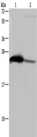
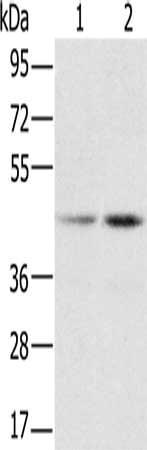
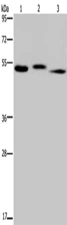
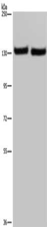
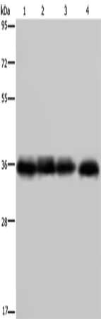
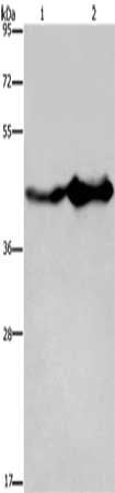 SDS-PAGE
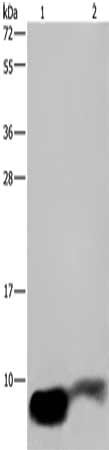
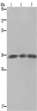
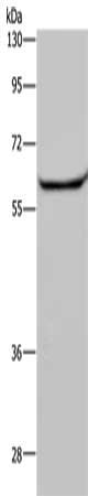
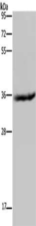
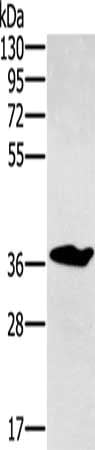
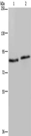
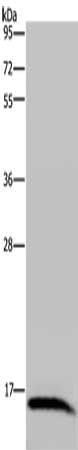
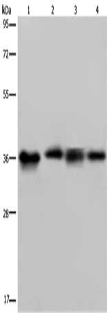
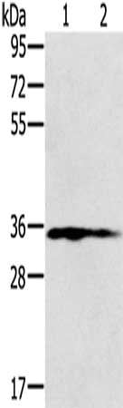
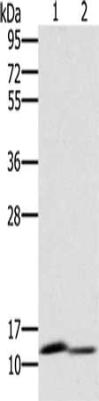
(Gel: 8%SDS-PAGE, Lysate: 40 ug, Lane 1-2: Mouse liver tissue, Mouse brain tissue, Primary antibody: AAA238960(GLUL Antibody) at dilution 1/600, Secondary antibody: Goat anti rabbit IgG at 1/8000 dilution, Exposure time: 10 seconds)
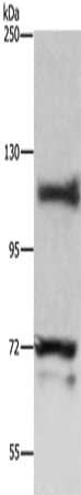
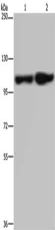
SDS-PAGE
(Gel: 8%SDS-PAGE, Lysate: 40 ug, Lane 1-2: Mouse liver tissue, Mouse brain tissue, Primary antibody: AAA238960(GLUL Antibody) at dilution 1/600, Secondary antibody: Goat anti rabbit IgG at 1/8000 dilution, Exposure time: 10 seconds)
GLUL, Polyclonal Antibody (Cat# AAA238960)
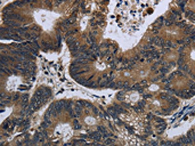
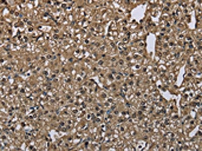
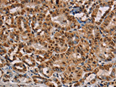
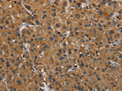
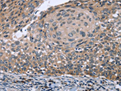
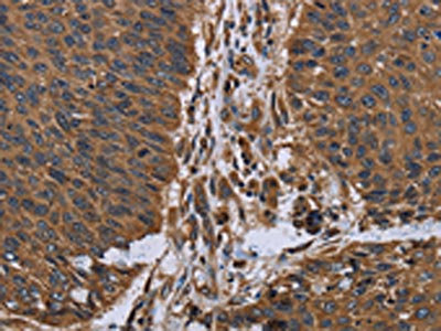
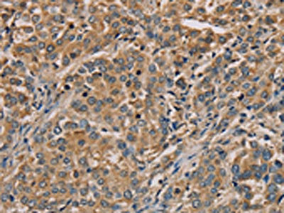
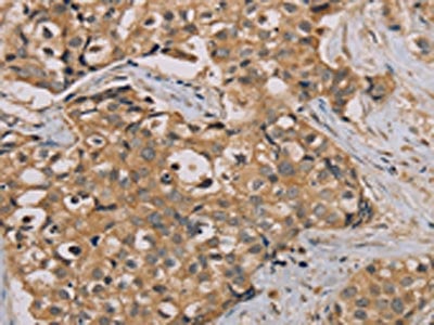
 IHC (Immunohiostchemistry)
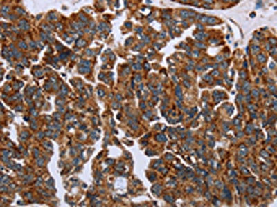
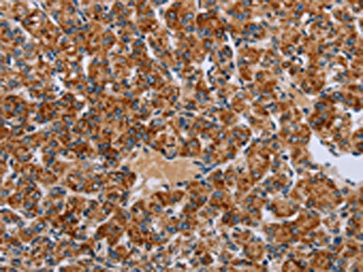
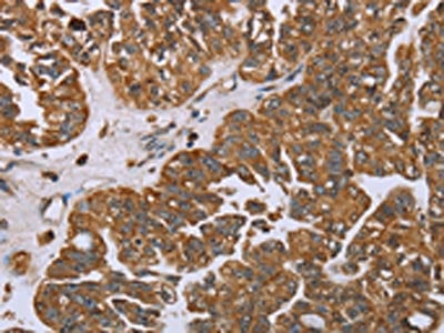
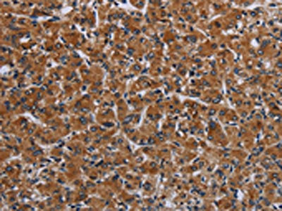
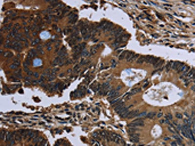
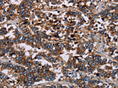
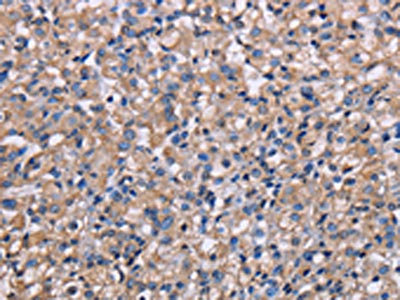
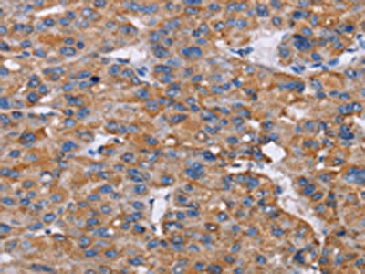
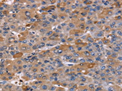
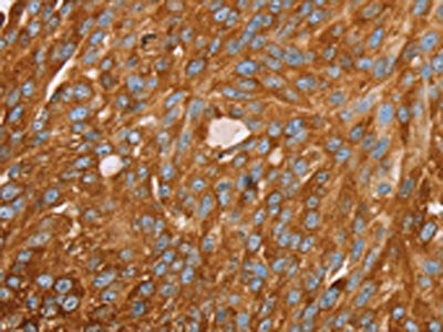
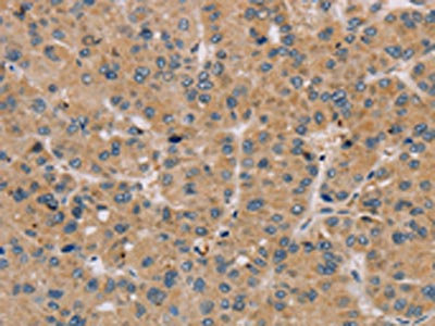
(The image on the left is immunohistochemistry of paraffin-embedded Human liver cancer tissue using AAA238963(GK Antibody) at dilution 1/60, on the right is treated with fusion protein. (Original magnification: ×200))
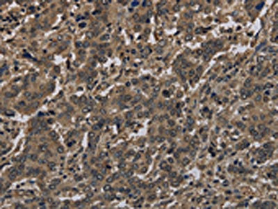
IHC (Immunohiostchemistry)
(The image on the left is immunohistochemistry of paraffin-embedded Human liver cancer tissue using AAA238963(GK Antibody) at dilution 1/60, on the right is treated with fusion protein. (Original magnification: ×200))
GK, Polyclonal Antibody (Cat# AAA238963)
 SDS-PAGE
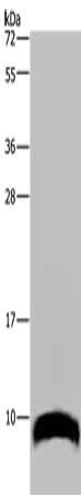
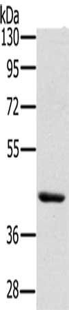
(Gel: 10%SDS-PAGE, Lysate: 40 ug, Lane: Mouse liver tissue, Primary antibody: AAA238964(PAEP Antibody) at dilution 1/300, Secondary antibody: Goat anti rabbit IgG at 1/8000 dilution, Exposure time: 5 minutes)
SDS-PAGE
(Gel: 10%SDS-PAGE, Lysate: 40 ug, Lane: Mouse liver tissue, Primary antibody: AAA238964(PAEP Antibody) at dilution 1/300, Secondary antibody: Goat anti rabbit IgG at 1/8000 dilution, Exposure time: 5 minutes)
PAEP, Polyclonal Antibody (Cat# AAA238964)
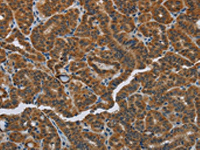
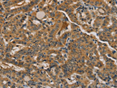
 IHC (Immunohiostchemistry)
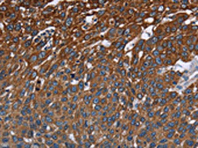
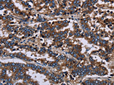
(The image on the left is immunohistochemistry of paraffin-embedded Human tonsil tissue using AAA238971(GYPC Antibody) at dilution 1/60, on the right is treated with fusion protein. (Original magnification: ×200))
IHC (Immunohiostchemistry)
(The image on the left is immunohistochemistry of paraffin-embedded Human tonsil tissue using AAA238971(GYPC Antibody) at dilution 1/60, on the right is treated with fusion protein. (Original magnification: ×200))
GYPC, Polyclonal Antibody (Cat# AAA238971)
 SDS-PAGE
(Gel: 10%SDS-PAGE, Lysate: 40 ug, Lane: Mouse paranephros tissue, Primary antibody: AAA238974(GNG2 Antibody) at dilution 1/300, Secondary antibody: Goat anti rabbit IgG at 1/8000 dilution, Exposure time: 5 minutes)
SDS-PAGE
(Gel: 10%SDS-PAGE, Lysate: 40 ug, Lane: Mouse paranephros tissue, Primary antibody: AAA238974(GNG2 Antibody) at dilution 1/300, Secondary antibody: Goat anti rabbit IgG at 1/8000 dilution, Exposure time: 5 minutes)
GNG2, Polyclonal Antibody (Cat# AAA238974)
 SDS-PAGE
(Gel: 10%SDS-PAGE, Lysate: 40 ug, Lane 1-2: Human fetal brain tissue, mouse brain tissue, Primary antibody: AAA238975(GNG2 Antibody) at dilution 1/350, Secondary antibody: Goat anti rabbit IgG at 1/8000 dilution, Exposure time: 2 minutes)
SDS-PAGE
(Gel: 10%SDS-PAGE, Lysate: 40 ug, Lane 1-2: Human fetal brain tissue, mouse brain tissue, Primary antibody: AAA238975(GNG2 Antibody) at dilution 1/350, Secondary antibody: Goat anti rabbit IgG at 1/8000 dilution, Exposure time: 2 minutes)
GNG2, Polyclonal Antibody (Cat# AAA238975)
 SDS-PAGE
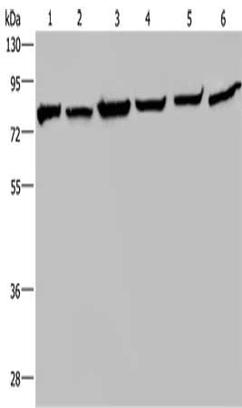
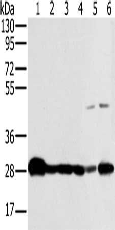
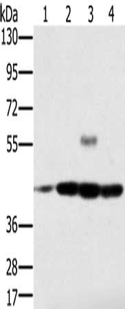
(Gel: 8%SDS-PAGE, Lysate: 40 ug, Lane 1-6: Mouse kidney tissue, HT29 cells, A549 cells, 293T cells, MCF7 cells, Hela cells, Primary antibody: AAA238981(HSPA9 Antibody) at dilution 1/400, Secondary antibody: Goat anti rabbit IgG at 1/8000 dilution, Exposure time: 10 seconds)
SDS-PAGE
(Gel: 8%SDS-PAGE, Lysate: 40 ug, Lane 1-6: Mouse kidney tissue, HT29 cells, A549 cells, 293T cells, MCF7 cells, Hela cells, Primary antibody: AAA238981(HSPA9 Antibody) at dilution 1/400, Secondary antibody: Goat anti rabbit IgG at 1/8000 dilution, Exposure time: 10 seconds)
HSPA9, Polyclonal Antibody (Cat# AAA238981)
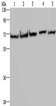 SDS-PAGE
(Gel: 8%SDS-PAGE, Lysate: 40 ug, Lane 1-5: HT29 cells, A549 cells, 293T cells, MCF7 cells, Hela cells, Primary antibody: AAA238982(HSPA9 Antibody) at dilution 1/300, Secondary antibody: Goat anti rabbit IgG at 1/8000 dilution, Exposure time: 10 seconds)
SDS-PAGE
(Gel: 8%SDS-PAGE, Lysate: 40 ug, Lane 1-5: HT29 cells, A549 cells, 293T cells, MCF7 cells, Hela cells, Primary antibody: AAA238982(HSPA9 Antibody) at dilution 1/300, Secondary antibody: Goat anti rabbit IgG at 1/8000 dilution, Exposure time: 10 seconds)
HSPA9, Polyclonal Antibody (Cat# AAA238982)
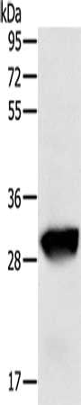 SDS-PAGE
(Gel: 8%SDS-PAGE, Lysate: 50 ug, Lane: Human liver cancer tissue, Primary antibody: AAA238983(GSTO1 Antibody) at dilution 1/200, Secondary antibody: Goat anti rabbit IgG at 1/8000 dilution, Exposure time: 20 seconds)
SDS-PAGE
(Gel: 8%SDS-PAGE, Lysate: 50 ug, Lane: Human liver cancer tissue, Primary antibody: AAA238983(GSTO1 Antibody) at dilution 1/200, Secondary antibody: Goat anti rabbit IgG at 1/8000 dilution, Exposure time: 20 seconds)
GSTO1, Polyclonal Antibody (Cat# AAA238983)
 SDS-PAGE
(Gel: 10%SDS-PAGE, Lysate: 40 ug, Lane 1-2: Human testis tissue, HepG2 cells, Primary antibody: AAA238985(GSTA3 Antibody) at dilution 1/250, Secondary antibody: Goat anti rabbit IgG at 1/8000 dilution, Exposure time: 10 seconds)
SDS-PAGE
(Gel: 10%SDS-PAGE, Lysate: 40 ug, Lane 1-2: Human testis tissue, HepG2 cells, Primary antibody: AAA238985(GSTA3 Antibody) at dilution 1/250, Secondary antibody: Goat anti rabbit IgG at 1/8000 dilution, Exposure time: 10 seconds)
GSTA3, Polyclonal Antibody (Cat# AAA238985)
 SDS-PAGE
(Gel: 10%SDS-PAGE, Lysate: 40 ug, Lane: Human testis tissue, Primary antibody: AAA238986(GSTA3 Antibody) at dilution 1/250, Secondary antibody: Goat anti rabbit IgG at 1/8000 dilution, Exposure time: 10 seconds)
SDS-PAGE
(Gel: 10%SDS-PAGE, Lysate: 40 ug, Lane: Human testis tissue, Primary antibody: AAA238986(GSTA3 Antibody) at dilution 1/250, Secondary antibody: Goat anti rabbit IgG at 1/8000 dilution, Exposure time: 10 seconds)
GSTA3, Polyclonal Antibody (Cat# AAA238986)
 SDS-PAGE
(Gel: 8%SDS-PAGE, Lysate: 60 ug, Lane: Hela cells, Primary antibody: AAA238991(HIF1A Antibody) at dilution 1/500, Secondary antibody: Goat anti rabbit IgG at 1/8000 dilution, Exposure time: 10 minutes)
SDS-PAGE
(Gel: 8%SDS-PAGE, Lysate: 60 ug, Lane: Hela cells, Primary antibody: AAA238991(HIF1A Antibody) at dilution 1/500, Secondary antibody: Goat anti rabbit IgG at 1/8000 dilution, Exposure time: 10 minutes)
HIF1A, Polyclonal Antibody (Cat# AAA238991)
 SDS-PAGE
(Gel: 10%SDS-PAGE, Lysate: 40 ug, Lane 1-7: K562 cells, mouse pancreas tissue, Hela cells, mouse thymus tissue, 293T cells, NIH/3T3 cells, LoVo cells, Primary antibody: AAA238994(HIST4H4 Antibody) at dilution 1/300, Secondary antibody: Goat anti rabbit IgG at 1/8000 dilution, Exposure time: 10 seconds)
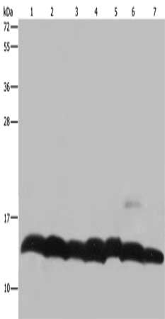
SDS-PAGE
(Gel: 10%SDS-PAGE, Lysate: 40 ug, Lane 1-7: K562 cells, mouse pancreas tissue, Hela cells, mouse thymus tissue, 293T cells, NIH/3T3 cells, LoVo cells, Primary antibody: AAA238994(HIST4H4 Antibody) at dilution 1/300, Secondary antibody: Goat anti rabbit IgG at 1/8000 dilution, Exposure time: 10 seconds)
HIST1H4A, Polyclonal Antibody (Cat# AAA238994)
 SDS-PAGE
(Gel: 8%SDS-PAGE, Lysate: 40 ug, Lane 1-3: SKOV3 cells, mouse heart tissue, Mouse brain tissue, Primary antibody: AAA238997(HMGCL Antibody) at dilution 1/150, Secondary antibody: Goat anti rabbit IgG at 1/8000 dilution, Exposure time: 5 seconds)
SDS-PAGE
(Gel: 8%SDS-PAGE, Lysate: 40 ug, Lane 1-3: SKOV3 cells, mouse heart tissue, Mouse brain tissue, Primary antibody: AAA238997(HMGCL Antibody) at dilution 1/150, Secondary antibody: Goat anti rabbit IgG at 1/8000 dilution, Exposure time: 5 seconds)
HMGCL, Polyclonal Antibody (Cat# AAA238997)
 SDS-PAGE
(Gel: 8%SDS-PAGE, Lysate: 40 ug, Lane 1-6: Mouse thymus tissue, human ovarian cancer tissue, mouse heart tissue, human fetal liver tissue, mouse liver tissue, SKOV3 cells, Primary antibody: AAA238998(HMGCL Antibody) at dilution 1/300, Secondary antibody: Goat anti rabbit IgG at 1/8000 dilution, Exposure time: 5 seconds)
SDS-PAGE
(Gel: 8%SDS-PAGE, Lysate: 40 ug, Lane 1-6: Mouse thymus tissue, human ovarian cancer tissue, mouse heart tissue, human fetal liver tissue, mouse liver tissue, SKOV3 cells, Primary antibody: AAA238998(HMGCL Antibody) at dilution 1/300, Secondary antibody: Goat anti rabbit IgG at 1/8000 dilution, Exposure time: 5 seconds)
HMGCL, Polyclonal Antibody (Cat# AAA238998)
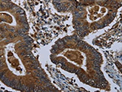
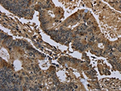
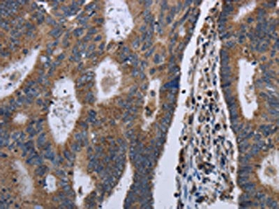
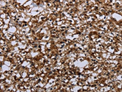
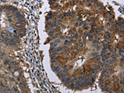
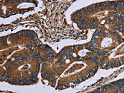
 IHC (Immunohiostchemistry)
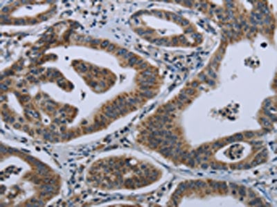
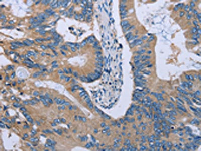
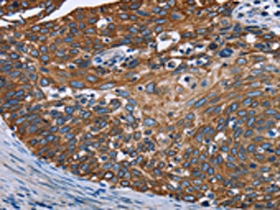
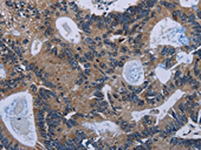
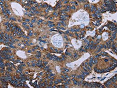
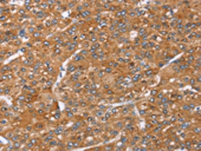
(The image on the left is immunohistochemistry of paraffin-embedded Human colon cancer tissue using AAA238999(HMGCR Antibody) at dilution 1/20, on the right is treated with fusion protein. (Original magnification: ×200))
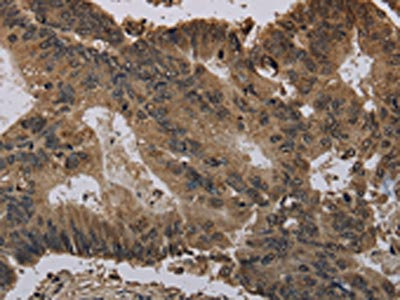
IHC (Immunohiostchemistry)
(The image on the left is immunohistochemistry of paraffin-embedded Human colon cancer tissue using AAA238999(HMGCR Antibody) at dilution 1/20, on the right is treated with fusion protein. (Original magnification: ×200))
HMGCR, Polyclonal Antibody (Cat# AAA238999)
 SDS-PAGE
(Gel: 8%SDS-PAGE, Lysate: 40 ug, Lane 1-2: Hela cells, K562 cells, Primary antibody: AAA239001(RBMX Antibody) at dilution 1/800, Secondary antibody: Goat anti rabbit IgG at 1/8000 dilution, Exposure time: 5 seconds)
SDS-PAGE
(Gel: 8%SDS-PAGE, Lysate: 40 ug, Lane 1-2: Hela cells, K562 cells, Primary antibody: AAA239001(RBMX Antibody) at dilution 1/800, Secondary antibody: Goat anti rabbit IgG at 1/8000 dilution, Exposure time: 5 seconds)
RBMX, Polyclonal Antibody (Cat# AAA239001)
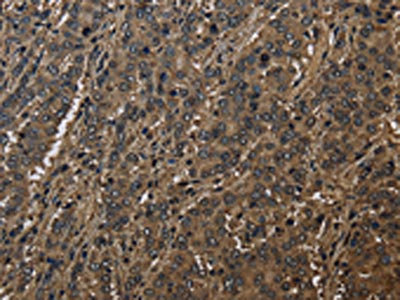
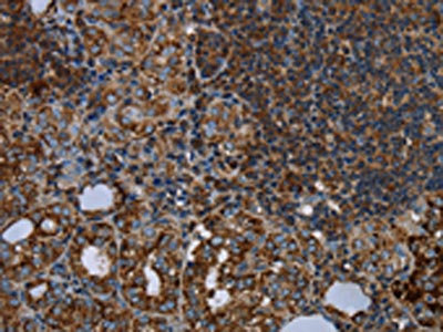
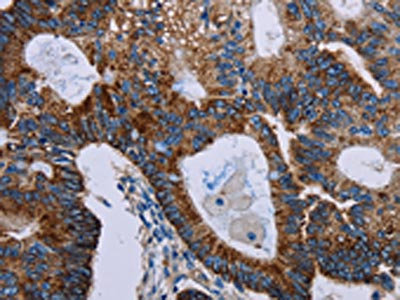
 IHC (Immunohiostchemistry)
(The image on the left is immunohistochemistry of paraffin-embedded Human gastric cancer tissue using AAA239005(HOMER2 Antibody) at dilution 1/30, on the right is treated with fusion protein. (Original magnification: ×200))
IHC (Immunohiostchemistry)
(The image on the left is immunohistochemistry of paraffin-embedded Human gastric cancer tissue using AAA239005(HOMER2 Antibody) at dilution 1/30, on the right is treated with fusion protein. (Original magnification: ×200))
HOMER2, Polyclonal Antibody (Cat# AAA239005)
 SDS-PAGE
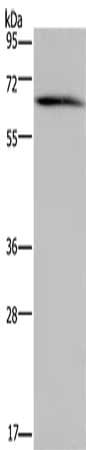
(Gel: 8%SDS-PAGE, Lysate: 40 ug, Lane: Human testis tissue, Primary antibody: AAA239006(HACL1 Antibody) at dilution 1/200, Secondary antibody: Goat anti rabbit IgG at 1/8000 dilution, Exposure time: 30 seconds)
SDS-PAGE
(Gel: 8%SDS-PAGE, Lysate: 40 ug, Lane: Human testis tissue, Primary antibody: AAA239006(HACL1 Antibody) at dilution 1/200, Secondary antibody: Goat anti rabbit IgG at 1/8000 dilution, Exposure time: 30 seconds)
HACL1, Polyclonal Antibody (Cat# AAA239006)
 SDS-PAGE
(Gel: 10%SDS-PAGE, Lysate: 40 ug, Lane: Mouse liver tissue, Primary antibody: AAA239020(HSD17B6 Antibody) at dilution 1/200, Secondary antibody: Goat anti rabbit IgG at 1/8000 dilution, Exposure time: 1 minute)
SDS-PAGE
(Gel: 10%SDS-PAGE, Lysate: 40 ug, Lane: Mouse liver tissue, Primary antibody: AAA239020(HSD17B6 Antibody) at dilution 1/200, Secondary antibody: Goat anti rabbit IgG at 1/8000 dilution, Exposure time: 1 minute)
HSD17B6, Polyclonal Antibody (Cat# AAA239020)
 SDS-PAGE
(Gel: 8%SDS-PAGE, Lysate: 40 ug, Lane: Human adrenal gland tissue, Primary antibody: AAA239025(HSD3B1 Antibody) at dilution 1/250, Secondary antibody: Goat anti rabbit IgG at 1/8000 dilution, Exposure time: 3 minutes)
SDS-PAGE
(Gel: 8%SDS-PAGE, Lysate: 40 ug, Lane: Human adrenal gland tissue, Primary antibody: AAA239025(HSD3B1 Antibody) at dilution 1/250, Secondary antibody: Goat anti rabbit IgG at 1/8000 dilution, Exposure time: 3 minutes)
HSD3B1, Polyclonal Antibody (Cat# AAA239025)
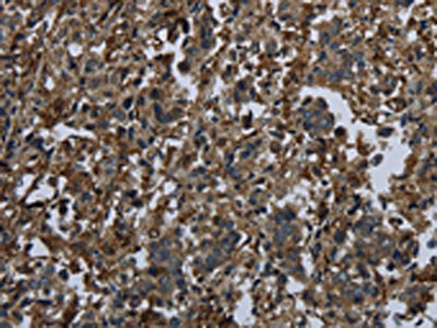
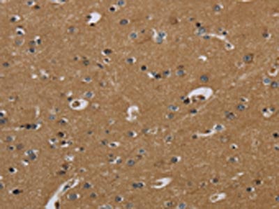
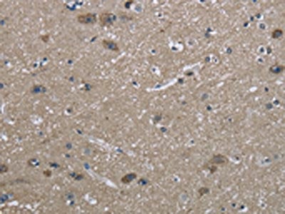
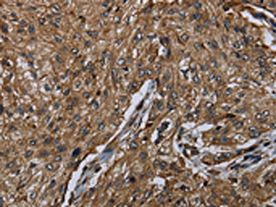
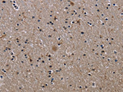
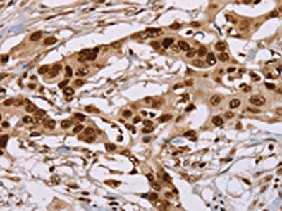
 IHC (Immunohiostchemistry)
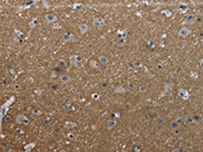
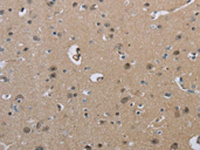
(The image on the left is immunohistochemistry of paraffin-embedded Human brain tissue using AAA239026(HSD3B7 Antibody) at dilution 1/40, on the right is treated with fusion protein. (Original magnification: ×200))
IHC (Immunohiostchemistry)
(The image on the left is immunohistochemistry of paraffin-embedded Human brain tissue using AAA239026(HSD3B7 Antibody) at dilution 1/40, on the right is treated with fusion protein. (Original magnification: ×200))
HSD3B7, Polyclonal Antibody (Cat# AAA239026)
 SDS-PAGE
(Gel: 8%SDS-PAGE, Lysate: 40 ug, Lane: Skov3 cells, Primary antibody: AAA239027(HSD3B7 Antibody) at dilution 1/400, Secondary antibody: Goat anti rabbit IgG at 1/8000 dilution, Exposure time: 10 seconds)
SDS-PAGE
(Gel: 8%SDS-PAGE, Lysate: 40 ug, Lane: Skov3 cells, Primary antibody: AAA239027(HSD3B7 Antibody) at dilution 1/400, Secondary antibody: Goat anti rabbit IgG at 1/8000 dilution, Exposure time: 10 seconds)
HSD3B7, Polyclonal Antibody (Cat# AAA239027)
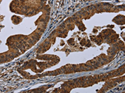
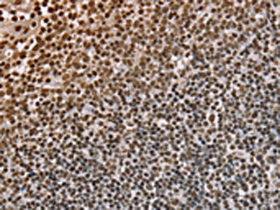
 IHC (Immunohiostchemistry)
(The image on the left is immunohistochemistry of paraffin-embedded Human prostate cancer tissue using AAA239028(HSD3B2 Antibody) at dilution 1/20, on the right is treated with fusion protein. (Original magnification: ×200))
IHC (Immunohiostchemistry)
(The image on the left is immunohistochemistry of paraffin-embedded Human prostate cancer tissue using AAA239028(HSD3B2 Antibody) at dilution 1/20, on the right is treated with fusion protein. (Original magnification: ×200))
HSD3B2, Polyclonal Antibody (Cat# AAA239028)
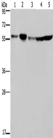
 SDS-PAGE
(Gel: 8%SDS-PAGE, Lysate: 40 ug, Lane 1-3: Human kidney tissue, HepG2 cells, human testis tissue, Primary antibody: AAA239033(HYAL3 Antibody) at dilution 1/400, Secondary antibody: Goat anti rabbit IgG at 1/8000 dilution, Exposure time: 1 minute)
SDS-PAGE
(Gel: 8%SDS-PAGE, Lysate: 40 ug, Lane 1-3: Human kidney tissue, HepG2 cells, human testis tissue, Primary antibody: AAA239033(HYAL3 Antibody) at dilution 1/400, Secondary antibody: Goat anti rabbit IgG at 1/8000 dilution, Exposure time: 1 minute)
HYAL3, Polyclonal Antibody (Cat# AAA239033)
 SDS-PAGE
(Gel: 8%SDS-PAGE, Lysate: 40 ug, Lane: TM4 cells, Primary antibody: AAA239035(RNF144B Antibody) at dilution 1/400, Secondary antibody: Goat anti rabbit IgG at 1/8000 dilution, Exposure time: 3 minutes)
SDS-PAGE
(Gel: 8%SDS-PAGE, Lysate: 40 ug, Lane: TM4 cells, Primary antibody: AAA239035(RNF144B Antibody) at dilution 1/400, Secondary antibody: Goat anti rabbit IgG at 1/8000 dilution, Exposure time: 3 minutes)
RNF144B, Polyclonal Antibody (Cat# AAA239035)
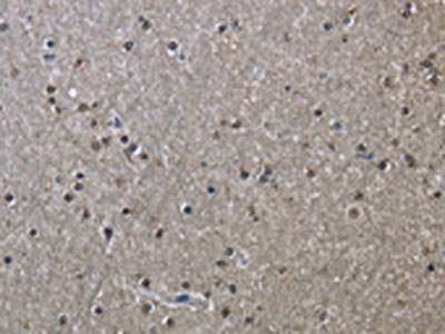
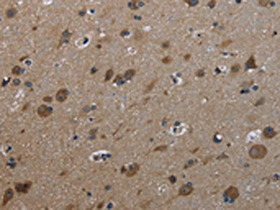
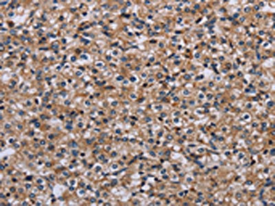
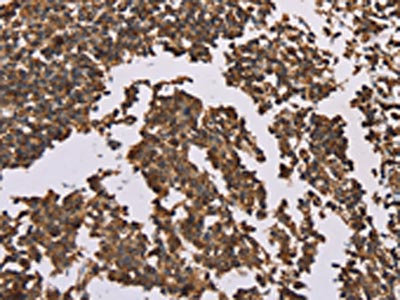
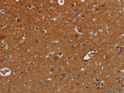 IHC (Immunohiostchemistry)
(The image on the left is immunohistochemistry of paraffin-embedded Human brain tissue using AAA239043(IFRD1 Antibody) at dilution 1/30, on the right is treated with fusion protein. (Original magnification: ×200))
IHC (Immunohiostchemistry)
(The image on the left is immunohistochemistry of paraffin-embedded Human brain tissue using AAA239043(IFRD1 Antibody) at dilution 1/30, on the right is treated with fusion protein. (Original magnification: ×200))
IFRD1, Polyclonal Antibody (Cat# AAA239043)
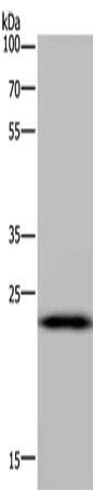
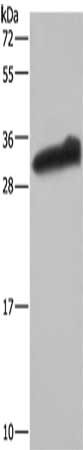
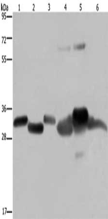
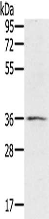
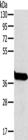
 SDS-PAGE
(Gel: 8%SDS-PAGE, Lysate: 40 ug, Lane: Mouse liver tissue, Primary antibody: AAA239044(NOV Antibody) at dilution 1/300, Secondary antibody: Goat anti rabbit IgG at 1/8000 dilution, Exposure time: 3 minutes)
SDS-PAGE
(Gel: 8%SDS-PAGE, Lysate: 40 ug, Lane: Mouse liver tissue, Primary antibody: AAA239044(NOV Antibody) at dilution 1/300, Secondary antibody: Goat anti rabbit IgG at 1/8000 dilution, Exposure time: 3 minutes)
NOV, Polyclonal Antibody (Cat# AAA239044)
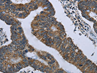
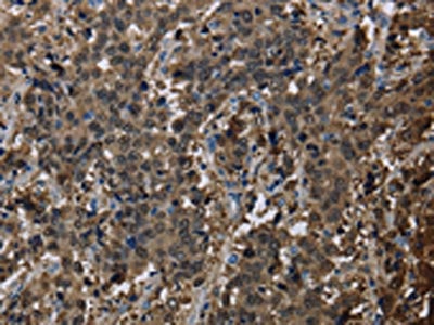
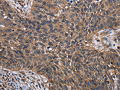
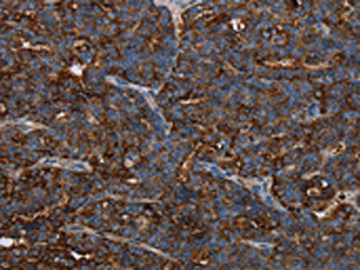
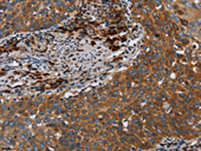
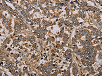
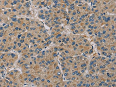
 IHC (Immunohiostchemistry)
(The image on the left is immunohistochemistry of paraffin-embedded Human cervical cancer tissue using AAA239045(FCGR3A Antibody) at dilution 1/40, on the right is treated with fusion protein. (Original magnification: ×200))
IHC (Immunohiostchemistry)
(The image on the left is immunohistochemistry of paraffin-embedded Human cervical cancer tissue using AAA239045(FCGR3A Antibody) at dilution 1/40, on the right is treated with fusion protein. (Original magnification: ×200))
FCGR3A, Polyclonal Antibody (Cat# AAA239045)
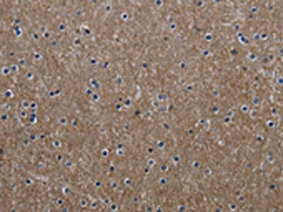
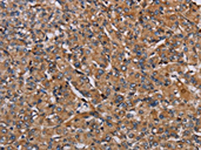
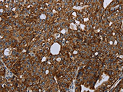
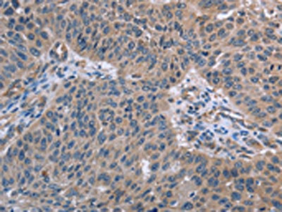
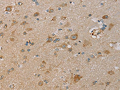
 IHC (Immunohiostchemistry)
(The image on the left is immunohistochemistry of paraffin-embedded Human lung cancer tissue using AAA239047(IHH Antibody) at dilution 1/30, on the right is treated with fusion protein. (Original magnification: ×200))
IHC (Immunohiostchemistry)
(The image on the left is immunohistochemistry of paraffin-embedded Human lung cancer tissue using AAA239047(IHH Antibody) at dilution 1/30, on the right is treated with fusion protein. (Original magnification: ×200))
IHH, Polyclonal Antibody (Cat# AAA239047)
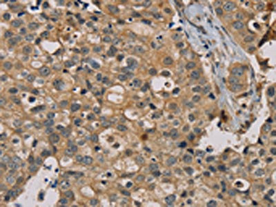
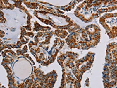
 IHC (Immunohiostchemistry)
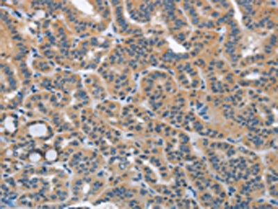
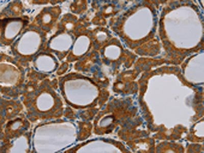
(The image on the left is immunohistochemistry of paraffin-embedded Human thyroid cancer tissue using AAA239050(IKBKE Antibody) at dilution 1/30, on the right is treated with fusion protein. (Original magnification: ×200))
IHC (Immunohiostchemistry)
(The image on the left is immunohistochemistry of paraffin-embedded Human thyroid cancer tissue using AAA239050(IKBKE Antibody) at dilution 1/30, on the right is treated with fusion protein. (Original magnification: ×200))
IKBKE, Polyclonal Antibody (Cat# AAA239050)
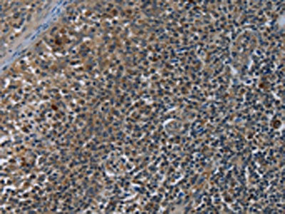
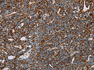
 IHC (Immunohiostchemistry)
(The image on the left is immunohistochemistry of paraffin-embedded Human prostate cancer tissue using AAA239059(IL31RA Antibody) at dilution 1/20, on the right is treated with fusion protein. (Original magnification: ×200))
IHC (Immunohiostchemistry)
(The image on the left is immunohistochemistry of paraffin-embedded Human prostate cancer tissue using AAA239059(IL31RA Antibody) at dilution 1/20, on the right is treated with fusion protein. (Original magnification: ×200))
IL31RA, Polyclonal Antibody (Cat# AAA239059)
 SDS-PAGE
(Gel: 6%SDS-PAGE, Lysate: 40 ug, Lane 1-2: A549 cells, human testis tissue, Primary antibody: AAA239064(IPO4 Antibody) at dilution 1/550, Secondary antibody: Goat anti rabbit IgG at 1/8000 dilution, Exposure time: 40 seconds)
SDS-PAGE
(Gel: 6%SDS-PAGE, Lysate: 40 ug, Lane 1-2: A549 cells, human testis tissue, Primary antibody: AAA239064(IPO4 Antibody) at dilution 1/550, Secondary antibody: Goat anti rabbit IgG at 1/8000 dilution, Exposure time: 40 seconds)
IPO4, Polyclonal Antibody (Cat# AAA239064)
 SDS-PAGE
(Gel: 6%SDS-PAGE, Lysate: 40 ug, Lane 1-2: Human fetal liver tissue, Human placenta tissue, Primary antibody: AAA239066(ITGA2B Antibody) at dilution 1/200, Secondary antibody: Goat anti rabbit IgG at 1/8000 dilution, Exposure time: 3 minutes)
SDS-PAGE
(Gel: 6%SDS-PAGE, Lysate: 40 ug, Lane 1-2: Human fetal liver tissue, Human placenta tissue, Primary antibody: AAA239066(ITGA2B Antibody) at dilution 1/200, Secondary antibody: Goat anti rabbit IgG at 1/8000 dilution, Exposure time: 3 minutes)
ITGA2B, Polyclonal Antibody (Cat# AAA239066)
 SDS-PAGE
(Gel: 6%SDS-PAGE, Lysate: 40 ug, Lane 1-2: 293T cells, K562 cells, Primary antibody: AAA239069(ITGB2 Antibody) at dilution 1/600, Secondary antibody: Goat anti rabbit IgG at 1/8000 dilution, Exposure time: 3 minutes)
SDS-PAGE
(Gel: 6%SDS-PAGE, Lysate: 40 ug, Lane 1-2: 293T cells, K562 cells, Primary antibody: AAA239069(ITGB2 Antibody) at dilution 1/600, Secondary antibody: Goat anti rabbit IgG at 1/8000 dilution, Exposure time: 3 minutes)
ITGB2, Polyclonal Antibody (Cat# AAA239069)
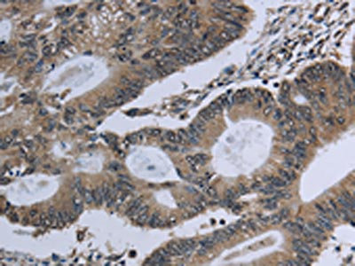
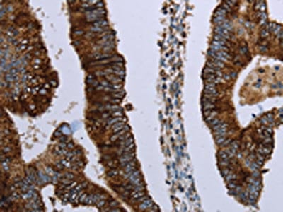
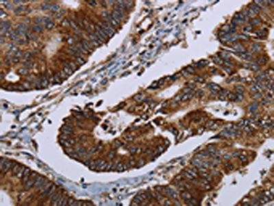
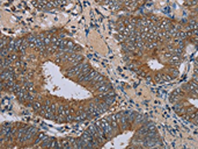
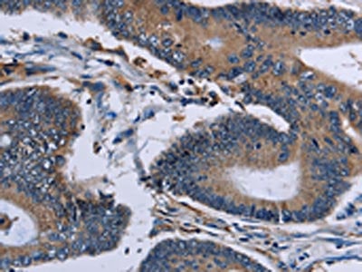
 IHC (Immunohiostchemistry)
(The image on the left is immunohistochemistry of paraffin-embedded Human colon cancer tissue using AAA239070(IRGM Antibody) at dilution 1/50, on the right is treated with fusion protein. (Original magnification: ×200))
IHC (Immunohiostchemistry)
(The image on the left is immunohistochemistry of paraffin-embedded Human colon cancer tissue using AAA239070(IRGM Antibody) at dilution 1/50, on the right is treated with fusion protein. (Original magnification: ×200))
IRGM, Polyclonal Antibody (Cat# AAA239070)
 SDS-PAGE
(Gel: 10%SDS-PAGE, Lysate: 40 ug, Lane: Human hepatocellular carcinoma tissue, Primary antibody: AAA239071(IRGM Antibody) at dilution 1/200, Secondary antibody: Goat anti rabbit IgG at 1/8000 dilution, Exposure time: 5 minutes)
SDS-PAGE
(Gel: 10%SDS-PAGE, Lysate: 40 ug, Lane: Human hepatocellular carcinoma tissue, Primary antibody: AAA239071(IRGM Antibody) at dilution 1/200, Secondary antibody: Goat anti rabbit IgG at 1/8000 dilution, Exposure time: 5 minutes)
IRGM, Polyclonal Antibody (Cat# AAA239071)
 SDS-PAGE
(Gel: 8%SDS-PAGE, Lysate: 40 ug, Lane 1-4: HepG2 cells, 293T cells, human kidney cancer tissue, K562 cells, Primary antibody: AAA239073(F11R Antibody) at dilution 1/500, Secondary antibody: Goat anti rabbit IgG at 1/8000 dilution, Exposure time: 5 seconds)
SDS-PAGE
(Gel: 8%SDS-PAGE, Lysate: 40 ug, Lane 1-4: HepG2 cells, 293T cells, human kidney cancer tissue, K562 cells, Primary antibody: AAA239073(F11R Antibody) at dilution 1/500, Secondary antibody: Goat anti rabbit IgG at 1/8000 dilution, Exposure time: 5 seconds)
F11R, Polyclonal Antibody (Cat# AAA239073)
 SDS-PAGE
(Gel: 8%SDS-PAGE, Lysate: 40 ug, Lane 1-4: K562 cells, human kidney cancer tissue, 293T cells, HepG2 cells, Primary antibody: AAA239074(F11R Antibody) at dilution 1/600, Secondary antibody: Goat anti rabbit IgG at 1/8000 dilution, Exposure time: 5 seconds)
SDS-PAGE
(Gel: 8%SDS-PAGE, Lysate: 40 ug, Lane 1-4: K562 cells, human kidney cancer tissue, 293T cells, HepG2 cells, Primary antibody: AAA239074(F11R Antibody) at dilution 1/600, Secondary antibody: Goat anti rabbit IgG at 1/8000 dilution, Exposure time: 5 seconds)
F11R, Polyclonal Antibody (Cat# AAA239074)
 IHC (Immunohiostchemistry)
(The image on the left is immunohistochemistry of paraffin-embedded Human breast cancer tissue using AAA239193(NANOS2 Antibody) at dilution 1/30, on the right is treated with fusion protein. (Original magnification: ×200))
IHC (Immunohiostchemistry)
(The image on the left is immunohistochemistry of paraffin-embedded Human breast cancer tissue using AAA239193(NANOS2 Antibody) at dilution 1/30, on the right is treated with fusion protein. (Original magnification: ×200))
NANOS2, Polyclonal Antibody (Cat# AAA239193)
 IHC (Immunohiostchemistry)
(The image on the left is immunohistochemistry of paraffin-embedded Human breast cancer tissue using AAA239194(NANOS2 Antibody) at dilution 1/20, on the right is treated with fusion protein. (Original magnification: ×200))
IHC (Immunohiostchemistry)
(The image on the left is immunohistochemistry of paraffin-embedded Human breast cancer tissue using AAA239194(NANOS2 Antibody) at dilution 1/20, on the right is treated with fusion protein. (Original magnification: ×200))
NANOS2, Polyclonal Antibody (Cat# AAA239194)
 SDS-PAGE
(Gel: 8%SDS-PAGE, Lysate: 40 ug, Lane 1-6: Mouse brain tissue, human fetal brain tissue, hela cells, Raw264.7 cells, NIH/3T3 cells, 293T cells, Primary antibody: AAA239209(NDUFS3 Antibody) at dilution 1/300, Secondary antibody: Goat anti rabbit IgG at 1/8000 dilution, Exposure time: 5 seconds)
SDS-PAGE
(Gel: 8%SDS-PAGE, Lysate: 40 ug, Lane 1-6: Mouse brain tissue, human fetal brain tissue, hela cells, Raw264.7 cells, NIH/3T3 cells, 293T cells, Primary antibody: AAA239209(NDUFS3 Antibody) at dilution 1/300, Secondary antibody: Goat anti rabbit IgG at 1/8000 dilution, Exposure time: 5 seconds)
NDUFS3, Polyclonal Antibody (Cat# AAA239209)
 SDS-PAGE
(Gel: 8%SDS-PAGE, Lysate: 40 ug, Lane 1-2: Human placenta tissue, Human fetal liver tissue, Primary antibody: AAA239225(NIT1 Antibody) at dilution 1/250, Secondary antibody: Goat anti rabbit IgG at 1/8000 dilution, Exposure time: 2 minutes)
SDS-PAGE
(Gel: 8%SDS-PAGE, Lysate: 40 ug, Lane 1-2: Human placenta tissue, Human fetal liver tissue, Primary antibody: AAA239225(NIT1 Antibody) at dilution 1/250, Secondary antibody: Goat anti rabbit IgG at 1/8000 dilution, Exposure time: 2 minutes)
NIT1, Polyclonal Antibody (Cat# AAA239225)
 SDS-PAGE
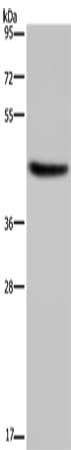
(Gel: 8%SDS-PAGE, Lysate: 40 ug, Lane: Human brain malignant glioma tissue, Primary antibody: AAA239229(NCF2 Antibody) at dilution 1/200, Secondary antibody: Goat anti rabbit IgG at 1/8000 dilution, Exposure time: 5 seconds)
SDS-PAGE
(Gel: 8%SDS-PAGE, Lysate: 40 ug, Lane: Human brain malignant glioma tissue, Primary antibody: AAA239229(NCF2 Antibody) at dilution 1/200, Secondary antibody: Goat anti rabbit IgG at 1/8000 dilution, Exposure time: 5 seconds)
NCF2, Polyclonal Antibody (Cat# AAA239229)
 SDS-PAGE
(Gel: 8%SDS-PAGE, Lysate: 40 ug, Lane 1-5: Hela cells, 293T cells, A549 cells, HT29 cells, K562 cells, Primary antibody: AAA239238(NUP50 Antibody) at dilution 1/200, Secondary antibody: Goat anti rabbit IgG at 1/8000 dilution, Exposure time: 20 seconds)
SDS-PAGE
(Gel: 8%SDS-PAGE, Lysate: 40 ug, Lane 1-5: Hela cells, 293T cells, A549 cells, HT29 cells, K562 cells, Primary antibody: AAA239238(NUP50 Antibody) at dilution 1/200, Secondary antibody: Goat anti rabbit IgG at 1/8000 dilution, Exposure time: 20 seconds)
NUP50, Polyclonal Antibody (Cat# AAA239238)
 SDS-PAGE
(Gel: 8%SDS-PAGE, Lysate: 40 ug, Lane: Mouse liver tissue, Primary antibody: AAA239241(OTC Antibody) at dilution 1/800, Secondary antibody: Goat anti rabbit IgG at 1/8000 dilution, Exposure time: 1 second)
SDS-PAGE
(Gel: 8%SDS-PAGE, Lysate: 40 ug, Lane: Mouse liver tissue, Primary antibody: AAA239241(OTC Antibody) at dilution 1/800, Secondary antibody: Goat anti rabbit IgG at 1/8000 dilution, Exposure time: 1 second)
OTC, Polyclonal Antibody (Cat# AAA239241)
 SDS-PAGE
(Gel: 10%SDS-PAGE, Lysate: 40 ug, Lane 1-2: Mouse brain tissue, Mouse kidney tissue, Primary antibody: AAA239247(PAGE2 Antibody) at dilution 1/400, Secondary antibody: Goat anti rabbit IgG at 1/8000 dilution, Exposure time: 1 minute)
SDS-PAGE
(Gel: 10%SDS-PAGE, Lysate: 40 ug, Lane 1-2: Mouse brain tissue, Mouse kidney tissue, Primary antibody: AAA239247(PAGE2 Antibody) at dilution 1/400, Secondary antibody: Goat anti rabbit IgG at 1/8000 dilution, Exposure time: 1 minute)
PAGE2, Polyclonal Antibody (Cat# AAA239247)
 SDS-PAGE
(Gel: 8%SDS-PAGE, Lysate: 40 ug, Lane 1-4: Jurkat cells, Raji cells, K562 cells, hela cells, Primary antibody: AAA239248(PAICS Antibody) at dilution 1/400, Secondary antibody: Goat anti rabbit IgG at 1/8000 dilution, Exposure time: 20 seconds)
SDS-PAGE
(Gel: 8%SDS-PAGE, Lysate: 40 ug, Lane 1-4: Jurkat cells, Raji cells, K562 cells, hela cells, Primary antibody: AAA239248(PAICS Antibody) at dilution 1/400, Secondary antibody: Goat anti rabbit IgG at 1/8000 dilution, Exposure time: 20 seconds)
PAICS, Polyclonal Antibody (Cat# AAA239248)
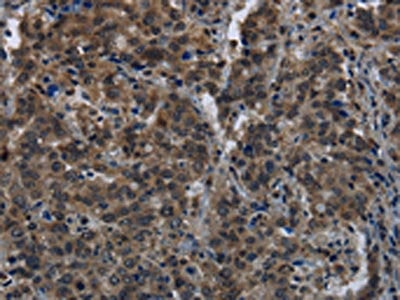
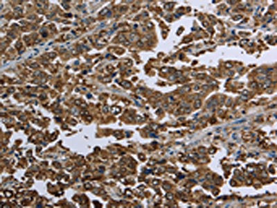
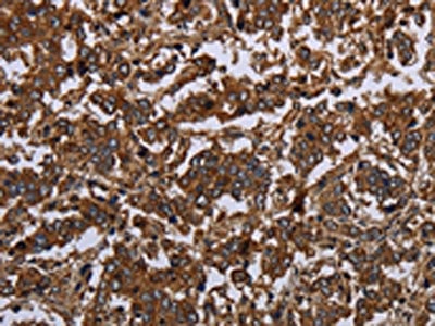
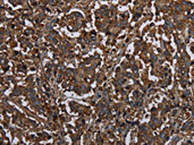
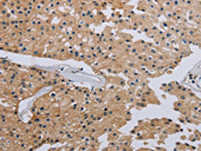
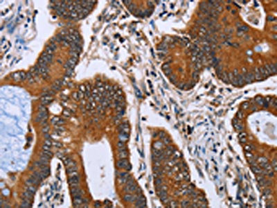
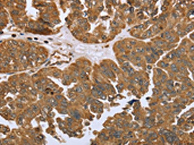
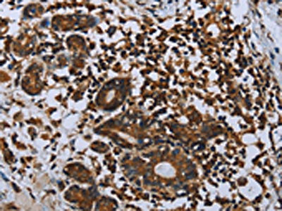
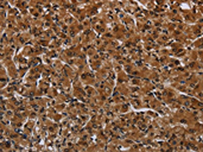
 IHC (Immunohiostchemistry)
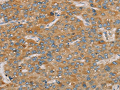
(The image on the left is immunohistochemistry of paraffin-embedded Human liver cancer tissue using AAA239251(PARL Antibody) at dilution 1/25, on the right is treated with fusion protein. (Original magnification: ×200))
IHC (Immunohiostchemistry)
(The image on the left is immunohistochemistry of paraffin-embedded Human liver cancer tissue using AAA239251(PARL Antibody) at dilution 1/25, on the right is treated with fusion protein. (Original magnification: ×200))
PARL, Polyclonal Antibody (Cat# AAA239251)
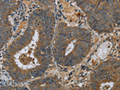
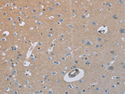
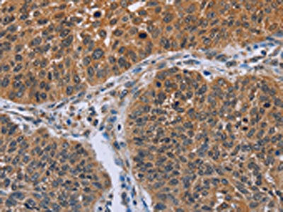
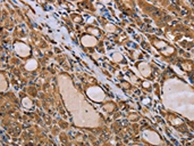
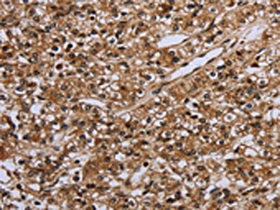
 IHC (Immunohiostchemistry)
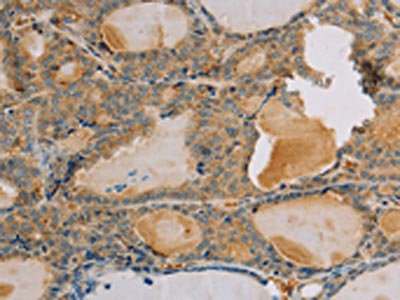
(The image on the left is immunohistochemistry of paraffin-embedded Human thyroid cancer tissue using AAA239254(PARP3 Antibody) at dilution 1/25, on the right is treated with fusion protein. (Original magnification: ×200))
IHC (Immunohiostchemistry)
(The image on the left is immunohistochemistry of paraffin-embedded Human thyroid cancer tissue using AAA239254(PARP3 Antibody) at dilution 1/25, on the right is treated with fusion protein. (Original magnification: ×200))
PARP3, Polyclonal Antibody (Cat# AAA239254)
What are Polyclonal Antibodies?
Polyclonal antibodies are antibodies that come from multiple B cell clones of a host animal. The typical hosts used for the majority of polyclonal antibody production are rabbits, goats, sheep, and donkeys. These polyclonal antibodies, once having identified their target, will bind to different epitopes located at different regions or sequences on the same protein/antigen. As a result, they are ideal at locating and binding to the target, even if the target is in very low concentrations (due to many different antibodies being able to bind to the same target molecule, which allows for significant amplification of a downstream signal).
Polyclonal antibodies are typically produced by injecting an antigen into a host animal, which causes the animal’s immune system to attack the foreign antigen by mass generating antibodies against it. After a period of time, serum is collected from the animal and purified using physicochemical fractionation, class-specific affinity purification, and/or antigen-affinity purification.
Key Uses of Polyclonal Antibodies
- Western Blotting: This method is used to find specific proteins in biological samples after separating them by size.
- Immunohistochemistry: IHC helps visualize the location of proteins in tissue sections using various staining techniques.
- ELISA: (Enzyme-Linked Immunosorbent Assay) is typically used to identify specific protein quantities in a sample. ELISAs can be either “Quantitative” or “Qualitative”.
- Flow Cytometry: technique that identifies and measures the specific protein on the surface or inside the cells in a fluid suspension.
- Immunoprecipitation: IP isolates and studies a specific protein from a complex mixture using antibodies.
Why Buy Polyclonal Antibodies from AAA Biotech?
1. Ideal for Various Applications
Our antibodies are generally going to be validated for use in multiple types of assays, including ELISA, Western Blotting, Immunohistochemistry, Immunoprecipitation, amongst others. They are ideal for a wide range of research applications.
2. Rigorous Quality Control
All of the antibodies in our catalog undergo strict quality testing to ensure specificity, sensitivity, and consistent performance. We are confident in the ability of our antibodies to provide you with accurate results.
3. Wide Assortment of Antibodies
Antibodies in are catalog can be found for both common and exotic species, and these antibodies are also available in both conjugated and recombinant forms to suit many diverse experimental needs.
4. Highly Purified
Our antibodies are available in purified forms with over 85% purity, as confirmed by SDS-PAGE. They are also available with tags such as His, Flag, GST, or MBP. We cater to customers worldwide.
FAQ
1. How are polyclonal antibodies produced?
Traditionally, polyclonal antibodies are produced by injecting an antigen into a host animal (such as a rabbit or goat), which then triggers an immune response from the host animal. The animal’s B cells produce antibodies that will recognize different parts of the injected antigen. These antibodies are then collected from the animal’s blood and purified for use.
2. How do polyclonal antibodies differ from monoclonal antibodies?
Polyclonal antibodies are a mix of antibodies that bind to different locations (epitopes) of the same antigen, while monoclonal antibodies are identical and bind to just one specific epitope. This makes polyclonal antibodies more versatile and better at detecting proteins that may be present in low quantities or in altered/modified forms.
3. How should I store polyclonal antibodies?
Polyclonal antibodies should be stored at 4°C for short-term use (up to a few weeks) and at -20°C or -80°C for long-term storage. Avoid repeated freeze-thaw cycles by dividing them into small aliquots. Always check the datasheet for specific storage instructions.