Filters
▼Clonality
▼Type
▼Reactivity
▼Gene Name
▼Isotype
▼Host
▼Application
▼Clone
▼Polyclonal Antibodies
At AAA Biotech also known as AAA Bio or AAABio, we provide a broad range of purified polyclonal antibodies (pAbs) that are able to all be browsed online through our website. Due to their high specificity and strong binding affinity, these antibodies are ideal for wide swathes of research and experimental applications.
Our polyclonal antibodies can easily support your work, whether you use them for Western Blotting, Immunocytochemistry (with or without Immunofluorescence used in conjunction), Immunohistochemistry, Immunoprecipitation, and ELISA tests. We highly encourage you to browse our range of pAbs and choose the one that best suits your experimental model.
Viewing 7050-7100 of 96805 product results
 IHC (Immunohiostchemistry)
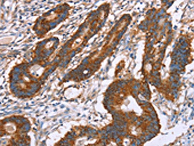
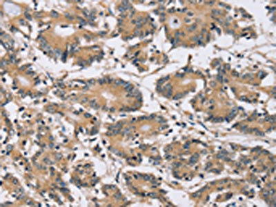
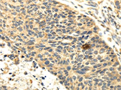
(The image on the left is immunohistochemistry of paraffin-embedded Human lung cancer tissue using AAA239255(PARP11 Antibody) at dilution 1/30, on the right is treated with fusion protein. (Original magnification: ×200))
IHC (Immunohiostchemistry)
(The image on the left is immunohistochemistry of paraffin-embedded Human lung cancer tissue using AAA239255(PARP11 Antibody) at dilution 1/30, on the right is treated with fusion protein. (Original magnification: ×200))
PARP11, Polyclonal Antibody (Cat# AAA239255)
 IHC (Immunohiostchemistry)
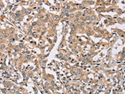
(The image on the left is immunohistochemistry of paraffin-embedded Human breast cancer tissue using AAA239258(SCGB2A1 Antibody) at dilution 1/20, on the right is treated with fusion protein. (Original magnification: ×200))
IHC (Immunohiostchemistry)
(The image on the left is immunohistochemistry of paraffin-embedded Human breast cancer tissue using AAA239258(SCGB2A1 Antibody) at dilution 1/20, on the right is treated with fusion protein. (Original magnification: ×200))
SCGB2A1, Polyclonal Antibody (Cat# AAA239258)
 SDS-PAGE
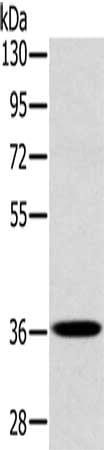
(Gel: 8%SDS-PAGE, Lysate: 40 ug, Lane: A172 cells, Primary antibody: AAA239259(PDLIM1 Antibody) at dilution 1/400, Secondary antibody: Goat anti rabbit IgG at 1/8000 dilution, Exposure time: 10 seconds)
SDS-PAGE
(Gel: 8%SDS-PAGE, Lysate: 40 ug, Lane: A172 cells, Primary antibody: AAA239259(PDLIM1 Antibody) at dilution 1/400, Secondary antibody: Goat anti rabbit IgG at 1/8000 dilution, Exposure time: 10 seconds)
PDLIM1, Polyclonal Antibody (Cat# AAA239259)
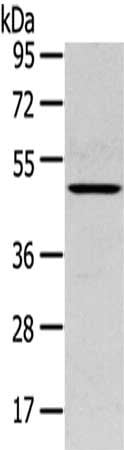
 SDS-PAGE
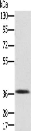
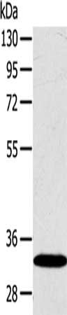
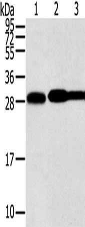
(Gel: 8%SDS-PAGE, Lysate: 40 ug, Lane: Mouse liver tissue, Primary antibody: AAA239260(PDLIM2 Antibody) at dilution 1/250, Secondary antibody: Goat anti rabbit IgG at 1/8000 dilution, Exposure time: 3 minutes)
SDS-PAGE
(Gel: 8%SDS-PAGE, Lysate: 40 ug, Lane: Mouse liver tissue, Primary antibody: AAA239260(PDLIM2 Antibody) at dilution 1/250, Secondary antibody: Goat anti rabbit IgG at 1/8000 dilution, Exposure time: 3 minutes)
PDLIM2, Polyclonal Antibody (Cat# AAA239260)
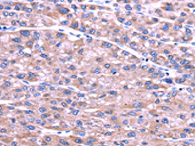
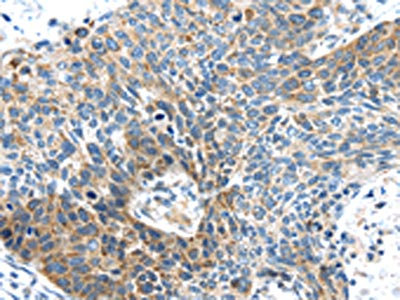
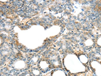
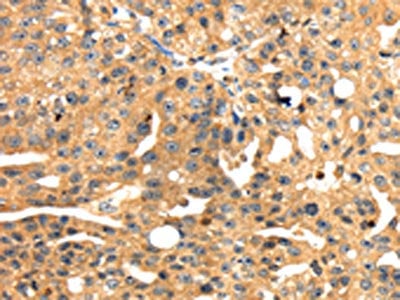
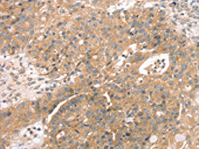
 IHC (Immunohiostchemistry)
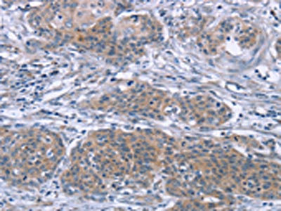
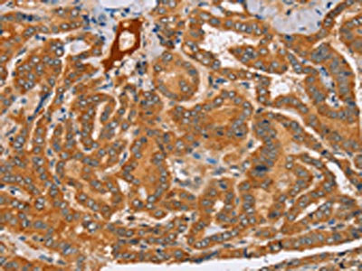
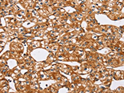
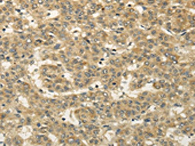
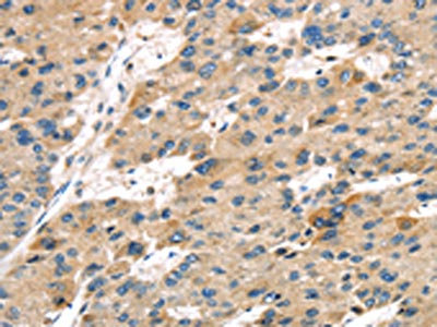
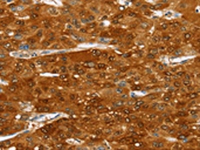
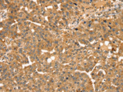
(The image on the left is immunohistochemistry of paraffin-embedded Human liver cancer tissue using AAA239262(PDLIM7 Antibody) at dilution 1/20, on the right is treated with fusion protein. (Original magnification: ×200))
IHC (Immunohiostchemistry)
(The image on the left is immunohistochemistry of paraffin-embedded Human liver cancer tissue using AAA239262(PDLIM7 Antibody) at dilution 1/20, on the right is treated with fusion protein. (Original magnification: ×200))
PDLIM7, Polyclonal Antibody (Cat# AAA239262)
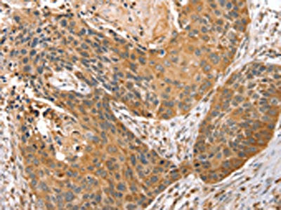
 IHC (Immunohiostchemistry)
(The image on the left is immunohistochemistry of paraffin-embedded Human prostate cancer tissue using AAA239263(PEA15 Antibody) at dilution 1/25, on the right is treated with fusion protein. (Original magnification: ×200))
IHC (Immunohiostchemistry)
(The image on the left is immunohistochemistry of paraffin-embedded Human prostate cancer tissue using AAA239263(PEA15 Antibody) at dilution 1/25, on the right is treated with fusion protein. (Original magnification: ×200))
PEA15, Polyclonal Antibody (Cat# AAA239263)
 SDS-PAGE
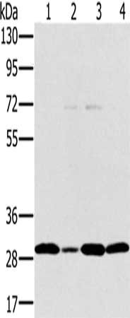
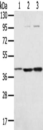
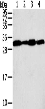
(Gel: 8%SDS-PAGE, Lysate: 40 ug, Lane 1-4: Jurkat cells, hela cells, 293T cells, human thyroid cancer tissue, Primary antibody: AAA239265(PEF1 Antibody) at dilution 1/200, Secondary antibody: Goat anti rabbit IgG at 1/8000 dilution, Exposure time: 20 seconds)
SDS-PAGE
(Gel: 8%SDS-PAGE, Lysate: 40 ug, Lane 1-4: Jurkat cells, hela cells, 293T cells, human thyroid cancer tissue, Primary antibody: AAA239265(PEF1 Antibody) at dilution 1/200, Secondary antibody: Goat anti rabbit IgG at 1/8000 dilution, Exposure time: 20 seconds)
PEF1, Polyclonal Antibody (Cat# AAA239265)
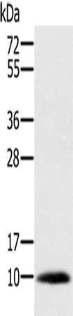 SDS-PAGE
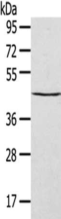
(Gel: 10%SDS-PAGE, Lysate: 40 ug, Lane: Human prostate tissue, Primary antibody: AAA239267(MSMB Antibody) at dilution 1/200, Secondary antibody: Goat anti rabbit IgG at 1/8000 dilution, Exposure time: 1 second)
SDS-PAGE
(Gel: 10%SDS-PAGE, Lysate: 40 ug, Lane: Human prostate tissue, Primary antibody: AAA239267(MSMB Antibody) at dilution 1/200, Secondary antibody: Goat anti rabbit IgG at 1/8000 dilution, Exposure time: 1 second)
MSMB, Polyclonal Antibody (Cat# AAA239267)
 SDS-PAGE
(Gel: 12%SDS-PAGE, Lysate: 40 ug, Lane 1-2: K562 cells, hepg2 cells, Primary antibody: AAA239360(RPLP1 Antibody) at dilution 1/400, Secondary antibody: Goat anti rabbit IgG at 1/8000 dilution, Exposure time: 2 minutes)
SDS-PAGE
(Gel: 12%SDS-PAGE, Lysate: 40 ug, Lane 1-2: K562 cells, hepg2 cells, Primary antibody: AAA239360(RPLP1 Antibody) at dilution 1/400, Secondary antibody: Goat anti rabbit IgG at 1/8000 dilution, Exposure time: 2 minutes)
RPLP1, Polyclonal Antibody (Cat# AAA239360)
 SDS-PAGE
(Gel: 6%SDS-PAGE, Lysate: 40 ug, Lane: K562 cells, Primary antibody: AAA239364(RPS6KA1 Antibody) at dilution 1/400, Secondary antibody: Goat anti rabbit IgG at 1/8000 dilution, Exposure time: 5 seconds)
SDS-PAGE
(Gel: 6%SDS-PAGE, Lysate: 40 ug, Lane: K562 cells, Primary antibody: AAA239364(RPS6KA1 Antibody) at dilution 1/400, Secondary antibody: Goat anti rabbit IgG at 1/8000 dilution, Exposure time: 5 seconds)
RPS6KA1, Polyclonal Antibody (Cat# AAA239364)
 SDS-PAGE
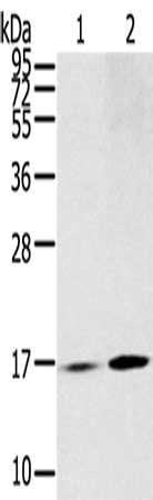
(Gel: 12%SDS-PAGE, Lysate: 40 ug, Lane 1-2: Hepg2 cells, A549 cells, Primary antibody: AAA239370(S100A11 Antibody) at dilution 1/600, Secondary antibody: Goat anti rabbit IgG at 1/8000 dilution, Exposure time: 1 seconds)
SDS-PAGE
(Gel: 12%SDS-PAGE, Lysate: 40 ug, Lane 1-2: Hepg2 cells, A549 cells, Primary antibody: AAA239370(S100A11 Antibody) at dilution 1/600, Secondary antibody: Goat anti rabbit IgG at 1/8000 dilution, Exposure time: 1 seconds)
S100A11, Polyclonal Antibody (Cat# AAA239370)
 IHC (Immunohiostchemistry)
(The image on the left is immunohistochemistry of paraffin-embedded Human esophagus cancer tissue using AAA239372(S100A14 Antibody) at dilution 1/30, on the right is treated with fusion protein. (Original magnification: ×200))
IHC (Immunohiostchemistry)
(The image on the left is immunohistochemistry of paraffin-embedded Human esophagus cancer tissue using AAA239372(S100A14 Antibody) at dilution 1/30, on the right is treated with fusion protein. (Original magnification: ×200))
S100A14, Polyclonal Antibody (Cat# AAA239372)
 IHC (Immunohiostchemistry)
(The image on the left is immunohistochemistry of paraffin-embedded Human esophagus cancer tissue using AAA239374(SAMD3 Antibody) at dilution 1/30, on the right is treated with fusion protein. (Original magnification: ×200))
IHC (Immunohiostchemistry)
(The image on the left is immunohistochemistry of paraffin-embedded Human esophagus cancer tissue using AAA239374(SAMD3 Antibody) at dilution 1/30, on the right is treated with fusion protein. (Original magnification: ×200))
SAMD3, Polyclonal Antibody (Cat# AAA239374)
 SDS-PAGE
(Gel: 8%SDS-PAGE, Lysate: 40 ug, Lane 1-4: Mouse muscle tissue, Jurkat cells, K562 cells, A431 cells, Primary antibody: AAA239378(MAPK12 Antibody) at dilution 1/350, Secondary antibody: Goat anti rabbit IgG at 1/8000 dilution, Exposure time: 30 seconds)
SDS-PAGE
(Gel: 8%SDS-PAGE, Lysate: 40 ug, Lane 1-4: Mouse muscle tissue, Jurkat cells, K562 cells, A431 cells, Primary antibody: AAA239378(MAPK12 Antibody) at dilution 1/350, Secondary antibody: Goat anti rabbit IgG at 1/8000 dilution, Exposure time: 30 seconds)
MAPK12, Polyclonal Antibody (Cat# AAA239378)
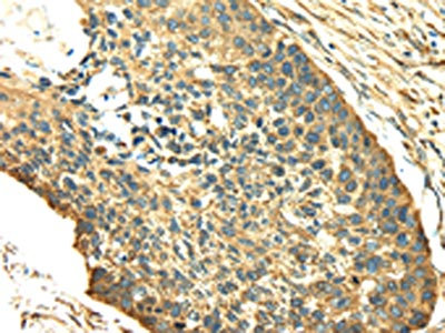
 IHC (Immunohiostchemistry)
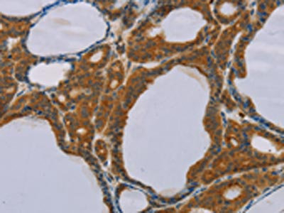
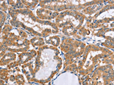
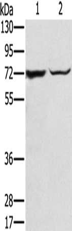
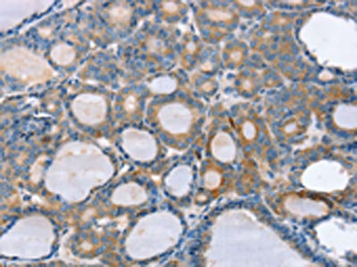
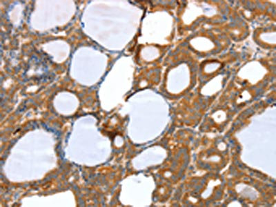
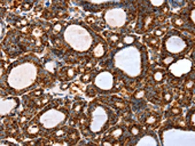
(The image on the left is immunohistochemistry of paraffin-embedded Human thyroid cancer tissue using AAA239384(SDF4 Antibody) at dilution 1/40, on the right is treated with fusion protein. (Original magnification: ×200))
IHC (Immunohiostchemistry)
(The image on the left is immunohistochemistry of paraffin-embedded Human thyroid cancer tissue using AAA239384(SDF4 Antibody) at dilution 1/40, on the right is treated with fusion protein. (Original magnification: ×200))
SDF4, Polyclonal Antibody (Cat# AAA239384)
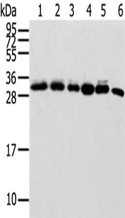
 SDS-PAGE
(Gel: 8%SDS-PAGE,Lysate: 40 ug,Lane 1-2: Mouse heart tissue, Mouse brain tissue,Primary antibody: AAA239385(SDHA Antibody) at dilution 1/600 dilution,Secondary antibody: Goat anti rabbit IgG at 1/8000 dilution,Exposure time: 5 seconds)
SDS-PAGE
(Gel: 8%SDS-PAGE,Lysate: 40 ug,Lane 1-2: Mouse heart tissue, Mouse brain tissue,Primary antibody: AAA239385(SDHA Antibody) at dilution 1/600 dilution,Secondary antibody: Goat anti rabbit IgG at 1/8000 dilution,Exposure time: 5 seconds)
SDHA, Polyclonal Antibody (Cat# AAA239385)
 SDS-PAGE
(Gel: 8%SDS-PAGE,Lysate: 40 ug,Lane 1-2: Jurkat cells, HepG2 cells,Primary antibody: AAA239388(DHCR24 Antibody) at dilution 1/600 dilution,Secondary antibody: Goat anti rabbit IgG at 1/8000 dilution,Exposure time: 2 minutes)
SDS-PAGE
(Gel: 8%SDS-PAGE,Lysate: 40 ug,Lane 1-2: Jurkat cells, HepG2 cells,Primary antibody: AAA239388(DHCR24 Antibody) at dilution 1/600 dilution,Secondary antibody: Goat anti rabbit IgG at 1/8000 dilution,Exposure time: 2 minutes)
DHCR24, Polyclonal Antibody (Cat# AAA239388)
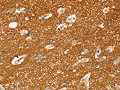
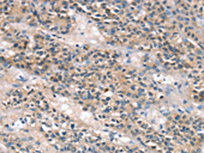
 IHC (Immunohiostchemistry)
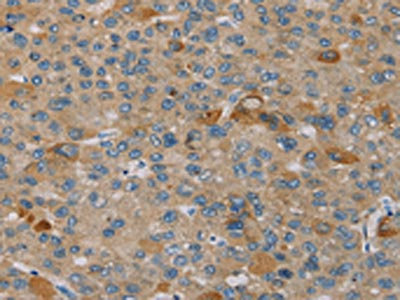
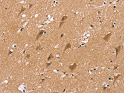
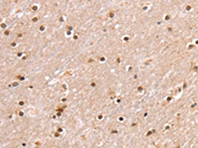
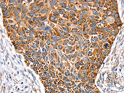
(The image on the left is immunohistochemistry of paraffin-embedded Human brain tissue using AAA239391(SEMA4F Antibody) at dilution 1/35, on the right is treated with fusion protein. (Original magnification: ×200))
IHC (Immunohiostchemistry)
(The image on the left is immunohistochemistry of paraffin-embedded Human brain tissue using AAA239391(SEMA4F Antibody) at dilution 1/35, on the right is treated with fusion protein. (Original magnification: ×200))
SEMA4F, Polyclonal Antibody (Cat# AAA239391)
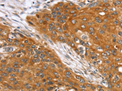
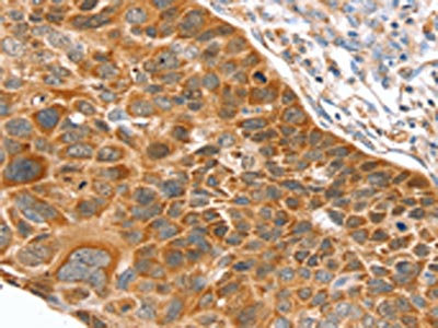
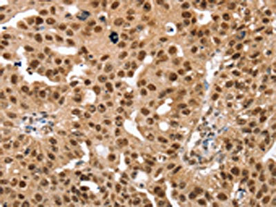
 IHC (Immunohiostchemistry)
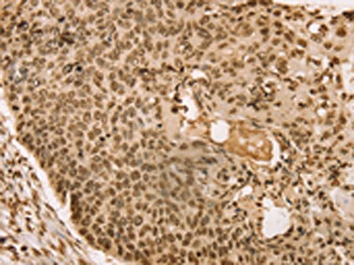
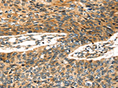
(The image on the left is immunohistochemistry of paraffin-embedded Human prostate cancer tissue using AAA239392(SEMA4F Antibody) at dilution 1/30, on the right is treated with fusion protein. (Original magnification: ×200))
IHC (Immunohiostchemistry)
(The image on the left is immunohistochemistry of paraffin-embedded Human prostate cancer tissue using AAA239392(SEMA4F Antibody) at dilution 1/30, on the right is treated with fusion protein. (Original magnification: ×200))
SEMA4F, Polyclonal Antibody (Cat# AAA239392)
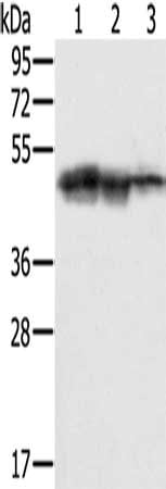
 SDS-PAGE
(Gel: 12%SDS-PAGE,Lysate: 40 ug,Lane 1-6: 293T cells, Hela cells, Lovo cells, Raji cells, HUEVC cells, Mouse spleen tissue,Primary antibody: AAA239395(SENP8 Antibody) at dilution 1/600 dilution,Secondary antibody: Goat anti rabbit IgG at 1/8000 dilution,Exposure time: 20 seconds)
SDS-PAGE
(Gel: 12%SDS-PAGE,Lysate: 40 ug,Lane 1-6: 293T cells, Hela cells, Lovo cells, Raji cells, HUEVC cells, Mouse spleen tissue,Primary antibody: AAA239395(SENP8 Antibody) at dilution 1/600 dilution,Secondary antibody: Goat anti rabbit IgG at 1/8000 dilution,Exposure time: 20 seconds)
SENP8, Polyclonal Antibody (Cat# AAA239395)
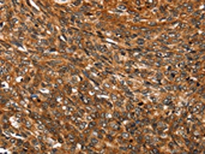
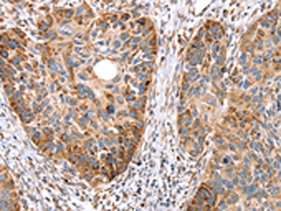
 IHC (Immunohiostchemistry)
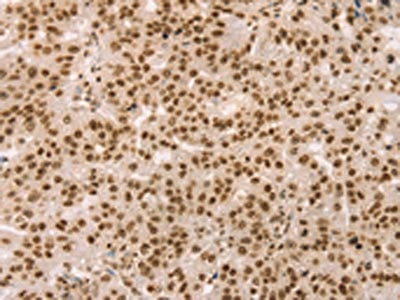
(The image on the left is immunohistochemistry of paraffin-embedded Human breast cancer tissue using AAA239398(SENP2 Antibody) at dilution 1/20, on the right is treated with fusion protein. (Original magnification: ×200))
IHC (Immunohiostchemistry)
(The image on the left is immunohistochemistry of paraffin-embedded Human breast cancer tissue using AAA239398(SENP2 Antibody) at dilution 1/20, on the right is treated with fusion protein. (Original magnification: ×200))
SENP2, Polyclonal Antibody (Cat# AAA239398)
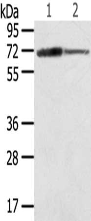
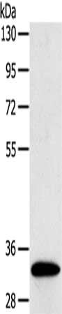
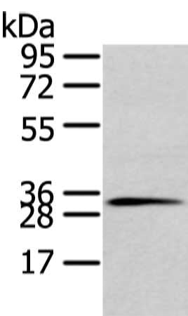
 SDS-PAGE
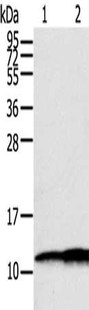
(Gel: 8%SDS-PAGE,Lysate: 40 ug,,Primary antibody: AAA239399(SEPT2 Antibody) at dilution 1/400 dilution,Secondary antibody: Goat anti rabbit IgG at 1/8000 dilution,Exposure time: 10S)
SDS-PAGE
(Gel: 8%SDS-PAGE,Lysate: 40 ug,,Primary antibody: AAA239399(SEPT2 Antibody) at dilution 1/400 dilution,Secondary antibody: Goat anti rabbit IgG at 1/8000 dilution,Exposure time: 10S)
SEPT2, Polyclonal Antibody (Cat# AAA239399)
 SDS-PAGE
(Gel: 8%SDS-PAGE,Lysate: 40 ug,,Primary antibody: AAA239402(SERPINB8 Antibody) at dilution 1/400 dilution,Secondary antibody: Goat anti rabbit IgG at 1/8000 dilution,Exposure time: 15 seconds)
SDS-PAGE
(Gel: 8%SDS-PAGE,Lysate: 40 ug,,Primary antibody: AAA239402(SERPINB8 Antibody) at dilution 1/400 dilution,Secondary antibody: Goat anti rabbit IgG at 1/8000 dilution,Exposure time: 15 seconds)
SERPINB8, Polyclonal Antibody (Cat# AAA239402)
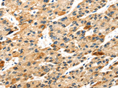
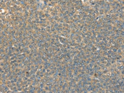
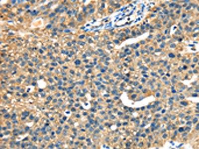
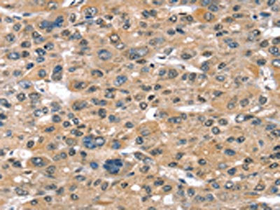
 IHC (Immunohiostchemistry)
(The image on the left is immunohistochemistry of paraffin-embedded Human esophagus cancer tissue using AAA239403(DNAJA3 Antibody) at dilution 1/30, on the right is treated with fusion protein. (Original magnification: ×200))
IHC (Immunohiostchemistry)
(The image on the left is immunohistochemistry of paraffin-embedded Human esophagus cancer tissue using AAA239403(DNAJA3 Antibody) at dilution 1/30, on the right is treated with fusion protein. (Original magnification: ×200))
DNAJA3, Polyclonal Antibody (Cat# AAA239403)
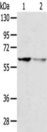
 SDS-PAGE
(Gel: 8%SDS-PAGE,Lysate: 40 ug,,Primary antibody: AAA239404(SERPINB11 Antibody) at dilution 1/400 dilution,Secondary antibody: Goat anti rabbit IgG at 1/8000 dilution,Exposure time: 30 seconds)
SDS-PAGE
(Gel: 8%SDS-PAGE,Lysate: 40 ug,,Primary antibody: AAA239404(SERPINB11 Antibody) at dilution 1/400 dilution,Secondary antibody: Goat anti rabbit IgG at 1/8000 dilution,Exposure time: 30 seconds)
SERPINB11, Polyclonal Antibody (Cat# AAA239404)
 SDS-PAGE
(Gel: 8%SDS-PAGE,Lysate: 40 ug,,Primary antibody: AAA239406(SH3KBP1 Antibody) at dilution 1/800 dilution,Secondary antibody: Goat anti rabbit IgG at 1/8000 dilution,Exposure time: 30 seconds)
SDS-PAGE
(Gel: 8%SDS-PAGE,Lysate: 40 ug,,Primary antibody: AAA239406(SH3KBP1 Antibody) at dilution 1/800 dilution,Secondary antibody: Goat anti rabbit IgG at 1/8000 dilution,Exposure time: 30 seconds)
SH3KBP1, Polyclonal Antibody (Cat# AAA239406)
 SDS-PAGE
(Gel: 8%SDS-PAGE,Lysate: 40 ug,,Primary antibody: AAA239409(SIGLEC12 Antibody) at dilution 1/500 dilution,Secondary antibody: Goat anti rabbit IgG at 1/8000 dilution,Exposure time: 30 seconds)
SDS-PAGE
(Gel: 8%SDS-PAGE,Lysate: 40 ug,,Primary antibody: AAA239409(SIGLEC12 Antibody) at dilution 1/500 dilution,Secondary antibody: Goat anti rabbit IgG at 1/8000 dilution,Exposure time: 30 seconds)
SIGLEC12, Polyclonal Antibody (Cat# AAA239409)
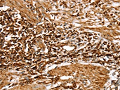
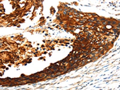
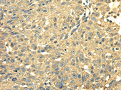
 IHC (Immunohiostchemistry)
(The image on the left is immunohistochemistry of paraffin-embedded Human breast cancer tissue using AAA239412(SRPK1 Antibody) at dilution 1/35, on the right is treated with fusion protein. (Original magnification: ×200))
IHC (Immunohiostchemistry)
(The image on the left is immunohistochemistry of paraffin-embedded Human breast cancer tissue using AAA239412(SRPK1 Antibody) at dilution 1/35, on the right is treated with fusion protein. (Original magnification: ×200))
SRPK1, Polyclonal Antibody (Cat# AAA239412)
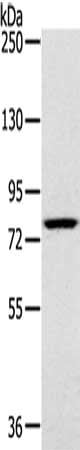
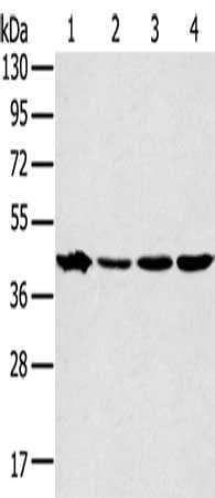
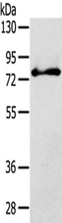
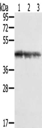
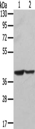
 SDS-PAGE
(Gel: 8%SDS-PAGE, Lysate: 40 ug, Lane 1-2: Mouse liver tissue, hepg2 cells, Primary antibody: AAA239417(SLC25A13 Antibody) at dilution 1/450, Secondary antibody: Goat anti rabbit IgG at 1/8000 dilution, Exposure time: 20 seconds)
SDS-PAGE
(Gel: 8%SDS-PAGE, Lysate: 40 ug, Lane 1-2: Mouse liver tissue, hepg2 cells, Primary antibody: AAA239417(SLC25A13 Antibody) at dilution 1/450, Secondary antibody: Goat anti rabbit IgG at 1/8000 dilution, Exposure time: 20 seconds)
SLC25A13, Polyclonal Antibody (Cat# AAA239417)
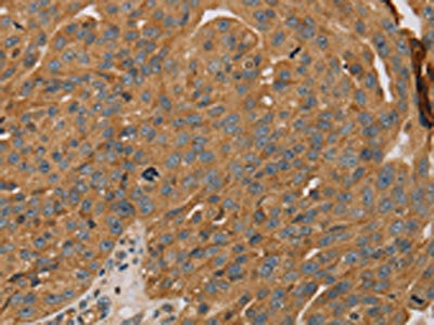
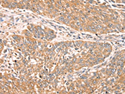
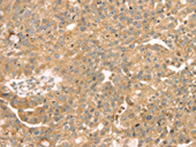
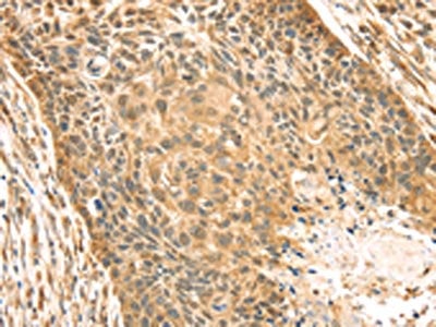
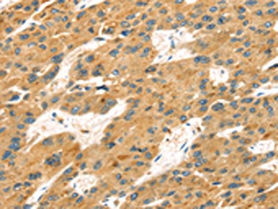
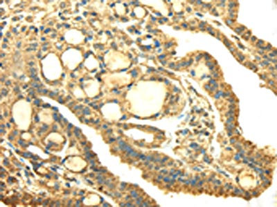
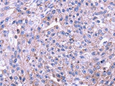
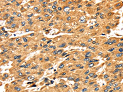
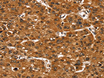
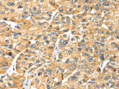
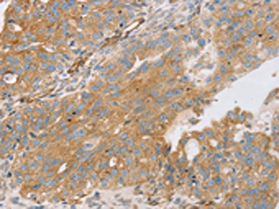
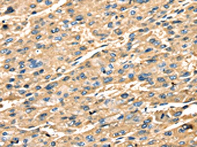
 IHC (Immunohiostchemistry)
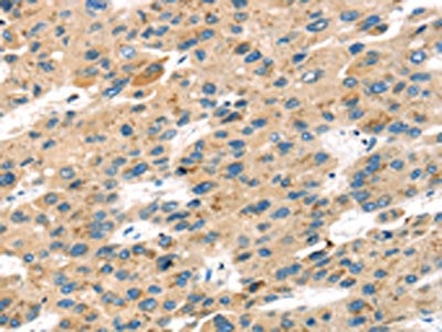
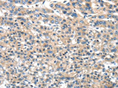
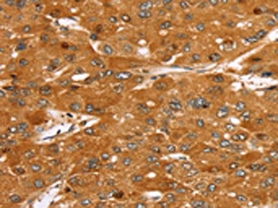
(The image on the left is immunohistochemistry of paraffin-embedded Human liver cancer tissue using AAA239418(SLC25A1 Antibody) at dilution 1/25, on the right is treated with fusion protein. (Original magnification: ×200))
IHC (Immunohiostchemistry)
(The image on the left is immunohistochemistry of paraffin-embedded Human liver cancer tissue using AAA239418(SLC25A1 Antibody) at dilution 1/25, on the right is treated with fusion protein. (Original magnification: ×200))
SLC25A1, Polyclonal Antibody (Cat# AAA239418)

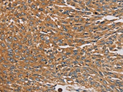
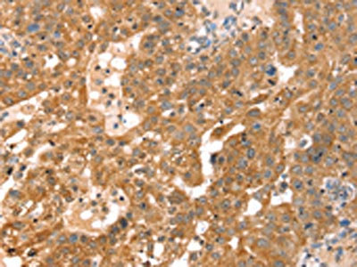
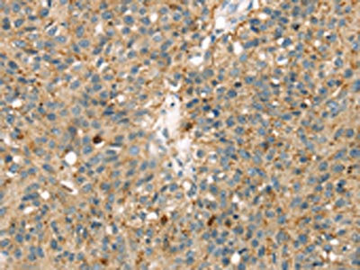
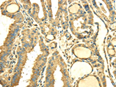
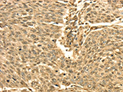
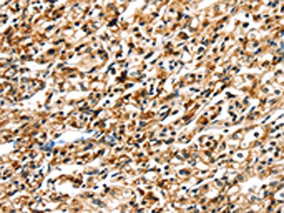
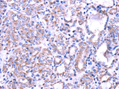
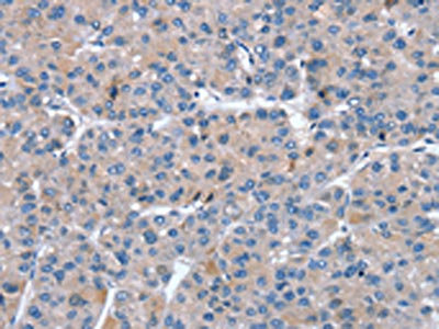
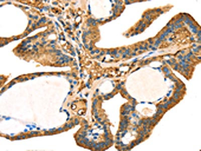
 IHC (Immunohiostchemistry)
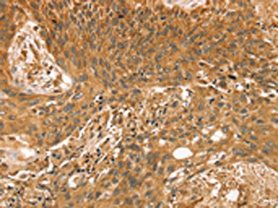
(The image on the left is immunohistochemistry of paraffin-embedded Human esophagus cancer tissue using AAA239419(SLC27A2 Antibody) at dilution 1/20, on the right is treated with fusion protein. (Original magnification: ×200))
IHC (Immunohiostchemistry)
(The image on the left is immunohistochemistry of paraffin-embedded Human esophagus cancer tissue using AAA239419(SLC27A2 Antibody) at dilution 1/20, on the right is treated with fusion protein. (Original magnification: ×200))
SLC27A2, Polyclonal Antibody (Cat# AAA239419)
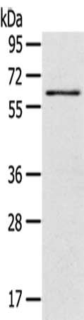
 SDS-PAGE
(Gel: 8%SDS-PAGE, Lysate: 40 ug, Lane 1-3: Hepg2 cells, K562 cells, Jurkat cells, Primary antibody: AAA239427(SMARCB1 Antibody) at dilution 1/400, Secondary antibody: Goat anti rabbit IgG at 1/8000 dilution, Exposure time: 10 seconds)
SDS-PAGE
(Gel: 8%SDS-PAGE, Lysate: 40 ug, Lane 1-3: Hepg2 cells, K562 cells, Jurkat cells, Primary antibody: AAA239427(SMARCB1 Antibody) at dilution 1/400, Secondary antibody: Goat anti rabbit IgG at 1/8000 dilution, Exposure time: 10 seconds)
SMARCB1, Polyclonal Antibody (Cat# AAA239427)
 SDS-PAGE
(Gel: 8%SDS-PAGE, Lysate: 40 ug, Lane 1-3: Jurkat cells, K562 cells, hepg2 cells, Primary antibody: AAA239428(SMARCB1 Antibody) at dilution 1/450, Secondary antibody: Goat anti rabbit IgG at 1/8000 dilution, Exposure time: 10 seconds)
SDS-PAGE
(Gel: 8%SDS-PAGE, Lysate: 40 ug, Lane 1-3: Jurkat cells, K562 cells, hepg2 cells, Primary antibody: AAA239428(SMARCB1 Antibody) at dilution 1/450, Secondary antibody: Goat anti rabbit IgG at 1/8000 dilution, Exposure time: 10 seconds)
SMARCB1, Polyclonal Antibody (Cat# AAA239428)
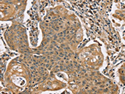
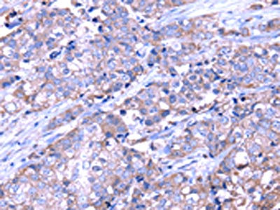
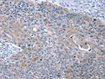 IHC (Immunohiostchemistry)
(The image on the left is immunohistochemistry of paraffin-embedded Human esophagus cancer tissue using AAA239431(SNX27 Antibody) at dilution 1/20, on the right is treated with fusion protein. (Original magnification: ×200))
IHC (Immunohiostchemistry)
(The image on the left is immunohistochemistry of paraffin-embedded Human esophagus cancer tissue using AAA239431(SNX27 Antibody) at dilution 1/20, on the right is treated with fusion protein. (Original magnification: ×200))
SNX27, Polyclonal Antibody (Cat# AAA239431)
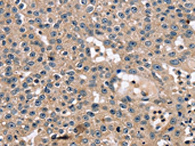
 IHC (Immunohiostchemistry)
(The image on the left is immunohistochemistry of paraffin-embedded Human esophagus cancer tissue using AAA239432(SNX11 Antibody) at dilution 1/30, on the right is treated with fusion protein. (Original magnification: ×200))
IHC (Immunohiostchemistry)
(The image on the left is immunohistochemistry of paraffin-embedded Human esophagus cancer tissue using AAA239432(SNX11 Antibody) at dilution 1/30, on the right is treated with fusion protein. (Original magnification: ×200))
SNX11, Polyclonal Antibody (Cat# AAA239432)
 IHC (Immunohiostchemistry)
(The image on the left is immunohistochemistry of paraffin-embedded Human breast cancer tissue using AAA239435(SNX8 Antibody) at dilution 1/30, on the right is treated with fusion protein. (Original magnification: ×200))
IHC (Immunohiostchemistry)
(The image on the left is immunohistochemistry of paraffin-embedded Human breast cancer tissue using AAA239435(SNX8 Antibody) at dilution 1/30, on the right is treated with fusion protein. (Original magnification: ×200))
SNX8, Polyclonal Antibody (Cat# AAA239435)
 IHC (Immunohiostchemistry)
(The image on the left is immunohistochemistry of paraffin-embedded Human esophagus cancer tissue using AAA239439(SSBP1 Antibody) at dilution 1/20, on the right is treated with fusion protein. (Original magnification: ×200))
IHC (Immunohiostchemistry)
(The image on the left is immunohistochemistry of paraffin-embedded Human esophagus cancer tissue using AAA239439(SSBP1 Antibody) at dilution 1/20, on the right is treated with fusion protein. (Original magnification: ×200))
SSBP1, Polyclonal Antibody (Cat# AAA239439)
 SDS-PAGE
(Gel: 8%SDS-PAGE, Lysate: 40 ug, Lane: Human fetal intestines tissue, Primary antibody: AAA239453(SULT1B1 Antibody) at dilution 1/800, Secondary antibody: Goat anti rabbit IgG at 1/8000 dilution, Exposure time: 3 seconds)
SDS-PAGE
(Gel: 8%SDS-PAGE, Lysate: 40 ug, Lane: Human fetal intestines tissue, Primary antibody: AAA239453(SULT1B1 Antibody) at dilution 1/800, Secondary antibody: Goat anti rabbit IgG at 1/8000 dilution, Exposure time: 3 seconds)
SULT1B1, Polyclonal Antibody (Cat# AAA239453)
 SDS-PAGE
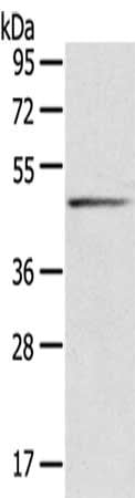
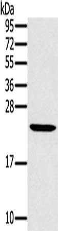
(Gel: 8%SDS-PAGE, Lysate: 40 ug, Lane: Human fetal liver tissue, Primary antibody: AAA239455(SULT2A1 Antibody) at dilution 1/650, Secondary antibody: Goat anti rabbit IgG at 1/8000 dilution, Exposure time: 1 second)
SDS-PAGE
(Gel: 8%SDS-PAGE, Lysate: 40 ug, Lane: Human fetal liver tissue, Primary antibody: AAA239455(SULT2A1 Antibody) at dilution 1/650, Secondary antibody: Goat anti rabbit IgG at 1/8000 dilution, Exposure time: 1 second)
SULT2A1, Polyclonal Antibody (Cat# AAA239455)
 SDS-PAGE
(Gel: 12%SDS-PAGE, Lysate: 40 ug, Lane: Mouse liver tissue, Primary antibody: AAA239457(MED22 Antibody) at dilution 1/800, Secondary antibody: Goat anti rabbit IgG at 1/8000 dilution, Exposure time: 15 seconds)
SDS-PAGE
(Gel: 12%SDS-PAGE, Lysate: 40 ug, Lane: Mouse liver tissue, Primary antibody: AAA239457(MED22 Antibody) at dilution 1/800, Secondary antibody: Goat anti rabbit IgG at 1/8000 dilution, Exposure time: 15 seconds)
MED22, Polyclonal Antibody (Cat# AAA239457)
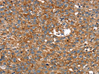
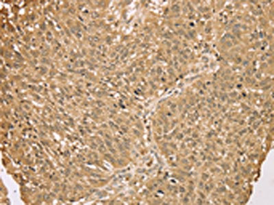
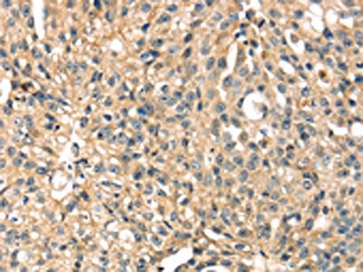
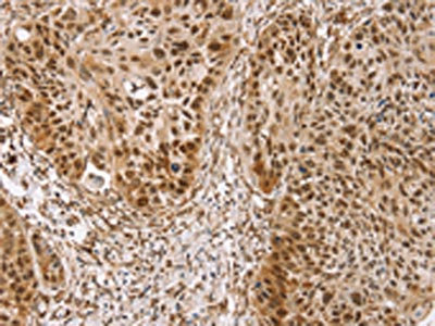
 IHC (Immunohiostchemistry)
(The image on the left is immunohistochemistry of paraffin-embedded Human lung cancer tissue using AAA239458(ARMC10 Antibody) at dilution 1/30, on the right is treated with fusion protein. (Original magnification: ×200))
IHC (Immunohiostchemistry)
(The image on the left is immunohistochemistry of paraffin-embedded Human lung cancer tissue using AAA239458(ARMC10 Antibody) at dilution 1/30, on the right is treated with fusion protein. (Original magnification: ×200))
ARMC10, Polyclonal Antibody (Cat# AAA239458)
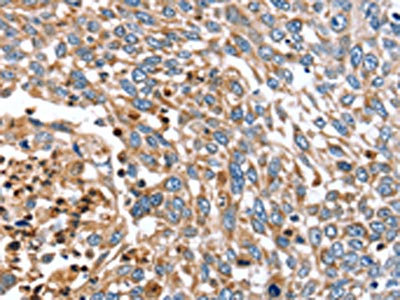
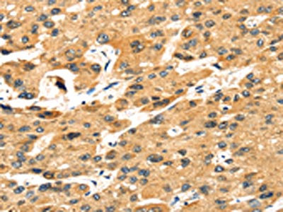
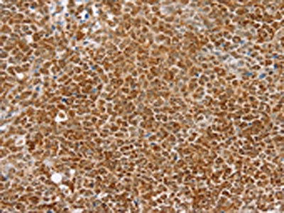
 IHC (Immunohiostchemistry)
(The image on the left is immunohistochemistry of paraffin-embedded Human breast cancer tissue using AAA239460(STX7 Antibody) at dilution 1/25, on the right is treated with fusion protein. (Original magnification: ×200))
IHC (Immunohiostchemistry)
(The image on the left is immunohistochemistry of paraffin-embedded Human breast cancer tissue using AAA239460(STX7 Antibody) at dilution 1/25, on the right is treated with fusion protein. (Original magnification: ×200))
STX7, Polyclonal Antibody (Cat# AAA239460)
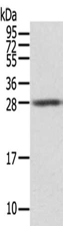 SDS-PAGE
(Gel: 12%SDS-PAGE,Lysate: 40 ug,,Primary antibody: AAA239461(STX8 Antibody) at dilution 1/500 dilution,Secondary antibody: Goat anti rabbit IgG at 1/8000 dilution,Exposure time: 5seconds)
SDS-PAGE
(Gel: 12%SDS-PAGE,Lysate: 40 ug,,Primary antibody: AAA239461(STX8 Antibody) at dilution 1/500 dilution,Secondary antibody: Goat anti rabbit IgG at 1/8000 dilution,Exposure time: 5seconds)
STX8, Polyclonal Antibody (Cat# AAA239461)
 SDS-PAGE
(Gel: 8%SDS-PAGE, Lysate: 40 ug, Lane 1-2: Hela cells, hepg2 cells, Primary antibody: AAA239463(STX16 Antibody) at dilution 1/400, Secondary antibody: Goat anti rabbit IgG at 1/8000 dilution, Exposure time: 5 seconds)
SDS-PAGE
(Gel: 8%SDS-PAGE, Lysate: 40 ug, Lane 1-2: Hela cells, hepg2 cells, Primary antibody: AAA239463(STX16 Antibody) at dilution 1/400, Secondary antibody: Goat anti rabbit IgG at 1/8000 dilution, Exposure time: 5 seconds)
STX16, Polyclonal Antibody (Cat# AAA239463)
 SDS-PAGE
(Gel: 8%SDS-PAGE, Lysate: 40 ug, Lane 1-3: Hepg2 cells, TM4 cells, Raw264.7 cells, Primary antibody: AAA239465(STX18 Antibody) at dilution 1/300, Secondary antibody: Goat anti rabbit IgG at 1/8000 dilution, Exposure time: 20 seconds)
SDS-PAGE
(Gel: 8%SDS-PAGE, Lysate: 40 ug, Lane 1-3: Hepg2 cells, TM4 cells, Raw264.7 cells, Primary antibody: AAA239465(STX18 Antibody) at dilution 1/300, Secondary antibody: Goat anti rabbit IgG at 1/8000 dilution, Exposure time: 20 seconds)
STX18, Polyclonal Antibody (Cat# AAA239465)
 SDS-PAGE
(Gel: 12%SDS-PAGE, Lysate: 40 ug, Lane 1-3: Hela cells, lovo cells, A431 cells, Primary antibody: AAA239467(STX10 Antibody) at dilution 1/800, Secondary antibody: Goat anti rabbit IgG at 1/8000 dilution, Exposure time: 5 seconds)
SDS-PAGE
(Gel: 12%SDS-PAGE, Lysate: 40 ug, Lane 1-3: Hela cells, lovo cells, A431 cells, Primary antibody: AAA239467(STX10 Antibody) at dilution 1/800, Secondary antibody: Goat anti rabbit IgG at 1/8000 dilution, Exposure time: 5 seconds)
STX10, Polyclonal Antibody (Cat# AAA239467)
 SDS-PAGE
(Gel: 12%SDS-PAGE, Lysate: 40 ug, Lane 1-4: A375 cells, A431 cells, lovo cells, hela cells, Primary antibody: AAA239468(STX10 Antibody) at dilution 1/800, Secondary antibody: Goat anti rabbit IgG at 1/8000 dilution, Exposure time: 5 seconds)
SDS-PAGE
(Gel: 12%SDS-PAGE, Lysate: 40 ug, Lane 1-4: A375 cells, A431 cells, lovo cells, hela cells, Primary antibody: AAA239468(STX10 Antibody) at dilution 1/800, Secondary antibody: Goat anti rabbit IgG at 1/8000 dilution, Exposure time: 5 seconds)
STX10, Polyclonal Antibody (Cat# AAA239468)
 SDS-PAGE
(Gel: 10%SDS-PAGE, Lysate: 80 ug, Lane: 231 cell, Primary antibody: AAA239471(STX19 Antibody) at dilution 1/800 dilution, Secondary antibody: Goat anti rabbit IgG at 1/8000 dilution, Exposure time: 10 seconds)
SDS-PAGE
(Gel: 10%SDS-PAGE, Lysate: 80 ug, Lane: 231 cell, Primary antibody: AAA239471(STX19 Antibody) at dilution 1/800 dilution, Secondary antibody: Goat anti rabbit IgG at 1/8000 dilution, Exposure time: 10 seconds)
STX19, Polyclonal Antibody (Cat# AAA239471)
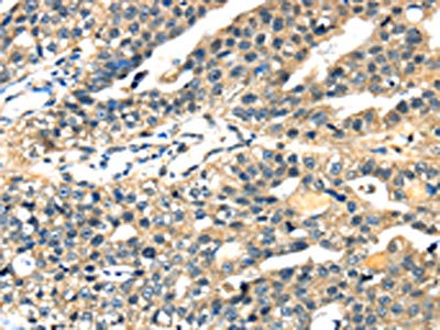
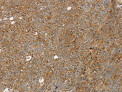
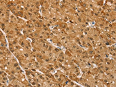
 IHC (Immunohiostchemistry)
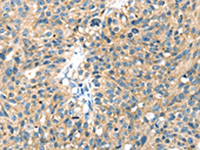
(The image on the left is immunohistochemistry of paraffin-embedded Human ovarian cancer tissue using AAA239473(TAF12 Antibody) at dilution 1/30, on the right is treated with fusion protein. (Original magnification: ×200))
IHC (Immunohiostchemistry)
(The image on the left is immunohistochemistry of paraffin-embedded Human ovarian cancer tissue using AAA239473(TAF12 Antibody) at dilution 1/30, on the right is treated with fusion protein. (Original magnification: ×200))
TAF12, Polyclonal Antibody (Cat# AAA239473)
 IHC (Immunohiostchemistry)
(The image on the left is immunohistochemistry of paraffin-embedded Human esophagus cancer tissue using AAA239474(TAF12 Antibody) at dilution 1/35, on the right is treated with fusion protein. (Original magnification: ×200))
IHC (Immunohiostchemistry)
(The image on the left is immunohistochemistry of paraffin-embedded Human esophagus cancer tissue using AAA239474(TAF12 Antibody) at dilution 1/35, on the right is treated with fusion protein. (Original magnification: ×200))
TAF12, Polyclonal Antibody (Cat# AAA239474)
What are Polyclonal Antibodies?
Polyclonal antibodies are antibodies that come from multiple B cell clones of a host animal. The typical hosts used for the majority of polyclonal antibody production are rabbits, goats, sheep, and donkeys. These polyclonal antibodies, once having identified their target, will bind to different epitopes located at different regions or sequences on the same protein/antigen. As a result, they are ideal at locating and binding to the target, even if the target is in very low concentrations (due to many different antibodies being able to bind to the same target molecule, which allows for significant amplification of a downstream signal).
Polyclonal antibodies are typically produced by injecting an antigen into a host animal, which causes the animal’s immune system to attack the foreign antigen by mass generating antibodies against it. After a period of time, serum is collected from the animal and purified using physicochemical fractionation, class-specific affinity purification, and/or antigen-affinity purification.
Key Uses of Polyclonal Antibodies
- Western Blotting: This method is used to find specific proteins in biological samples after separating them by size.
- Immunohistochemistry: IHC helps visualize the location of proteins in tissue sections using various staining techniques.
- ELISA: (Enzyme-Linked Immunosorbent Assay) is typically used to identify specific protein quantities in a sample. ELISAs can be either “Quantitative” or “Qualitative”.
- Flow Cytometry: technique that identifies and measures the specific protein on the surface or inside the cells in a fluid suspension.
- Immunoprecipitation: IP isolates and studies a specific protein from a complex mixture using antibodies.
Why Buy Polyclonal Antibodies from AAA Biotech?
1. Ideal for Various Applications
Our antibodies are generally going to be validated for use in multiple types of assays, including ELISA, Western Blotting, Immunohistochemistry, Immunoprecipitation, amongst others. They are ideal for a wide range of research applications.
2. Rigorous Quality Control
All of the antibodies in our catalog undergo strict quality testing to ensure specificity, sensitivity, and consistent performance. We are confident in the ability of our antibodies to provide you with accurate results.
3. Wide Assortment of Antibodies
Antibodies in are catalog can be found for both common and exotic species, and these antibodies are also available in both conjugated and recombinant forms to suit many diverse experimental needs.
4. Highly Purified
Our antibodies are available in purified forms with over 85% purity, as confirmed by SDS-PAGE. They are also available with tags such as His, Flag, GST, or MBP. We cater to customers worldwide.
FAQ
1. How are polyclonal antibodies produced?
Traditionally, polyclonal antibodies are produced by injecting an antigen into a host animal (such as a rabbit or goat), which then triggers an immune response from the host animal. The animal’s B cells produce antibodies that will recognize different parts of the injected antigen. These antibodies are then collected from the animal’s blood and purified for use.
2. How do polyclonal antibodies differ from monoclonal antibodies?
Polyclonal antibodies are a mix of antibodies that bind to different locations (epitopes) of the same antigen, while monoclonal antibodies are identical and bind to just one specific epitope. This makes polyclonal antibodies more versatile and better at detecting proteins that may be present in low quantities or in altered/modified forms.
3. How should I store polyclonal antibodies?
Polyclonal antibodies should be stored at 4°C for short-term use (up to a few weeks) and at -20°C or -80°C for long-term storage. Avoid repeated freeze-thaw cycles by dividing them into small aliquots. Always check the datasheet for specific storage instructions.