Filters
▼Clonality
▼Type
▼Reactivity
▼Gene Name
▼Isotype
▼Host
▼Application
▼Clone
▼Phospho Antibodies
Phospho-specific antibodies’ typical purpose is to enable researchers to detect changes in proteins. They will exclusively bind to the amino acid sequence on a protein that has been phosphorylated (which is both a physical & chemical change) and do not bind to the same amino acid sequence on said protein if it lacks said phosphorylation. This aids in being able to clearly see and understand the data produced from this particular protein modification.
Viewing 4150-4200 of 5297 product results
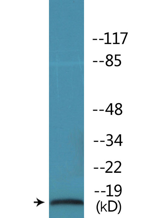
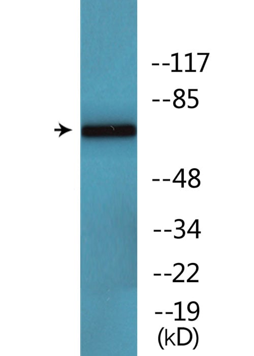
 WB (Western Blot)
(Western blot analysis of p53 phosphorylation expression in ovary cancer whole cell lysates, The lane on the left is treated with the antigen-specific peptide.)
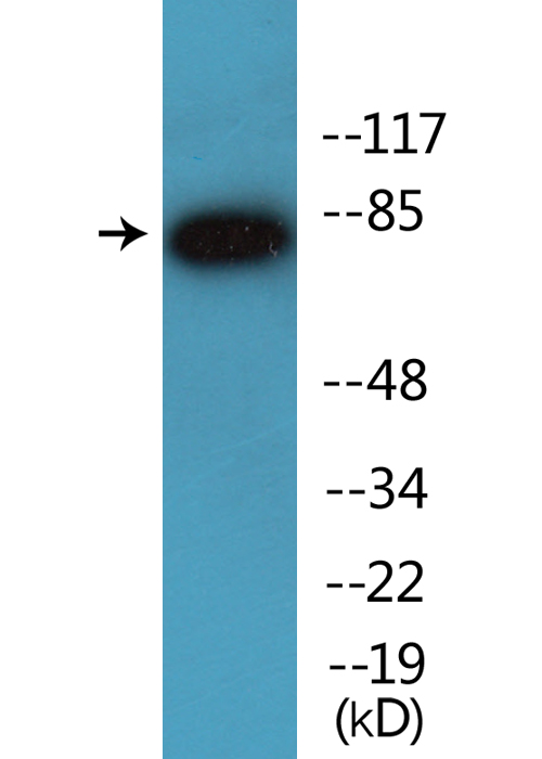
WB (Western Blot)
(Western blot analysis of p53 phosphorylation expression in ovary cancer whole cell lysates, The lane on the left is treated with the antigen-specific peptide.)
p53, Polyclonal Antibody (Cat# AAA321468)
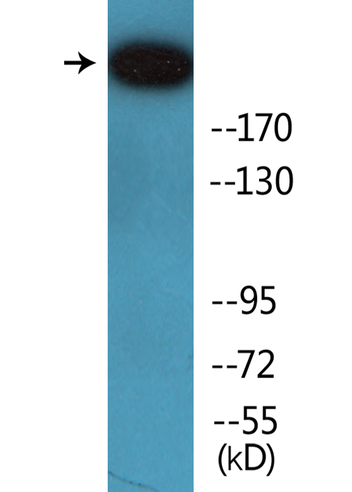
 WB (Western Blot)
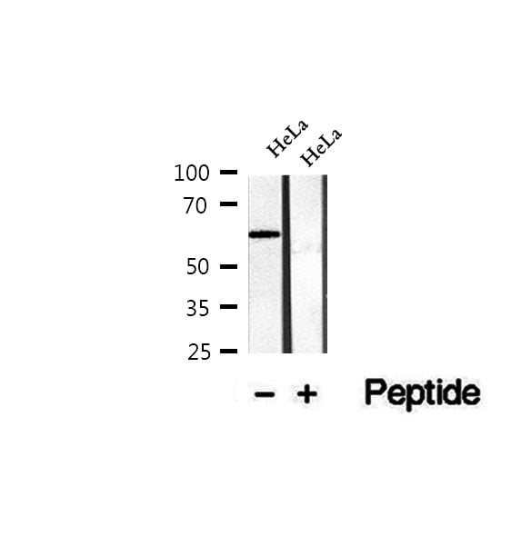
(Western blot analysis of extracts of HeLa cells, using Phospho-CTDSPL2 (Ser104) Antibody.)
WB (Western Blot)
(Western blot analysis of extracts of HeLa cells, using Phospho-CTDSPL2 (Ser104) Antibody.)
CTDSPL2, Polyclonal Antibody (Cat# AAA321317)
 WB (Western Blot)
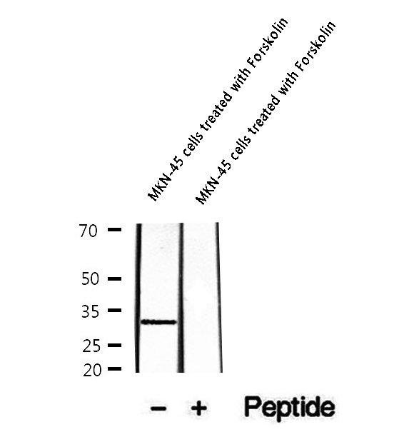
(Western blot analysis of extracts of MKN-45 cells treated with Forskolin, using Phospho-DARPP-32 (Ser97) Antibody.)
WB (Western Blot)
(Western blot analysis of extracts of MKN-45 cells treated with Forskolin, using Phospho-DARPP-32 (Ser97) Antibody.)
DARPP-32, Polyclonal Antibody (Cat# AAA321320)
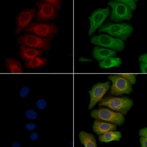 IF (Immunofluorescence)
(AAA321321 staining Heat-shock treated Hela cells by IF/ICC. The samples were fixed with PFA and permeabilized in 0.1% Triton X-100,then blocked in 10% serum for 45 minutes at 25°C. Samples were then incubated with primary Ab and mouse anti-beta tubulin Ab for 1 hour at 37°C. An AlexaFluor594 conjugated goat anti-rabbit IgG(H+L) Ab(Red) and an AlexaFluor488 conjugated goat anti-mouse IgG(H+L) Ab(Green) were used as the secondary Ab. The nuclear counter stain is DAPI (blue).)
IF (Immunofluorescence)
(AAA321321 staining Heat-shock treated Hela cells by IF/ICC. The samples were fixed with PFA and permeabilized in 0.1% Triton X-100,then blocked in 10% serum for 45 minutes at 25°C. Samples were then incubated with primary Ab and mouse anti-beta tubulin Ab for 1 hour at 37°C. An AlexaFluor594 conjugated goat anti-rabbit IgG(H+L) Ab(Red) and an AlexaFluor488 conjugated goat anti-mouse IgG(H+L) Ab(Green) were used as the secondary Ab. The nuclear counter stain is DAPI (blue).)
DDR1, Polyclonal Antibody (Cat# AAA321321)
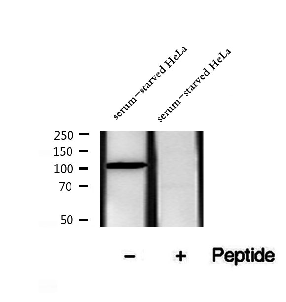 WB (Western Blot)
(Western blot analysis of extracts of serum-starved HeLa cells, using Phospho-eEF2k (Ser366) Antibody.)
WB (Western Blot)
(Western blot analysis of extracts of serum-starved HeLa cells, using Phospho-eEF2k (Ser366) Antibody.)
eEF2k, Polyclonal Antibody (Cat# AAA321324)
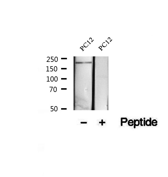 WB (Western Blot)
(Western blot analysis of extracts of PC12 cells, using Phospho-eIF4G (Ser1147) Antibody.)
WB (Western Blot)
(Western blot analysis of extracts of PC12 cells, using Phospho-eIF4G (Ser1147) Antibody.)
eIF4G, Polyclonal Antibody (Cat# AAA321325)
 IF (Immunofluorescence)
(Immunofluorescent analysis of Histone H3 (pS10) staining in Raw264.7 cells. Formalin-fixed cells were permeabilized with 0.1% Triton X-100 in TBS for 5-10 minutes and blocked with 3% BSA-PBS for 30 minutes at room temperature. Cells were probed with the primary antibody in 3% BSA-PBS and incubated overnight at 4 °C in a humidified chamber. Cells were washed with PBST and incubated with a DyLight 594-conjugated secondary antibody (red) in PBS at room temperature in the dark.)
IF (Immunofluorescence)
(Immunofluorescent analysis of Histone H3 (pS10) staining in Raw264.7 cells. Formalin-fixed cells were permeabilized with 0.1% Triton X-100 in TBS for 5-10 minutes and blocked with 3% BSA-PBS for 30 minutes at room temperature. Cells were probed with the primary antibody in 3% BSA-PBS and incubated overnight at 4 °C in a humidified chamber. Cells were washed with PBST and incubated with a DyLight 594-conjugated secondary antibody (red) in PBS at room temperature in the dark.)
Histone H3 (pS10), Polyclonal Antibody (Cat# AAA105161)
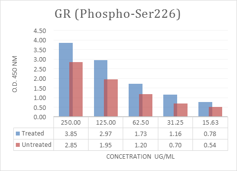
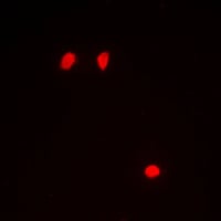
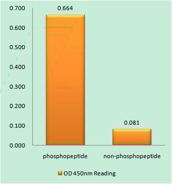
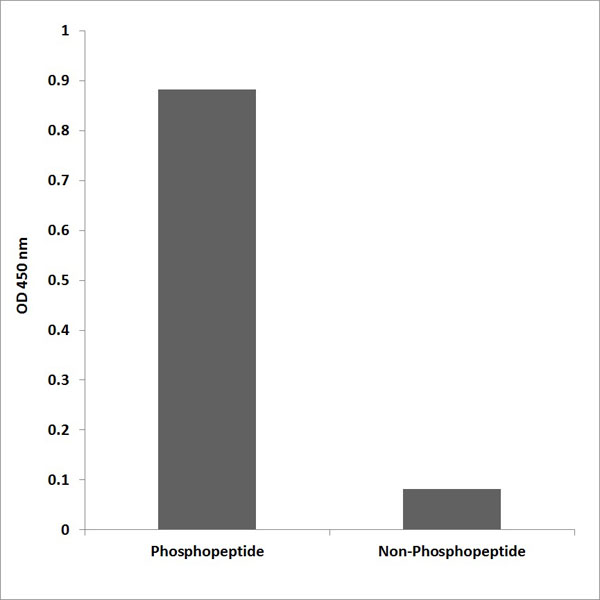
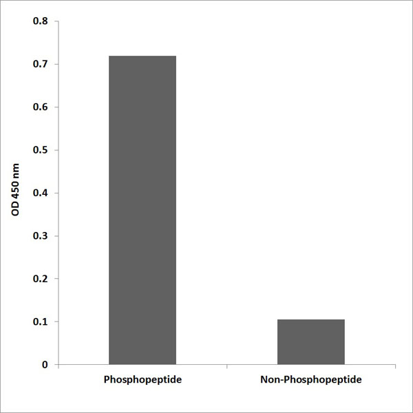
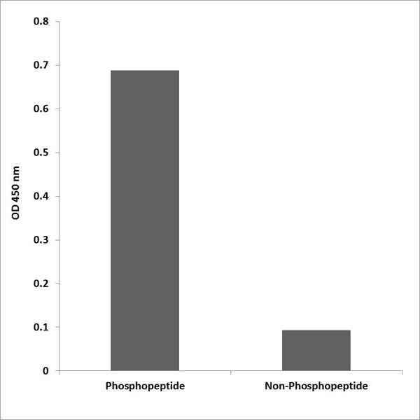
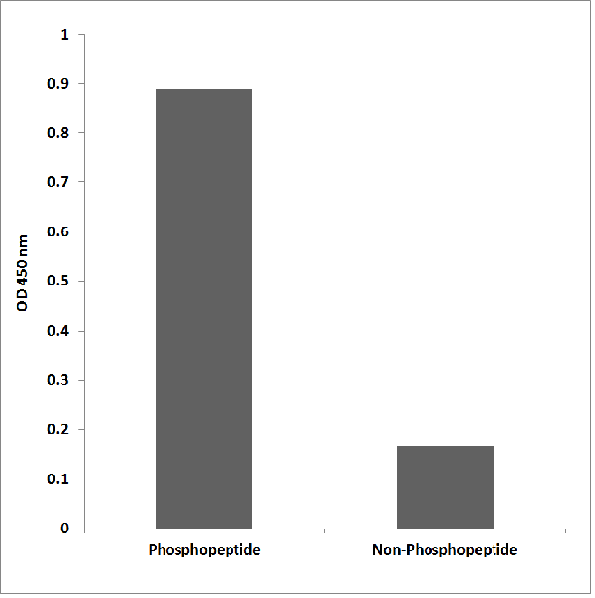
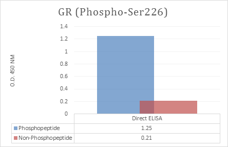 ELISA
(Enzyme-Linked Immunosorbent Assay (ELISA) for immunogen phosphor-peptide (left) and non-phospho peptide (right), using Anti-GR (Phospho-Ser226) Antibody.)
ELISA
(Enzyme-Linked Immunosorbent Assay (ELISA) for immunogen phosphor-peptide (left) and non-phospho peptide (right), using Anti-GR (Phospho-Ser226) Antibody.)
GR, ELISA Kit (Cat# AAA318615)
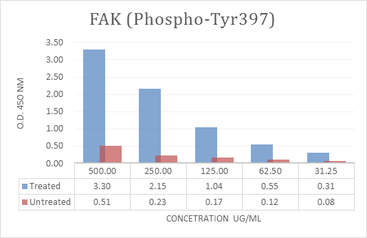
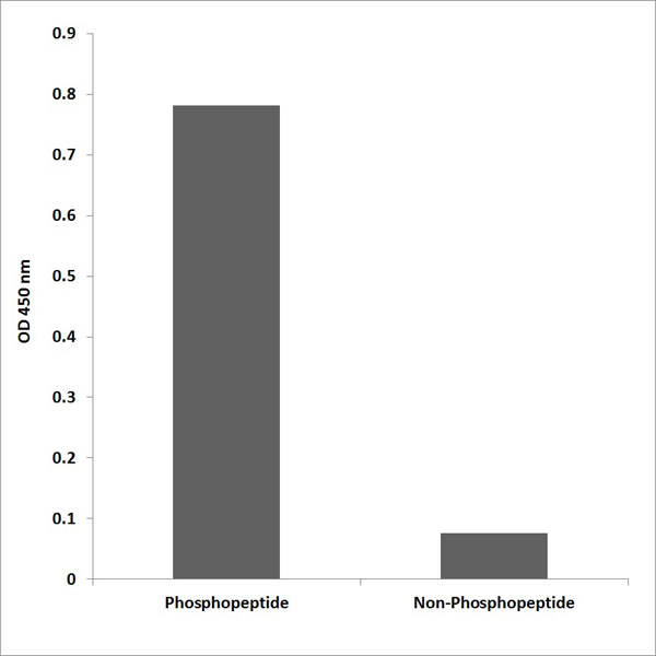
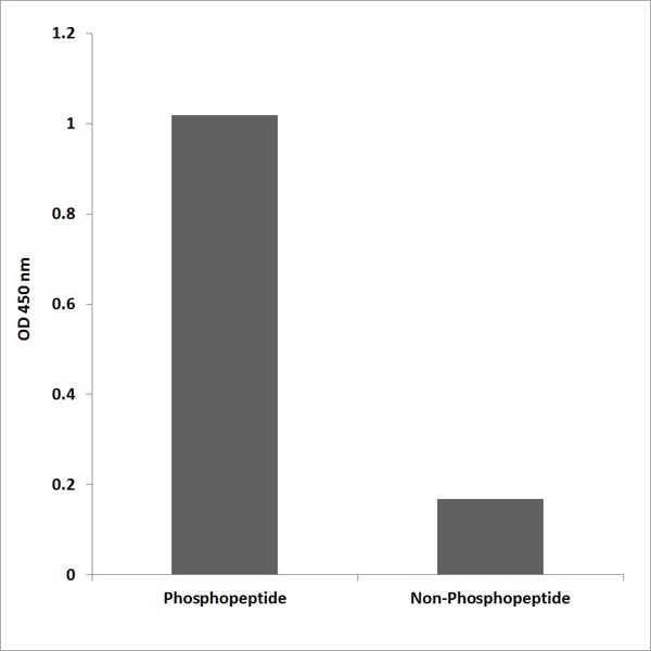
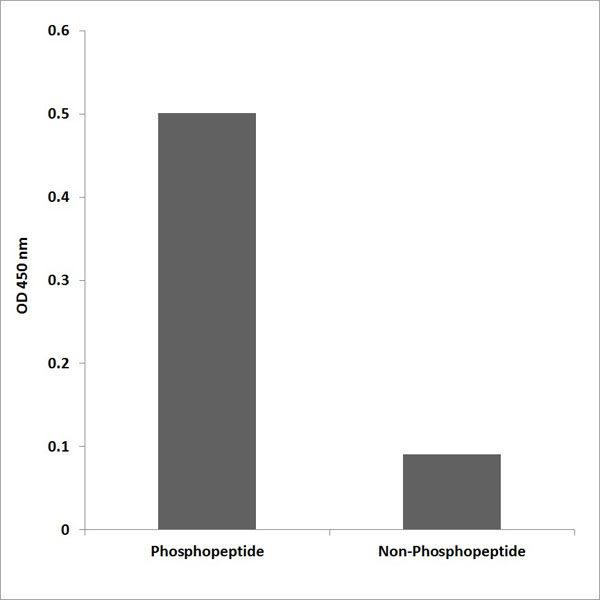
 ELISA
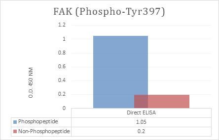
(Enzyme-Linked Immunosorbent Assay (ELISA) for immunogen phosphor-peptide (left) and non-phospho peptide (right), using Anti-FAK (Phospho-Tyr397) Antibody.)
ELISA
(Enzyme-Linked Immunosorbent Assay (ELISA) for immunogen phosphor-peptide (left) and non-phospho peptide (right), using Anti-FAK (Phospho-Tyr397) Antibody.)
FAK, ELISA Kit (Cat# AAA318617)
 ELISA
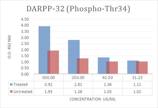
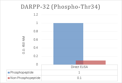
(Enzyme-Linked Immunosorbent Assay (ELISA) for immunogen phosphor-peptide (left) and non-phospho peptide (right), using Anti-DARPP-32 (Phospho-Thr34) Antibody.)
ELISA
(Enzyme-Linked Immunosorbent Assay (ELISA) for immunogen phosphor-peptide (left) and non-phospho peptide (right), using Anti-DARPP-32 (Phospho-Thr34) Antibody.)
DARPP-32, ELISA Kit (Cat# AAA318622)
 ELISA
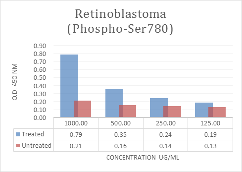
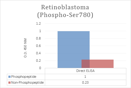
(Enzyme-Linked Immunosorbent Assay (ELISA) for immunogen phosphor-peptide (left) and non-phospho peptide (right), using Anti-Retinoblastoma (Phospho-Ser780) Antibody.)
ELISA
(Enzyme-Linked Immunosorbent Assay (ELISA) for immunogen phosphor-peptide (left) and non-phospho peptide (right), using Anti-Retinoblastoma (Phospho-Ser780) Antibody.)
Retinoblastoma, ELISA Kit (Cat# AAA318628)
 Application Data
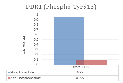
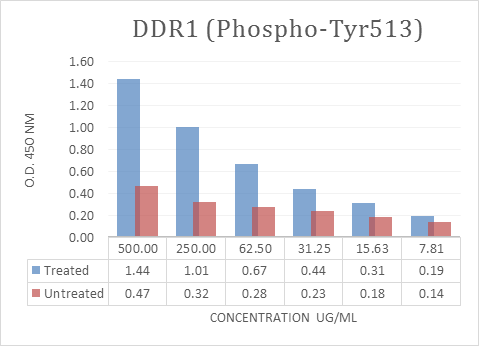
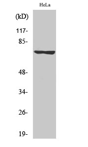
(HepG2 cells were grown to 90% confluency and were stimulated with Vanadate 1mM and incubated for 30 mins. Cells were immediately lysed thereafter and measured for Total Protein Concentration and O.D. 450 nm of DDR1 P-Tyr513 versus untreated HepG2 lysates.)
Application Data
(HepG2 cells were grown to 90% confluency and were stimulated with Vanadate 1mM and incubated for 30 mins. Cells were immediately lysed thereafter and measured for Total Protein Concentration and O.D. 450 nm of DDR1 P-Tyr513 versus untreated HepG2 lysates.)
DDR1, ELISA Kit (Cat# AAA318631)
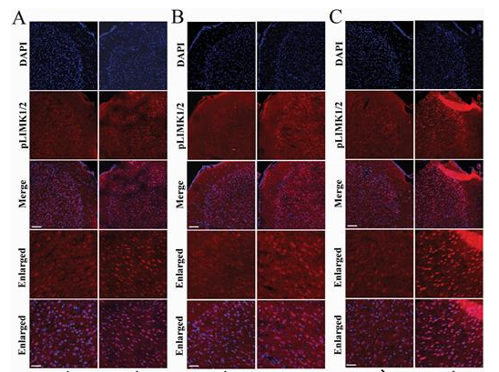 Application Data
(Gao, Ting-Ting, et al. "LIMK1/2 in the mPFC plays a role in chronic stress-induced depressive-like effects in mice." International Journal of Neuropsychopharmacology 23.12 (2020): 821-836.)
Application Data
(Gao, Ting-Ting, et al. "LIMK1/2 in the mPFC plays a role in chronic stress-induced depressive-like effects in mice." International Journal of Neuropsychopharmacology 23.12 (2020): 821-836.)
LIMK-1/2, Polyclonal Antibody (Cat# AAA318640)
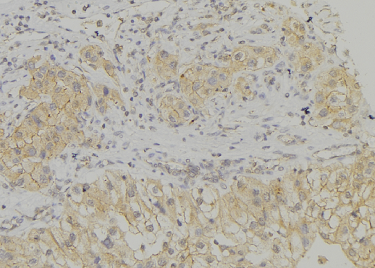
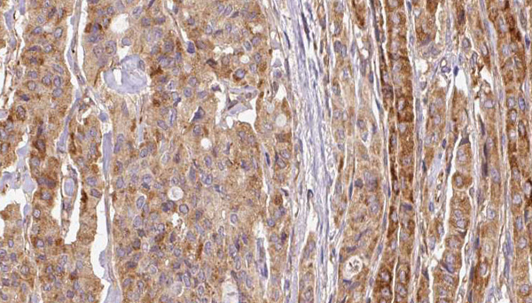
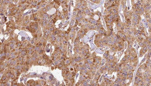
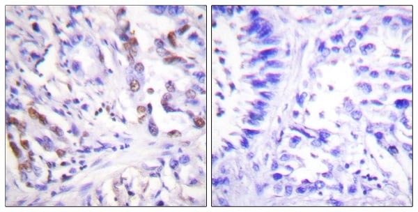
 IHC (Immunohistochemisry)
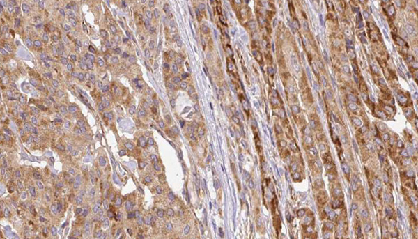
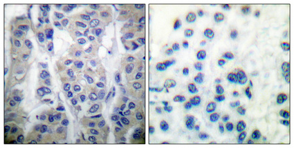
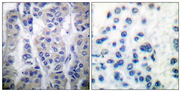
(Immunohistochemical analysis of paraffin-embedded Human breast cancer. Antibody was diluted at 1:100(4 degree overnight). High-pressure and temperature Tris-EDTA,pH8.0 was used for antigen retrieval. Negetive contrl (right) obtaned from antibody was pre-absorbed by immunogen peptide.)
IHC (Immunohistochemisry)
(Immunohistochemical analysis of paraffin-embedded Human breast cancer. Antibody was diluted at 1:100(4 degree overnight). High-pressure and temperature Tris-EDTA,pH8.0 was used for antigen retrieval. Negetive contrl (right) obtaned from antibody was pre-absorbed by immunogen peptide.)
Stat4, Polyclonal Antibody (Cat# AAA318641)
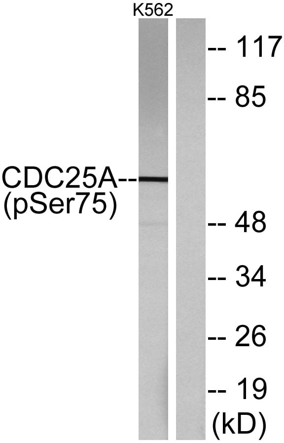
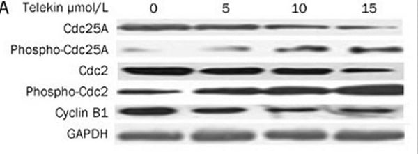 Application Data
(Li, Lin, et al. "Telekin suppresses human hepatocellular carcinoma cells in vitro by inducing G 2/M phase arrest via the p38 MAPK signaling pathway." Acta Pharmacologica Sinica 35.10 (2014): 1311.)
Application Data
(Li, Lin, et al. "Telekin suppresses human hepatocellular carcinoma cells in vitro by inducing G 2/M phase arrest via the p38 MAPK signaling pathway." Acta Pharmacologica Sinica 35.10 (2014): 1311.)
Cdc25A, Polyclonal Antibody (Cat# AAA318643)
 WB (Western Blot)
(Western blot analysis of lysates from HepG2 cells treated with EGF 200ng/ml 30', using RSK1/2/3/4 (Phospho-Ser221/227/S218/232) Antibody.)
WB (Western Blot)
(Western blot analysis of lysates from HepG2 cells treated with EGF 200ng/ml 30', using RSK1/2/3/4 (Phospho-Ser221/227/S218/232) Antibody.)
RSK1/2/3/4, Polyclonal Antibody (Cat# AAA318382)
 WB (Western Blot)
(Western blot analysis of lysates from COS7 cells treated with nocodazole 1ug/ml 16h, using STMN1 (Phospho-Ser62) Antibody.)
WB (Western Blot)
(Western blot analysis of lysates from COS7 cells treated with nocodazole 1ug/ml 16h, using STMN1 (Phospho-Ser62) Antibody.)
STMN1, Polyclonal Antibody (Cat# AAA318391)
 WB (Western Blot)
(Western blot analysis of lysates from NIH/3T3 cells treated with TNF 20ng/ml 30', using IKK-alpha (Phospho-Ser176) /IKK-beta (Phospho-Ser177) Antibody.)
WB (Western Blot)
(Western blot analysis of lysates from NIH/3T3 cells treated with TNF 20ng/ml 30', using IKK-alpha (Phospho-Ser176) /IKK-beta (Phospho-Ser177) Antibody.)
IKK-alpha, Polyclonal Antibody (Cat# AAA318329)
 WB (Western Blot)
(Western blot analysis of lysates from Jurkat cells, using Lck (Phospho-Tyr192) Antibody.)
WB (Western Blot)
(Western blot analysis of lysates from Jurkat cells, using Lck (Phospho-Tyr192) Antibody.)
Lck, Polyclonal Antibody (Cat# AAA318344)
 WB (Western Blot)
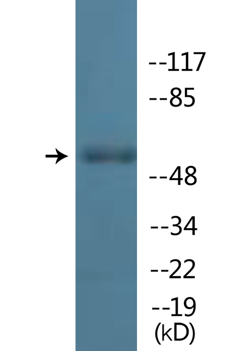
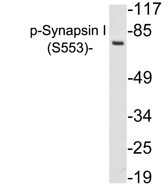
(Western blot analysis of lysates from 293 cells treated with PMA, using p-Serynapsin I (Phospho-Ser553) antibody.)
WB (Western Blot)
(Western blot analysis of lysates from 293 cells treated with PMA, using p-Serynapsin I (Phospho-Ser553) antibody.)
Synapsin I, Polyclonal Antibody (Cat# AAA318273)
 WB (Western Blot)
(Western blot analysis of lysates from rat brain, using CaMK2-beta/gamma/delta (Phospho-Thr287) Antibody.)
WB (Western Blot)
(Western blot analysis of lysates from rat brain, using CaMK2-beta/gamma/delta (Phospho-Thr287) Antibody.)
CaMK2-beta/gamma/delta, Polyclonal Antibody (Cat# AAA318293)
 WB (Western Blot)
(Western blot analysis of lysates from COS7 cells treated with UV 15', using IGF2R (Phospho-Ser2409) Antibody.)
WB (Western Blot)
(Western blot analysis of lysates from COS7 cells treated with UV 15', using IGF2R (Phospho-Ser2409) Antibody.)
IGF2R, Polyclonal Antibody (Cat# AAA318328)
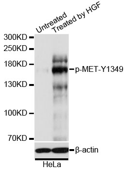
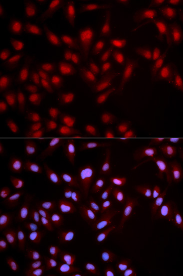 IF (Immunofluorescence)
(Immunofluorescence analysis of U2OS cells using Phospho-MET-Y1349 antibody. Blue: DAPI for nuclear staining.)
IF (Immunofluorescence)
(Immunofluorescence analysis of U2OS cells using Phospho-MET-Y1349 antibody. Blue: DAPI for nuclear staining.)
MET-Y1349, Antibody (Cat# AAA36657)
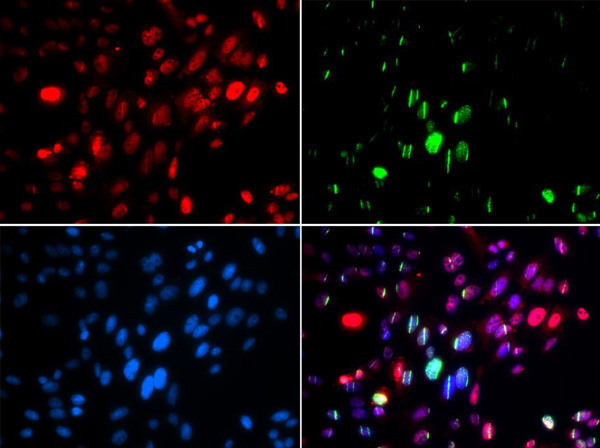
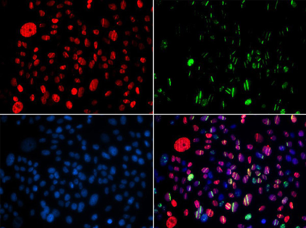
 IF (Immunofluorescence)
(Immunofluorescence analysis of U2OS cells using Phospho-ABL1-Y204 antibody. Blue: DAPI for nuclear staining.)
IF (Immunofluorescence)
(Immunofluorescence analysis of U2OS cells using Phospho-ABL1-Y204 antibody. Blue: DAPI for nuclear staining.)
ABL1-Y204, Antibody (Cat# AAA36690)
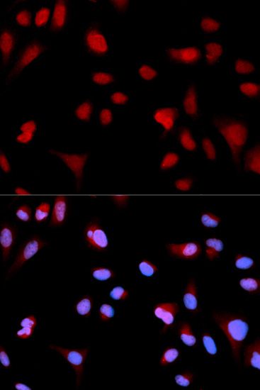
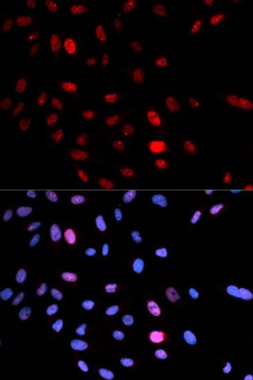
 IF (Immunofluorescence)
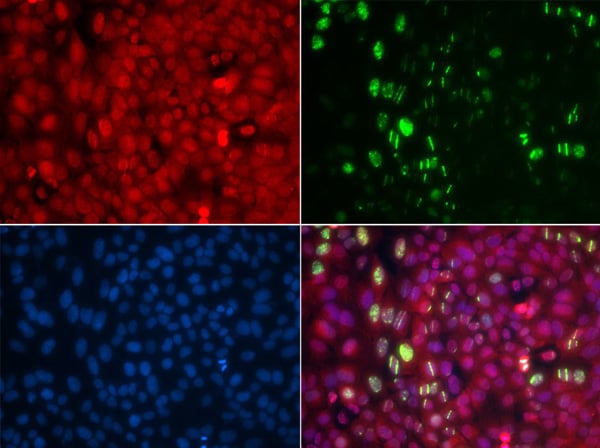
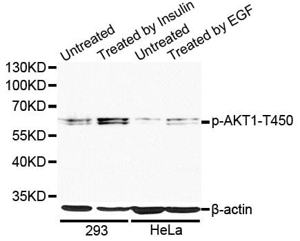
(Immunofluorescence analysis of MCF-7 cells using Phospho-AKT1-T450 antibody. Blue: DAPI for nuclear staining.)
IF (Immunofluorescence)
(Immunofluorescence analysis of MCF-7 cells using Phospho-AKT1-T450 antibody. Blue: DAPI for nuclear staining.)
AKT1-T450, Antibody (Cat# AAA36692)
 IF (Immunofluorescence)
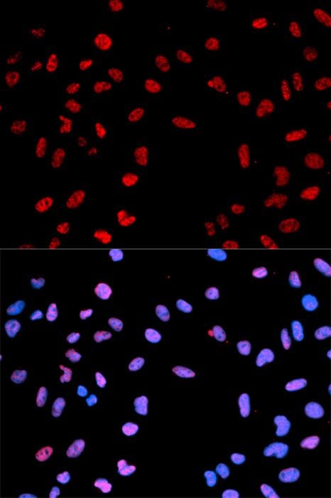
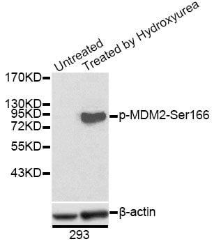
(Immunofluorescence analysis of U2OS cells using Phospho-MDM2-S166 antibody. Blue: DAPI for nuclear staining.)
IF (Immunofluorescence)
(Immunofluorescence analysis of U2OS cells using Phospho-MDM2-S166 antibody. Blue: DAPI for nuclear staining.)
MDM2-S166, Antibody (Cat# AAA36703)
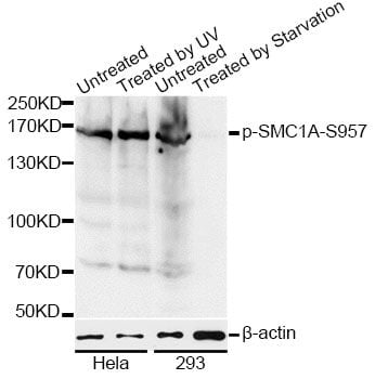
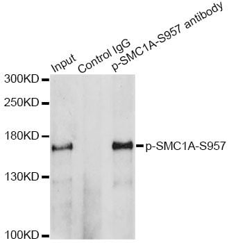 IP (Immunoprecipitation)
(Immunoprecipitation analysis of 200ug extracts of HeLa cells treated by UV using 2.5ug Phospho-SMC1A-S957 antibody. Western blot was performed from the immunoprecipitate using Phospho-SMC1A-S957 antibody at a dilition of 1:1000.)
IP (Immunoprecipitation)
(Immunoprecipitation analysis of 200ug extracts of HeLa cells treated by UV using 2.5ug Phospho-SMC1A-S957 antibody. Western blot was performed from the immunoprecipitate using Phospho-SMC1A-S957 antibody at a dilition of 1:1000.)
SMC1A-S957, Antibody (Cat# AAA36761)
 WB (Western Blot)
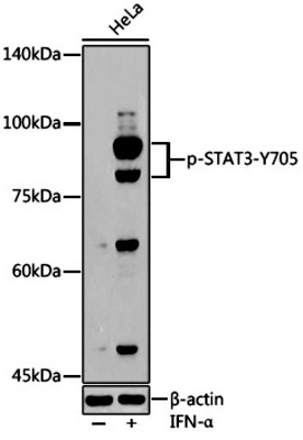
(Western blot analysis of extracts of HeLa cells, using Phospho-STAT3-Y705 antibody (AAA36779) at 1:500 dilution.HeLa cells were treated by IFN-α (100 ng/ml) at 37°C for 30 minutes after serum-starvation overnight. Secondary antibody: HRP Goat Anti-Rabbit IgG (H+L) at 1:10000 dilution. Lysates/proteins: 25ug per lane. Blocking buffer: 3% nonfat dry milk in TBST. Detection: ECL Basic Kit. Exposure time: 180s.)
WB (Western Blot)
(Western blot analysis of extracts of HeLa cells, using Phospho-STAT3-Y705 antibody (AAA36779) at 1:500 dilution.HeLa cells were treated by IFN-α (100 ng/ml) at 37°C for 30 minutes after serum-starvation overnight. Secondary antibody: HRP Goat Anti-Rabbit IgG (H+L) at 1:10000 dilution. Lysates/proteins: 25ug per lane. Blocking buffer: 3% nonfat dry milk in TBST. Detection: ECL Basic Kit. Exposure time: 180s.)
Stat3-Y705, Antibody (Cat# AAA36779)
 IF (Immunofluorescence)
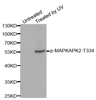
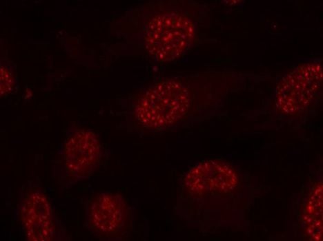
(Immunofluorescence staining of methanol-fixed HeLa cells using Phospho-MAPKAPK2-T334 antibody.)
IF (Immunofluorescence)
(Immunofluorescence staining of methanol-fixed HeLa cells using Phospho-MAPKAPK2-T334 antibody.)
MAPKAPK2-T334, Antibody (Cat# AAA37351)
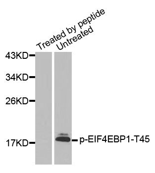
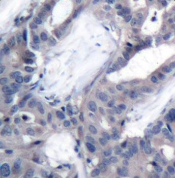
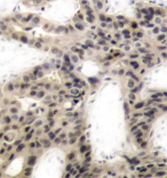 IHC (Immunohiostchemistry)
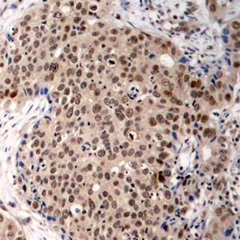
(Immunohistochemistry of paraffin-embedded human breast carcinoma using Phospho-EIF4EBP1-T45 antibody.)
IHC (Immunohiostchemistry)
(Immunohistochemistry of paraffin-embedded human breast carcinoma using Phospho-EIF4EBP1-T45 antibody.)
EIF4EBP1-T45, Antibody (Cat# AAA37362)
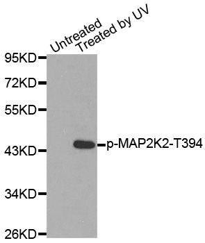
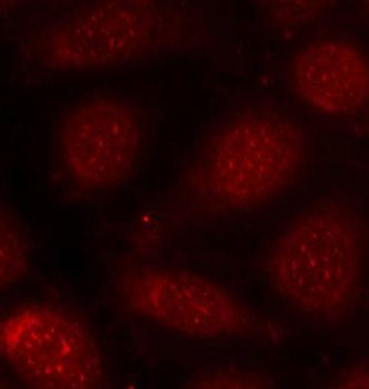
 IF (Immunofluorescence)
(Immunofluorescence staining of methanol-fixed HeLa cells using Phospho-MAP2K2-T394 antibody.)
IF (Immunofluorescence)
(Immunofluorescence staining of methanol-fixed HeLa cells using Phospho-MAP2K2-T394 antibody.)
MAP2K2-T394, Antibody (Cat# AAA37364)
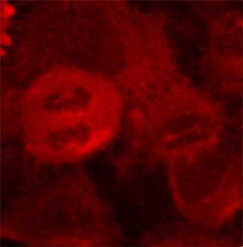
 IF (Immunofluorescence)
(Immunofluorescence staining of methanol-fixed MCF-7 cells using Phospho-PRKCB-T641 antibody.)
IF (Immunofluorescence)
(Immunofluorescence staining of methanol-fixed MCF-7 cells using Phospho-PRKCB-T641 antibody.)
PRKCB-T641, Antibody (Cat# AAA37367)
 WB (Western Blot)
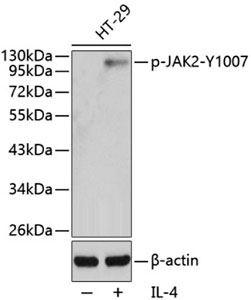
(Western blot analysis of extracts from HT29 cells using Phospho-JAK2-Y1007 antibody.Secondary antibody: HRP Goat Anti-Rabbit IgG (H+L) at 1:10000 dilution.Lysates/proteins: 25ug per lane.Blocking buffer: 3% BSA.)
WB (Western Blot)
(Western blot analysis of extracts from HT29 cells using Phospho-JAK2-Y1007 antibody.Secondary antibody: HRP Goat Anti-Rabbit IgG (H+L) at 1:10000 dilution.Lysates/proteins: 25ug per lane.Blocking buffer: 3% BSA.)
JAK2-Y1007, Antibody (Cat# AAA37372)
 WB (Western Blot)
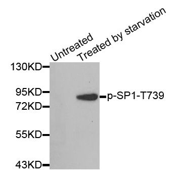
(Western blot analysis of extracts from 3T3 cells, using Phospho-SP1-T739 antibody.Secondary antibody: HRP Goat Anti-Rabbit IgG (H+L) at 1:10000 dilution.Lysates/proteins: 25ug per lane.Blocking buffer: 3% BSA.)
WB (Western Blot)
(Western blot analysis of extracts from 3T3 cells, using Phospho-SP1-T739 antibody.Secondary antibody: HRP Goat Anti-Rabbit IgG (H+L) at 1:10000 dilution.Lysates/proteins: 25ug per lane.Blocking buffer: 3% BSA.)
SP1-T739, Antibody (Cat# AAA37376)
 IHC (Immunohiostchemistry)
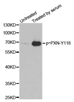
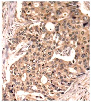
(Immunohistochemistry of paraffin-embedded human breast carcinoma tissue, using Phospho-PXN-Y118 antibody.)
IHC (Immunohiostchemistry)
(Immunohistochemistry of paraffin-embedded human breast carcinoma tissue, using Phospho-PXN-Y118 antibody.)
PXN-Y118, Antibody (Cat# AAA37380)
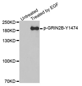
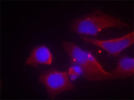 IF (Immunofluorescence)
(Immunofluorescence staining of methanol-fixed HeLa cells using Phospho-GRIN2B-Y1474 antibody.)
IF (Immunofluorescence)
(Immunofluorescence staining of methanol-fixed HeLa cells using Phospho-GRIN2B-Y1474 antibody.)
GRIN2B-Y1474, Antibody (Cat# AAA37384)
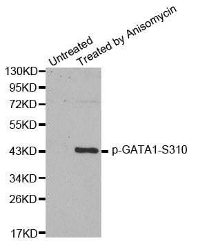
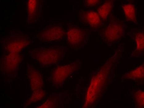 IF (Immunofluorescence)
(Immunofluorescence staining of methanol-fixed HeLa cells using Phospho-GATA1-S310 antibody.)
IF (Immunofluorescence)
(Immunofluorescence staining of methanol-fixed HeLa cells using Phospho-GATA1-S310 antibody.)
GATA1-S310, Antibody (Cat# AAA37404)

 IF (Immunofluorescence)
(Immunofluorescence staining of methanol-fixed HeLa cells using Phospho-YWHAZ-S58 antibody.)
IF (Immunofluorescence)
(Immunofluorescence staining of methanol-fixed HeLa cells using Phospho-YWHAZ-S58 antibody.)
YWHAZ-S58, Antibody (Cat# AAA37407)

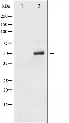
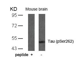
 WB (Western Blot)
(Western blot analysis of extracts from JK cells (Lane 2), using ALOX5 (Phospho-Ser523) Antibody. The lane on the left is treated with synthesized peptide.)
WB (Western Blot)
(Western blot analysis of extracts from JK cells (Lane 2), using ALOX5 (Phospho-Ser523) Antibody. The lane on the left is treated with synthesized peptide.)
ALOX5, Polyclonal Antibody (Cat# AAA243241)
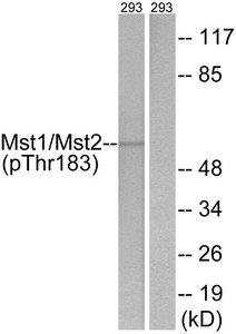
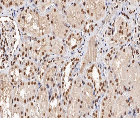
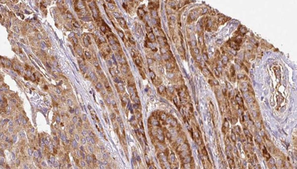
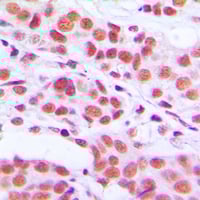
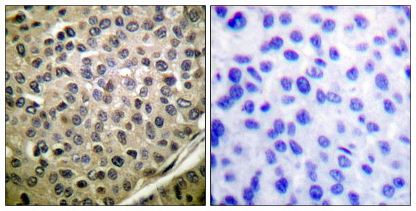
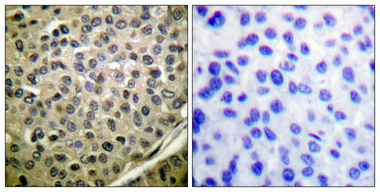
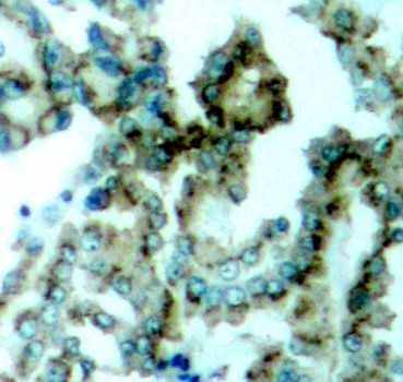
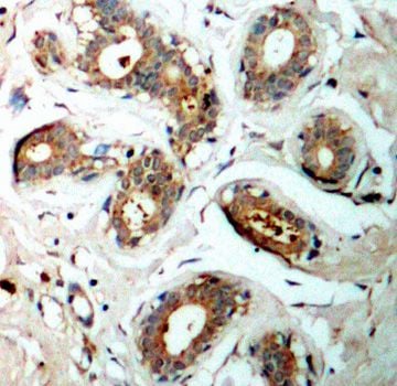
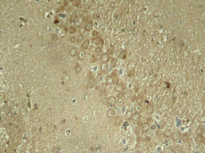
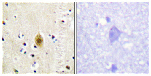 IHC (Immunohiostchemistry)
(Immunohistochemistry analysis of paraffin-embedded human brain tissue, using Mst1/2 (Phospho-Thr183) antibody. The picture on the right is treated with the synthesized peptide.)
IHC (Immunohiostchemistry)
(Immunohistochemistry analysis of paraffin-embedded human brain tissue, using Mst1/2 (Phospho-Thr183) antibody. The picture on the right is treated with the synthesized peptide.)
STK4, Polyclonal Antibody (Cat# AAA243255)
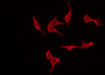
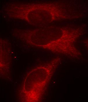
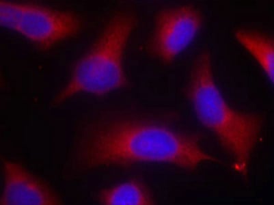 IF (Immunofluorescence)
(Immunofluorescence staining of methanol-fixed Hela cells using Tau(Phospho-Ser262) Antibody.)
IF (Immunofluorescence)
(Immunofluorescence staining of methanol-fixed Hela cells using Tau(Phospho-Ser262) Antibody.)
MAPT, Polyclonal Antibody (Cat# AAA243277)
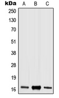
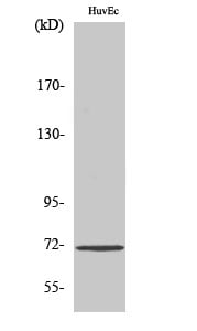
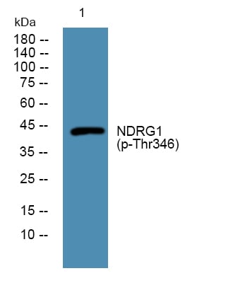 WB (Western Blot)
(Western blot analysis of lysates from PC12 cells, primary antibody was diluted at 1:1000, 4 degree C over night)
WB (Western Blot)
(Western blot analysis of lysates from PC12 cells, primary antibody was diluted at 1:1000, 4 degree C over night)
NDRG1, ELISA Kit (Cat# AAA319085)
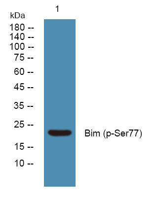 WB (Western Blot)
(Western blot analysis of lysates from U2OS cells, primary antibody was diluted at 1:1000, 4 degree C over night)
WB (Western Blot)
(Western blot analysis of lysates from U2OS cells, primary antibody was diluted at 1:1000, 4 degree C over night)
Bim, ELISA Kit (Cat# AAA319095)
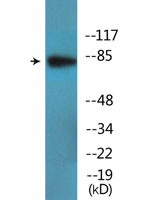
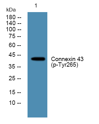 WB (Western Blot)
(Western blot analysis of lysates from K562 cells, primary antibody was diluted at 1:1000, 4 degree C over night)
WB (Western Blot)
(Western blot analysis of lysates from K562 cells, primary antibody was diluted at 1:1000, 4 degree C over night)
Connexin 43, ELISA Kit (Cat# AAA319101)
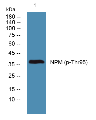 WB (Western Blot)
(Western blot analysis of lysates from SW480 cells, primary antibody was diluted at 1:1000, 4 degree C over night)
WB (Western Blot)
(Western blot analysis of lysates from SW480 cells, primary antibody was diluted at 1:1000, 4 degree C over night)
NPM, ELISA Kit (Cat# AAA319115)
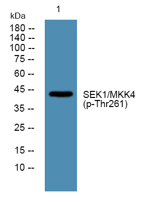
 WB (Western Blot)
(Western blot analysis of lysates from K562 cells, primary antibody was diluted at 1:1000, 4 degree C over night)
WB (Western Blot)
(Western blot analysis of lysates from K562 cells, primary antibody was diluted at 1:1000, 4 degree C over night)
SEK1/MKK4, ELISA Kit (Cat# AAA319122)
 WB (Western Blot)
(Western blot analysis of lysates from HCT116 cells, primary antibody was diluted at 1:1000, 4 degree C over night)
WB (Western Blot)
(Western blot analysis of lysates from HCT116 cells, primary antibody was diluted at 1:1000, 4 degree C over night)
SEK1/MKK4, ELISA Kit (Cat# AAA319124)
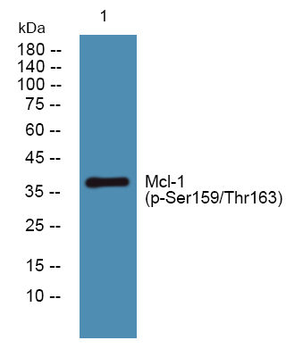 WB (Western Blot)
(Western blot analysis of lysates from SH-SY5Y cells, primary antibody was diluted at 1:1000, 4 degree C over night)
WB (Western Blot)
(Western blot analysis of lysates from SH-SY5Y cells, primary antibody was diluted at 1:1000, 4 degree C over night)
Mcl-1, ELISA Kit (Cat# AAA319137)
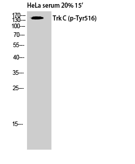 WB (Western Blot)
(Western Blot analysis of HELA cells using Phospho-Trk C (Y516) Polyclonal Antibody)
WB (Western Blot)
(Western Blot analysis of HELA cells using Phospho-Trk C (Y516) Polyclonal Antibody)
Trk C, ELISA Kit (Cat# AAA319140)
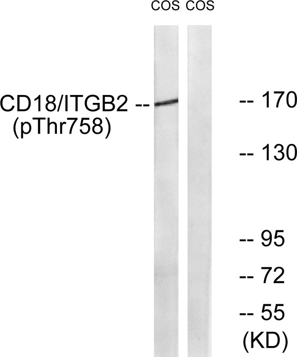 WB (Western Blot)
(Western blot analysis of lysates from COS7 cells treated with EGF 200ng/ml 30', using CD18/ITGB2 (Phospho-Thr758) Antibody. The lane on the right is blocked with the phospho peptide.)
WB (Western Blot)
(Western blot analysis of lysates from COS7 cells treated with EGF 200ng/ml 30', using CD18/ITGB2 (Phospho-Thr758) Antibody. The lane on the right is blocked with the phospho peptide.)
Integrin beta2, ELISA Kit (Cat# AAA319143)
What Are Phospho Antibodies?
Protein phosphorylation is a process where a phosphate group is added to certain amino acid residues of a protein – usually serine (S), threonine (T), or tyrosine (Y) - by enzymes called kinases. This process is integral in controlling cellular signaling, cellular growth, and other biological functions.
Our catalog includes a wide range of phospho-specific antibodies that can accurately detect this important marker. They perform strongly in widely-used laboratory applications such as Western blot, flow cytometry, immunohistochemistry, and immunofluorescence microscopy. We value your trust in us and are committed to providing top-quality products and services. All of our antibodies are guaranteed to work for the applications and species indicated on our website & associated product pages.
What Are The Key Applications of Phospho Antibodies?
1. Western Blotting
One of the first steps a researcher can take in utilizing these phospho-specific antibodies, is to check if the antibody works using a technique referred to as “Western blot”. For those unfamiliar, Western Blot aids in showing whether the protein that the antibody recognizes is appearing at the correct/expected size. These phospho-specific antibodies should also be able to detect changes in the target protein’s phosphorylation (on/off state) when cells are stimulated in certain ways.
2. Staining of Fixed Cells (Immunocytochemistry)
Another routine use of these phospho-specific antibodies, is to test if the antibody is able to demonstrate similar performance when used on fixed cells (intact cells that have been preserved) as it did in the Western blot tests. It is an important aspect in many cases to confirm that the antibody works in actual intact cell samples. Ideally, the method used for cellular fixation should be the same as what is used in pathology labs (like using 10% formalin). To check if the antibody works well in tissue sections (FFPE), researchers will often test it on fixed cells that are processed similar to tissue samples.
3. Specificity Tests Using Peptides
In order to make sure that the antibody is only binding to the right target:
- Laboratory technicians will mix the antibody with phospho-peptides (short segments of the protein containing the phosphate group modification).
- If the antibody signal disappears, it is confirmation that it is binding to the correct phosphorylated location.
- A more robust test is to use both the phosphorylated and non-phosphorylated (dephosphorylated) versions of the protein. The antibody should react only with the phosphorylated one.
- Another method sometimes utilized is to treat the sample with an enzyme, such as alkaline phosphatase, that specifically removes phosphate groups. If the antibody signal disappears after this, it also confirms specificity.
4. Genetic Confirmation
As a final step, scientists can genetically manipulate the nucleotide sequence and alter the target protein by removing the exact site where phosphorylation happens. If the antibody no longer appears to detect the modified protein, it is strong evidence supporting the antibody being specific for that phosphorylated site.
Why Buy Phospho Antibodies Through Us?
- The production laboratory adheres to strict and consistent protocols prior to releasing any of these phospho-specific antibodies:
- Standard methods and proper controls in all tests to ensure high quality.
- These antibodies are tested and validated in different cell types and species.
- High quality control criterion to ensure each batch is consistent, so you will obtain reliable results every time.
FAQ
1. What Are Phospho-Specific Antibodies?
Phospho-specific antibodies are made to detect proteins only when they have a phosphate group linked to a specific amino acid residue. This empowers scientists understand if a protein is "turned on" or active, based on its phosphorylation state.
2. How to Detect Phosphorylated Proteins in a Western Blot?
To find out if a protein is phosphorylated using Western blot:
- Use a phospho-specific antibody that binds only to the phosphorylated form of the protein.
- You can also use a “regular” antibody for the same amino acid sequence of the protein that the phospho-specific antibody is binding to (but in this case, this antibody will not bind if there is a phosphate group present) in order to compare how much of it is phosphorylated versus how much is non-phosphorylated (or “total” protein, if the “normal” antibody’s epitopes are non-phospho-site-specific).
3. How to Choose the Best Antibody?
Here are some simple tips to help you pick the right antibody:
- Know your target
- Match your sample characteristics
- Confirm the intended use is appropriate
- Check “host” and “type”
- Check the “quality” of the presented data/images
- Appraise whether the available validation meets your needs