Filters
▼Clonality
▼Type
▼Reactivity
▼Gene Name
▼Isotype
▼Host
▼Application
▼Clone
▼Phospho Antibodies
Phospho-specific antibodies’ typical purpose is to enable researchers to detect changes in proteins. They will exclusively bind to the amino acid sequence on a protein that has been phosphorylated (which is both a physical & chemical change) and do not bind to the same amino acid sequence on said protein if it lacks said phosphorylation. This aids in being able to clearly see and understand the data produced from this particular protein modification.
Viewing 4050-4100 of 5297 product results
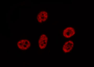
 IF (Immunofluorescence)
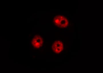
(AAA321568 staining 293 cells by ICC/IF. Cells were fixed with PFA and permeabilized in 0.1% saponin prior to blocking in 10% serum for 45 minutes at 37 degree C. The primary antibody was diluted 1/400 and incubated with the sample for 1 hour at 37 degree C. A Alexa Fluor 594 conjugated goat polyclonal to rabbit IgG (H+L), diluted 1/600 was used as secondary antibody.)
IF (Immunofluorescence)
(AAA321568 staining 293 cells by ICC/IF. Cells were fixed with PFA and permeabilized in 0.1% saponin prior to blocking in 10% serum for 45 minutes at 37 degree C. The primary antibody was diluted 1/400 and incubated with the sample for 1 hour at 37 degree C. A Alexa Fluor 594 conjugated goat polyclonal to rabbit IgG (H+L), diluted 1/600 was used as secondary antibody.)
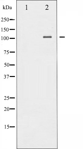
p70 S6 Kinase, Polyclonal Antibody (Cat# AAA321568)
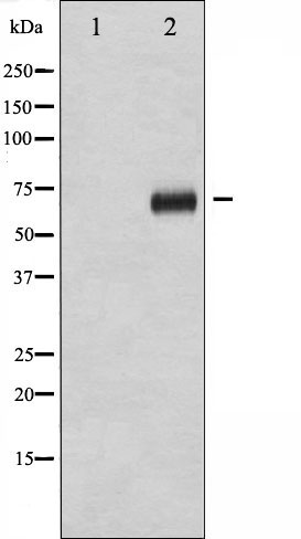
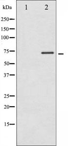
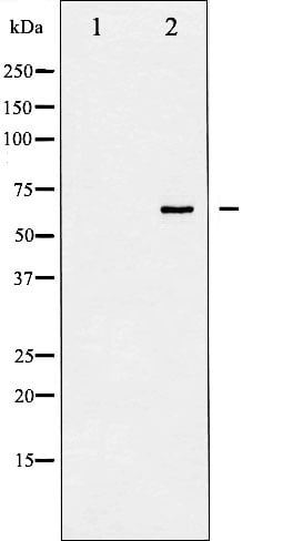
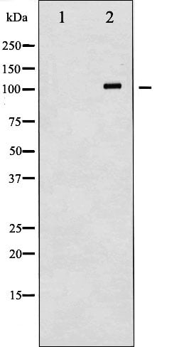
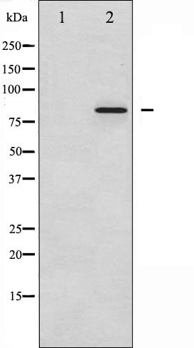
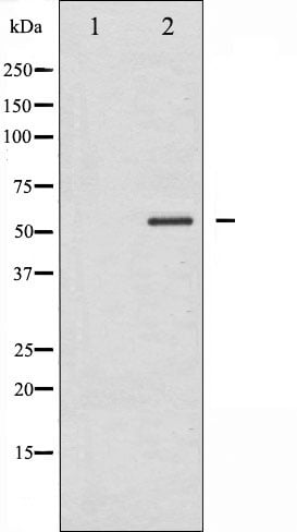
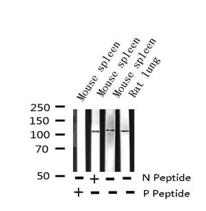
 WB (Western Blot)
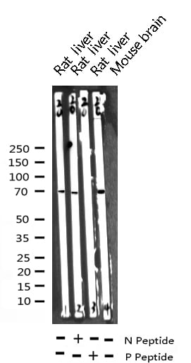
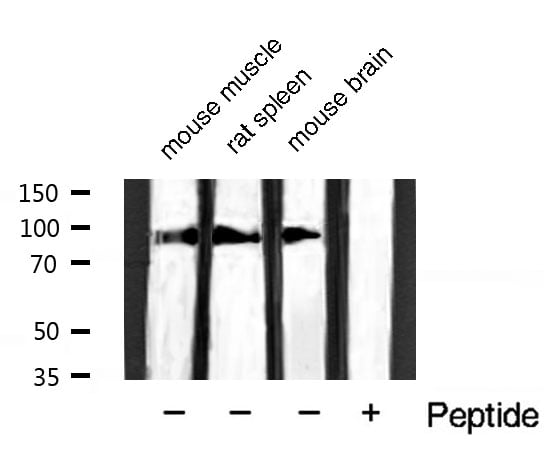
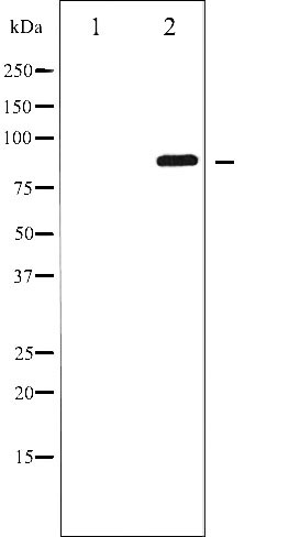
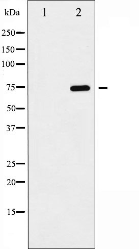
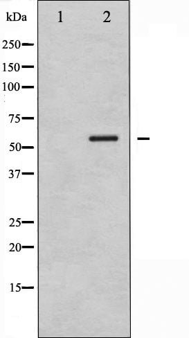
(Western blot analysis of Phospho-p70 S6 Kinase (Ser411) expression in various lysates)
WB (Western Blot)
(Western blot analysis of Phospho-p70 S6 Kinase (Ser411) expression in various lysates)
p70 S6 Kinase, Polyclonal Antibody (Cat# AAA321569)

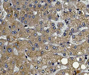
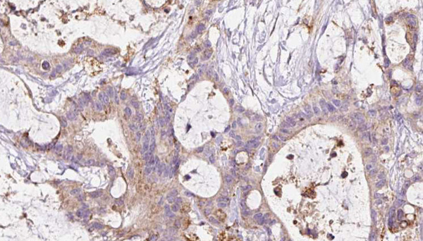
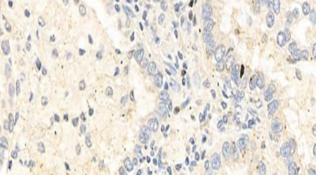
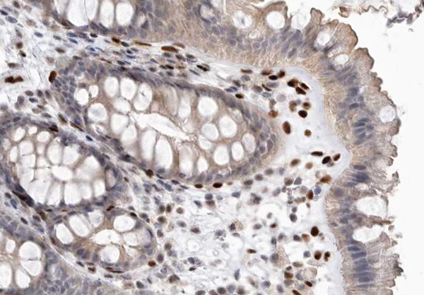
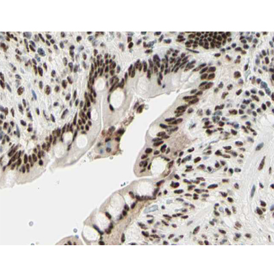
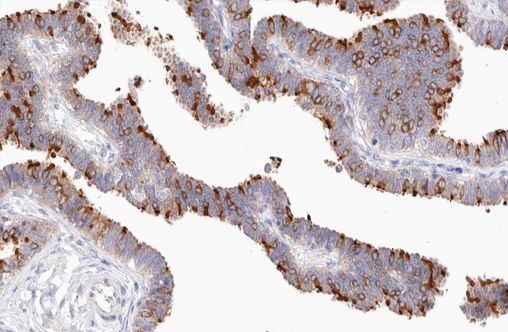
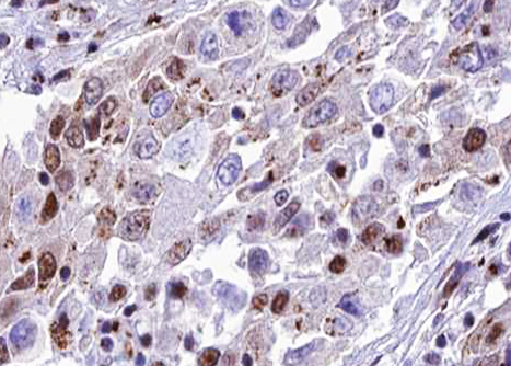
 IHC (Immunohistochemistry)
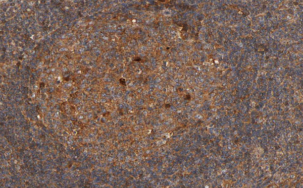
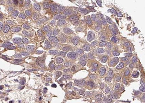
(AAA321571 at 1/100 staining human Lymphoma tissue sections by IHC-P. The tissue was formaldehyde fixed and a heat mediated antigen retrieval step in citrate buffer was performed. The tissue was then blocked and incubated with the antibody for 1.5 hours at 22 degree C. An HRP conjugated goat anti-rabbit antibody was used as the secondary.)
IHC (Immunohistochemistry)
(AAA321571 at 1/100 staining human Lymphoma tissue sections by IHC-P. The tissue was formaldehyde fixed and a heat mediated antigen retrieval step in citrate buffer was performed. The tissue was then blocked and incubated with the antibody for 1.5 hours at 22 degree C. An HRP conjugated goat anti-rabbit antibody was used as the secondary.)
p70 S6 Kinase, Polyclonal Antibody (Cat# AAA321571)
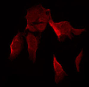
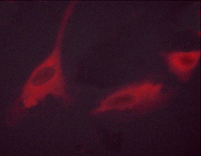
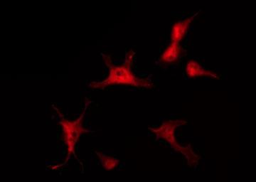
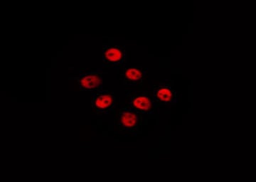
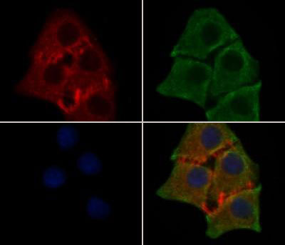
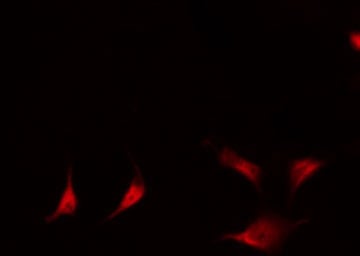
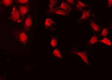
 IF (Immunofluorescence)
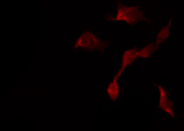
(AAA321575 staining A2780 by IF/ICC. The sample were fixed with PFA and permeabilized in 0.1% Triton X-100,then blocked in 10% serum for 45 minutes at 25°C. The primary antibody was diluted at 1/200 and incubated with the sample for 1 hour at 37°C. An Alexa Fluor 594 conjugated goat anti-rabbit IgG (H+L) Ab, diluted at 1/600, was used as the secondary antibody.)
IF (Immunofluorescence)
(AAA321575 staining A2780 by IF/ICC. The sample were fixed with PFA and permeabilized in 0.1% Triton X-100,then blocked in 10% serum for 45 minutes at 25°C. The primary antibody was diluted at 1/200 and incubated with the sample for 1 hour at 37°C. An Alexa Fluor 594 conjugated goat anti-rabbit IgG (H+L) Ab, diluted at 1/600, was used as the secondary antibody.)
CDK1/CDC2, Polyclonal Antibody (Cat# AAA321575)
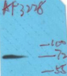
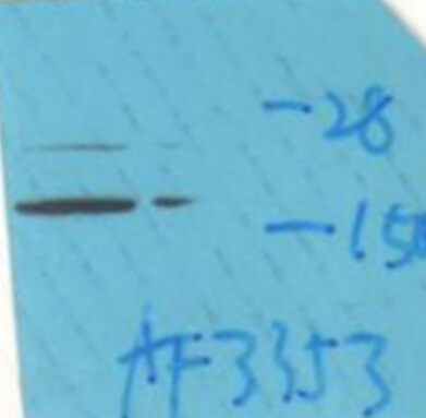
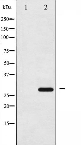
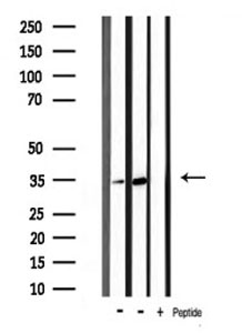
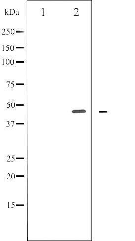
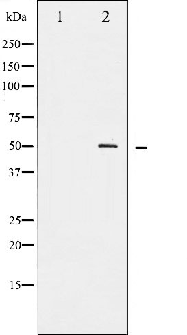
 WB (Western Blot)
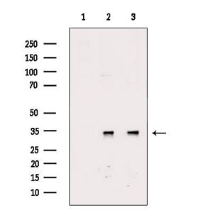
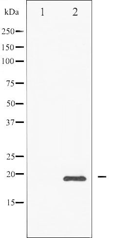
(Western blot analysis of CDK2 phosphorylation expression in A2780 whole cell lysates, The lane on the left is treated with the antigen-specific peptide.)
WB (Western Blot)
(Western blot analysis of CDK2 phosphorylation expression in A2780 whole cell lysates, The lane on the left is treated with the antigen-specific peptide.)
CDK2, Polyclonal Antibody (Cat# AAA321576)
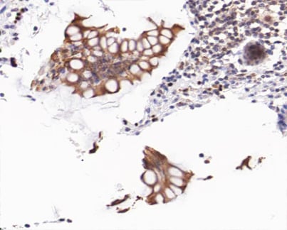
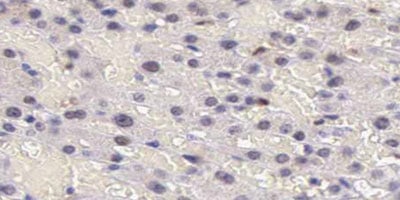
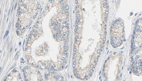
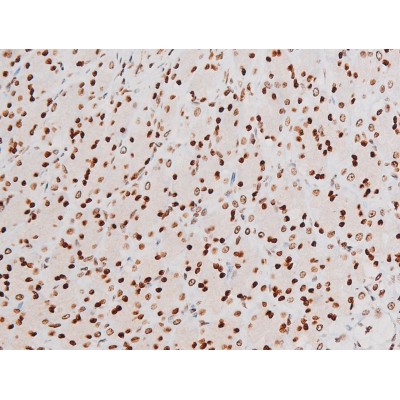
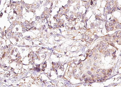 IHC (Immunohistochemistry)
(AAA321582 at 1/100 staining human Breast carcinoma tissue sections by IHC-P. The tissue was formaldehyde fixed and a heat mediated antigen retrieval step in citrate buffer was performed. The tissue was then blocked and incubated with the antibody for 1.5 hours at 22 degree C. An HRP conjugated goat anti-rabbit antibody was used as the secondary.)
IHC (Immunohistochemistry)
(AAA321582 at 1/100 staining human Breast carcinoma tissue sections by IHC-P. The tissue was formaldehyde fixed and a heat mediated antigen retrieval step in citrate buffer was performed. The tissue was then blocked and incubated with the antibody for 1.5 hours at 22 degree C. An HRP conjugated goat anti-rabbit antibody was used as the secondary.)
Shc, Polyclonal Antibody (Cat# AAA321582)
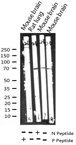
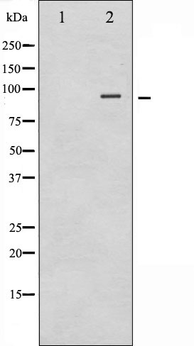
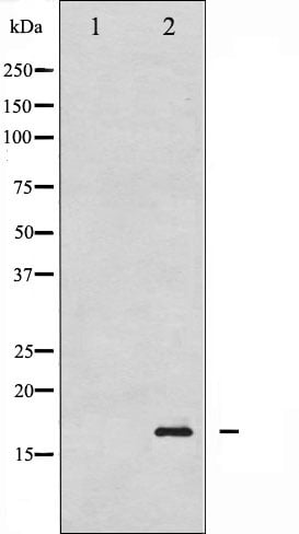
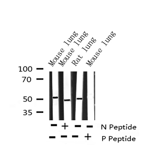
 WB (Western Blot)
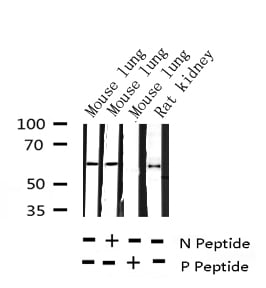
(Western blot analysis of Phospho-eNOS (Thr494) expression in various lysates)
WB (Western Blot)
(Western blot analysis of Phospho-eNOS (Thr494) expression in various lysates)
eNOS, Polyclonal Antibody (Cat# AAA321584)
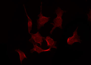
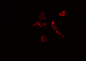
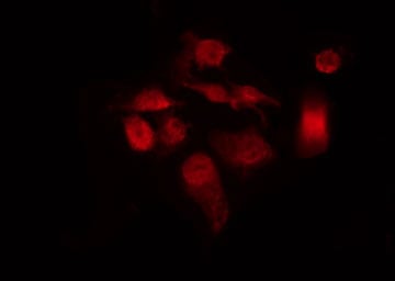
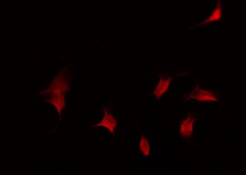
 IF (Immunofluorescence)
(AAA321588 staining 293 by IF/ICC. The sample were fixed with PFA and permeabilized in 0.1% Triton X-100, then blocked in 10% serum for 45 minutes at 25 degree C. The primary antibody was diluted at 1/200 and incubated with the sample for 1 hour at 37 degree C. An Alexa Fluor 594 conjugated goat anti-rabbit IgG (H+L) Ab, diluted at 1/600, was used as the secondary antibody.)
IF (Immunofluorescence)
(AAA321588 staining 293 by IF/ICC. The sample were fixed with PFA and permeabilized in 0.1% Triton X-100, then blocked in 10% serum for 45 minutes at 25 degree C. The primary antibody was diluted at 1/200 and incubated with the sample for 1 hour at 37 degree C. An Alexa Fluor 594 conjugated goat anti-rabbit IgG (H+L) Ab, diluted at 1/600, was used as the secondary antibody.)
Akt, Polyclonal Antibody (Cat# AAA321588)
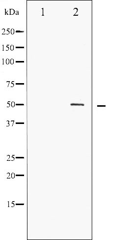
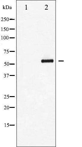
 WB (Western Blot)
(Western blot analysis of VE-Cadherin phosphorylation expression in Na3VO4 treated HepG2 whole cell lysates, The lane on the left is treated with the antigen-specific peptide.)
WB (Western Blot)
(Western blot analysis of VE-Cadherin phosphorylation expression in Na3VO4 treated HepG2 whole cell lysates, The lane on the left is treated with the antigen-specific peptide.)
VE-Cadherin, Polyclonal Antibody (Cat# AAA321592)
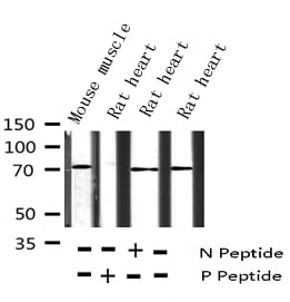
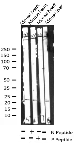
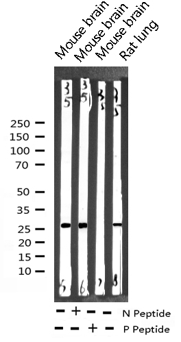
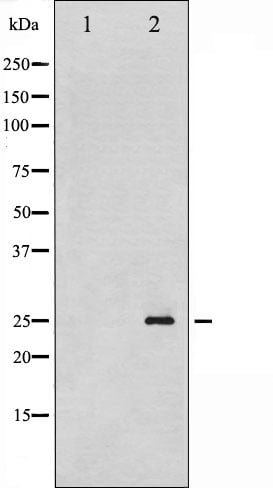
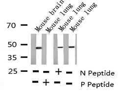
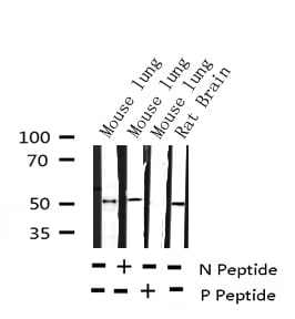
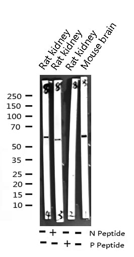
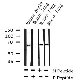
 WB (Western Blot)
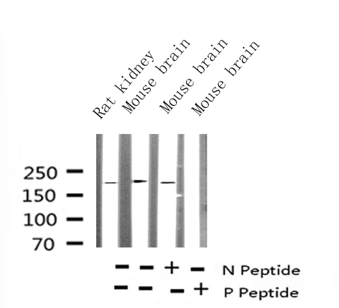
(Western blot analysis of extracts of various tissue sample, using Phospho-Catenin-beta (Ser37) Antibody.)
WB (Western Blot)
(Western blot analysis of extracts of various tissue sample, using Phospho-Catenin-beta (Ser37) Antibody.)
Catenin-beta, Polyclonal Antibody (Cat# AAA321593)
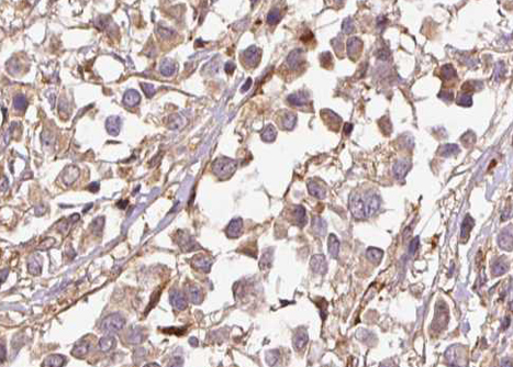
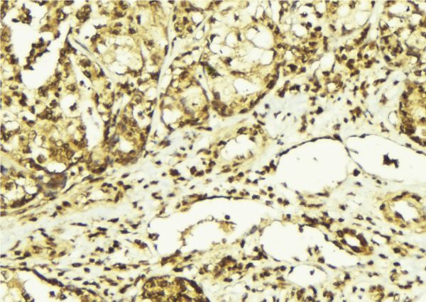
 IHC (Immunohistochemistry)
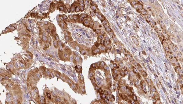
(AAA321600 at 1/100 staining human Breast carcinoma tissue sections by IHC-P. The tissue was formaldehyde fixed and a heat mediated antigen retrieval step in citrate buffer was performed. The tissue was then blocked and incubated with the antibody for 1.5 hours at 22 degree C. An HRP conjugated goat anti-rabbit antibody was used as the secondary.)
IHC (Immunohistochemistry)
(AAA321600 at 1/100 staining human Breast carcinoma tissue sections by IHC-P. The tissue was formaldehyde fixed and a heat mediated antigen retrieval step in citrate buffer was performed. The tissue was then blocked and incubated with the antibody for 1.5 hours at 22 degree C. An HRP conjugated goat anti-rabbit antibody was used as the secondary.)
CHOP, Polyclonal Antibody (Cat# AAA321600)
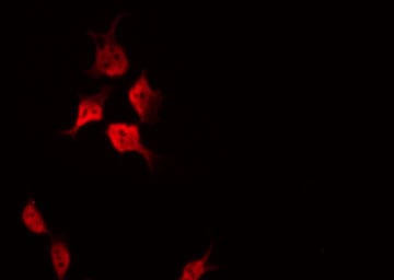
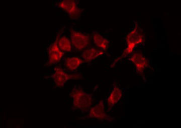
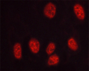
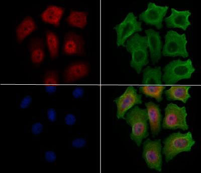 IF (Immunofluorescence)
(AAA321612 staining HepG2 cells(4h of LPS treatment) by IF/ICC. The samples were fixed with PFA and permeabilized in 0.1% Triton X-100,then blocked in 10% serum for 45 minutes at 25°C. Samples were then incubated with primary Ab(AAA321612 1:200) and mouse anti-beta tubulin Ab for 1 hour at 37°C. An AlexaFluor594 conjugated goat anti-rabbit IgG(H+L) Ab(Red)and an AlexaFluor488 conjugated goat anti-mouse IgG(H+L) Ab(Green) were used as the secondary Ab.The nuclear counter stain is DAPI(blue).)
IF (Immunofluorescence)
(AAA321612 staining HepG2 cells(4h of LPS treatment) by IF/ICC. The samples were fixed with PFA and permeabilized in 0.1% Triton X-100,then blocked in 10% serum for 45 minutes at 25°C. Samples were then incubated with primary Ab(AAA321612 1:200) and mouse anti-beta tubulin Ab for 1 hour at 37°C. An AlexaFluor594 conjugated goat anti-rabbit IgG(H+L) Ab(Red)and an AlexaFluor488 conjugated goat anti-mouse IgG(H+L) Ab(Green) were used as the secondary Ab.The nuclear counter stain is DAPI(blue).)
p21 Cip1, Polyclonal Antibody (Cat# AAA321612)

 WB (Western Blot)
(Western blot analysis of STAT3 phosphorylation expression in HeLa whole cell lysates, The lane on the left is treated with the antigen-specific peptide.)
WB (Western Blot)
(Western blot analysis of STAT3 phosphorylation expression in HeLa whole cell lysates, The lane on the left is treated with the antigen-specific peptide.)
STAT3, Polyclonal Antibody (Cat# AAA321613)
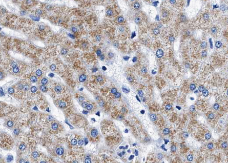
 IHC (Immunohistochemisry)
(AAA321618 at 1/100 staining human liver tissue sections by IHC-P. The tissue was formaldehyde fixed and a heat mediated antigen retrieval step in citrate buffer was performed. The tissue was then blocked and incubated with the antibody for 1.5 hours at 22°C. An HRP conjugated goat anti-rabbit antibody was used as the secondary.)
IHC (Immunohistochemisry)
(AAA321618 at 1/100 staining human liver tissue sections by IHC-P. The tissue was formaldehyde fixed and a heat mediated antigen retrieval step in citrate buffer was performed. The tissue was then blocked and incubated with the antibody for 1.5 hours at 22°C. An HRP conjugated goat anti-rabbit antibody was used as the secondary.)
STAT5A, Polyclonal Antibody (Cat# AAA321618)
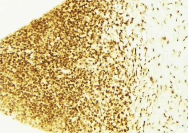
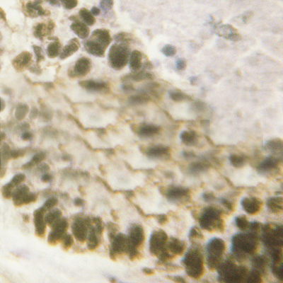
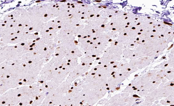
 IHC (Immunohistochemisry)
(AAA321620 at 1/100 staining human brain tissue sections by IHC-P. The tissue was formaldehyde fixed and a heat mediated antigen retrieval step in citrate buffer was performed. The tissue was then blocked and incubated with the antibody for 1.5 hours at 22 degree C. An HRP conjugated goat anti-rabbit antibody was used as the secondary.)
IHC (Immunohistochemisry)
(AAA321620 at 1/100 staining human brain tissue sections by IHC-P. The tissue was formaldehyde fixed and a heat mediated antigen retrieval step in citrate buffer was performed. The tissue was then blocked and incubated with the antibody for 1.5 hours at 22 degree C. An HRP conjugated goat anti-rabbit antibody was used as the secondary.)
GluR2, Polyclonal Antibody (Cat# AAA321620)
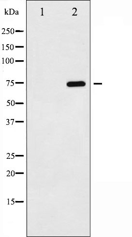
 WB (Western Blot)
(Western blot analysis of mTOR phosphorylation expression in Insulin treated NIH-3T3 whole cell lysates, The lane on the left is treated with the antigen-specific peptide.)
WB (Western Blot)
(Western blot analysis of mTOR phosphorylation expression in Insulin treated NIH-3T3 whole cell lysates, The lane on the left is treated with the antigen-specific peptide.)
mTOR, Polyclonal Antibody (Cat# AAA321622)
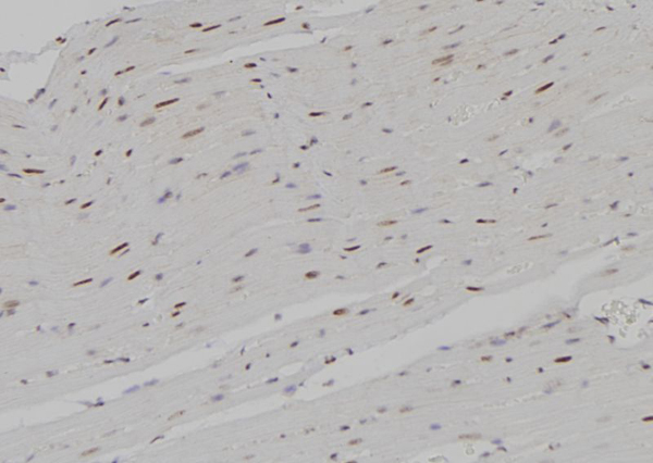
 IHC (Immunohiostchemistry)
(AAA321628 at 1/100 staining human Heart muscle tissue sections by IHC-P. The tissue was formaldehyde fixed and a heat mediated antigen retrieval step in citrate buffer was performed. The tissue was then blocked and incubated with the antibody for 1.5 hours at 22 degree C. An HRP conjugated goat anti-rabbit antibody was used as the secondary.)
IHC (Immunohiostchemistry)
(AAA321628 at 1/100 staining human Heart muscle tissue sections by IHC-P. The tissue was formaldehyde fixed and a heat mediated antigen retrieval step in citrate buffer was performed. The tissue was then blocked and incubated with the antibody for 1.5 hours at 22 degree C. An HRP conjugated goat anti-rabbit antibody was used as the secondary.)
SEK1/MKK4, Polyclonal Antibody (Cat# AAA321628)
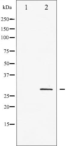
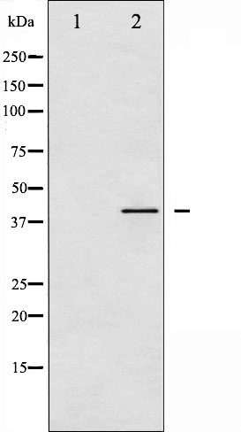
 WB (Western Blot)
(Western blot analysis of p27 Kip1 phosphorylation expression in EGF treated HeLa whole cell lysates, The lane on the left is treated with the antigen-specific peptide.)
WB (Western Blot)
(Western blot analysis of p27 Kip1 phosphorylation expression in EGF treated HeLa whole cell lysates, The lane on the left is treated with the antigen-specific peptide.)
p27 Kip1, Polyclonal Antibody (Cat# AAA321630)
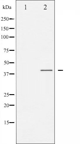
 WB (Western Blot)
(Western blot analysis of p27 Kip1 phosphorylation expression in HeLa whole cell lysates, The lane on the left is treated with the antigen-specific peptide.)
WB (Western Blot)
(Western blot analysis of p27 Kip1 phosphorylation expression in HeLa whole cell lysates, The lane on the left is treated with the antigen-specific peptide.)
p27 Kip1, Polyclonal Antibody (Cat# AAA321631)
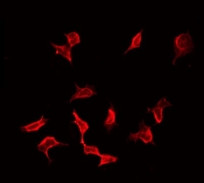
 IF (Immunofluorescence)
(AAA321639 staining HepG2 by IF/ICC. The sample were fixed with PFA and permeabilized in 0.1% Triton X-100, then blocked in 10% serum for 45 minutes at 25 degree C. The primary antibody was diluted at 1/200 and incubated with the sample for 1 hour at 37 degree C. An Alexa Fluor 594 conjugated goat anti-rabbit IgG (H+L) Ab, diluted at 1/600, was used as the secondary antibody.)
IF (Immunofluorescence)
(AAA321639 staining HepG2 by IF/ICC. The sample were fixed with PFA and permeabilized in 0.1% Triton X-100, then blocked in 10% serum for 45 minutes at 25 degree C. The primary antibody was diluted at 1/200 and incubated with the sample for 1 hour at 37 degree C. An Alexa Fluor 594 conjugated goat anti-rabbit IgG (H+L) Ab, diluted at 1/600, was used as the secondary antibody.)
C/EBP alpha, Polyclonal Antibody (Cat# AAA321639)
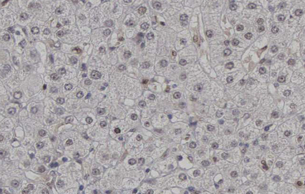
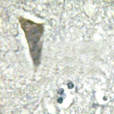
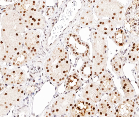
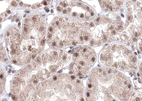
 IHC (Immunohistochemisry)
(AAA321640 at 1/100 staining human liver tissue sections by IHC-P. The tissue was formaldehyde fixed and a heat mediated antigen retrieval step in citrate buffer was performed. The tissue was then blocked and incubated with the antibody for 1.5 hours at 22 degree C. An HRP conjugated goat anti-rabbit antibody was used as the secondary.)
IHC (Immunohistochemisry)
(AAA321640 at 1/100 staining human liver tissue sections by IHC-P. The tissue was formaldehyde fixed and a heat mediated antigen retrieval step in citrate buffer was performed. The tissue was then blocked and incubated with the antibody for 1.5 hours at 22 degree C. An HRP conjugated goat anti-rabbit antibody was used as the secondary.)
TSC2, Polyclonal Antibody (Cat# AAA321640)

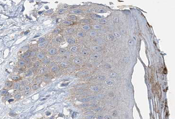 IHC (Immunohistochemistry)
(AAA321643 at 1/100 staining human Skin cancer tissue sections by IHC-P. The tissue was formaldehyde fixed and a heat mediated antigen retrieval step in citrate buffer was performed. The tissue was then blocked and incubated with the antibody for 1.5 hours at 22 degree C. An HRP conjugated goat anti-rabbit antibody was used as the secondary.)
IHC (Immunohistochemistry)
(AAA321643 at 1/100 staining human Skin cancer tissue sections by IHC-P. The tissue was formaldehyde fixed and a heat mediated antigen retrieval step in citrate buffer was performed. The tissue was then blocked and incubated with the antibody for 1.5 hours at 22 degree C. An HRP conjugated goat anti-rabbit antibody was used as the secondary.)
VASP, Polyclonal Antibody (Cat# AAA321643)
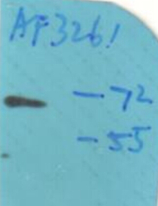
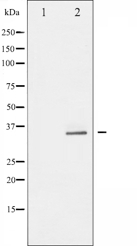
 WB (Western Blot)
(Western blot analysis of PTEN phosphorylation expression in Vanadate treated HeLa whole cell lysates, The lane on the left is treated with the antigen-specific peptide.)
WB (Western Blot)
(Western blot analysis of PTEN phosphorylation expression in Vanadate treated HeLa whole cell lysates, The lane on the left is treated with the antigen-specific peptide.)
PTEN, Polyclonal Antibody (Cat# AAA321655)
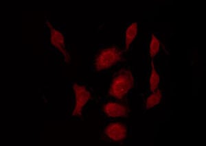
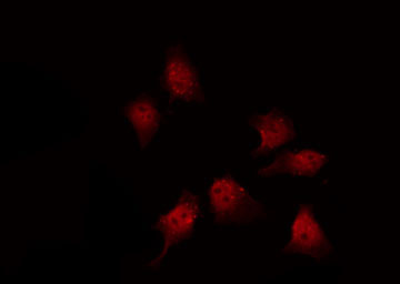
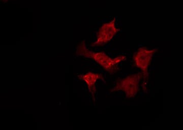
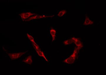
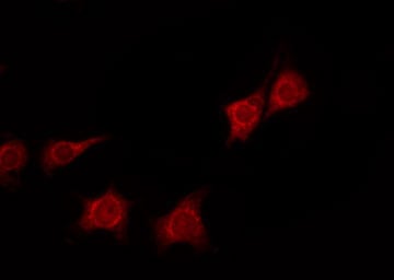
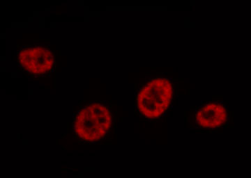
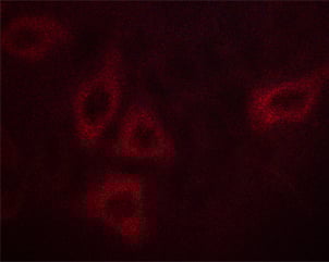 IF (Immunofluorescence)
(AAA321657 staining HepG2 cells by ICC/IF. Cells were fixed with PFA and permeabilized in 0.1% saponin prior to blocking in 10% serum for 45 minutes at 37 degree C. The primary antibody was diluted 1/400 and incubated with the sample for 1 hour at 37 degree C. A Alexa Fluor 594 conjugated goat polyclonal to rabbit IgG (H+L), diluted 1/600 was used as secondary antibody.)
IF (Immunofluorescence)
(AAA321657 staining HepG2 cells by ICC/IF. Cells were fixed with PFA and permeabilized in 0.1% saponin prior to blocking in 10% serum for 45 minutes at 37 degree C. The primary antibody was diluted 1/400 and incubated with the sample for 1 hour at 37 degree C. A Alexa Fluor 594 conjugated goat polyclonal to rabbit IgG (H+L), diluted 1/600 was used as secondary antibody.)
Calmodulin, Polyclonal Antibody (Cat# AAA321657)
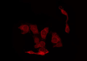
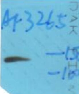
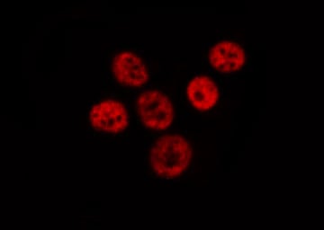
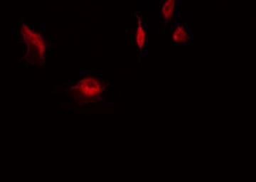 IF (Immunofluorescence)
(AAA321658 staining HeLa by IF/ICC. The sample were fixed with PFA and permeabilized in 0.1% Triton X-100, then blocked in 10% serum for 45 minutes at 25 degree C. The primary antibody was diluted at 1/200 and incubated with the sample for 1 hour at 37 degree C. An Alexa Fluor 594 conjugated goat anti-rabbit IgG (H+L) Ab, diluted at 1/600, was used as the secondary antibody.)
IF (Immunofluorescence)
(AAA321658 staining HeLa by IF/ICC. The sample were fixed with PFA and permeabilized in 0.1% Triton X-100, then blocked in 10% serum for 45 minutes at 25 degree C. The primary antibody was diluted at 1/200 and incubated with the sample for 1 hour at 37 degree C. An Alexa Fluor 594 conjugated goat anti-rabbit IgG (H+L) Ab, diluted at 1/600, was used as the secondary antibody.)
S6 Ribosomal Protein, Polyclonal Antibody (Cat# AAA321658)
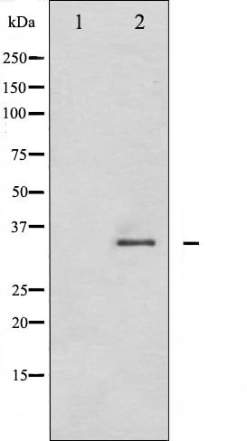
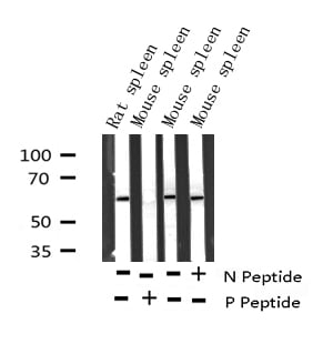
 WB (Western Blot)
(Western blot analysis of 14-3-3 zeta phosphorylation expression in UV treated NIH-3T3 whole cell lysates, The lane on the left is treated with the antigen-specific peptide.)
WB (Western Blot)
(Western blot analysis of 14-3-3 zeta phosphorylation expression in UV treated NIH-3T3 whole cell lysates, The lane on the left is treated with the antigen-specific peptide.)
14-3-3 zeta, Polyclonal Antibody (Cat# AAA321660)
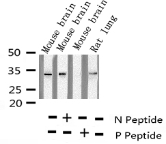
 WB (Western Blot)
(Western blot analysis of Histone H3.1 phosphorylation expression in COLO205 whole cell lysates, The lane on the left is treated with the antigen-specific peptide.)
WB (Western Blot)
(Western blot analysis of Histone H3.1 phosphorylation expression in COLO205 whole cell lysates, The lane on the left is treated with the antigen-specific peptide.)
Histone H3.1, Polyclonal Antibody (Cat# AAA321662)
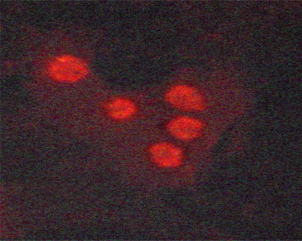
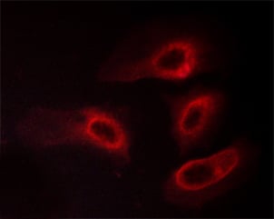
 IF (Immunofluorescence)
(AAA321663 staining HUVEC cells treated with serum 20% 30' by ICC/IF. Cells were fixed with PFA and permeabilized in 0.1% saponin prior to blocking in 10% serum for 45 minutes at 37 degree C. The primary antibody was diluted 1/400 and incubated with the sample for 1 hour at 37 degree C. A Alexa Fluor 594 conjugated goat polyclonal to rabbit IgG (H+L), diluted 1/600 was used as secondary antibody.)
IF (Immunofluorescence)
(AAA321663 staining HUVEC cells treated with serum 20% 30' by ICC/IF. Cells were fixed with PFA and permeabilized in 0.1% saponin prior to blocking in 10% serum for 45 minutes at 37 degree C. The primary antibody was diluted 1/400 and incubated with the sample for 1 hour at 37 degree C. A Alexa Fluor 594 conjugated goat polyclonal to rabbit IgG (H+L), diluted 1/600 was used as secondary antibody.)
DNA-PK, Polyclonal Antibody (Cat# AAA321663)
 WB (Western Blot)
(Western blot analysis of Smad3 phosphorylation expression in Jurkat whole cell lysates, The lane on the left is treated with the antigen-specific peptide.)
WB (Western Blot)
(Western blot analysis of Smad3 phosphorylation expression in Jurkat whole cell lysates, The lane on the left is treated with the antigen-specific peptide.)
Smad3, Polyclonal Antibody (Cat# AAA321668)
 IF (Immunofluorescence)
(AAA321669 staining HepG2 cells(30min of 4uM Forskolin treatment) by IF/ICC. The samples were fixed with PFA and permeabilized in 0.1% Triton X-100,then blocked in 10% serum for 45 minutes at 25°C. Samples were then incubated with primary Ab(AAA321669) and mouse anti-beta tubulin Ab for 1 hour at 37°C. An AlexaFluor594 conjugated goat anti-rabbit IgG(H+L) Ab(Red) and an AlexaFluor488 conjugated goat anti-mouse IgG(H+L) Ab(Green) were used as the secondary antibody. The nuclear counter stain is DAPI(blue).)
IF (Immunofluorescence)
(AAA321669 staining HepG2 cells(30min of 4uM Forskolin treatment) by IF/ICC. The samples were fixed with PFA and permeabilized in 0.1% Triton X-100,then blocked in 10% serum for 45 minutes at 25°C. Samples were then incubated with primary Ab(AAA321669) and mouse anti-beta tubulin Ab for 1 hour at 37°C. An AlexaFluor594 conjugated goat anti-rabbit IgG(H+L) Ab(Red) and an AlexaFluor488 conjugated goat anti-mouse IgG(H+L) Ab(Green) were used as the secondary antibody. The nuclear counter stain is DAPI(blue).)
CDK5, Polyclonal Antibody (Cat# AAA321669)
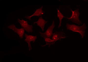
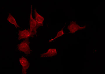
 IF (Immunofluorescence)
(AAA321672 staining RAW264.7 by IF/ICC. The sample were fixed with PFA and permeabilized in 0.1% Triton X-100, then blocked in 10% serum for 45 minutes at 25 degree C. The primary antibody was diluted at 1/200 and incubated with the sample for 1 hour at 37 degree C. An Alexa Fluor 594 conjugated goat anti-rabbit IgG (H+L) Ab, diluted at 1/600, was used as the secondary antibody.)
IF (Immunofluorescence)
(AAA321672 staining RAW264.7 by IF/ICC. The sample were fixed with PFA and permeabilized in 0.1% Triton X-100, then blocked in 10% serum for 45 minutes at 25 degree C. The primary antibody was diluted at 1/200 and incubated with the sample for 1 hour at 37 degree C. An Alexa Fluor 594 conjugated goat anti-rabbit IgG (H+L) Ab, diluted at 1/600, was used as the secondary antibody.)
NF kappaB p100/p52, Polyclonal Antibody (Cat# AAA321672)
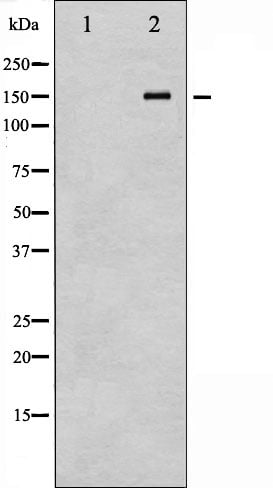
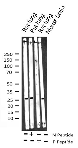
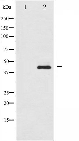
 WB (Western Blot)
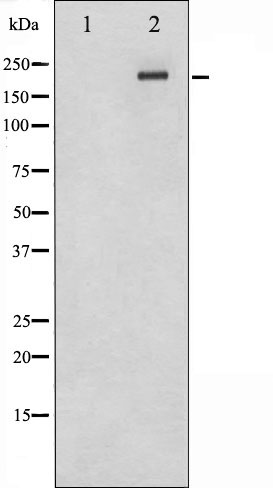
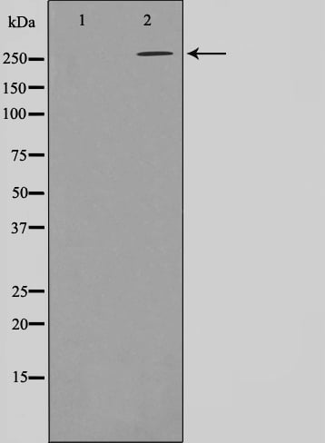
(Western blot analysis of Phospho-E2F1 (Thr433) expression in various lysates)
WB (Western Blot)
(Western blot analysis of Phospho-E2F1 (Thr433) expression in various lysates)
E2F1, Polyclonal Antibody (Cat# AAA321675)
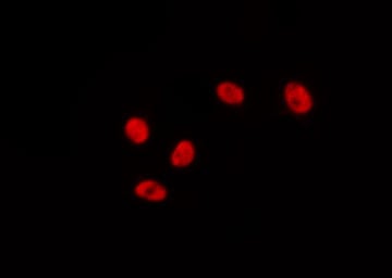
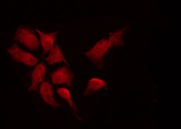
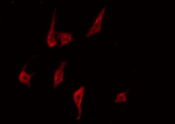
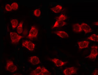
 IF (Immunofluorescence)
(AAA321676 staining Hela by IF/ICC. The sample were fixed with PFA and permeabilized in 0.1% Triton X-100, then blocked in 10% serum for 45 minutes at 25 degree C. The primary antibody was diluted at 1/200 and incubated with the sample for 1 hour at 37 degree C. An Alexa Fluor 594 conjugated goat anti-rabbit IgG (H+L) Ab, diluted at 1/600, was used as the secondary antibody.)
IF (Immunofluorescence)
(AAA321676 staining Hela by IF/ICC. The sample were fixed with PFA and permeabilized in 0.1% Triton X-100, then blocked in 10% serum for 45 minutes at 25 degree C. The primary antibody was diluted at 1/200 and incubated with the sample for 1 hour at 37 degree C. An Alexa Fluor 594 conjugated goat anti-rabbit IgG (H+L) Ab, diluted at 1/600, was used as the secondary antibody.)
AML1, Polyclonal Antibody (Cat# AAA321676)
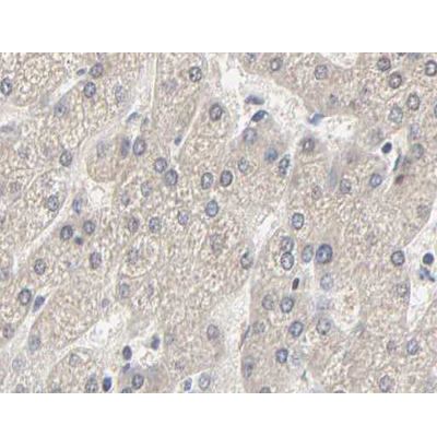
 IF (Immunofluorescence)
(AAA321677 staining Hela by IF/ICC. The sample were fixed with PFA and permeabilized in 0.1% Triton X-100, then blocked in 10% serum for 45 minutes at 25 degree C. The primary antibody was diluted at 1/200 and incubated with the sample for 1 hour at 37 degree C. An Alexa Fluor 594 conjugated goat anti-rabbit IgG (H+L) Ab, diluted at 1/600, was used as the secondary antibody.)
IF (Immunofluorescence)
(AAA321677 staining Hela by IF/ICC. The sample were fixed with PFA and permeabilized in 0.1% Triton X-100, then blocked in 10% serum for 45 minutes at 25 degree C. The primary antibody was diluted at 1/200 and incubated with the sample for 1 hour at 37 degree C. An Alexa Fluor 594 conjugated goat anti-rabbit IgG (H+L) Ab, diluted at 1/600, was used as the secondary antibody.)
AML1, Polyclonal Antibody (Cat# AAA321677)
 IF (Immunofluorescence)
(AAA321679 staining HeLa by IF/ICC. The sample were fixed with PFA and permeabilized in 0.1% Triton X-100, then blocked in 10% serum for 45 minutes at 25 degree C. The primary antibody was diluted at 1/200 and incubated with the sample for 1 hour at 37 degree C. An Alexa Fluor 594 conjugated goat anti-rabbit IgG (H+L) Ab, diluted at 1/600, was used as the secondary antibody.)
IF (Immunofluorescence)
(AAA321679 staining HeLa by IF/ICC. The sample were fixed with PFA and permeabilized in 0.1% Triton X-100, then blocked in 10% serum for 45 minutes at 25 degree C. The primary antibody was diluted at 1/200 and incubated with the sample for 1 hour at 37 degree C. An Alexa Fluor 594 conjugated goat anti-rabbit IgG (H+L) Ab, diluted at 1/600, was used as the secondary antibody.)
MEF2A, Polyclonal Antibody (Cat# AAA321679)
 IF (Immunofluorescence)
(AAA321686 staining HeLa by IF/ICC. The sample were fixed with PFA and permeabilized in 0.1% Triton X-100, then blocked in 10% serum for 45 minutes at 25 degree C. The primary antibody was diluted at 1/200 and incubated with the sample for 1 hour at 37 degree C. An Alexa Fluor 594 conjugated goat anti-rabbit IgG (H+L) Ab, diluted at 1/600, was used as the secondary antibody.)
IF (Immunofluorescence)
(AAA321686 staining HeLa by IF/ICC. The sample were fixed with PFA and permeabilized in 0.1% Triton X-100, then blocked in 10% serum for 45 minutes at 25 degree C. The primary antibody was diluted at 1/200 and incubated with the sample for 1 hour at 37 degree C. An Alexa Fluor 594 conjugated goat anti-rabbit IgG (H+L) Ab, diluted at 1/600, was used as the secondary antibody.)
NF kappaB p65, Polyclonal Antibody (Cat# AAA321686)
 IF (Immunofluorescence)
(AAA321688 staining HeLa by IF/ICC. The sample were fixed with PFA and permeabilized in 0.1% Triton X-100, then blocked in 10% serum for 45 minutes at 25 degree C. The primary antibody was diluted at 1/200 and incubated with the sample for 1 hour at 37 degree C. An Alexa Fluor 594 conjugated goat anti-rabbit IgG (H+L) Ab, diluted at 1/600, was used as the secondary antibody.)
IF (Immunofluorescence)
(AAA321688 staining HeLa by IF/ICC. The sample were fixed with PFA and permeabilized in 0.1% Triton X-100, then blocked in 10% serum for 45 minutes at 25 degree C. The primary antibody was diluted at 1/200 and incubated with the sample for 1 hour at 37 degree C. An Alexa Fluor 594 conjugated goat anti-rabbit IgG (H+L) Ab, diluted at 1/600, was used as the secondary antibody.)
NF kappaB p65, Polyclonal Antibody (Cat# AAA321688)
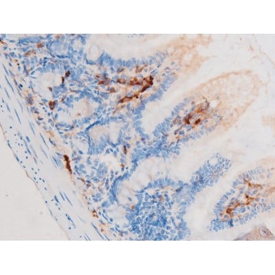
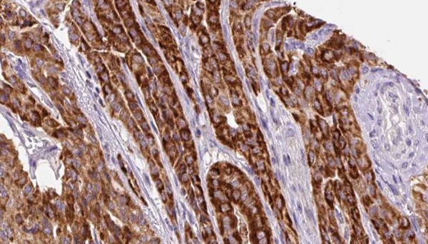
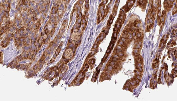
 IHC (Immunohistochemistry)
(AAA321690 at 1/200 staining Rat ganstric tissue sections by IHC-P. The tissue was formaldehyde fixed and a heat mediated antigen retrieval step in citrate buffer was performed. The tissue was then blocked and incubated with the antibody for 1.5 hours at 22 degree C. An HRP conjugated goat anti-rabbit antibody was used as the secondary.)
IHC (Immunohistochemistry)
(AAA321690 at 1/200 staining Rat ganstric tissue sections by IHC-P. The tissue was formaldehyde fixed and a heat mediated antigen retrieval step in citrate buffer was performed. The tissue was then blocked and incubated with the antibody for 1.5 hours at 22 degree C. An HRP conjugated goat anti-rabbit antibody was used as the secondary.)
NF kappaB p65, Polyclonal Antibody (Cat# AAA321690)

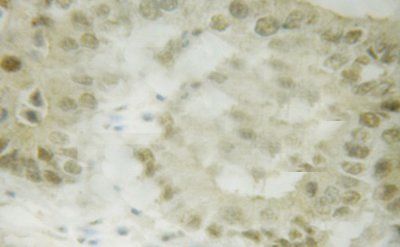
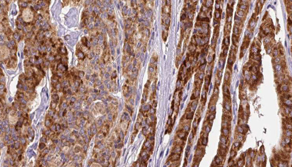
 IHC (Immunohistochemistry)
(AAA321702 at 1/100 staining Human breast cancer tissue by IHC-P. The sample was formaldehyde fixed and a heat mediated antigen retrieval step in citrate buffer was performed. The sample was then blocked and incubated with the antibody for 1.5 hours at 22 degree C. An HRP conjugated goat anti-rabbit antibody was used as the secondary.)
IHC (Immunohistochemistry)
(AAA321702 at 1/100 staining Human breast cancer tissue by IHC-P. The sample was formaldehyde fixed and a heat mediated antigen retrieval step in citrate buffer was performed. The sample was then blocked and incubated with the antibody for 1.5 hours at 22 degree C. An HRP conjugated goat anti-rabbit antibody was used as the secondary.)
PKC delta, Polyclonal Antibody (Cat# AAA321702)
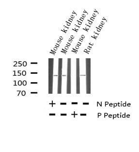
 WB (Western Blot)
(Western blot analysis of Phospho-ACC1 (Ser80) expression in various lysates)
WB (Western Blot)
(Western blot analysis of Phospho-ACC1 (Ser80) expression in various lysates)
ACC1, Polyclonal Antibody (Cat# AAA321712)
 WB (Western Blot)
(Western blot analysis of Phospho-G3BP-1 (Ser232) expression in various lysates)
WB (Western Blot)
(Western blot analysis of Phospho-G3BP-1 (Ser232) expression in various lysates)
G3BP-1, Polyclonal Antibody (Cat# AAA321717)
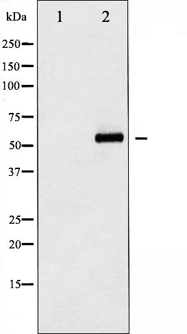
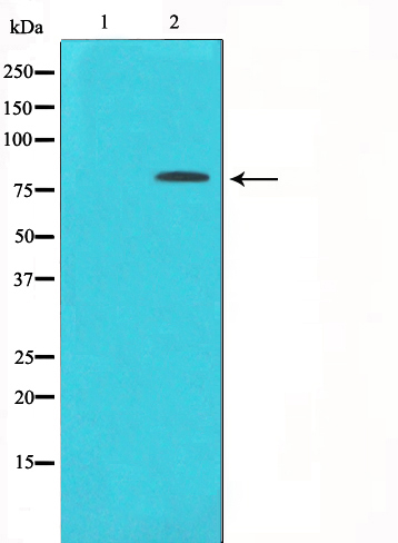
 WB (Western Blot)
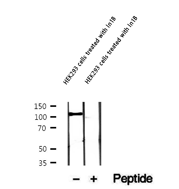
(Western blot analysis of extracts of HEK293 cells treated with In1B, using Phospho-Gab1 (Tyr307) Antibody.)
WB (Western Blot)
(Western blot analysis of extracts of HEK293 cells treated with In1B, using Phospho-Gab1 (Tyr307) Antibody.)
Gab1, Polyclonal Antibody (Cat# AAA321329)
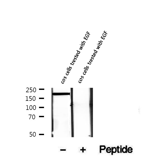 WB (Western Blot)
(Western blot analysis of extracts of cos cells trested with EGF, using Phospho-HER2/ErbB2 (Tyr1221/1222) Antibody.)
WB (Western Blot)
(Western blot analysis of extracts of cos cells trested with EGF, using Phospho-HER2/ErbB2 (Tyr1221/1222) Antibody.)
HER2/ErbB2, Polyclonal Antibody (Cat# AAA321333)
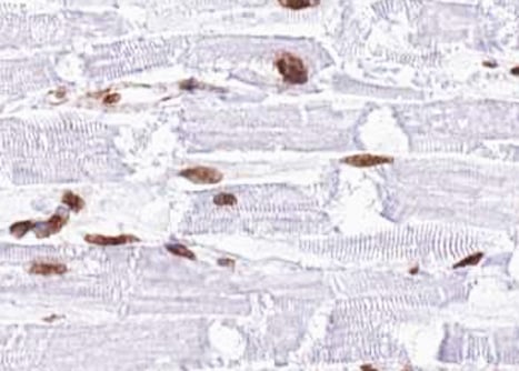
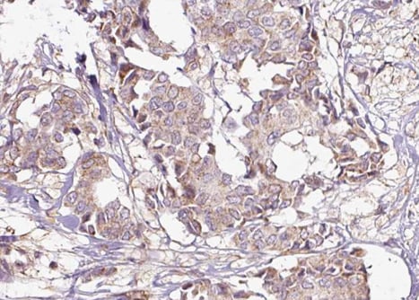
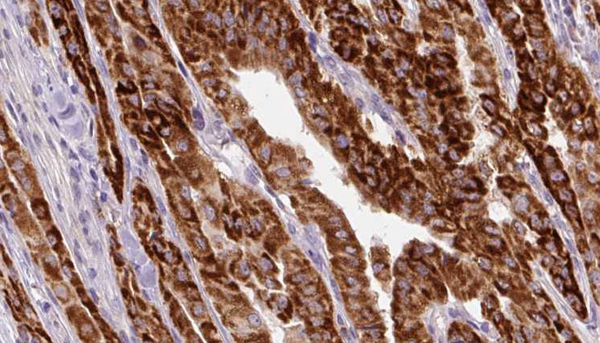
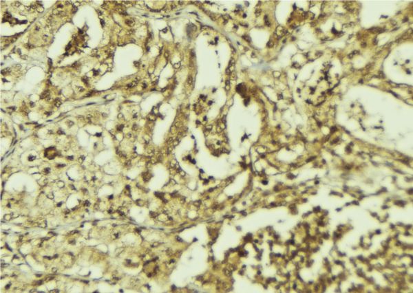
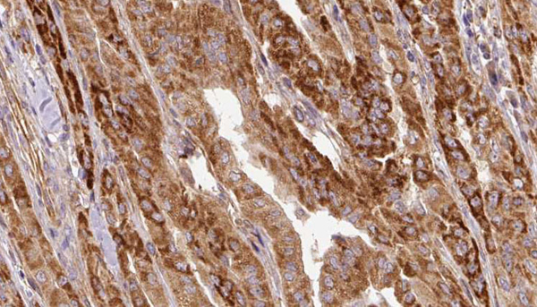 IHC (Immunohistochemistry)
(AAA321334 at 1/100 staining Human thyroid cancer tissue by IHC-P. The sample was formaldehyde fixed and a heat mediated antigen retrieval step in citrate buffer was performed. The sample was then blocked and incubated with the antibody for 1.5 hours at 22 degree C. An HRP conjugated goat anti-rabbit antibody was used as the secondary.)
IHC (Immunohistochemistry)
(AAA321334 at 1/100 staining Human thyroid cancer tissue by IHC-P. The sample was formaldehyde fixed and a heat mediated antigen retrieval step in citrate buffer was performed. The sample was then blocked and incubated with the antibody for 1.5 hours at 22 degree C. An HRP conjugated goat anti-rabbit antibody was used as the secondary.)
HSL, Polyclonal Antibody (Cat# AAA321334)
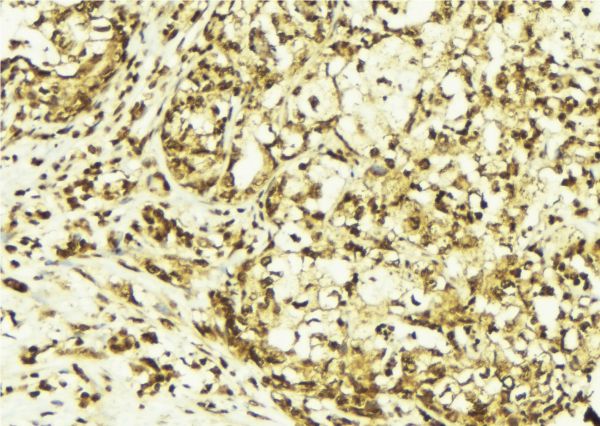
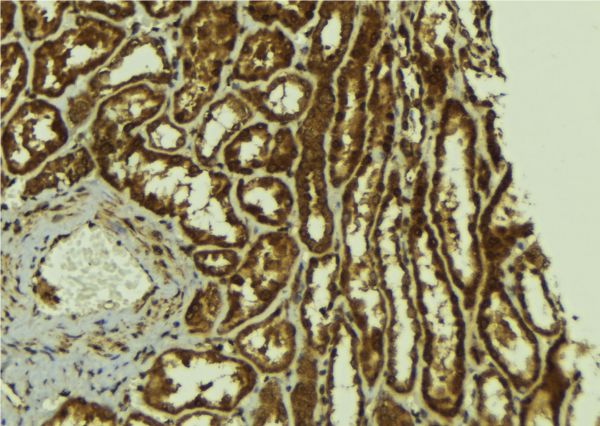
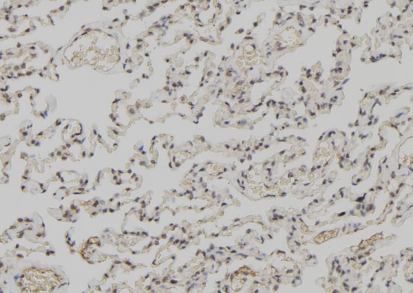 IHC (Immunohiostchemistry)
(AAA321337 at 1/100 staining Human lung tissue by IHC-P. The sample was formaldehyde fixed and a heat mediated antigen retrieval step in citrate buffer was performed. The sample was then blocked and incubated with the antibody for 1.5 hours at 22 degree C. An HRP conjugated goat anti-rabbit antibody was used as the secondary.)
IHC (Immunohiostchemistry)
(AAA321337 at 1/100 staining Human lung tissue by IHC-P. The sample was formaldehyde fixed and a heat mediated antigen retrieval step in citrate buffer was performed. The sample was then blocked and incubated with the antibody for 1.5 hours at 22 degree C. An HRP conjugated goat anti-rabbit antibody was used as the secondary.)
IRF-3, Polyclonal Antibody (Cat# AAA321337)
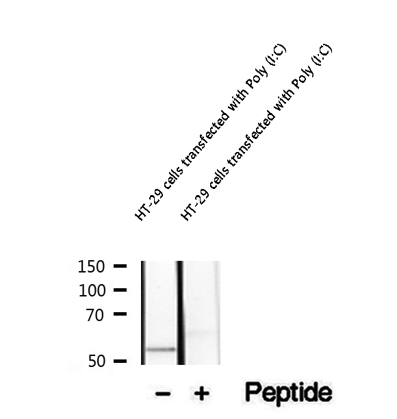
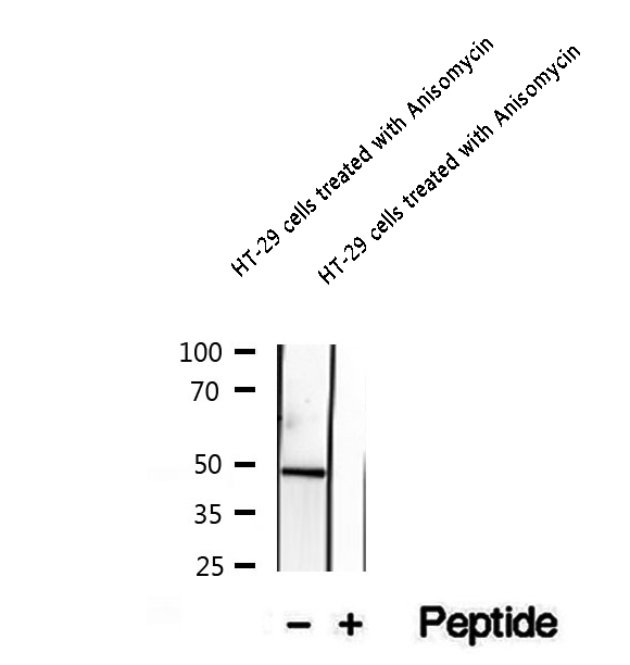 WB (Western Blot)
(Western blot analysis of extracts of HT-29 cells treated with Anisomycin, using Phospho-Keratin 20 (Ser13) Antibody.)
WB (Western Blot)
(Western blot analysis of extracts of HT-29 cells treated with Anisomycin, using Phospho-Keratin 20 (Ser13) Antibody.)
Keratin 20, Polyclonal Antibody (Cat# AAA321340)
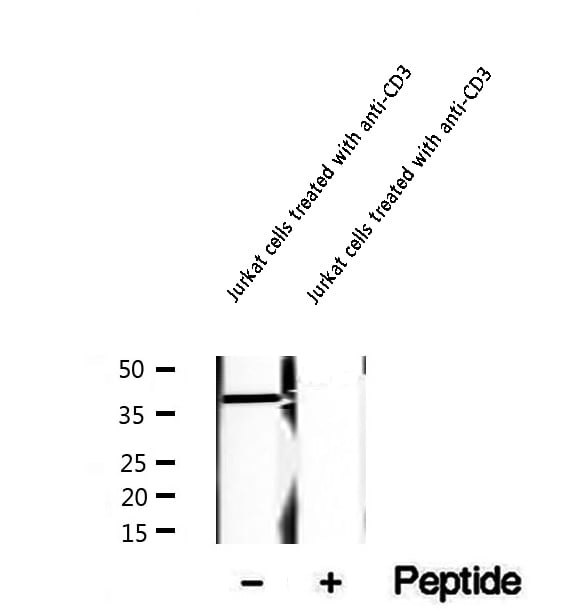 WB (Western Blot)
(Western blot analysis of extracts of Jurkat cells treated with anti-CD3, using Phospho-LAT (Tyr200) Antibody.)
WB (Western Blot)
(Western blot analysis of extracts of Jurkat cells treated with anti-CD3, using Phospho-LAT (Tyr200) Antibody.)
LAT, Polyclonal Antibody (Cat# AAA321343)
 WB (Western Blot)
(Western blot analysis of extracts of A431 cells, using Phospho-MEK1 (Thr286) Antibody.)
WB (Western Blot)
(Western blot analysis of extracts of A431 cells, using Phospho-MEK1 (Thr286) Antibody.)
MEK1, Polyclonal Antibody (Cat# AAA321348)
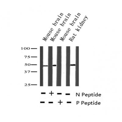
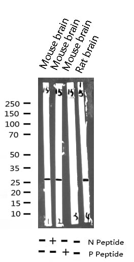
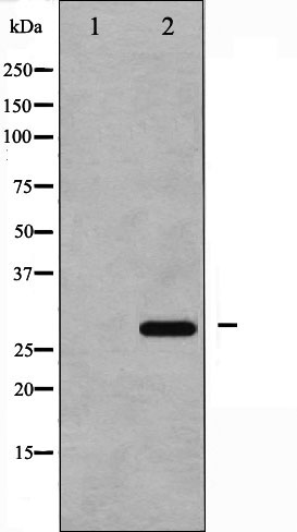
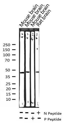
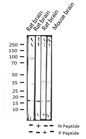
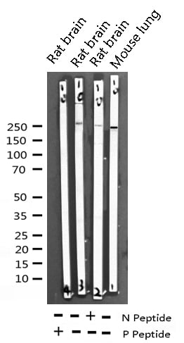
 WB (Western Blot)
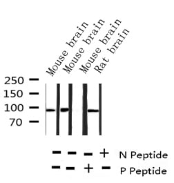
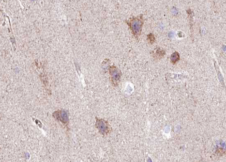
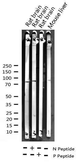
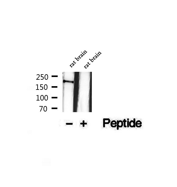
(Western blot analysis of extracts of rat brain tissue, using Phospho-NMDAR2B (Ser1284) Antibody.)
WB (Western Blot)
(Western blot analysis of extracts of rat brain tissue, using Phospho-NMDAR2B (Ser1284) Antibody.)
NMDAR2B, Polyclonal Antibody (Cat# AAA321355)
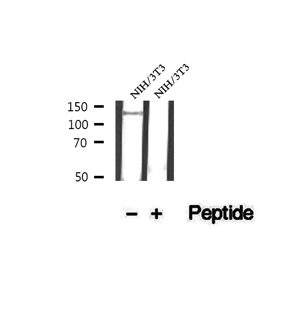 WB (Western Blot)
(Western blot analysis of extracts of NIH/3T3 cells, using Phospho-p130 Cas (Tyr249) Antibody.)
WB (Western Blot)
(Western blot analysis of extracts of NIH/3T3 cells, using Phospho-p130 Cas (Tyr249) Antibody.)
p130 Cas, Polyclonal Antibody (Cat# AAA321359)
What Are Phospho Antibodies?
Protein phosphorylation is a process where a phosphate group is added to certain amino acid residues of a protein – usually serine (S), threonine (T), or tyrosine (Y) - by enzymes called kinases. This process is integral in controlling cellular signaling, cellular growth, and other biological functions.
Our catalog includes a wide range of phospho-specific antibodies that can accurately detect this important marker. They perform strongly in widely-used laboratory applications such as Western blot, flow cytometry, immunohistochemistry, and immunofluorescence microscopy. We value your trust in us and are committed to providing top-quality products and services. All of our antibodies are guaranteed to work for the applications and species indicated on our website & associated product pages.
What Are The Key Applications of Phospho Antibodies?
1. Western Blotting
One of the first steps a researcher can take in utilizing these phospho-specific antibodies, is to check if the antibody works using a technique referred to as “Western blot”. For those unfamiliar, Western Blot aids in showing whether the protein that the antibody recognizes is appearing at the correct/expected size. These phospho-specific antibodies should also be able to detect changes in the target protein’s phosphorylation (on/off state) when cells are stimulated in certain ways.
2. Staining of Fixed Cells (Immunocytochemistry)
Another routine use of these phospho-specific antibodies, is to test if the antibody is able to demonstrate similar performance when used on fixed cells (intact cells that have been preserved) as it did in the Western blot tests. It is an important aspect in many cases to confirm that the antibody works in actual intact cell samples. Ideally, the method used for cellular fixation should be the same as what is used in pathology labs (like using 10% formalin). To check if the antibody works well in tissue sections (FFPE), researchers will often test it on fixed cells that are processed similar to tissue samples.
3. Specificity Tests Using Peptides
In order to make sure that the antibody is only binding to the right target:
- Laboratory technicians will mix the antibody with phospho-peptides (short segments of the protein containing the phosphate group modification).
- If the antibody signal disappears, it is confirmation that it is binding to the correct phosphorylated location.
- A more robust test is to use both the phosphorylated and non-phosphorylated (dephosphorylated) versions of the protein. The antibody should react only with the phosphorylated one.
- Another method sometimes utilized is to treat the sample with an enzyme, such as alkaline phosphatase, that specifically removes phosphate groups. If the antibody signal disappears after this, it also confirms specificity.
4. Genetic Confirmation
As a final step, scientists can genetically manipulate the nucleotide sequence and alter the target protein by removing the exact site where phosphorylation happens. If the antibody no longer appears to detect the modified protein, it is strong evidence supporting the antibody being specific for that phosphorylated site.
Why Buy Phospho Antibodies Through Us?
- The production laboratory adheres to strict and consistent protocols prior to releasing any of these phospho-specific antibodies:
- Standard methods and proper controls in all tests to ensure high quality.
- These antibodies are tested and validated in different cell types and species.
- High quality control criterion to ensure each batch is consistent, so you will obtain reliable results every time.
FAQ
1. What Are Phospho-Specific Antibodies?
Phospho-specific antibodies are made to detect proteins only when they have a phosphate group linked to a specific amino acid residue. This empowers scientists understand if a protein is "turned on" or active, based on its phosphorylation state.
2. How to Detect Phosphorylated Proteins in a Western Blot?
To find out if a protein is phosphorylated using Western blot:
- Use a phospho-specific antibody that binds only to the phosphorylated form of the protein.
- You can also use a “regular” antibody for the same amino acid sequence of the protein that the phospho-specific antibody is binding to (but in this case, this antibody will not bind if there is a phosphate group present) in order to compare how much of it is phosphorylated versus how much is non-phosphorylated (or “total” protein, if the “normal” antibody’s epitopes are non-phospho-site-specific).
3. How to Choose the Best Antibody?
Here are some simple tips to help you pick the right antibody:
- Know your target
- Match your sample characteristics
- Confirm the intended use is appropriate
- Check “host” and “type”
- Check the “quality” of the presented data/images
- Appraise whether the available validation meets your needs