Filters
▼Clonality
▼Type
▼Reactivity
▼Gene Name
▼Isotype
▼Host
▼Application
▼Clone
▼Phospho Antibodies
Phospho-specific antibodies’ typical purpose is to enable researchers to detect changes in proteins. They will exclusively bind to the amino acid sequence on a protein that has been phosphorylated (which is both a physical & chemical change) and do not bind to the same amino acid sequence on said protein if it lacks said phosphorylation. This aids in being able to clearly see and understand the data produced from this particular protein modification.
Viewing 3900-3950 of 5297 product results
 WB (Western Blot)
(Western blot analysis of Phospho-CDC37 (Ser13) expression in various lysates)
WB (Western Blot)
(Western blot analysis of Phospho-CDC37 (Ser13) expression in various lysates)
CDC37, Polyclonal Antibody (Cat# AAA321746)
 WB (Western Blot)
(Western blot analysis of Phospho-WNK1 (Thr58) expression in various lysates)
WB (Western Blot)
(Western blot analysis of Phospho-WNK1 (Thr58) expression in various lysates)
WNK1, Polyclonal Antibody (Cat# AAA321771)
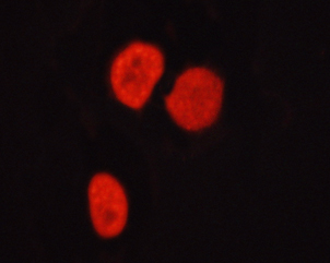 IF (Immunofluorescence)
(AAA321773 staining HepG2 cells by ICC/IF. Cells were fixed with PFA and permeabilized in 0.1% saponin prior to blocking in 10% serum for 45 minutes at 37 degree C. The primary antibody was diluted 1/400 and incubated with the sample for 1 hour at 37 degree C. A Alexa Fluor 594 conjugated goat polyclonal to rabbit IgG (H+L), diluted 1/600 was used as secondary antibody.)
IF (Immunofluorescence)
(AAA321773 staining HepG2 cells by ICC/IF. Cells were fixed with PFA and permeabilized in 0.1% saponin prior to blocking in 10% serum for 45 minutes at 37 degree C. The primary antibody was diluted 1/400 and incubated with the sample for 1 hour at 37 degree C. A Alexa Fluor 594 conjugated goat polyclonal to rabbit IgG (H+L), diluted 1/600 was used as secondary antibody.)
HDAC6, Polyclonal Antibody (Cat# AAA321773)
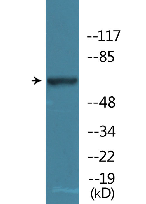
 WB (Western Blot)
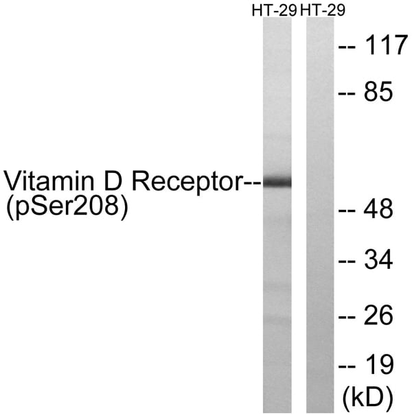
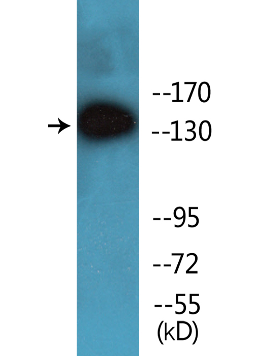
(Western blot analysis of lysates from HT29 cells treated with heat shock, using Vitamin D Receptor (Phospho-Ser208) Antibody. The lane on the right is blocked with the phospho peptide.)
WB (Western Blot)
(Western blot analysis of lysates from HT29 cells treated with heat shock, using Vitamin D Receptor (Phospho-Ser208) Antibody. The lane on the right is blocked with the phospho peptide.)
Vitamin D Receptor, Polyclonal Antibody (Cat# AAA316449)
 Application Data
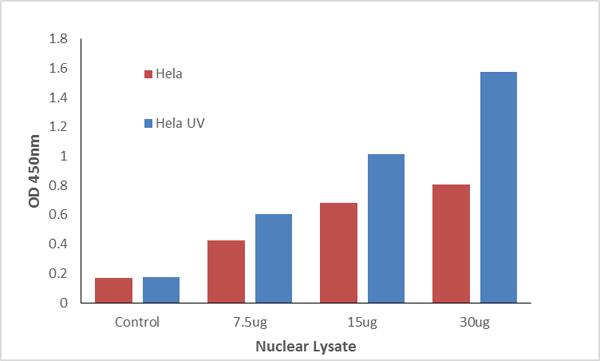
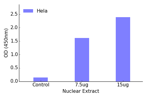
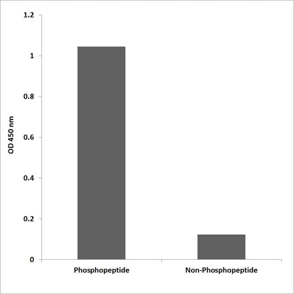
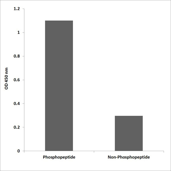
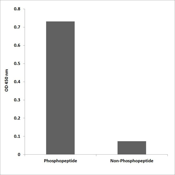
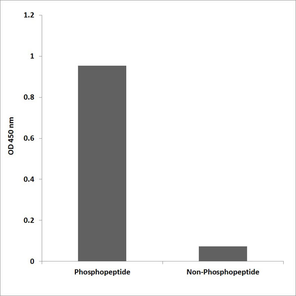
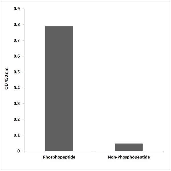
(The TFact Estrogen Receptor-beta (Phospho-Ser105) DNA-Binding ELISA detects active Estrogen Receptor-beta (Phospho-Ser105) in Hela Nuclear Extract. The Hela cells were grown 3 days in DMEM with 10% FBS and harvested for nuclear extract. The Hela cells were stimulated by UV (100J/M2) or directly harvested without UV stimulation.)
Application Data
(The TFact Estrogen Receptor-beta (Phospho-Ser105) DNA-Binding ELISA detects active Estrogen Receptor-beta (Phospho-Ser105) in Hela Nuclear Extract. The Hela cells were grown 3 days in DMEM with 10% FBS and harvested for nuclear extract. The Hela cells were stimulated by UV (100J/M2) or directly harvested without UV stimulation.)
Estrogen Receptor-beta, ELISA Kit (Cat# AAA315883)
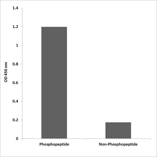
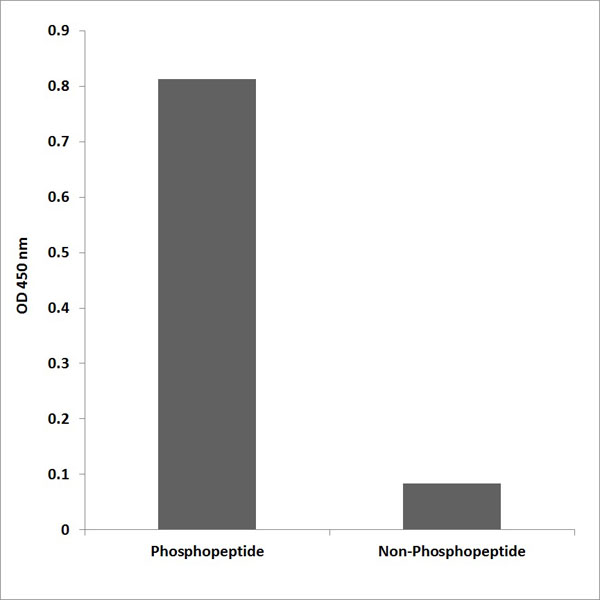
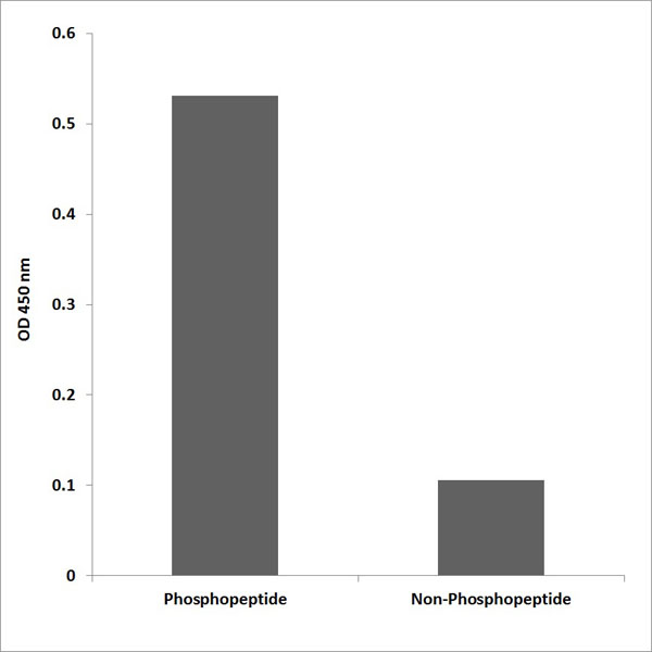
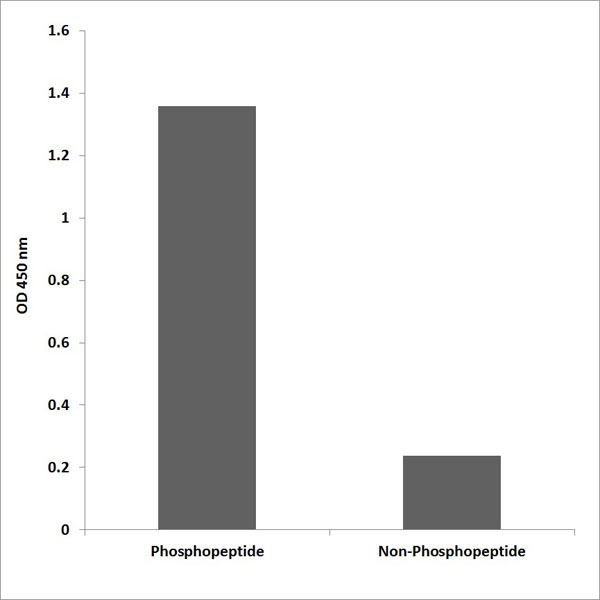
 Application Data
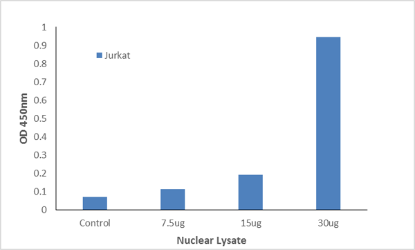
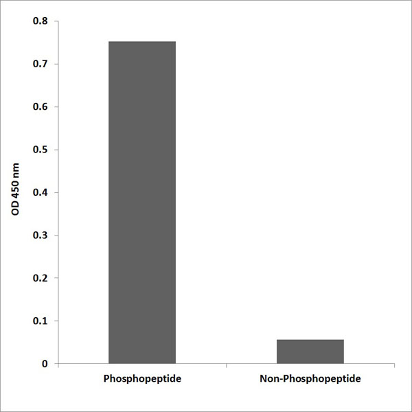
(The TFact Elk1 (Phospho-Ser389) DNA-Binding ELISA detects active Elk1 (Phospho-Ser389) in Jurkat Nuclear Extract. The Jurkat cells were grown 3 days in RPMI 1640 with 10% FBS and harvested for nuclear extract. The DNA-transcription factor complex is treated with stablization buffer.)
Application Data
(The TFact Elk1 (Phospho-Ser389) DNA-Binding ELISA detects active Elk1 (Phospho-Ser389) in Jurkat Nuclear Extract. The Jurkat cells were grown 3 days in RPMI 1640 with 10% FBS and harvested for nuclear extract. The DNA-transcription factor complex is treated with stablization buffer.)
Elk1, ELISA Kit (Cat# AAA315888)
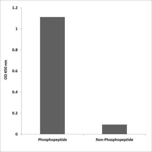
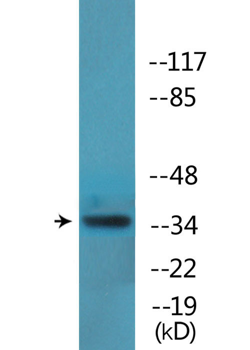
 Application Data
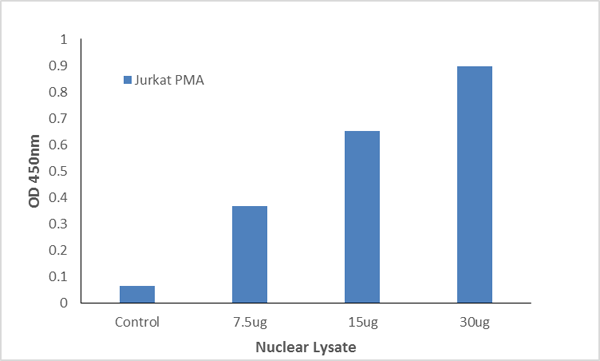
(The TFact ETS1 (Phospho-Thr38) DNA-Binding ELISA detects active ETS1 (Phospho-Thr38) in Jurkat Nuclear Extract. The Jurkat cells were grown 3 days in RPMI 1640 with 10% FBS and harvested for nuclear extract. Jurkat cells were stimulated by PMA (200nM) before harvest.)
Application Data
(The TFact ETS1 (Phospho-Thr38) DNA-Binding ELISA detects active ETS1 (Phospho-Thr38) in Jurkat Nuclear Extract. The Jurkat cells were grown 3 days in RPMI 1640 with 10% FBS and harvested for nuclear extract. Jurkat cells were stimulated by PMA (200nM) before harvest.)
ETS1, ELISA Kit (Cat# AAA315893)
 Application Data
(The TFact FOXO1 (Phospho-Ser319) DNA-Binding ELISA detects active FKHR (Phospho-Ser319) in Hela Nuclear Extract. The Hela cells were grown 3 days in DMEM with 10% FBS and harvested for nuclear extract. The Hela cells were stimulated by serum (20% FBS) before harvest.)
Application Data
(The TFact FOXO1 (Phospho-Ser319) DNA-Binding ELISA detects active FKHR (Phospho-Ser319) in Hela Nuclear Extract. The Hela cells were grown 3 days in DMEM with 10% FBS and harvested for nuclear extract. The Hela cells were stimulated by serum (20% FBS) before harvest.)
FOXO1, ELISA Kit (Cat# AAA315902)
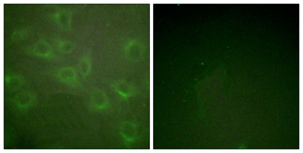
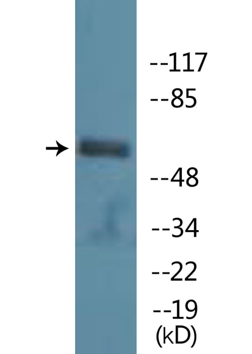
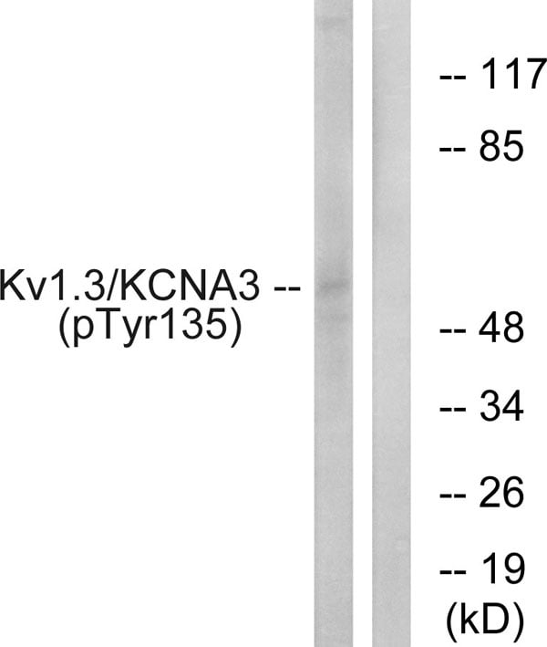 WB (Western Blot)
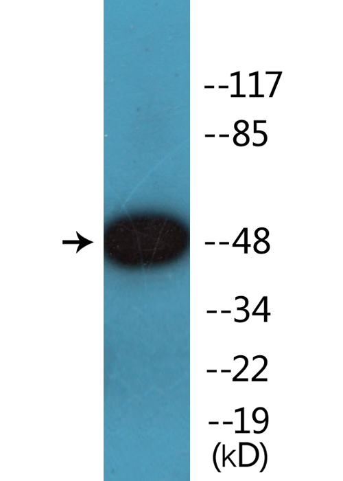
(Western blot analysis of lysates from Jurkat cells treated with starved 24h, using Kv1.3/KCNA3 (Phospho-Tyr135) Antibody. The lane on the right is blocked with the phospho peptide.)
WB (Western Blot)
(Western blot analysis of lysates from Jurkat cells treated with starved 24h, using Kv1.3/KCNA3 (Phospho-Tyr135) Antibody. The lane on the right is blocked with the phospho peptide.)
Kv1.3/KCNA3, Polyclonal Antibody (Cat# AAA316415)
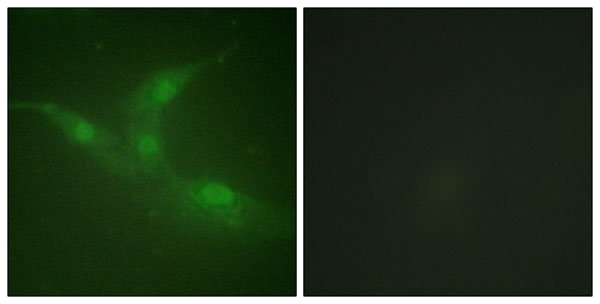
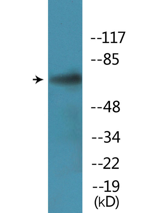
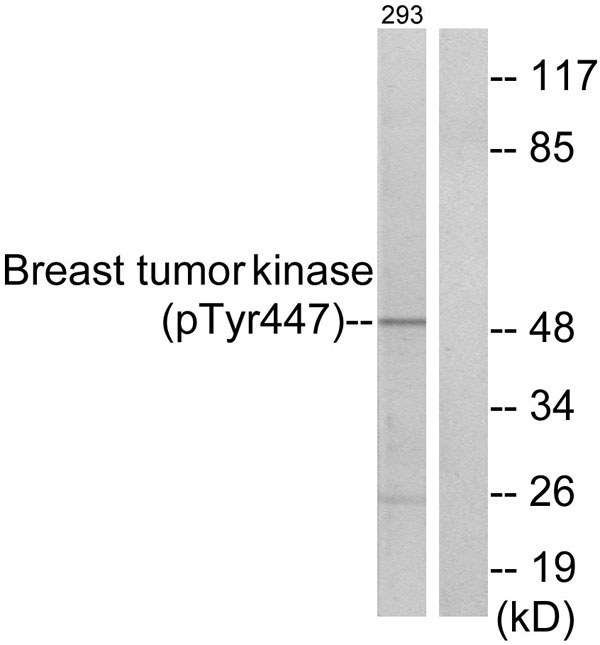 WB (Western Blot)
(Western blot analysis of lysates from 293 cells treated with EGF 200ng/ml 30', using Breast Tumor Kinase (Phospho-Tyr447) Antibody. The lane on the right is blocked with the phospho peptide.)
WB (Western Blot)
(Western blot analysis of lysates from 293 cells treated with EGF 200ng/ml 30', using Breast Tumor Kinase (Phospho-Tyr447) Antibody. The lane on the right is blocked with the phospho peptide.)
Breast Tumor Kinase, Polyclonal Antibody (Cat# AAA316298)
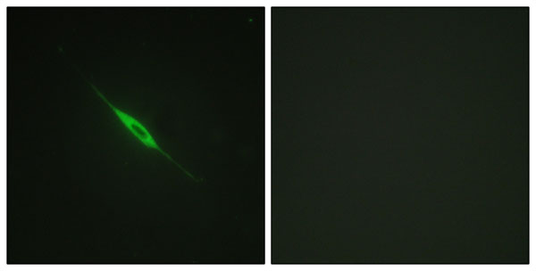
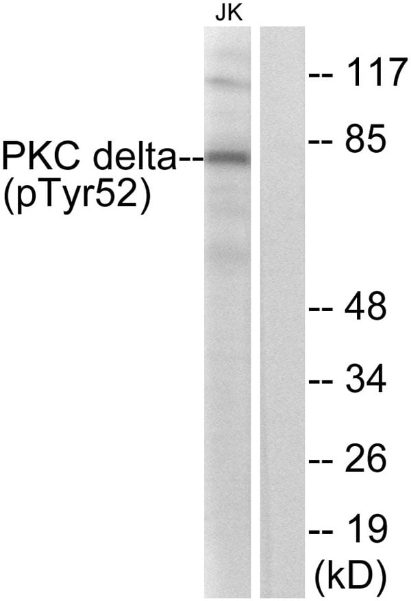 WB (Western Blot)
(Western blot analysis of lysates from Jurkat cells treated with starved 24h, using PKC delta (Phospho-Tyr52) Antibody. The lane on the right is blocked with the phospho peptide.)
WB (Western Blot)
(Western blot analysis of lysates from Jurkat cells treated with starved 24h, using PKC delta (Phospho-Tyr52) Antibody. The lane on the right is blocked with the phospho peptide.)
PKC delta, Polyclonal Antibody (Cat# AAA316302)
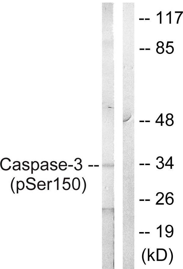 WB (Western Blot)
(Western blot analysis of lysates from Jurkat cells treated with Etoposide 25uM 60', using Caspase 3 (Phospho-Ser150) Antibody. The lane on the right is blocked with the phospho peptide.)
WB (Western Blot)
(Western blot analysis of lysates from Jurkat cells treated with Etoposide 25uM 60', using Caspase 3 (Phospho-Ser150) Antibody. The lane on the right is blocked with the phospho peptide.)
Caspase 3, Polyclonal Antibody (Cat# AAA316324)
 WB (Western Blot)
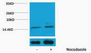
(Western blot analysis of extracts from Hela cells, untreated (-) or treated, 1:5000. Secondary antibody was diluted at 1:20000 cells nucleus extracted by Minute TM Cytoplasmic and Nuclear Fractionation kit (SC-003,Inventbiotech,MN,USA).)
WB (Western Blot)
(Western blot analysis of extracts from Hela cells, untreated (-) or treated, 1:5000. Secondary antibody was diluted at 1:20000 cells nucleus extracted by Minute TM Cytoplasmic and Nuclear Fractionation kit (SC-003,Inventbiotech,MN,USA).)
Histone H4, ELISA Kit (Cat# AAA319063)
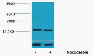
 WB (Western Blot)
(Western blot analysis of extracts from Hela cells, untreated (-) or treated, 1:2000. Secondary antibody was diluted at 1:20000 cells nucleus extracted by Minute TM Cytoplasmic and Nuclear Fractionation kit (SC-003,Inventbiotech,MN,USA).)
WB (Western Blot)
(Western blot analysis of extracts from Hela cells, untreated (-) or treated, 1:2000. Secondary antibody was diluted at 1:20000 cells nucleus extracted by Minute TM Cytoplasmic and Nuclear Fractionation kit (SC-003,Inventbiotech,MN,USA).)
Histone H3, ELISA Kit (Cat# AAA319067)
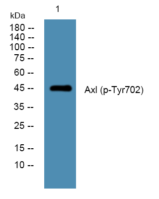 WB (Western Blot)
(Western blot analysis of lysates from K562 cells, primary antibody was diluted at 1:1000, 4 degree C over night)
WB (Western Blot)
(Western blot analysis of lysates from K562 cells, primary antibody was diluted at 1:1000, 4 degree C over night)
Axl, ELISA Kit (Cat# AAA319076)
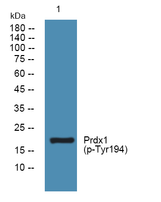 WB (Western Blot)
(Western blot analysis of lysates from K562 cells, primary antibody was diluted at 1:1000, 4 degree C over night)
WB (Western Blot)
(Western blot analysis of lysates from K562 cells, primary antibody was diluted at 1:1000, 4 degree C over night)
Prdx1, ELISA Kit (Cat# AAA319077)
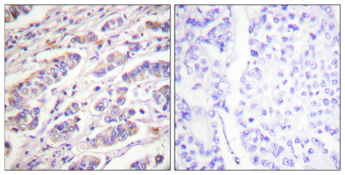
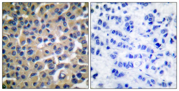
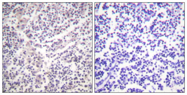
 IHC (Immunohistochemisry)
(Immunohistochemical analysis of paraffin-embedded Human breast cancer. Antibody was diluted at 1:100(4 degree overnight). High-pressure and temperature Tris-EDTA,pH8.0 was used for antigen retrieval. Negetive contrl (right) obtaned from antibody was pre-absorbed by immunogen peptide.)
IHC (Immunohistochemisry)
(Immunohistochemical analysis of paraffin-embedded Human breast cancer. Antibody was diluted at 1:100(4 degree overnight). High-pressure and temperature Tris-EDTA,pH8.0 was used for antigen retrieval. Negetive contrl (right) obtaned from antibody was pre-absorbed by immunogen peptide.)
Vav, Polyclonal Antibody (Cat# AAA318645)
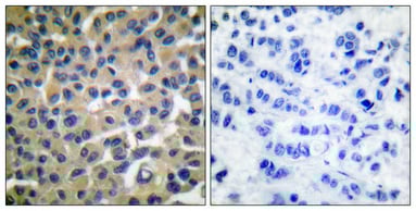
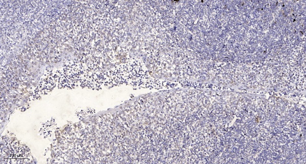 IHC (Immunohiostchemistry)
(Immunohistochemical analysis of paraffin-embedded human tonsil. 1, Antibody was diluted at 1:200(4 degree overnight). 2, Tris-EDTA,pH9.0 was used for antigen retrieval. 3,Secondary antibody was diluted at 1:200(room temperature, 30min).)
IHC (Immunohiostchemistry)
(Immunohistochemical analysis of paraffin-embedded human tonsil. 1, Antibody was diluted at 1:200(4 degree overnight). 2, Tris-EDTA,pH9.0 was used for antigen retrieval. 3,Secondary antibody was diluted at 1:200(room temperature, 30min).)
CEP55, Polyclonal Antibody (Cat# AAA318654)
 WB (Western Blot)
(Western blot analysis of lysates from K562 cells treated with EGF 200ng/ml 5', using p70 S6 Kinase beta (Phospho-Ser423) Antibody.)
WB (Western Blot)
(Western blot analysis of lysates from K562 cells treated with EGF 200ng/ml 5', using p70 S6 Kinase beta (Phospho-Ser423) Antibody.)
p70 S6 Kinase beta, Polyclonal Antibody (Cat# AAA318369)
 WB (Western Blot)
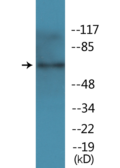
(Western blot analysis of lysates from LOVO cells treated with starved 24h, using PAK1 (Phospho-Ser199) Antibody.)
WB (Western Blot)
(Western blot analysis of lysates from LOVO cells treated with starved 24h, using PAK1 (Phospho-Ser199) Antibody.)
PAK1, Polyclonal Antibody (Cat# AAA318370)
 WB (Western Blot)
(Western blot analysis of lysates from Jurkat cells treated with PMA, using PLK1 (Phospho-Ser137) Antibody.)
WB (Western Blot)
(Western blot analysis of lysates from Jurkat cells treated with PMA, using PLK1 (Phospho-Ser137) Antibody.)
PLK1, Polyclonal Antibody (Cat# AAA318377)
 WB (Western Blot)
(Western blot analysis of lysates from HepG2 cells treated with nocodazole 1ug/ml 16h, using Retinoblastoma (Phospho-Thr826) Antibody.)
WB (Western Blot)
(Western blot analysis of lysates from HepG2 cells treated with nocodazole 1ug/ml 16h, using Retinoblastoma (Phospho-Thr826) Antibody.)
Retinoblastoma, Polyclonal Antibody (Cat# AAA318379)
S6 Ribosomal Protein, Polyclonal Antibody (Cat# AAA318383)
 WB (Western Blot)
(Western blot analysis of lysates from HepG2 cells treated with TNF 200NG/ML 30', using SHIP1 (Phospho-Tyr1021) Antibody.)
WB (Western Blot)
(Western blot analysis of lysates from HepG2 cells treated with TNF 200NG/ML 30', using SHIP1 (Phospho-Tyr1021) Antibody.)
SHIP1, Polyclonal Antibody (Cat# AAA318385)

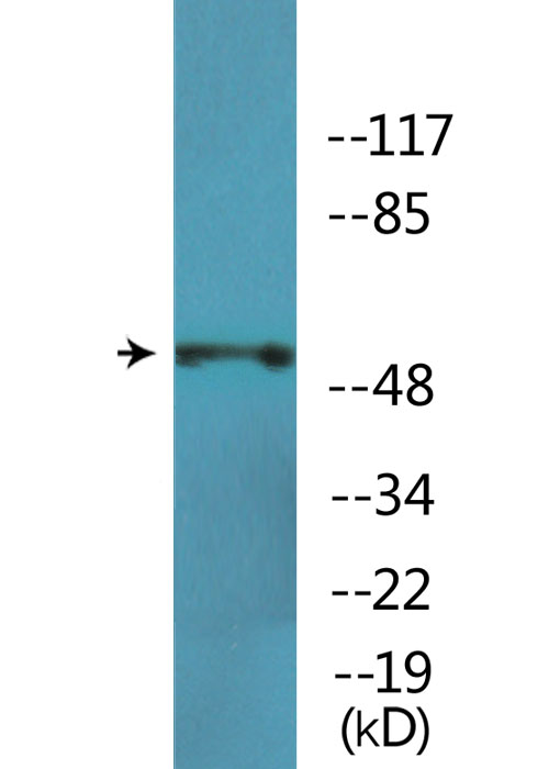
 WB (Western Blot)
(Western blot analysis of lysates from Jurkat cells, using Smad3 (Phospho-Ser208) Antibody.)
WB (Western Blot)
(Western blot analysis of lysates from Jurkat cells, using Smad3 (Phospho-Ser208) Antibody.)
Smad3, Polyclonal Antibody (Cat# AAA318387)
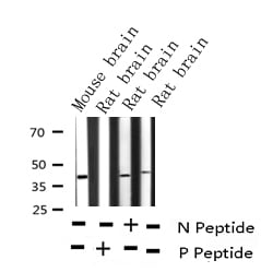
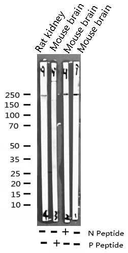
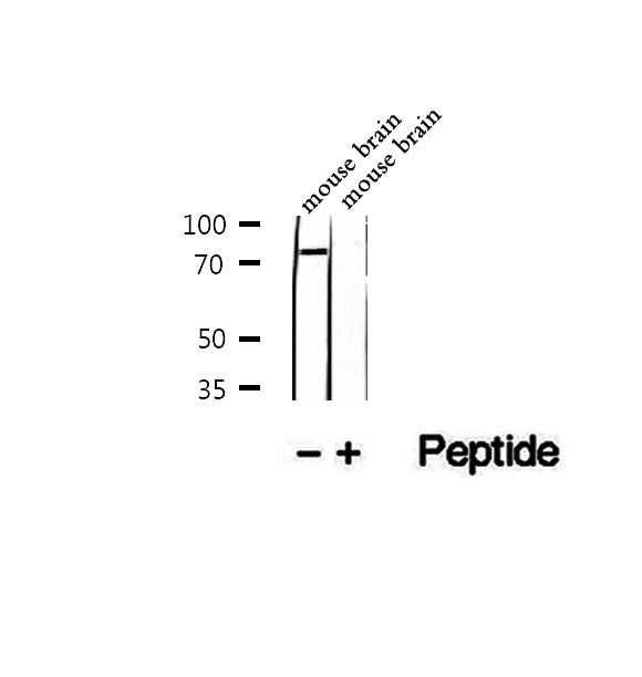
 WB (Western Blot)
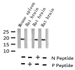
(Western blot analysis of lysates from mouse brain, using Synuclein-alpha (Phospho-Tyr136) Antibody.)
WB (Western Blot)
(Western blot analysis of lysates from mouse brain, using Synuclein-alpha (Phospho-Tyr136) Antibody.)
Synuclein-alpha, Polyclonal Antibody (Cat# AAA318393)
 WB (Western Blot)
(Western blot analysis of lysates from HepG2 cells treated with TNF 20ng/ml 5', using TAK1 (Phospho-Thr184) Antibody.)
WB (Western Blot)
(Western blot analysis of lysates from HepG2 cells treated with TNF 20ng/ml 5', using TAK1 (Phospho-Thr184) Antibody.)
TAK1, Polyclonal Antibody (Cat# AAA318394)
 WB (Western Blot)
(Western blot analysis of lysates from Jurkat cells treated with paclitaxel 1uM 24h, using TOP2A (Phospho-Ser1106) Antibody.)
WB (Western Blot)
(Western blot analysis of lysates from Jurkat cells treated with paclitaxel 1uM 24h, using TOP2A (Phospho-Ser1106) Antibody.)
TOP2A, Polyclonal Antibody (Cat# AAA318399)
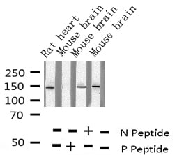
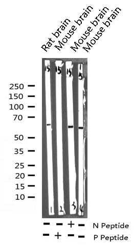
 WB (Western Blot)
(Western blot analysis of lysates from mouse brain, using Trk A (Phospho-Tyr701) Antibody.)
WB (Western Blot)
(Western blot analysis of lysates from mouse brain, using Trk A (Phospho-Tyr701) Antibody.)
Trk A, Polyclonal Antibody (Cat# AAA318403)
 WB (Western Blot)
(Western blot analysis of lysates from 293 cells treated with etoposide 25uM 60', using WEE1 (Phospho-Ser642) Antibody.)
WB (Western Blot)
(Western blot analysis of lysates from 293 cells treated with etoposide 25uM 60', using WEE1 (Phospho-Ser642) Antibody.)
WEE1, Polyclonal Antibody (Cat# AAA318407)
 WB (Western Blot)
(Western blot analysis of lysates from HepG2 cells treated with PMA 125ng/ml 15', using YB1 (Phospho-Ser102) Antibody.)
WB (Western Blot)
(Western blot analysis of lysates from HepG2 cells treated with PMA 125ng/ml 15', using YB1 (Phospho-Ser102) Antibody.)
YB1, Polyclonal Antibody (Cat# AAA318409)
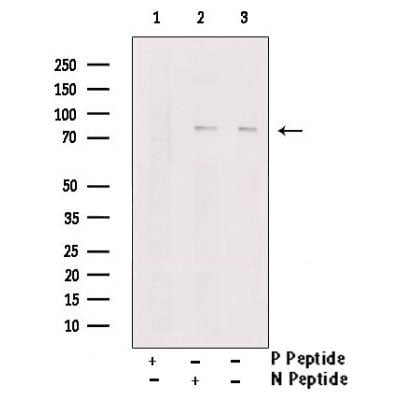
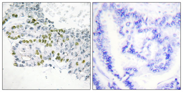
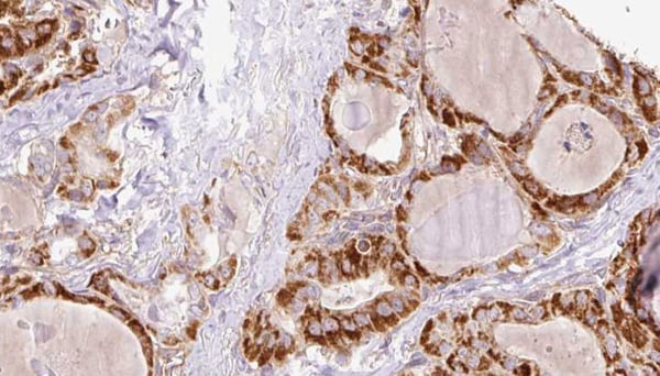 IHC (Immunohiostchemistry)
(AAA321408 at 1/100 staining Human thyroid cancer tissue by IHC-P. The sample was formaldehyde fixed and a heat mediated antigen retrieval step in citrate buffer was performed. The sample was then blocked and incubated with the antibody for 1.5 hours at 22 degree C. An HRP conjugated goat anti-rabbit antibody was used as the secondary.)
IHC (Immunohiostchemistry)
(AAA321408 at 1/100 staining Human thyroid cancer tissue by IHC-P. The sample was formaldehyde fixed and a heat mediated antigen retrieval step in citrate buffer was performed. The sample was then blocked and incubated with the antibody for 1.5 hours at 22 degree C. An HRP conjugated goat anti-rabbit antibody was used as the secondary.)
TNK1, Polyclonal Antibody (Cat# AAA321408)
 WB (Western Blot)
(Western blot analysis of Phospho-EGFR (Tyr1110) expression in various lysates)
WB (Western Blot)
(Western blot analysis of Phospho-EGFR (Tyr1110) expression in various lysates)
EGFR, Polyclonal Antibody (Cat# AAA321451)
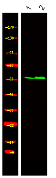
 WB (Western Blot)
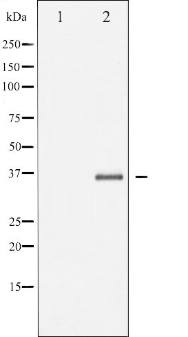
(Western blot analysis of Fos phosphorylation expression in forskolin treated K562 whole cell lysates, The lane on the left is treated with the antigen-specific peptide.)
WB (Western Blot)
(Western blot analysis of Fos phosphorylation expression in forskolin treated K562 whole cell lysates, The lane on the left is treated with the antigen-specific peptide.)
FOS, Polyclonal Antibody (Cat# AAA321455)
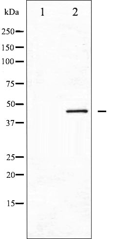
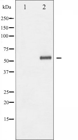
 WB (Western Blot)
(Western blot analysis of Estrogen Receptor alpha phosphorylation expression in EGF treated MCF7 whole cell lysates, The lane on the left is treated with the antigen-specific peptide.)
WB (Western Blot)
(Western blot analysis of Estrogen Receptor alpha phosphorylation expression in EGF treated MCF7 whole cell lysates, The lane on the left is treated with the antigen-specific peptide.)
Estrogen Receptor alpha, Polyclonal Antibody (Cat# AAA321456)
Predicted to cross-react with canine, goat, porcine and sheep based on sequence homology.
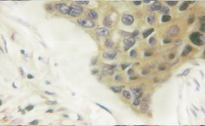
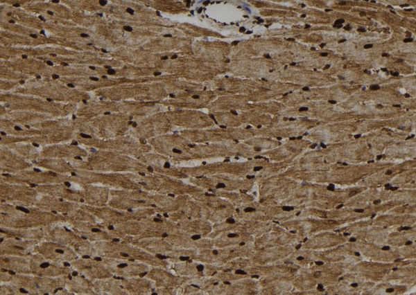 IHC (Immunohiostchemistry)
(AAA321310 at 1/100 staining Rat heart tissue by IHC-P. The sample was formaldehyde fixed and a heat mediated antigen retrieval step in citrate buffer was performed. The sample was then blocked and incubated with the antibody for 1.5 hours at 22 degree C. An HRP conjugated goat anti-rabbit antibody was used as the secondary.)
IHC (Immunohiostchemistry)
(AAA321310 at 1/100 staining Rat heart tissue by IHC-P. The sample was formaldehyde fixed and a heat mediated antigen retrieval step in citrate buffer was performed. The sample was then blocked and incubated with the antibody for 1.5 hours at 22 degree C. An HRP conjugated goat anti-rabbit antibody was used as the secondary.)
BMAL1, Polyclonal Antibody (Cat# AAA321310)
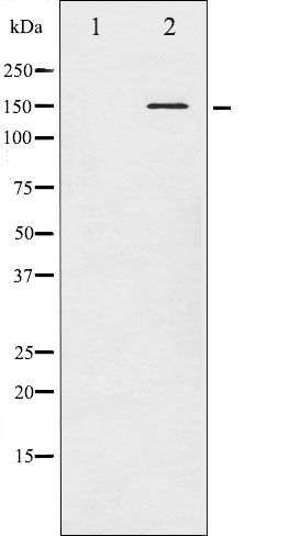
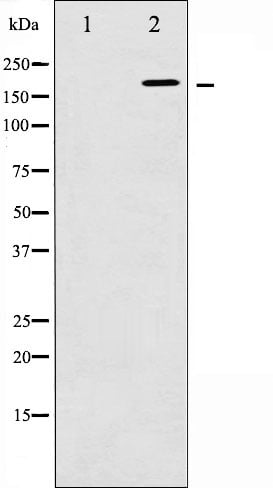
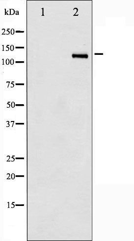
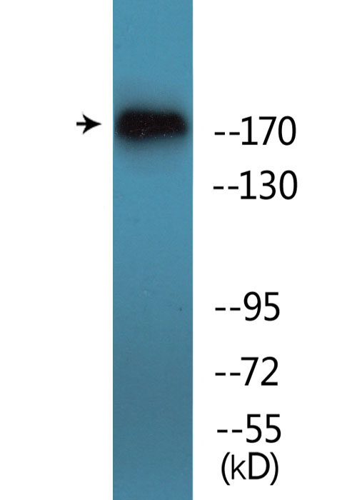
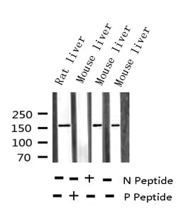
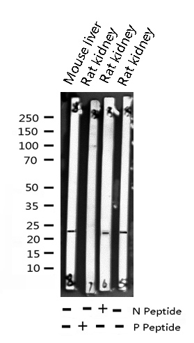
 WB (Western Blot)
(Western blot analysis of Phospho-Met (Tyr1349) expression in various lysates)
WB (Western Blot)
(Western blot analysis of Phospho-Met (Tyr1349) expression in various lysates)
Met, Polyclonal Antibody (Cat# AAA321502)
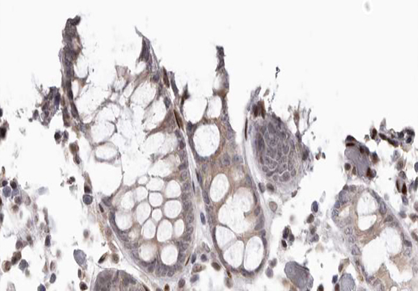
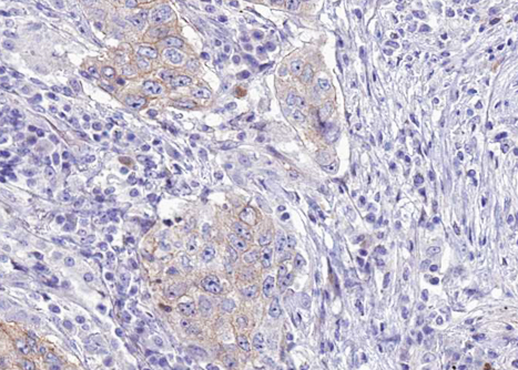
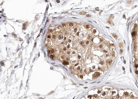
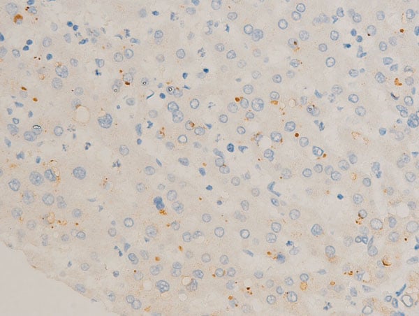
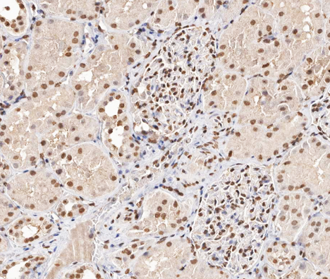
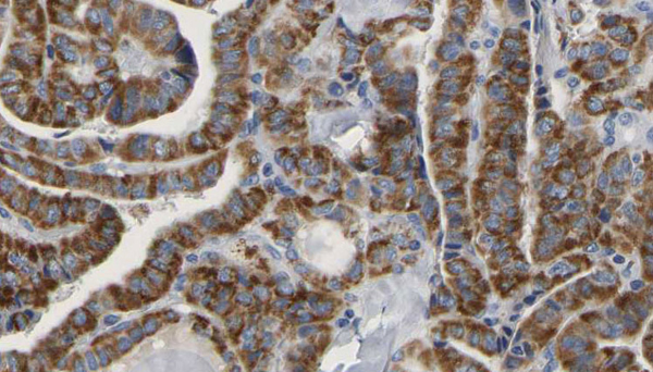
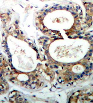
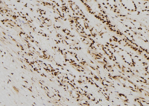 IHC (Immunohistochemistry)
(AAA321503 at 1/100 staining Human kidney tissue by IHC-P. The sample was formaldehyde fixed and a heat mediated antigen retrieval step in citrate buffer was performed. The sample was then blocked and incubated with the antibody for 1.5 hours at 22 degree C. An HRP conjugated goat anti-rabbit antibody was used as the secondary.)
IHC (Immunohistochemistry)
(AAA321503 at 1/100 staining Human kidney tissue by IHC-P. The sample was formaldehyde fixed and a heat mediated antigen retrieval step in citrate buffer was performed. The sample was then blocked and incubated with the antibody for 1.5 hours at 22 degree C. An HRP conjugated goat anti-rabbit antibody was used as the secondary.)
Vimentin, Polyclonal Antibody (Cat# AAA321503)
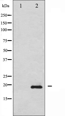
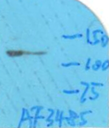
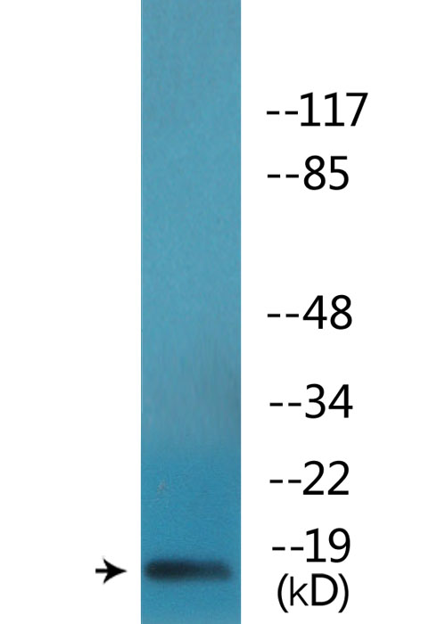
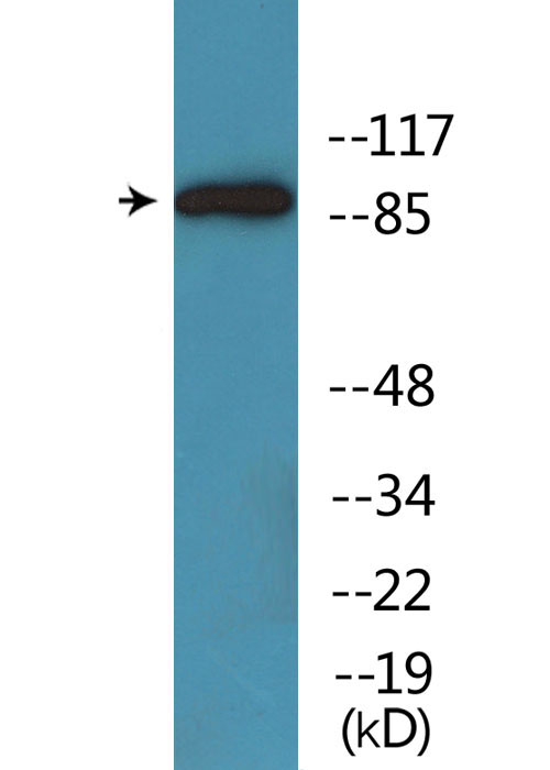
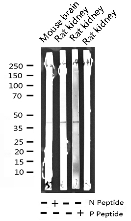
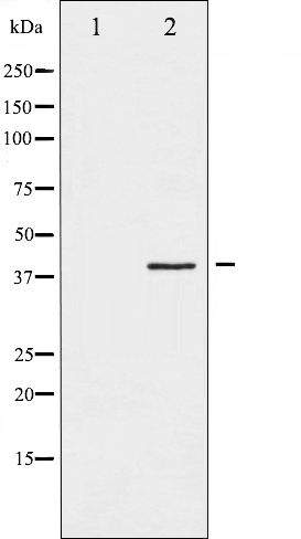
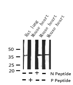
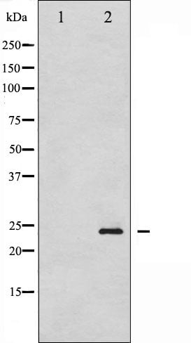
 WB (Western Blot)
(Western blot analysis of Phospho-Stathmin 1 (Ser15) expression in various lysates)
WB (Western Blot)
(Western blot analysis of Phospho-Stathmin 1 (Ser15) expression in various lysates)
Stathmin 1, Polyclonal Antibody (Cat# AAA321541)
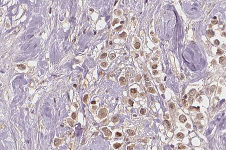
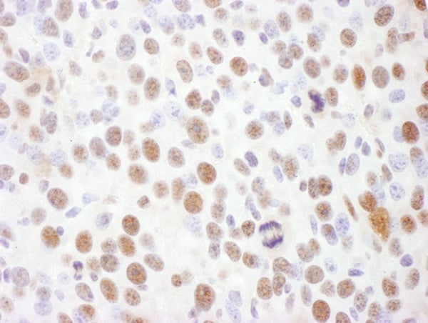
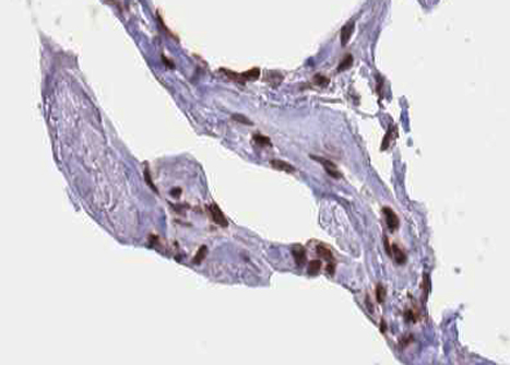 IHC (Immunohistochemistry)
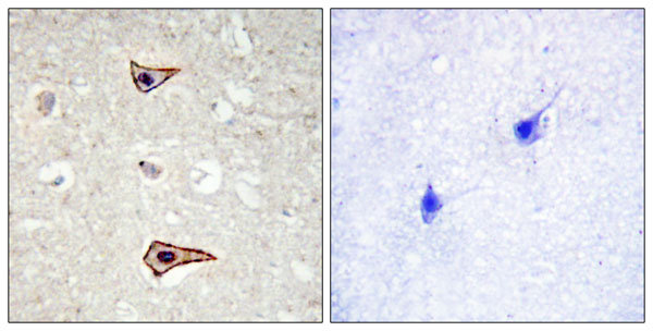
(AAA321556 at 1/100 staining human lung tissue sections by IHC-P. The tissue was formaldehyde fixed and a heat mediated antigen retrieval step in citrate buffer was performed. The tissue was then blocked and incubated with the antibody for 1.5 hours at 22 degree C. An HRP conjugated goat anti-rabbit antibody was used as the secondary.)
IHC (Immunohistochemistry)
(AAA321556 at 1/100 staining human lung tissue sections by IHC-P. The tissue was formaldehyde fixed and a heat mediated antigen retrieval step in citrate buffer was performed. The tissue was then blocked and incubated with the antibody for 1.5 hours at 22 degree C. An HRP conjugated goat anti-rabbit antibody was used as the secondary.)
Elk1, Polyclonal Antibody (Cat# AAA321556)
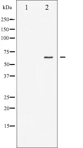
 WB (Western Blot)
(Western blot analysis of Phospho-EFNB1/2 (Tyr330) expression in various lysates)
WB (Western Blot)
(Western blot analysis of Phospho-EFNB1/2 (Tyr330) expression in various lysates)
EFNB1/2, Polyclonal Antibody (Cat# AAA321649)
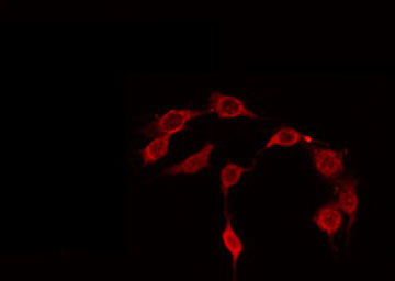
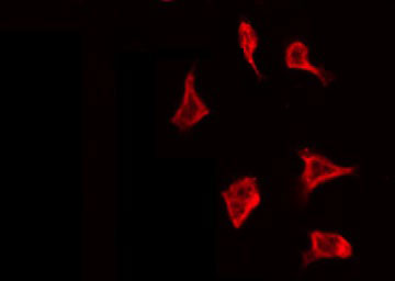
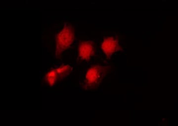
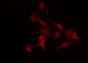
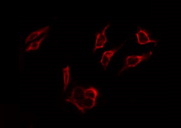
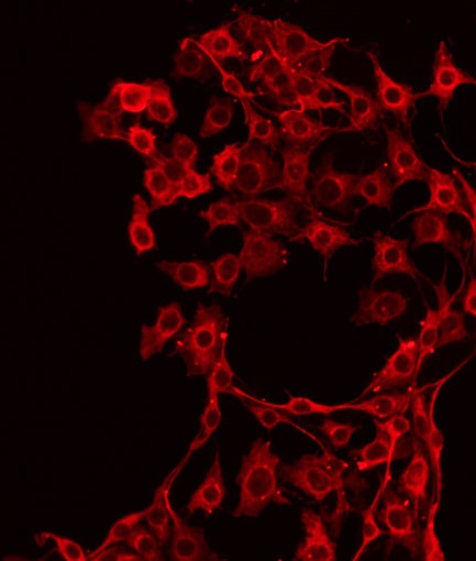
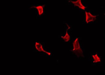
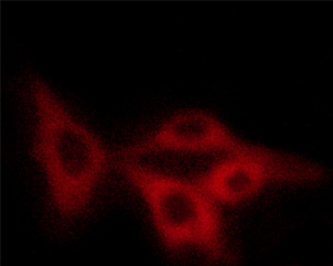
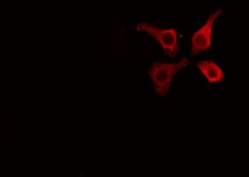 IF (Immunofluorescence)
(AAA321684 staining HuvEc cells by IF/ICC. The sample were fixed with PFA and permeabilized in 0.1% Triton X-100, then blocked in 10% serum for 45 minutes at 25 degree C. The primary antibody was diluted at 1/200 and incubated with the sample for 1 hour at 37 degree C. An Alexa Fluor 594 conjugated goat anti-rabbit IgG (H+L) antibody, diluted at 1/600, was used as secondary antibody.)
IF (Immunofluorescence)
(AAA321684 staining HuvEc cells by IF/ICC. The sample were fixed with PFA and permeabilized in 0.1% Triton X-100, then blocked in 10% serum for 45 minutes at 25 degree C. The primary antibody was diluted at 1/200 and incubated with the sample for 1 hour at 37 degree C. An Alexa Fluor 594 conjugated goat anti-rabbit IgG (H+L) antibody, diluted at 1/600, was used as secondary antibody.)
Caveolin-1, Polyclonal Antibody (Cat# AAA321684)
 WB (Western Blot)
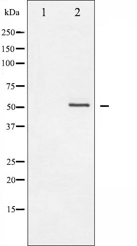
(Western blot analysis of extracts of Ramos cells, using Phospho-PLCgamma2 (Tyr1217) Antibody.)
WB (Western Blot)
(Western blot analysis of extracts of Ramos cells, using Phospho-PLCgamma2 (Tyr1217) Antibody.)
PLCgamma2, Polyclonal Antibody (Cat# AAA321368)
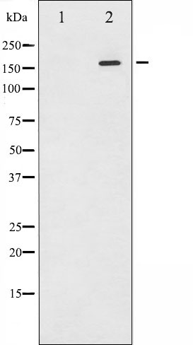
 WB (Western Blot)
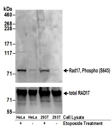
(Detection of human Rad17, Phospho (S645) by western blot. Samples: Whole cell lysate (50 ug) from HeLa and HEK293T cells treated with 100 uM etoposide for 2 hours (+) or mock treated (-) cells. Antibodies: Affinity purified rabbit anti-Rad17, Phospho (S645) antibody AAA210790 (lot AAA210790-2) used for WB at 0.4 ug/ml. To examine total Rad17, the membrane was reblotted with goat anti-Rad17 antibody BL239 (lower panel). Detection: Chemiluminescence with exposure times of 30 seconds (upper and lower panels).)
WB (Western Blot)
(Detection of human Rad17, Phospho (S645) by western blot. Samples: Whole cell lysate (50 ug) from HeLa and HEK293T cells treated with 100 uM etoposide for 2 hours (+) or mock treated (-) cells. Antibodies: Affinity purified rabbit anti-Rad17, Phospho (S645) antibody AAA210790 (lot AAA210790-2) used for WB at 0.4 ug/ml. To examine total Rad17, the membrane was reblotted with goat anti-Rad17 antibody BL239 (lower panel). Detection: Chemiluminescence with exposure times of 30 seconds (upper and lower panels).)
Rad17, Polyclonal Antibody (Cat# AAA210790)
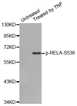
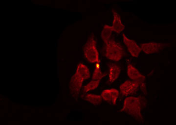
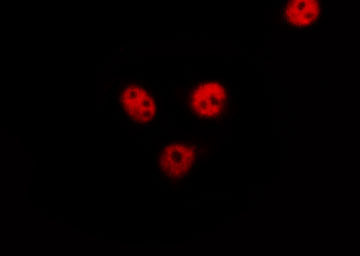
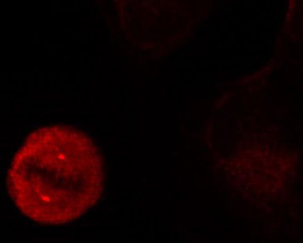
 IF (Immunofluorescence)
(Immunofluorescence staining of methanol-fixed HeLa cells using Phospho-RELA-S536 antibody.)
IF (Immunofluorescence)
(Immunofluorescence staining of methanol-fixed HeLa cells using Phospho-RELA-S536 antibody.)
RELA-S536, Antibody (Cat# AAA37345)
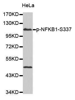
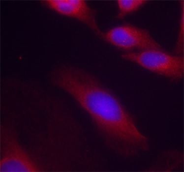
 IF (Immunofluorescence)
(Immunofluorescence staining of methanol-fixed HeLa cells using Phospho-NFKB1-S337 antibody.)
IF (Immunofluorescence)
(Immunofluorescence staining of methanol-fixed HeLa cells using Phospho-NFKB1-S337 antibody.)
NFKB1-S337, Antibody (Cat# AAA37350)
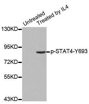
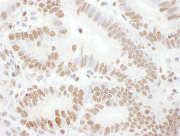
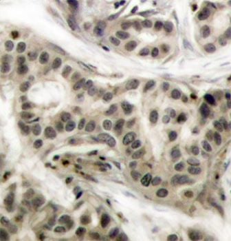
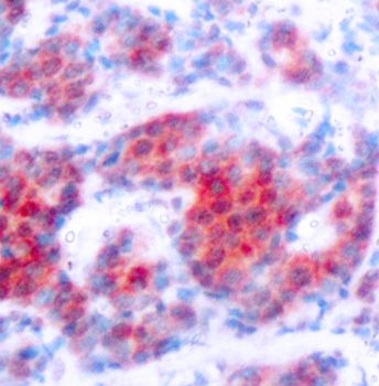 IHC (Immunohiostchemistry)
(Immunohistochemistry of paraffin-embedded human breast carcinoma using Phospho-STAT4-Y693 antibody.)
IHC (Immunohiostchemistry)
(Immunohistochemistry of paraffin-embedded human breast carcinoma using Phospho-STAT4-Y693 antibody.)
STAT4-Y693, Antibody (Cat# AAA37356)
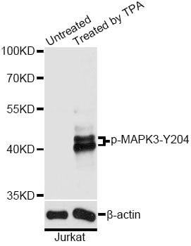
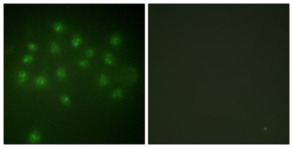
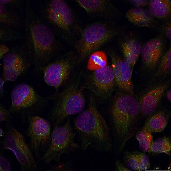
 IF (Immunofluorescence)
(Immunofluorescence staining of methanol-fixed HeLa cells showing centrosome and nuclear staining using Phospho-MAPK3-Y204 antibody.)
IF (Immunofluorescence)
(Immunofluorescence staining of methanol-fixed HeLa cells showing centrosome and nuclear staining using Phospho-MAPK3-Y204 antibody.)
MAPK3-Y204, Antibody (Cat# AAA37373)
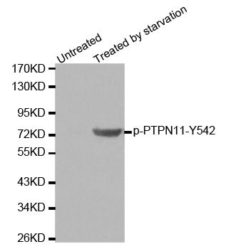
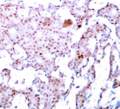
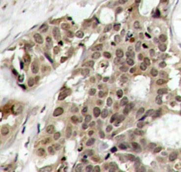
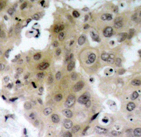
 IHC (Immunohiostchemistry)
(Immunohistochemistry of paraffin-embedded human breast carcinoma using Phospho-PTPN11-Y542 antibody.)
IHC (Immunohiostchemistry)
(Immunohistochemistry of paraffin-embedded human breast carcinoma using Phospho-PTPN11-Y542 antibody.)
PTPN11-Y542, Antibody (Cat# AAA37392)
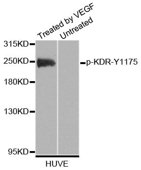
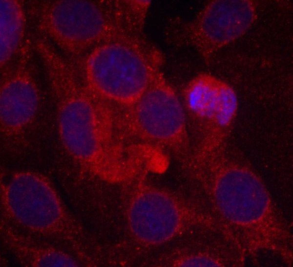 IF (Immunofluorescence)
(Immunofluorescence staining of methanol-fixed MCF-7 cells using Phospho-KDR-Y1175 antibody.)
IF (Immunofluorescence)
(Immunofluorescence staining of methanol-fixed MCF-7 cells using Phospho-KDR-Y1175 antibody.)
KDR-Y1175, Antibody (Cat# AAA37408)
What Are Phospho Antibodies?
Protein phosphorylation is a process where a phosphate group is added to certain amino acid residues of a protein – usually serine (S), threonine (T), or tyrosine (Y) - by enzymes called kinases. This process is integral in controlling cellular signaling, cellular growth, and other biological functions.
Our catalog includes a wide range of phospho-specific antibodies that can accurately detect this important marker. They perform strongly in widely-used laboratory applications such as Western blot, flow cytometry, immunohistochemistry, and immunofluorescence microscopy. We value your trust in us and are committed to providing top-quality products and services. All of our antibodies are guaranteed to work for the applications and species indicated on our website & associated product pages.
What Are The Key Applications of Phospho Antibodies?
1. Western Blotting
One of the first steps a researcher can take in utilizing these phospho-specific antibodies, is to check if the antibody works using a technique referred to as “Western blot”. For those unfamiliar, Western Blot aids in showing whether the protein that the antibody recognizes is appearing at the correct/expected size. These phospho-specific antibodies should also be able to detect changes in the target protein’s phosphorylation (on/off state) when cells are stimulated in certain ways.
2. Staining of Fixed Cells (Immunocytochemistry)
Another routine use of these phospho-specific antibodies, is to test if the antibody is able to demonstrate similar performance when used on fixed cells (intact cells that have been preserved) as it did in the Western blot tests. It is an important aspect in many cases to confirm that the antibody works in actual intact cell samples. Ideally, the method used for cellular fixation should be the same as what is used in pathology labs (like using 10% formalin). To check if the antibody works well in tissue sections (FFPE), researchers will often test it on fixed cells that are processed similar to tissue samples.
3. Specificity Tests Using Peptides
In order to make sure that the antibody is only binding to the right target:
- Laboratory technicians will mix the antibody with phospho-peptides (short segments of the protein containing the phosphate group modification).
- If the antibody signal disappears, it is confirmation that it is binding to the correct phosphorylated location.
- A more robust test is to use both the phosphorylated and non-phosphorylated (dephosphorylated) versions of the protein. The antibody should react only with the phosphorylated one.
- Another method sometimes utilized is to treat the sample with an enzyme, such as alkaline phosphatase, that specifically removes phosphate groups. If the antibody signal disappears after this, it also confirms specificity.
4. Genetic Confirmation
As a final step, scientists can genetically manipulate the nucleotide sequence and alter the target protein by removing the exact site where phosphorylation happens. If the antibody no longer appears to detect the modified protein, it is strong evidence supporting the antibody being specific for that phosphorylated site.
Why Buy Phospho Antibodies Through Us?
- The production laboratory adheres to strict and consistent protocols prior to releasing any of these phospho-specific antibodies:
- Standard methods and proper controls in all tests to ensure high quality.
- These antibodies are tested and validated in different cell types and species.
- High quality control criterion to ensure each batch is consistent, so you will obtain reliable results every time.
FAQ
1. What Are Phospho-Specific Antibodies?
Phospho-specific antibodies are made to detect proteins only when they have a phosphate group linked to a specific amino acid residue. This empowers scientists understand if a protein is "turned on" or active, based on its phosphorylation state.
2. How to Detect Phosphorylated Proteins in a Western Blot?
To find out if a protein is phosphorylated using Western blot:
- Use a phospho-specific antibody that binds only to the phosphorylated form of the protein.
- You can also use a “regular” antibody for the same amino acid sequence of the protein that the phospho-specific antibody is binding to (but in this case, this antibody will not bind if there is a phosphate group present) in order to compare how much of it is phosphorylated versus how much is non-phosphorylated (or “total” protein, if the “normal” antibody’s epitopes are non-phospho-site-specific).
3. How to Choose the Best Antibody?
Here are some simple tips to help you pick the right antibody:
- Know your target
- Match your sample characteristics
- Confirm the intended use is appropriate
- Check “host” and “type”
- Check the “quality” of the presented data/images
- Appraise whether the available validation meets your needs