Filters
▼Clonality
▼Type
▼Reactivity
▼Gene Name
▼Isotype
▼Host
▼Application
▼Clone
▼Phospho Antibodies
Phospho-specific antibodies’ typical purpose is to enable researchers to detect changes in proteins. They will exclusively bind to the amino acid sequence on a protein that has been phosphorylated (which is both a physical & chemical change) and do not bind to the same amino acid sequence on said protein if it lacks said phosphorylation. This aids in being able to clearly see and understand the data produced from this particular protein modification.
Viewing 1200-1250 of 5298 product results
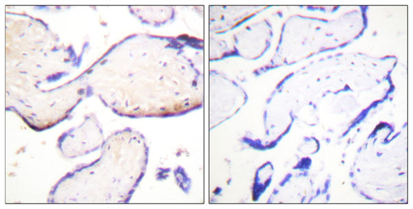
 IHC (Immunohistochemistry)
(Immunohistochemical analysis of paraffin-embedded human brain tissue using PDGF Receptor beta (phospho-Tyr751) antibody.P-peptide: -(left) +(right))
IHC (Immunohistochemistry)
(Immunohistochemical analysis of paraffin-embedded human brain tissue using PDGF Receptor beta (phospho-Tyr751) antibody.P-peptide: -(left) +(right))
PDGF Receptor beta, Antibody (Cat# AAA112061)
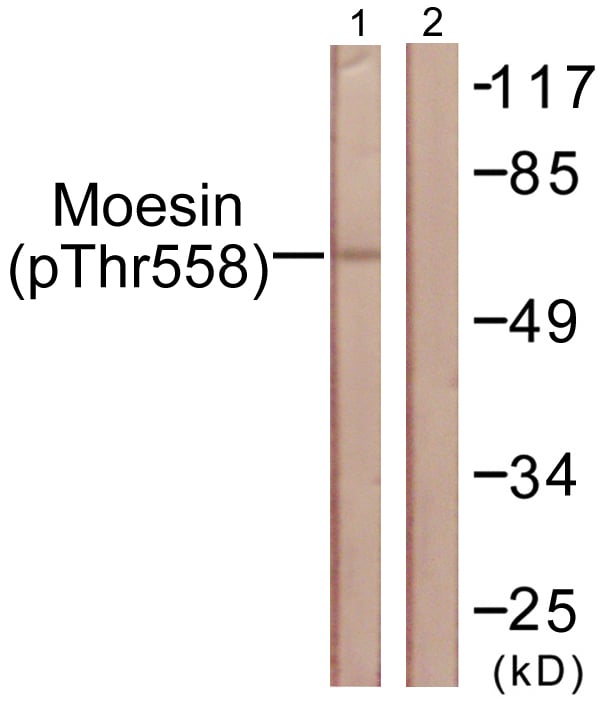
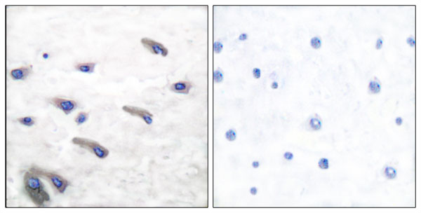
 IF (Immunofluorescence)
(P-peptide-+ Immunofluorescence analysis of A549 cells, using Moesin/Ezrin/Radixin (Phospho-Thr558) antibody.)
IF (Immunofluorescence)
(P-peptide-+ Immunofluorescence analysis of A549 cells, using Moesin/Ezrin/Radixin (Phospho-Thr558) antibody.)
Moesin/Ezrin/Radixin, Antibody (Cat# AAA112074)
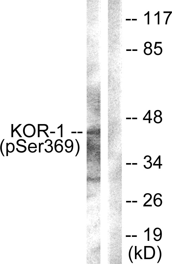 WB (Western Blot)
(Western blot analysis of extracts from NIH/3T3 cells, using KOR-1 (Phospho-Ser369) Antibody.)
WB (Western Blot)
(Western blot analysis of extracts from NIH/3T3 cells, using KOR-1 (Phospho-Ser369) Antibody.)
KOR-1, Antibody (Cat# AAA111929)
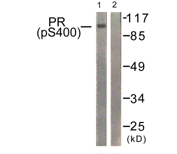
 WB (Western Blot)
(P-peptide-+ Western blot analysis of extracts from 293 cells, treated with heat shock, using PR (Phospho-Ser400) antibody. Immunofluorescence analysis of A549 cells, using PR (Phospho-Ser400) antibody.)
WB (Western Blot)
(P-peptide-+ Western blot analysis of extracts from 293 cells, treated with heat shock, using PR (Phospho-Ser400) antibody. Immunofluorescence analysis of A549 cells, using PR (Phospho-Ser400) antibody.)
Progesterone Receptor, Antibody (Cat# AAA111861)
 IHC (Immunohiostchemistry)
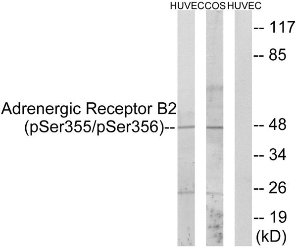
(Immunohistochemistry analysis of paraffin-embedded human brain tissue using Adrenergic Receptor B2 (Phospho-Ser355+Ser356) antibody. Western blot analysis of extracts from HUVEC cells and COS cells treated with serum (20%, 15mins), using Adrenergic Receptor B2 (Phospho-Ser355+Ser356) antibody.)
IHC (Immunohiostchemistry)
(Immunohistochemistry analysis of paraffin-embedded human brain tissue using Adrenergic Receptor B2 (Phospho-Ser355+Ser356) antibody. Western blot analysis of extracts from HUVEC cells and COS cells treated with serum (20%, 15mins), using Adrenergic Receptor B2 (Phospho-Ser355+Ser356) antibody.)
Adrenergic Receptor B2, Antibody (Cat# AAA111868)
 IHC (Immunohiostchemistry)
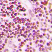
(Immunohistochemical analysis of p73 (pY99) staining in human lung cancer formalin fixed paraffin embedded tissue section. The section was pre-treated using heat mediated antigen retrieval with sodium citrate buffer (pH 6.0). The section was then incubated with the antibody at room temperature and detected using an HRP conjugated compact polymer system. DAB was used as the chromogen. The section was then counterstained with haematoxylin and mounted with DPX.)
IHC (Immunohiostchemistry)
(Immunohistochemical analysis of p73 (pY99) staining in human lung cancer formalin fixed paraffin embedded tissue section. The section was pre-treated using heat mediated antigen retrieval with sodium citrate buffer (pH 6.0). The section was then incubated with the antibody at room temperature and detected using an HRP conjugated compact polymer system. DAB was used as the chromogen. The section was then counterstained with haematoxylin and mounted with DPX.)
p73 (pY99), Polyclonal Antibody (Cat# AAA104646)

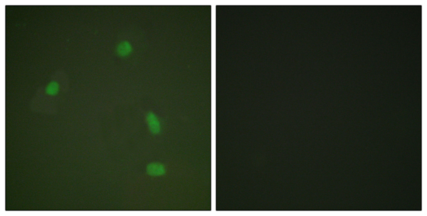
 IF (Immunofluorescence)
(Immunofluorescent analysis of FOXO4 (pS197) staining in HeLa cells. Formalin-fixed cells were permeabilized with 0.1% Triton X-100 in TBS for 5-10 minutes and blocked with 3% BSA-PBS for 30 minutes at room temperature. Cells were probed with the primary antibody in 3% BSA-PBS and incubated overnight at 4 °C in a humidified chamber. Cells were washed with PBST and incubated with a DyLight 594-conjugated secondary antibody (red) in PBS at room temperature in the dark.)
IF (Immunofluorescence)
(Immunofluorescent analysis of FOXO4 (pS197) staining in HeLa cells. Formalin-fixed cells were permeabilized with 0.1% Triton X-100 in TBS for 5-10 minutes and blocked with 3% BSA-PBS for 30 minutes at room temperature. Cells were probed with the primary antibody in 3% BSA-PBS and incubated overnight at 4 °C in a humidified chamber. Cells were washed with PBST and incubated with a DyLight 594-conjugated secondary antibody (red) in PBS at room temperature in the dark.)
FOXO4 (pS197), Polyclonal Antibody (Cat# AAA104654)
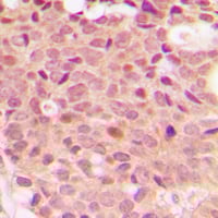
 IHC (Immunohiostchemistry)
(Immunohistochemical analysis of IRS1 (pS307) staining in human breast cancer formalin fixed paraffin embedded tissue section. The section was pre-treated using heat mediated antigen retrieval with sodium citrate buffer (pH 6.0). The section was then incubated with the antibody at room temperature and detected using an HRP conjugated compact polymer system. DAB was used as the chromogen. The section was then counterstained with haematoxylin and mounted with DPX.)
IHC (Immunohiostchemistry)
(Immunohistochemical analysis of IRS1 (pS307) staining in human breast cancer formalin fixed paraffin embedded tissue section. The section was pre-treated using heat mediated antigen retrieval with sodium citrate buffer (pH 6.0). The section was then incubated with the antibody at room temperature and detected using an HRP conjugated compact polymer system. DAB was used as the chromogen. The section was then counterstained with haematoxylin and mounted with DPX.)
IRS1 (pS307), Polyclonal Antibody (Cat# AAA104691)
 IHC (Immunohiostchemistry)
(Immunohistochemical analysis of Artemis (pS516) staining in human lung cancer formalin fixed paraffin embedded tissue section. The section was pre-treated using heat mediated antigen retrieval with sodium citrate buffer (pH 6.0). The section was then incubated with the antibody at room temperature and detected using an HRP conjugated compact polymer system. DAB was used as the chromogen. The section was then counterstained with haematoxylin and mounted with DPX.)
IHC (Immunohiostchemistry)
(Immunohistochemical analysis of Artemis (pS516) staining in human lung cancer formalin fixed paraffin embedded tissue section. The section was pre-treated using heat mediated antigen retrieval with sodium citrate buffer (pH 6.0). The section was then incubated with the antibody at room temperature and detected using an HRP conjugated compact polymer system. DAB was used as the chromogen. The section was then counterstained with haematoxylin and mounted with DPX.)
Artemis (pS516), Polyclonal Antibody (Cat# AAA104695)
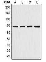
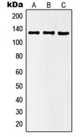
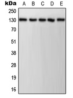 WB (Western Blot)
(Western blot analysis of CD31 (pY713) expression in HeLa (A), Jurkat (B), K562 (C), NIH3T3 (D), PC12 (E) whole cell lysates.)
WB (Western Blot)
(Western blot analysis of CD31 (pY713) expression in HeLa (A), Jurkat (B), K562 (C), NIH3T3 (D), PC12 (E) whole cell lysates.)
CD31 (pY713), Polyclonal Antibody (Cat# AAA105005)

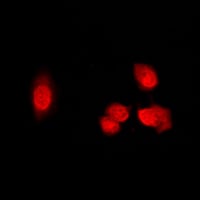
 IF (Immunofluorescence)
(Immunofluorescent analysis of CDK1/2/3 (pT14) staining in HeLa cells. Formalin-fixed cells were permeabilized with 0.1% Triton X-100 in TBS for 5-10 minutes and blocked with 3% BSA-PBS for 30 minutes at room temperature. Cells were probed with the primary antibody in 3% BSA-PBS and incubated overnight at 4 °C in a humidified chamber. Cells were washed with PBST and incubated with a DyLight 594-conjugated secondary antibody (red) in PBS at room temperature in the dark.)
IF (Immunofluorescence)
(Immunofluorescent analysis of CDK1/2/3 (pT14) staining in HeLa cells. Formalin-fixed cells were permeabilized with 0.1% Triton X-100 in TBS for 5-10 minutes and blocked with 3% BSA-PBS for 30 minutes at room temperature. Cells were probed with the primary antibody in 3% BSA-PBS and incubated overnight at 4 °C in a humidified chamber. Cells were washed with PBST and incubated with a DyLight 594-conjugated secondary antibody (red) in PBS at room temperature in the dark.)
CDK1/2/3 (pT14), Polyclonal Antibody (Cat# AAA105067)
 IHC (Immunohiostchemistry)
(Immunohistochemical analysis of CDC25A (pS75) staining in human tonsil formalin fixed paraffin embedded tissue section. The section was pre-treated using heat mediated antigen retrieval with sodium citrate buffer (pH 6.0). The section was then incubated with the antibody at room temperature and detected using an HRP conjugated compact polymer system. DAB was used as the chromogen. The section was then counterstained with haematoxylin and mounted with DPX.)
IHC (Immunohiostchemistry)
(Immunohistochemical analysis of CDC25A (pS75) staining in human tonsil formalin fixed paraffin embedded tissue section. The section was pre-treated using heat mediated antigen retrieval with sodium citrate buffer (pH 6.0). The section was then incubated with the antibody at room temperature and detected using an HRP conjugated compact polymer system. DAB was used as the chromogen. The section was then counterstained with haematoxylin and mounted with DPX.)
CDC25A (pS75), Polyclonal Antibody (Cat# AAA105125)
 IF (Immunofluorescence)
(Immunofluorescent analysis of Histone Deacetylase 5 (pS498) staining in Raw264.7 cells. Formalin-fixed cells were permeabilized with 0.1% Triton X-100 in TBS for 5-10 minutes and blocked with 3% BSA-PBS for 30 minutes at room temperature. Cells were probed with the primary antibody in 3% BSA-PBS and incubated overnight at 4 °C in a humidified chamber. Cells were washed with PBST and incubated with a DyLight 594-conjugated secondary antibody (red) in PBS at room temperature in the dark.)
IF (Immunofluorescence)
(Immunofluorescent analysis of Histone Deacetylase 5 (pS498) staining in Raw264.7 cells. Formalin-fixed cells were permeabilized with 0.1% Triton X-100 in TBS for 5-10 minutes and blocked with 3% BSA-PBS for 30 minutes at room temperature. Cells were probed with the primary antibody in 3% BSA-PBS and incubated overnight at 4 °C in a humidified chamber. Cells were washed with PBST and incubated with a DyLight 594-conjugated secondary antibody (red) in PBS at room temperature in the dark.)
Histone Deacetylase 5 (pS498), Polyclonal Antibody (Cat# AAA104896)
 IF (Immunofluorescence)
(Immunofluorescent analysis of ABL1/2 (pY393/439) staining in HeLa cells. Formalin-fixed cells were permeabilized with 0.1% Triton X-100 in TBS for 5-10 minutes and blocked with 3% BSA-PBS for 30 minutes at room temperature. Cells were probed with the primary antibody in 3% BSA-PBS and incubated overnight at 4 °C in a humidified chamber. Cells were washed with PBST and incubated with a DyLight 594-conjugated secondary antibody (red) in PBS at room temperature in the dark.)
IF (Immunofluorescence)
(Immunofluorescent analysis of ABL1/2 (pY393/439) staining in HeLa cells. Formalin-fixed cells were permeabilized with 0.1% Triton X-100 in TBS for 5-10 minutes and blocked with 3% BSA-PBS for 30 minutes at room temperature. Cells were probed with the primary antibody in 3% BSA-PBS and incubated overnight at 4 °C in a humidified chamber. Cells were washed with PBST and incubated with a DyLight 594-conjugated secondary antibody (red) in PBS at room temperature in the dark.)
ABL1/2 (pY393/439), Polyclonal Antibody (Cat# AAA104502)
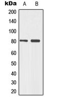
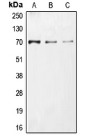
 WB (Western Blot)
(Western blot analysis of VEGFR1 (pY1213) expression in A10 (A), MDAMB231 (B) whole cell lysates.)
WB (Western Blot)
(Western blot analysis of VEGFR1 (pY1213) expression in A10 (A), MDAMB231 (B) whole cell lysates.)
VEGFR1 (pY1213), Polyclonal Antibody (Cat# AAA104516)
 IHC (Immunohiostchemistry)
(Immunohistochemical analysis of IRS1 (pS1101) staining in human breast cancer formalin fixed paraffin embedded tissue section. The section was pre-treated using heat mediated antigen retrieval with sodium citrate buffer (pH 6.0). The section was then incubated with the antibody at room temperature and detected using an HRP conjugated compact polymer system. DAB was used as the chromogen. The section was then counterstained with haematoxylin and mounted with DPX.)
IHC (Immunohiostchemistry)
(Immunohistochemical analysis of IRS1 (pS1101) staining in human breast cancer formalin fixed paraffin embedded tissue section. The section was pre-treated using heat mediated antigen retrieval with sodium citrate buffer (pH 6.0). The section was then incubated with the antibody at room temperature and detected using an HRP conjugated compact polymer system. DAB was used as the chromogen. The section was then counterstained with haematoxylin and mounted with DPX.)
IRS1 (pS1101), Polyclonal Antibody (Cat# AAA104471)
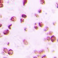
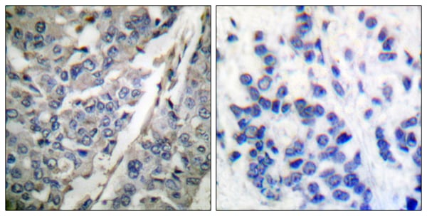
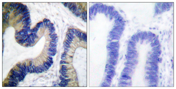
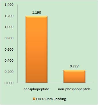 IHC (Immunohiostchemistry)
(Immunohistochemistry analysis of paraffin-embedded human brain tissue using CK-1 gamma 1/2/3 (Phospho-Tyr263) antibody. CK-1 gamma 1/2/3 (Phospho-Tyr263) antibody reacts with epitope-specific phosphopeptide and corresponding non-phosphopeptide. The absorbance readings at 450 nM are shown in the ELISA figure.)
IHC (Immunohiostchemistry)
(Immunohistochemistry analysis of paraffin-embedded human brain tissue using CK-1 gamma 1/2/3 (Phospho-Tyr263) antibody. CK-1 gamma 1/2/3 (Phospho-Tyr263) antibody reacts with epitope-specific phosphopeptide and corresponding non-phosphopeptide. The absorbance readings at 450 nM are shown in the ELISA figure.)
CK-1 gamma 1/2/3, Antibody (Cat# AAA109549)
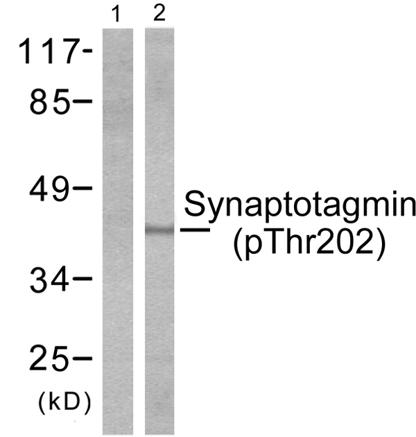 WB (Western Blot)
(Western blot analysis of extracts from Hela cells treated with Forskolin (40nM, 30mins), using Synaptotagmin (phospho-Thr202) antibody.)
WB (Western Blot)
(Western blot analysis of extracts from Hela cells treated with Forskolin (40nM, 30mins), using Synaptotagmin (phospho-Thr202) antibody.)
Synaptotagmin, Antibody (Cat# AAA109598)
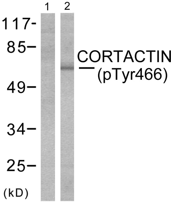 WB (Western Blot)
(Western blot analysis of extracts from cos-7cells using Cortactin (phospho-Tyr466) antibody (Line 1 and 2).)
WB (Western Blot)
(Western blot analysis of extracts from cos-7cells using Cortactin (phospho-Tyr466) antibody (Line 1 and 2).)
Cortactin, Antibody (Cat# AAA109686)
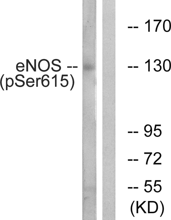 WB (Western Blot)
(Western blot analysis of extracts from K562 cells, treated with EGF (40nM, 30mins), using eNOS (Phospho-Ser615) antibody.)
WB (Western Blot)
(Western blot analysis of extracts from K562 cells, treated with EGF (40nM, 30mins), using eNOS (Phospho-Ser615) antibody.)
eNOS, Antibody (Cat# AAA109281)
 IHC (Immunohiostchemistry)
(Immunohistochemical analysis of IKK alpha (pT23) staining in human breast cancer formalin fixed paraffin embedded tissue section. The section was pre-treated using heat mediated antigen retrieval with sodium citrate buffer (pH 6.0). The section was then incubated with the antibody at room temperature and detected using an HRP conjugated compact polymer system. DAB was used as the chromogen. The section was then counterstained with haematoxylin and mounted with DPX.)
IHC (Immunohiostchemistry)
(Immunohistochemical analysis of IKK alpha (pT23) staining in human breast cancer formalin fixed paraffin embedded tissue section. The section was pre-treated using heat mediated antigen retrieval with sodium citrate buffer (pH 6.0). The section was then incubated with the antibody at room temperature and detected using an HRP conjugated compact polymer system. DAB was used as the chromogen. The section was then counterstained with haematoxylin and mounted with DPX.)
IKK alpha (pT23), Polyclonal Antibody (Cat# AAA105235)
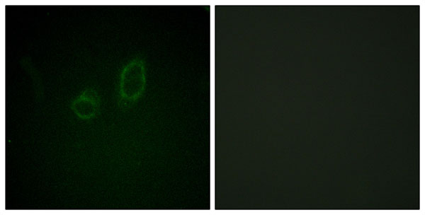
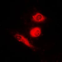
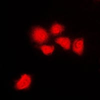
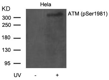
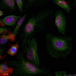
 IF (Immunofluorescence)
(Confocal immunofluorescent analysis ofPhospho-ATM-pS1981 Antibody with Hela cell followed by Alexa Fluor 488-conjugated goat anti-rabbit lgG (green). DAPI was used to stain the cell nuclear (blue).)
IF (Immunofluorescence)
(Confocal immunofluorescent analysis ofPhospho-ATM-pS1981 Antibody with Hela cell followed by Alexa Fluor 488-conjugated goat anti-rabbit lgG (green). DAPI was used to stain the cell nuclear (blue).)
Phospho-ATM (S1981), Polyclonal Antibody (Cat# AAA284603)
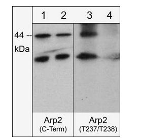
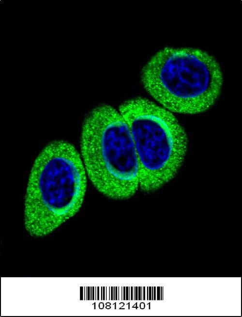
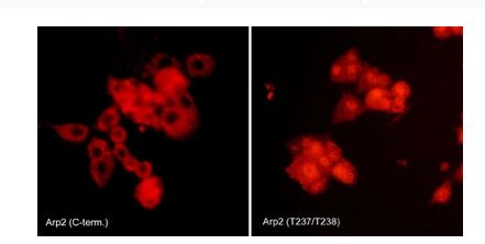 ICC (Immunocytochemistry)
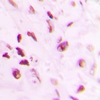
(Immunocytochemical labeling of Arp2 phosphorylation in rat PC12 cells differentiated with NGF. The cells were probed with Arp2 (C-terminal region) and Arp2 (Thr-237/Thr-238) rabbit polyclonal antibodies, then the antibodies were detected using appropriate secondary antibody conjugated to Cy3.)
ICC (Immunocytochemistry)
(Immunocytochemical labeling of Arp2 phosphorylation in rat PC12 cells differentiated with NGF. The cells were probed with Arp2 (C-terminal region) and Arp2 (Thr-237/Thr-238) rabbit polyclonal antibodies, then the antibodies were detected using appropriate secondary antibody conjugated to Cy3.)
Arp2, Polyclonal Antibody (Cat# AAA71568)
 Application Data
IkBa (pS32/36) (NFKB1A), Antibody (Cat# AAA71434)
 WB (Western Blot)
(Western blot of rat hippocampal lysate showing specific labeling of the ~135 kDa KCC2 protein phosphorylated at Thr1007 in the first lane (-). Phosphospecificity is shown in the second lane (+) where immunolabeling is completely eliminated by blot treatment with lambda phosphatase (lambda-Ptase, 1200 units for 30 min).)
WB (Western Blot)
(Western blot of rat hippocampal lysate showing specific labeling of the ~135 kDa KCC2 protein phosphorylated at Thr1007 in the first lane (-). Phosphospecificity is shown in the second lane (+) where immunolabeling is completely eliminated by blot treatment with lambda phosphatase (lambda-Ptase, 1200 units for 30 min).)
Potassium Chloride Cotransporter (KCC2), Polyclonal Antibody (Cat# AAA72760)
Expected Reactivity: Bovine, Human, Non-human primate, Sheep
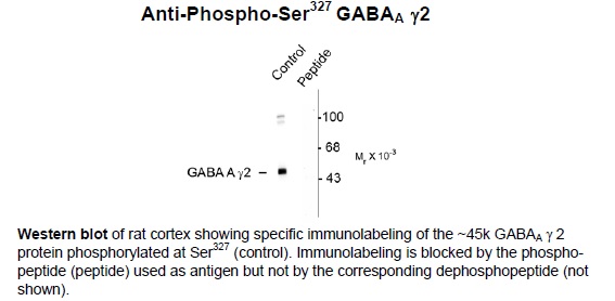 Application Data
GABAA Receptor, g 2 (Ser327), Polyclonal Antibody (Cat# AAA72791)
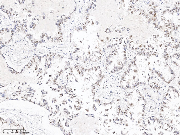
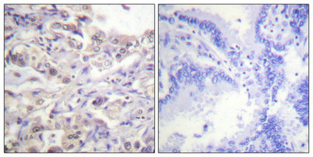
 IHC (Immunohistochemisry)
(Immunochemical staining of human MAPK1 in human breast carcinoma with rabbit polyclonal antibody at 1:100 dilution, formalin-fixed paraffin embedded sections.)
IHC (Immunohistochemisry)
(Immunochemical staining of human MAPK1 in human breast carcinoma with rabbit polyclonal antibody at 1:100 dilution, formalin-fixed paraffin embedded sections.)
ERK1/2, Polyclonal Antibody (Cat# AAA258879)
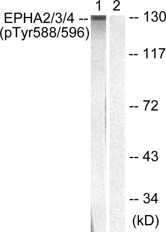
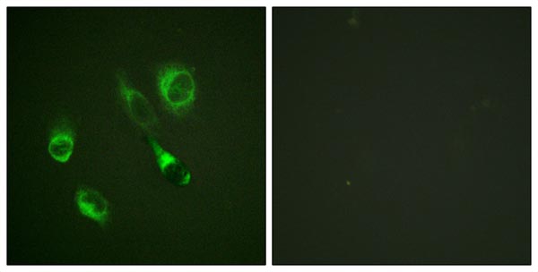 IF (Immunofluorescence)
(Immunofluorescence staining of methanol-fixed HeLa cells using EPHA2/3/4 (Phospho-Tyr588/596) Antibody.)
IF (Immunofluorescence)
(Immunofluorescence staining of methanol-fixed HeLa cells using EPHA2/3/4 (Phospho-Tyr588/596) Antibody.)
EPHA2/3/4, Polyclonal Antibody (Cat# AAA301787)
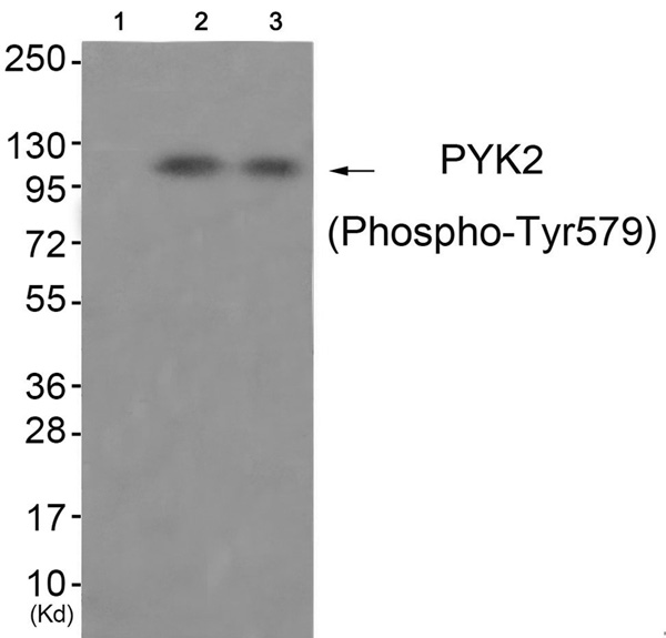
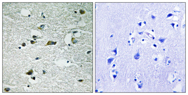
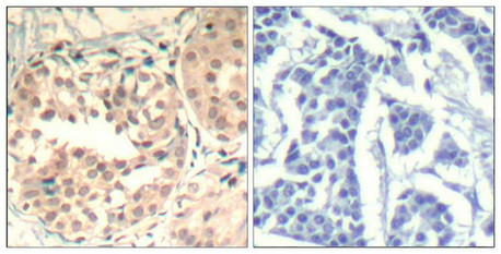
 IHC (Immunohiostchemistry)
(Immunohistochemical analysis of paraffin-embedded human brain tissue using PYK2 (Phospho-Tyr579) antibody (left)or the same antibody preincubated with blocking peptide (right).)
IHC (Immunohiostchemistry)
(Immunohistochemical analysis of paraffin-embedded human brain tissue using PYK2 (Phospho-Tyr579) antibody (left)or the same antibody preincubated with blocking peptide (right).)
PYK2, Polyclonal Antibody (Cat# AAA301955)
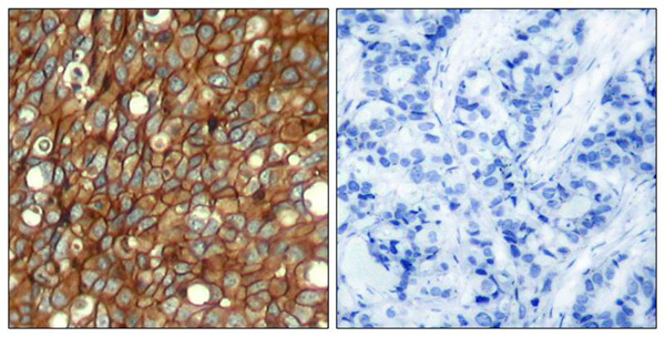
 IHC (Immunohistochemistry)
(Immunohistochemical analysis of paraffin-embedded human lung carcinoma tissue, using HER2 (Phospho-Tyr1248) Antibody.)
IHC (Immunohistochemistry)
(Immunohistochemical analysis of paraffin-embedded human lung carcinoma tissue, using HER2 (Phospho-Tyr1248) Antibody.)
HER2, Polyclonal Antibody (Cat# AAA301288)
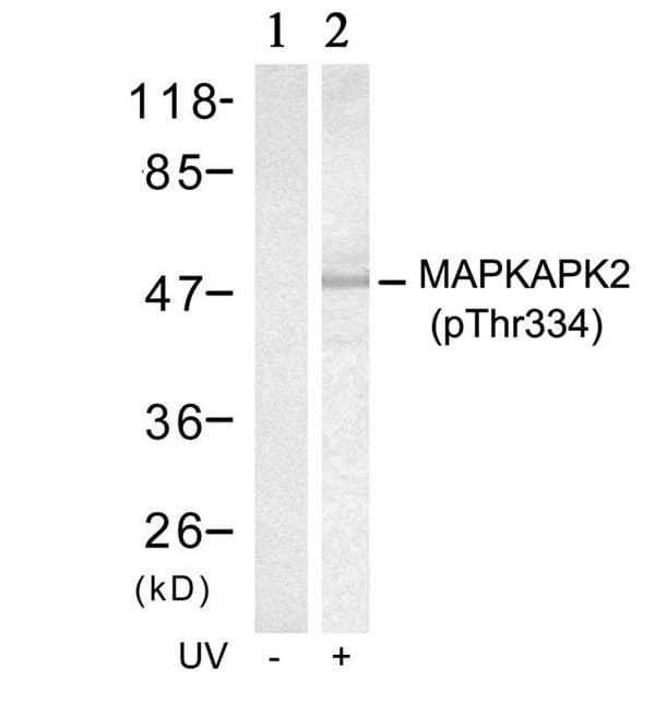
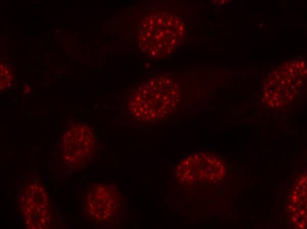 IF (Immunofluorescence)
(Immunofluorescence staining of methanol-fixed Hela cells using MAPKAPK-2(Phospho-Thr334) Antibody.)
IF (Immunofluorescence)
(Immunofluorescence staining of methanol-fixed Hela cells using MAPKAPK-2(Phospho-Thr334) Antibody.)
MAPKAPK-2, Polyclonal Antibody (Cat# AAA301290)
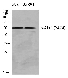
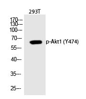
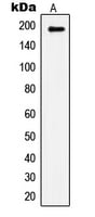
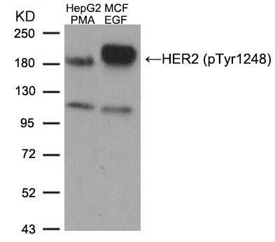
 WB (Western Blot)
(Western Blot analysis of KB using Phospho-Akt1 (Y474) Polyclonal Antibody)
WB (Western Blot)
(Western Blot analysis of KB using Phospho-Akt1 (Y474) Polyclonal Antibody)
Akt1, Polyclonal Antibody (Cat# AAA301508)
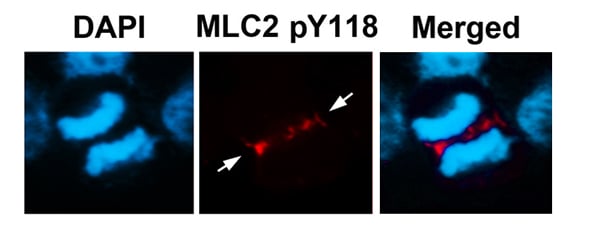
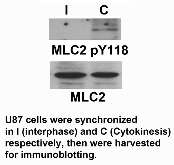 Application Data
MLC2, Polyclonal Antibody (Cat# AAA301575)
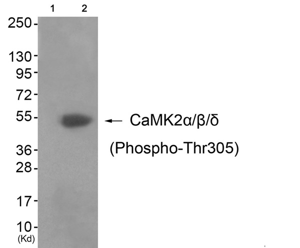
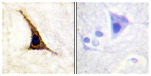
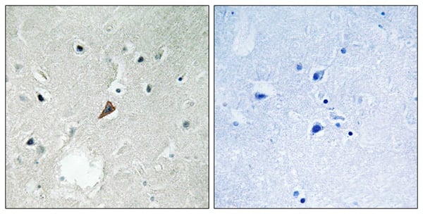
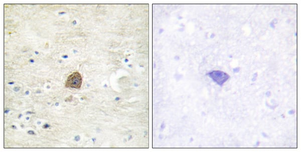 IHC (Immunohiostchemistry)
(Immunohistochemical analysis of paraffin-embedded human brain tissue using CaMKII (Phospho-Thr305) antibody (left)or the same antibody preincubated with blocking peptide (right).)
IHC (Immunohiostchemistry)
(Immunohistochemical analysis of paraffin-embedded human brain tissue using CaMKII (Phospho-Thr305) antibody (left)or the same antibody preincubated with blocking peptide (right).)
CaMK2alpha/beta/delta, Polyclonal Antibody (Cat# AAA301671)

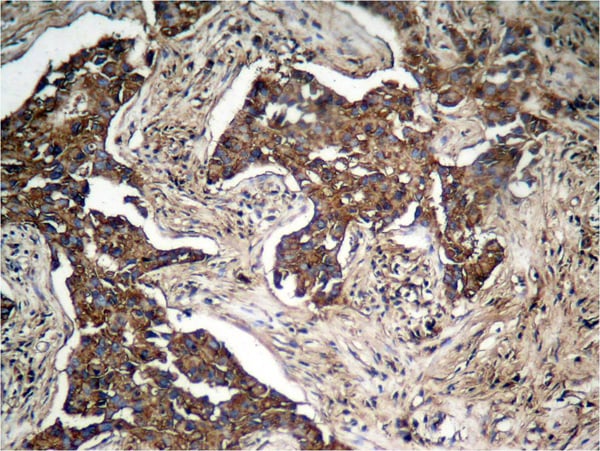
 IHC (Immunohiostchemistry)
(Immunohistochemical analysis of paraffin-embedded human breast carcinoma tissue using Gab2 (Phospho-Ser623) antibody (left)or the same antibody preincubated with blocking peptide (right).)
IHC (Immunohiostchemistry)
(Immunohistochemical analysis of paraffin-embedded human breast carcinoma tissue using Gab2 (Phospho-Ser623) antibody (left)or the same antibody preincubated with blocking peptide (right).)
Gab2, Polyclonal Antibody (Cat# AAA301684)
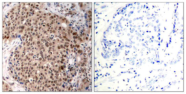
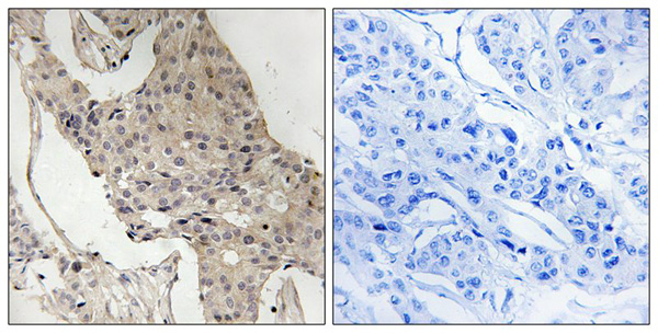
 IHC (Immunohiostchemistry)
(Immunohistochemical analysis of paraffin-embedded human breast carcinoma tissue using ATM(Phospho-Ser1981) Antibody (left) or the same antibody preincubated with blocking peptide(right).)
IHC (Immunohiostchemistry)
(Immunohistochemical analysis of paraffin-embedded human breast carcinoma tissue using ATM(Phospho-Ser1981) Antibody (left) or the same antibody preincubated with blocking peptide(right).)
ATM, Polyclonal Antibody (Cat# AAA301377)
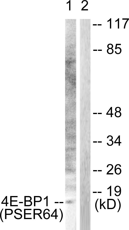
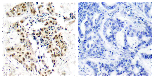
 IHC (Immunohiostchemistry)
(Immunohistochemical analysis of paraffin-embedded human colon carcinoma tissue using 4E-BP1 (Phospho-Ser64) antibody (#A0401). The picture on the right is treated with the synthesized peptide.)
IHC (Immunohiostchemistry)
(Immunohistochemical analysis of paraffin-embedded human colon carcinoma tissue using 4E-BP1 (Phospho-Ser64) antibody (#A0401). The picture on the right is treated with the synthesized peptide.)
4E-BP1, Polyclonal Antibody (Cat# AAA301416)
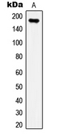
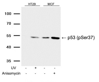 WB (Western Blot)
(Western blot analysis of extracts from HT29 cells untreated or treated with UV, and MCF cells untreated or treated with Anisomycin using p53 (Phospho-Ser37) Antibody.)
WB (Western Blot)
(Western blot analysis of extracts from HT29 cells untreated or treated with UV, and MCF cells untreated or treated with Anisomycin using p53 (Phospho-Ser37) Antibody.)
p53, Polyclonal Antibody (Cat# AAA301459)
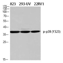
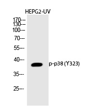
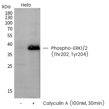
 WB (Western Blot)
(Western Blot analysis of HELA using Phospho-p38 (Y323) Polyclonal Antibody.)
WB (Western Blot)
(Western Blot analysis of HELA using Phospho-p38 (Y323) Polyclonal Antibody.)
p38, Polyclonal Antibody (Cat# AAA303106)
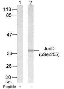
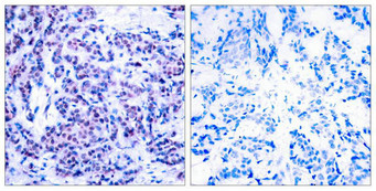 IHC (Immunohiostchemistry)
(Immunohistochemical analysis of paraffin-embedded human breast carcinoma tissue using JunD(Phospho-Ser255) Antibody(left) or the same antibody preincubated with blocking peptide(right).)
IHC (Immunohiostchemistry)
(Immunohistochemical analysis of paraffin-embedded human breast carcinoma tissue using JunD(Phospho-Ser255) Antibody(left) or the same antibody preincubated with blocking peptide(right).)
JUND, Polyclonal Antibody (Cat# AAA243047)
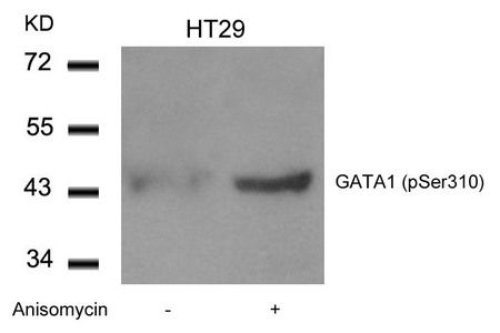
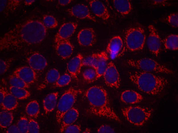
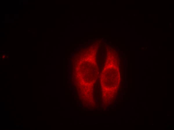
 IF (Immunofluorescence)
(Immunofluorescence staining of methanol-fixed Hela cells using GATA1(Phospho-Ser310) Antibody.)
IF (Immunofluorescence)
(Immunofluorescence staining of methanol-fixed Hela cells using GATA1(Phospho-Ser310) Antibody.)
GATA1, Polyclonal Antibody (Cat# AAA243054)
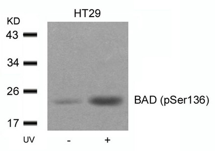
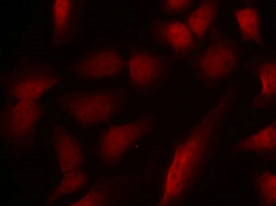
 IHC (Immunohiostchemistry)
(Immunohistochemical analysis of paraffin-embedded human breast carcinoma tissue using BAD(Phospho-Ser136) Antibody(left) or the same antibody preincubated with blocking peptide(right).)
IHC (Immunohiostchemistry)
(Immunohistochemical analysis of paraffin-embedded human breast carcinoma tissue using BAD(Phospho-Ser136) Antibody(left) or the same antibody preincubated with blocking peptide(right).)
Bad, Polyclonal Antibody (Cat# AAA243066)
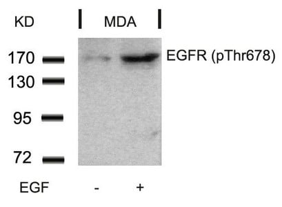
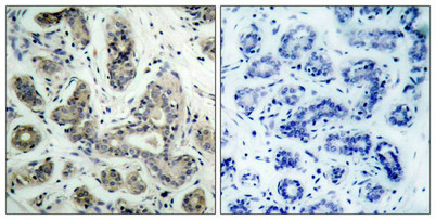
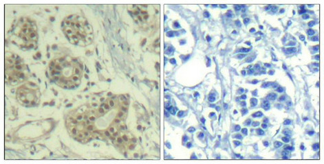
 IHC (Immunohiostchemistry)
(Immunohistochemical analysis of paraffin-embedded Rat Colorectal tissue using EGFR (Phospho-Thr678) Antibody.)
IHC (Immunohiostchemistry)
(Immunohistochemical analysis of paraffin-embedded Rat Colorectal tissue using EGFR (Phospho-Thr678) Antibody.)
EGFR, Polyclonal Antibody (Cat# AAA243107)
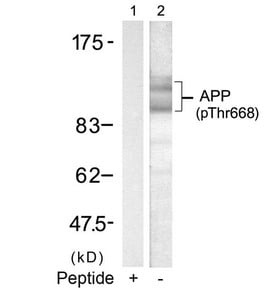
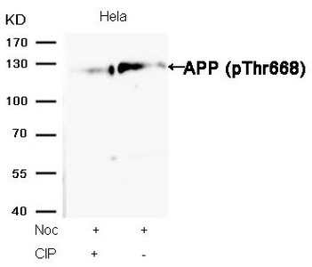 WB (Western Blot)
(Western blot analysis of extracts from Hela cells, treated with Noc or calf intestinal phosphatase (CIP), using APP (Phospho-Thr668) Antibody.)
WB (Western Blot)
(Western blot analysis of extracts from Hela cells, treated with Noc or calf intestinal phosphatase (CIP), using APP (Phospho-Thr668) Antibody.)
APP, Polyclonal Antibody (Cat# AAA243109)
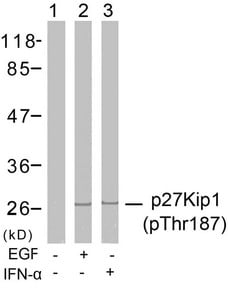
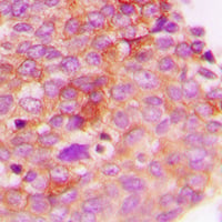
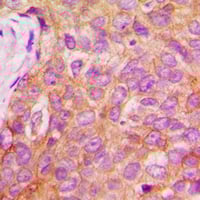
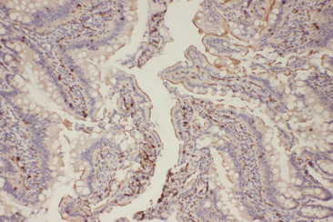
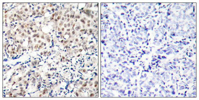 IHC (Immunohiostchemistry)
(Immunohistochemical analysis of paraffin-embedded human breast carcinoma tissue using p27Kip1(Phospho-Thr187) Antibody(left) or the same antibody preincubated with blocking peptide(right).)
IHC (Immunohiostchemistry)
(Immunohistochemical analysis of paraffin-embedded human breast carcinoma tissue using p27Kip1(Phospho-Thr187) Antibody(left) or the same antibody preincubated with blocking peptide(right).)
CDKN1B, Polyclonal Antibody (Cat# AAA243115)

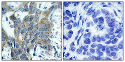 IHC (Immunohiostchemistry)
(Immunohistochemical analysis of paraffin-embedded human breast carcinoma tissue using b-Catenin(Phospho-Ser33) Antibody(left) or the same antibody preincubated with blocking peptide(right).)
IHC (Immunohiostchemistry)
(Immunohistochemical analysis of paraffin-embedded human breast carcinoma tissue using b-Catenin(Phospho-Ser33) Antibody(left) or the same antibody preincubated with blocking peptide(right).)
CTNNB1, Polyclonal Antibody (Cat# AAA243118)


 IF (Immunofluorescence)
(Immunofluorescence staining of methanol-fixed MCF7 cells using Her3/ErbB3(phospho-Tyr1328) Antibody.)
IF (Immunofluorescence)
(Immunofluorescence staining of methanol-fixed MCF7 cells using Her3/ErbB3(phospho-Tyr1328) Antibody.)
ERBB3, Polyclonal Antibody (Cat# AAA243323)
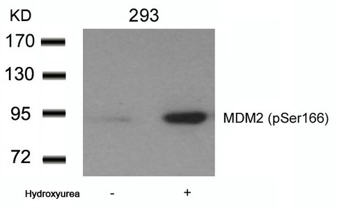
 IF (Immunofluorescence)
(Immunofluorescence staining of methanol-fixed Hela cells using MDM2(phospho-Ser166) Antibody.)
IF (Immunofluorescence)
(Immunofluorescence staining of methanol-fixed Hela cells using MDM2(phospho-Ser166) Antibody.)
MDM2, Polyclonal Antibody (Cat# AAA243331)

 IF (Immunofluorescence)
(Immunofluorescence staining of methanol-fixed HeLa cells using hnRPD (Phospho-Ser83) Antibody.)
IF (Immunofluorescence)
(Immunofluorescence staining of methanol-fixed HeLa cells using hnRPD (Phospho-Ser83) Antibody.)
HNRNPD, Polyclonal Antibody (Cat# AAA243345)


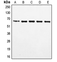
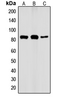
 WB (Western Blot)
(Western blot analysis of extracts from HUVEC cells, treated with calf intestinal phosphatase (CIP), using CDK6 (phospho-Tyr13) Antibody.)
WB (Western Blot)
(Western blot analysis of extracts from HUVEC cells, treated with calf intestinal phosphatase (CIP), using CDK6 (phospho-Tyr13) Antibody.)
CDK6, Polyclonal Antibody (Cat# AAA243375)
What Are Phospho Antibodies?
Protein phosphorylation is a process where a phosphate group is added to certain amino acid residues of a protein – usually serine (S), threonine (T), or tyrosine (Y) - by enzymes called kinases. This process is integral in controlling cellular signaling, cellular growth, and other biological functions.
Our catalog includes a wide range of phospho-specific antibodies that can accurately detect this important marker. They perform strongly in widely-used laboratory applications such as Western blot, flow cytometry, immunohistochemistry, and immunofluorescence microscopy. We value your trust in us and are committed to providing top-quality products and services. All of our antibodies are guaranteed to work for the applications and species indicated on our website & associated product pages.
What Are The Key Applications of Phospho Antibodies?
1. Western Blotting
One of the first steps a researcher can take in utilizing these phospho-specific antibodies, is to check if the antibody works using a technique referred to as “Western blot”. For those unfamiliar, Western Blot aids in showing whether the protein that the antibody recognizes is appearing at the correct/expected size. These phospho-specific antibodies should also be able to detect changes in the target protein’s phosphorylation (on/off state) when cells are stimulated in certain ways.
2. Staining of Fixed Cells (Immunocytochemistry)
Another routine use of these phospho-specific antibodies, is to test if the antibody is able to demonstrate similar performance when used on fixed cells (intact cells that have been preserved) as it did in the Western blot tests. It is an important aspect in many cases to confirm that the antibody works in actual intact cell samples. Ideally, the method used for cellular fixation should be the same as what is used in pathology labs (like using 10% formalin). To check if the antibody works well in tissue sections (FFPE), researchers will often test it on fixed cells that are processed similar to tissue samples.
3. Specificity Tests Using Peptides
In order to make sure that the antibody is only binding to the right target:
- Laboratory technicians will mix the antibody with phospho-peptides (short segments of the protein containing the phosphate group modification).
- If the antibody signal disappears, it is confirmation that it is binding to the correct phosphorylated location.
- A more robust test is to use both the phosphorylated and non-phosphorylated (dephosphorylated) versions of the protein. The antibody should react only with the phosphorylated one.
- Another method sometimes utilized is to treat the sample with an enzyme, such as alkaline phosphatase, that specifically removes phosphate groups. If the antibody signal disappears after this, it also confirms specificity.
4. Genetic Confirmation
As a final step, scientists can genetically manipulate the nucleotide sequence and alter the target protein by removing the exact site where phosphorylation happens. If the antibody no longer appears to detect the modified protein, it is strong evidence supporting the antibody being specific for that phosphorylated site.
Why Buy Phospho Antibodies Through Us?
- The production laboratory adheres to strict and consistent protocols prior to releasing any of these phospho-specific antibodies:
- Standard methods and proper controls in all tests to ensure high quality.
- These antibodies are tested and validated in different cell types and species.
- High quality control criterion to ensure each batch is consistent, so you will obtain reliable results every time.
FAQ
1. What Are Phospho-Specific Antibodies?
Phospho-specific antibodies are made to detect proteins only when they have a phosphate group linked to a specific amino acid residue. This empowers scientists understand if a protein is "turned on" or active, based on its phosphorylation state.
2. How to Detect Phosphorylated Proteins in a Western Blot?
To find out if a protein is phosphorylated using Western blot:
- Use a phospho-specific antibody that binds only to the phosphorylated form of the protein.
- You can also use a “regular” antibody for the same amino acid sequence of the protein that the phospho-specific antibody is binding to (but in this case, this antibody will not bind if there is a phosphate group present) in order to compare how much of it is phosphorylated versus how much is non-phosphorylated (or “total” protein, if the “normal” antibody’s epitopes are non-phospho-site-specific).
3. How to Choose the Best Antibody?
Here are some simple tips to help you pick the right antibody:
- Know your target
- Match your sample characteristics
- Confirm the intended use is appropriate
- Check “host” and “type”
- Check the “quality” of the presented data/images
- Appraise whether the available validation meets your needs