Filters
▼Clonality
▼Type
▼Reactivity
▼Gene Name
▼Isotype
▼Host
▼Application
▼Clone
▼Phospho Antibodies
Phospho-specific antibodies’ typical purpose is to enable researchers to detect changes in proteins. They will exclusively bind to the amino acid sequence on a protein that has been phosphorylated (which is both a physical & chemical change) and do not bind to the same amino acid sequence on said protein if it lacks said phosphorylation. This aids in being able to clearly see and understand the data produced from this particular protein modification.
Viewing 1050-1100 of 5298 product results
 WB (Western Blot)
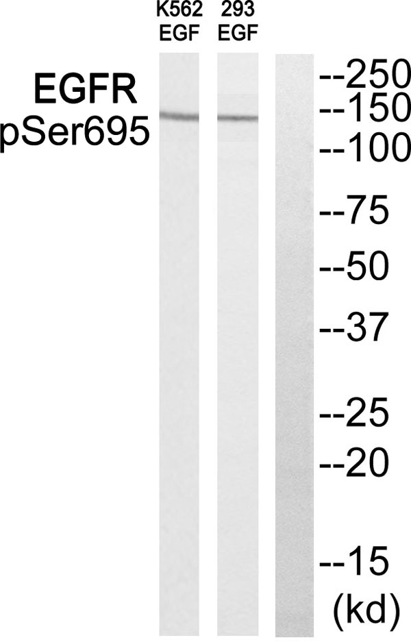
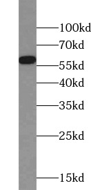
(Western blot analysis of extracts from 293 cells and K562 cells treated with EGF, using EGFR (Phospho-Ser695) antibody.)
WB (Western Blot)
(Western blot analysis of extracts from 293 cells and K562 cells treated with EGF, using EGFR (Phospho-Ser695) antibody.)
EGFR, Antibody (Cat# AAA109046)
Mouse (Identities = 100%, Positives = 100%)
Rat (Identities = 100%, Positives = 100%)
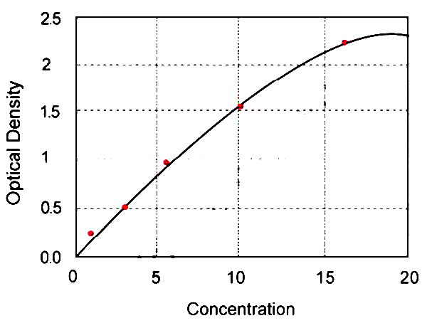 Standard Curve (Sample)
Phospho-NF-KB p65(Ser536), ELISA Kit (Cat# AAA122965)
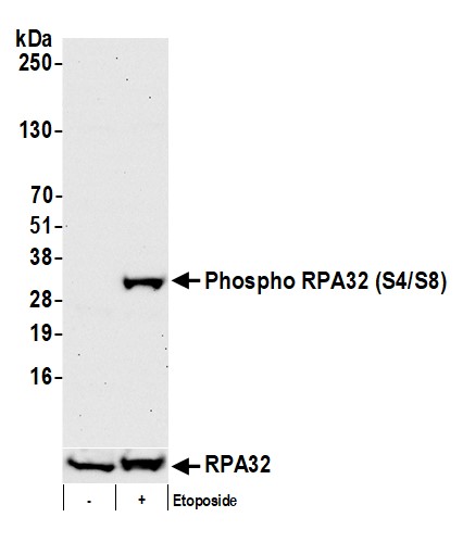 WB (Western Blot)
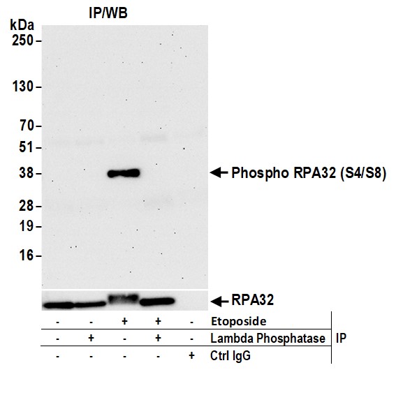
(Detection of human Phospho RPA32 (S4/S8) by western blot. Samples: Whole cell lysate (50 ug) from BeWo and AlphaTC1 Clone 9 cells prepared using NETN lysis buffer. Antibody: Affinity purified rabbit anti-Phospho RPA32 (S4/S8) antibody (AAA210838 lot 9) used for WB at 0.1 ug/ml. Detection: Chemiluminescence with an exposure time of 30 seconds.)
WB (Western Blot)
(Detection of human Phospho RPA32 (S4/S8) by western blot. Samples: Whole cell lysate (50 ug) from BeWo and AlphaTC1 Clone 9 cells prepared using NETN lysis buffer. Antibody: Affinity purified rabbit anti-Phospho RPA32 (S4/S8) antibody (AAA210838 lot 9) used for WB at 0.1 ug/ml. Detection: Chemiluminescence with an exposure time of 30 seconds.)
RPA32, Polyclonal Antibody (Cat# AAA210838)
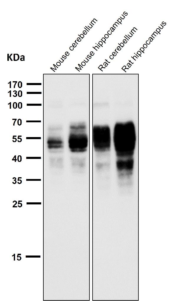
 WB (Western Blot)
(Western blot analysis of Phospho-Tau (S202) expression in mouse hippocampus cell lysate.)
WB (Western Blot)
(Western blot analysis of Phospho-Tau (S202) expression in mouse hippocampus cell lysate.)
Tau, Monoclonal Antibody (Cat# AAA128105)
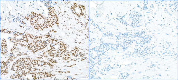
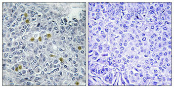 IHC (Immunohiostchemistry)
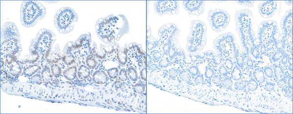
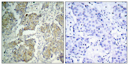
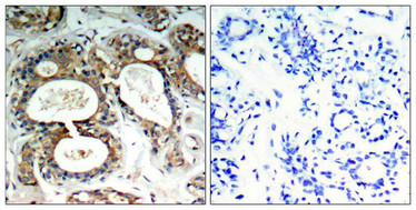
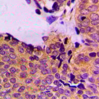
(Immunohistochemistry analysis of paraffin-embedded human breast carcinoma tissue using TOP2A (Phospho-Ser1106) antibody. The picture on the right is treated with the synthesized peptide.)
IHC (Immunohiostchemistry)
(Immunohistochemistry analysis of paraffin-embedded human breast carcinoma tissue using TOP2A (Phospho-Ser1106) antibody. The picture on the right is treated with the synthesized peptide.)
TOP2A, Polyclonal Antibody (Cat# AAA243234)
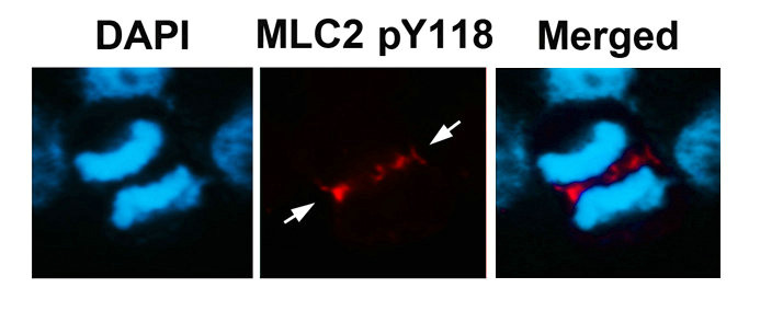
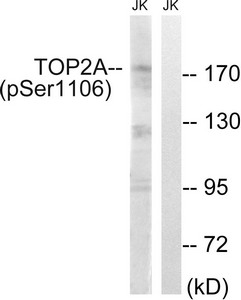
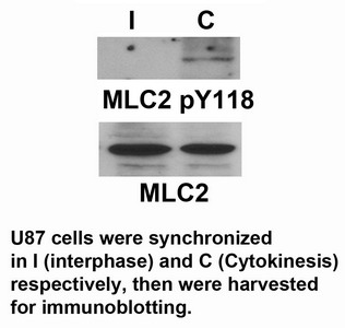 WB (Western Blot)
MYL9, Polyclonal Antibody (Cat# AAA243254)
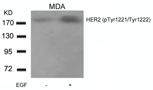
 WB (Western Blot)
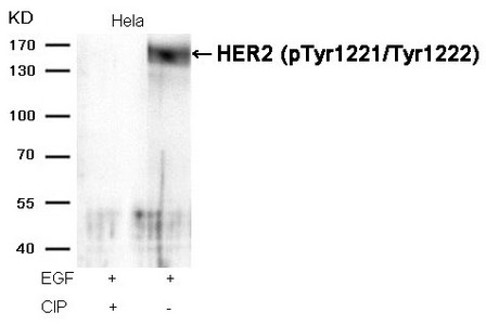
(Western blot analysis of extracts from Hela cells, treated with EGF or calf intestinal phosphatase (CIP), using HER2 (Phospho-Tyr1221/Tyr1222) Antibody.)
WB (Western Blot)
(Western blot analysis of extracts from Hela cells, treated with EGF or calf intestinal phosphatase (CIP), using HER2 (Phospho-Tyr1221/Tyr1222) Antibody.)
ERBB2, Polyclonal Antibody (Cat# AAA243269)
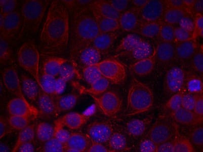
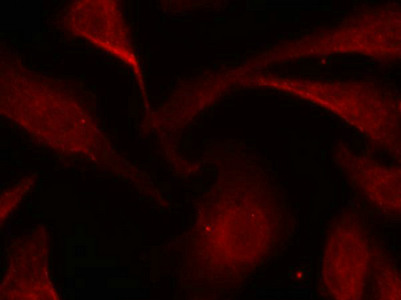
 IF (Immunofluorescence)
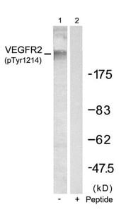
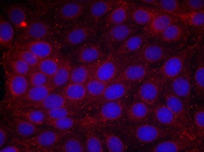
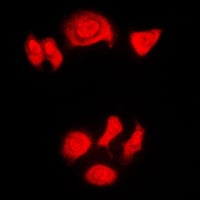
(Immunofluorescence staining of methanol-fixed MCF cells using VEGFR2(Phospho-Tyr1214) Antibody.)
IF (Immunofluorescence)
(Immunofluorescence staining of methanol-fixed MCF cells using VEGFR2(Phospho-Tyr1214) Antibody.)
KDR, Polyclonal Antibody (Cat# AAA243271)
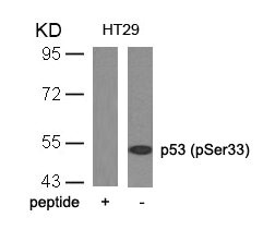
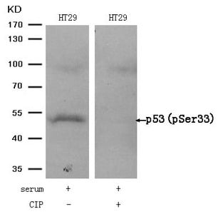 WB (Western Blot)
(Western blot analysis of extracts from HT29 cells, treated with serum or calf intestinal phosphatase (CIP), using p53 (Phospho-Ser33) Antibody.)
WB (Western Blot)
(Western blot analysis of extracts from HT29 cells, treated with serum or calf intestinal phosphatase (CIP), using p53 (Phospho-Ser33) Antibody.)
TP53, Polyclonal Antibody (Cat# AAA243273)
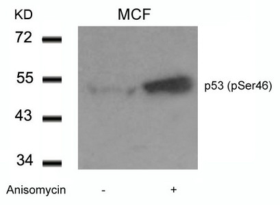
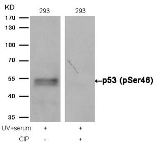 WB (Western Blot)
(Western blot analysis of extracts from 293 cells, treated with UV+serum or calf intestinal phosphatase (CIP), using p53 (Phospho-Ser46) Antibody.)
WB (Western Blot)
(Western blot analysis of extracts from 293 cells, treated with UV+serum or calf intestinal phosphatase (CIP), using p53 (Phospho-Ser46) Antibody.)
TP53, Polyclonal Antibody (Cat# AAA243274)
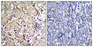
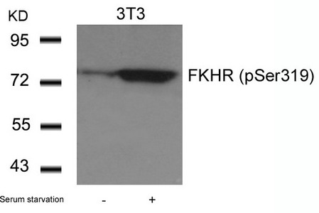
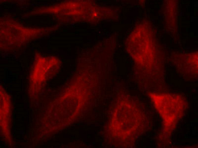
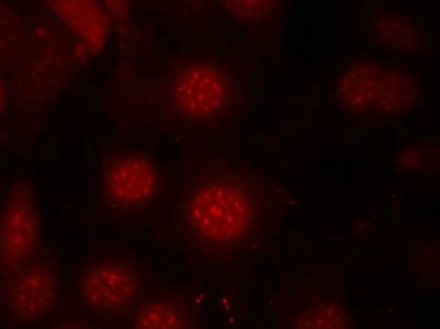
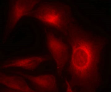
 IF (Immunofluorescence)
(Immunofluorescence staining of methanol-fixed Hela cells using FKHR(Phospho-Ser319) Antibody.)
IF (Immunofluorescence)
(Immunofluorescence staining of methanol-fixed Hela cells using FKHR(Phospho-Ser319) Antibody.)
FOXO1, Polyclonal Antibody (Cat# AAA243285)
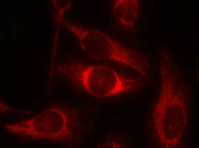
 IF (Immunofluorescence)
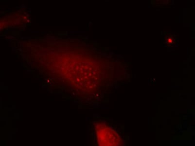
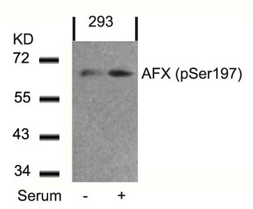
(Immunofluorescence staining of methanol-fixed MCF7 cells using AFX(Phospho-Ser197) Antibody.)
IF (Immunofluorescence)
(Immunofluorescence staining of methanol-fixed MCF7 cells using AFX(Phospho-Ser197) Antibody.)
FOXO4, Polyclonal Antibody (Cat# AAA243286)
 IF (Immunofluorescence)
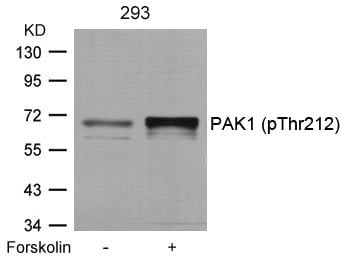
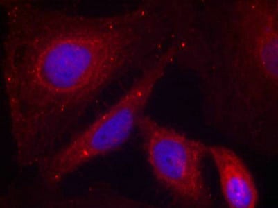
(Immunofluorescence staining of methanol-fixed Hela cells using PAK1(Phospho-Thr212) Antibody.)
IF (Immunofluorescence)
(Immunofluorescence staining of methanol-fixed Hela cells using PAK1(Phospho-Thr212) Antibody.)
PAK1, Polyclonal Antibody (Cat# AAA243290)
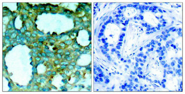
 IHC (Immunohiostchemistry)
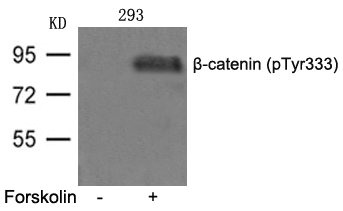
(Immunohistochemical analysis of paraffin-embedded human lung carcinoma tissue, using β-catenin (phospho-Tyr333) Antibody.)
IHC (Immunohiostchemistry)
(Immunohistochemical analysis of paraffin-embedded human lung carcinoma tissue, using β-catenin (phospho-Tyr333) Antibody.)
CTNNB1, Polyclonal Antibody (Cat# AAA243165)
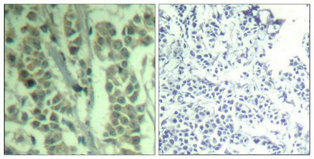
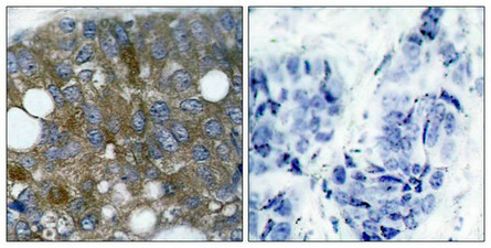
 IHC (Immunohiostchemistry)
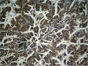
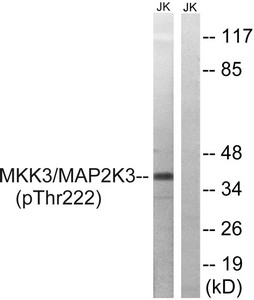
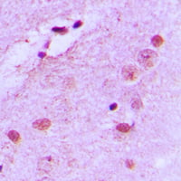
(Immunohistochemical analysis of paraffin-embedded human brain tissue using MAP2K3 (Phospho-Thr222) antibody (left)or the same antibody preincubated with blocking peptide (right).)
IHC (Immunohiostchemistry)
(Immunohistochemical analysis of paraffin-embedded human brain tissue using MAP2K3 (Phospho-Thr222) antibody (left)or the same antibody preincubated with blocking peptide (right).)
MAP2K3, Polyclonal Antibody (Cat# AAA243209)
 IHC (Immunohiostchemistry)
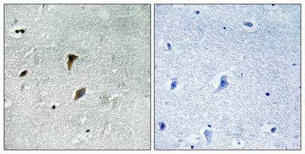
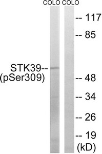
(Immunohistochemical analysis of paraffin-embedded human brain tissue using STK39 (Phospho-Ser309) antibody (left)or the same antibody preincubated with blocking peptide (right).)
IHC (Immunohiostchemistry)
(Immunohistochemical analysis of paraffin-embedded human brain tissue using STK39 (Phospho-Ser309) antibody (left)or the same antibody preincubated with blocking peptide (right).)
STK39, Polyclonal Antibody (Cat# AAA243213)
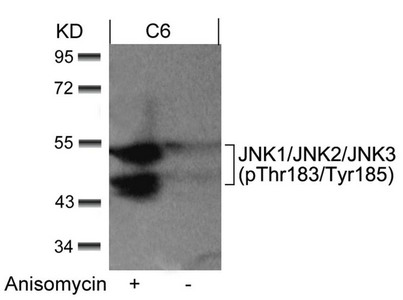
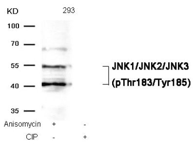 WB (Western Blot)
(Western blot analysis of extracts from 293 cells, treated with Anisomycin or calf intestinal phosphatase (CIP), using JNK1/JNK2/JNK3 (phospho-Thr183/Tyr185) Antibody.)
WB (Western Blot)
(Western blot analysis of extracts from 293 cells, treated with Anisomycin or calf intestinal phosphatase (CIP), using JNK1/JNK2/JNK3 (phospho-Thr183/Tyr185) Antibody.)
MAPK8/MAPK9/MAPK10, Polyclonal Antibody (Cat# AAA243322)
 IF (Immunofluorescence)
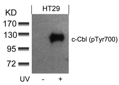
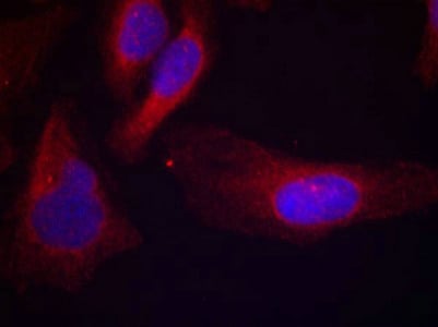
(Immunofluorescence staining of methanol-fixed Hela cells using c-Cbl(phospho-Tyr700) Antibody.)
IF (Immunofluorescence)
(Immunofluorescence staining of methanol-fixed Hela cells using c-Cbl(phospho-Tyr700) Antibody.)
CBL, Polyclonal Antibody (Cat# AAA243330)
 IF (Immunofluorescence)
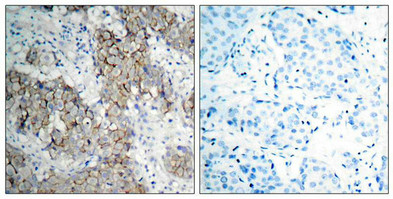
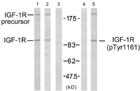
(Immunofluorescence staining of methanol-fixed MCF7 cells using IGF-1R (phospho-Tyr1161) antibody.)
IF (Immunofluorescence)
(Immunofluorescence staining of methanol-fixed MCF7 cells using IGF-1R (phospho-Tyr1161) antibody.)
IGF1R, Polyclonal Antibody (Cat# AAA243338)
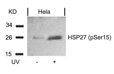
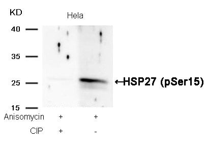 WB (Western Blot)
(Western blot analysis of extracts from Hela cells, treated with Anisomycin or calf intestinal phosphatase (CIP), using HSP27 (Phospho-Ser15) Antibody.)
WB (Western Blot)
(Western blot analysis of extracts from Hela cells, treated with Anisomycin or calf intestinal phosphatase (CIP), using HSP27 (Phospho-Ser15) Antibody.)
HSPB1, Polyclonal Antibody (Cat# AAA243364)
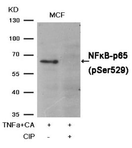
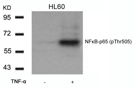
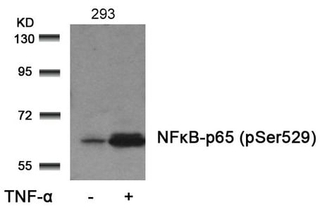 WB (Western Blot)
(Western blot analysis of extracts from 293 cells untreatedor treated with TNF-alpha using NFkappaB-p65 (Phospho-Ser529)Antibody.)
WB (Western Blot)
(Western blot analysis of extracts from 293 cells untreatedor treated with TNF-alpha using NFkappaB-p65 (Phospho-Ser529)Antibody.)
RELA, Polyclonal Antibody (Cat# AAA243368)
 IF (Immunofluorescence)
(Immunofluorescence staining of methanol-fixed Hela cells using JunB(Phospho-Ser79) Antibody.)
IF (Immunofluorescence)
(Immunofluorescence staining of methanol-fixed Hela cells using JunB(Phospho-Ser79) Antibody.)
JUNB, Polyclonal Antibody (Cat# AAA243045)
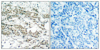
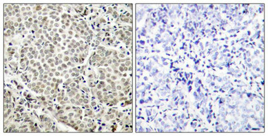
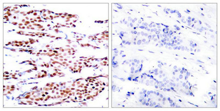
 IHC (Immunohiostchemistry)
(Immunohistochemical analysis of paraffin-embedded human breast carcinoma tissue using STAT5A(Phospho-Ser780) Antibody(left) or the same antibody preincubated with blocking peptide(right).)
IHC (Immunohiostchemistry)
(Immunohistochemical analysis of paraffin-embedded human breast carcinoma tissue using STAT5A(Phospho-Ser780) Antibody(left) or the same antibody preincubated with blocking peptide(right).)
STAT5A, Polyclonal Antibody (Cat# AAA243058)
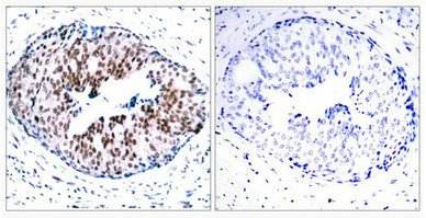
 IHC (Immunohiostchemistry)
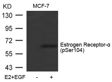
(Immunohistochemical analysis of paraffin-embedded human breast carcinoma tissue using Estrogen Receptor-a(Phospho-Ser104) Antibody(left) or the same antibody preincubated with blocking peptide(right).)
IHC (Immunohiostchemistry)
(Immunohistochemical analysis of paraffin-embedded human breast carcinoma tissue using Estrogen Receptor-a(Phospho-Ser104) Antibody(left) or the same antibody preincubated with blocking peptide(right).)
ESR1, Polyclonal Antibody (Cat# AAA243067)
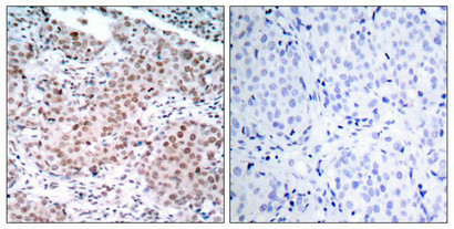
 IHC (Immunohiostchemistry)
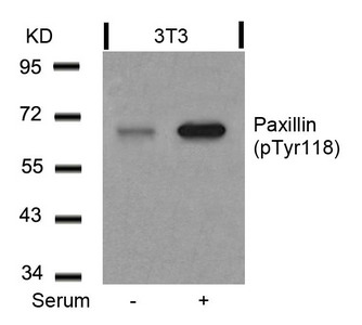
(Immunohistochemical analysis of paraffin-embedded human breast carcinoma tissue using Paxillin(Phospho-Tyr118) Antibody(left) or the same antibody preincubated with blocking peptide(right).)
IHC (Immunohiostchemistry)
(Immunohistochemical analysis of paraffin-embedded human breast carcinoma tissue using Paxillin(Phospho-Tyr118) Antibody(left) or the same antibody preincubated with blocking peptide(right).)
PXN, Polyclonal Antibody (Cat# AAA243073)
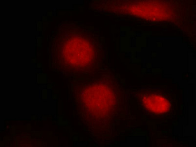 IF (Immunofluorescence)
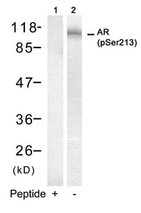
(Immunofluorescence staining of methanol-fixed Hela cells using Androgen Receptor(Phospho-Ser213) Antibody.)
IF (Immunofluorescence)
(Immunofluorescence staining of methanol-fixed Hela cells using Androgen Receptor(Phospho-Ser213) Antibody.)
AR, Polyclonal Antibody (Cat# AAA243085)
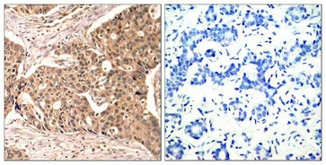
 IHC (Immunohiostchemistry)
(Immunohistochemical analysis of paraffin-embedded human breast carcinoma tissue using NFkB-p65(Phospho-Thr505) Antibody(left) or the same antibody preincubated with blocking peptide(right).)
IHC (Immunohiostchemistry)
(Immunohistochemical analysis of paraffin-embedded human breast carcinoma tissue using NFkB-p65(Phospho-Thr505) Antibody(left) or the same antibody preincubated with blocking peptide(right).)
RELA, Polyclonal Antibody (Cat# AAA243101)
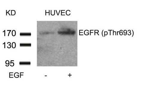
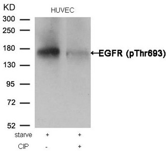 WB (Western Blot)
(Western blot analysis of extracts from HUVEC cells, treated with starve or calf intestinal phosphatase (CIP), using EGFR (Phospho-Thr693) Antibody.)
WB (Western Blot)
(Western blot analysis of extracts from HUVEC cells, treated with starve or calf intestinal phosphatase (CIP), using EGFR (Phospho-Thr693) Antibody.)
EGFR, Polyclonal Antibody (Cat# AAA243108)
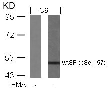
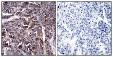
 IHC (Immunohiostchemistry)
(Immunohistochemical analysis of paraffin-embedded human tonsil carcinoma tissue using VASP(Phospho-Ser157) Antibody(left) or the same antibody preincubated with blocking peptide(right).)
IHC (Immunohiostchemistry)
(Immunohistochemical analysis of paraffin-embedded human tonsil carcinoma tissue using VASP(Phospho-Ser157) Antibody(left) or the same antibody preincubated with blocking peptide(right).)
VASP, Polyclonal Antibody (Cat# AAA243117)
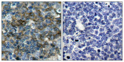
 IHC (Immunohiostchemistry)
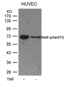
(Immunohistochemical analysis of paraffin-embedded human breast carcinoma tissue using RelB(Phospho-Ser573) Antibody(left) or the same antibody preincubated with blocking peptide(right).)
IHC (Immunohiostchemistry)
(Immunohistochemical analysis of paraffin-embedded human breast carcinoma tissue using RelB(Phospho-Ser573) Antibody(left) or the same antibody preincubated with blocking peptide(right).)
RELB, Polyclonal Antibody (Cat# AAA243130)
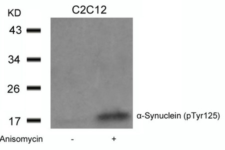
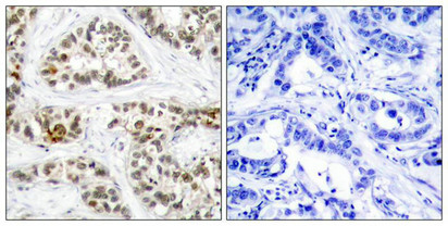
 IF (Immunofluorescence)
(Immunofluorescence staining of methanol-fixed Hela cells using a-Synuclein(Phospho-Tyr125) Antibody.)
IF (Immunofluorescence)
(Immunofluorescence staining of methanol-fixed Hela cells using a-Synuclein(Phospho-Tyr125) Antibody.)
SNCA, Polyclonal Antibody (Cat# AAA243131)
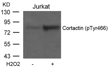
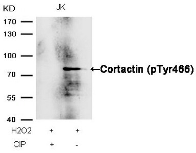 WB (Western Blot)
(Western blot analysis of extracts from JK cells, treated with H2O2 or calf intestinal phosphatase (CIP), using Cortactin (Phospho-Tyr466) Antibody.)
WB (Western Blot)
(Western blot analysis of extracts from JK cells, treated with H2O2 or calf intestinal phosphatase (CIP), using Cortactin (Phospho-Tyr466) Antibody.)
CTTN, Polyclonal Antibody (Cat# AAA243135)
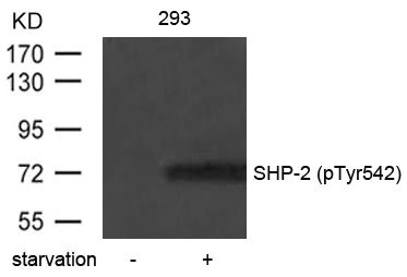
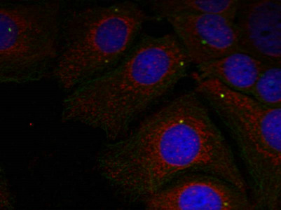
 IHC (Immunohiostchemistry)
(Immunohistochemical analysis of paraffin-embedded human breast carcinoma tissue using SHP-2(Phospho-Tyr542) Antibody(left) or the same antibody preincubated with blocking peptide(right).)
IHC (Immunohiostchemistry)
(Immunohistochemical analysis of paraffin-embedded human breast carcinoma tissue using SHP-2(Phospho-Tyr542) Antibody(left) or the same antibody preincubated with blocking peptide(right).)
PTPN11, Polyclonal Antibody (Cat# AAA243149)
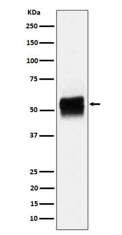
 WB (Western Blot)
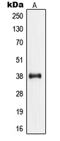
(Jurkat cells were subjected to SDS PAGE followed by western blot with AAA248043 (Phospho-Akt (S473) Antibody) at dilution of 1:2000)
WB (Western Blot)
(Jurkat cells were subjected to SDS PAGE followed by western blot with AAA248043 (Phospho-Akt (S473) Antibody) at dilution of 1:2000)
Phospho-Akt (Tyr315), Monoclonal Antibody (Cat# AAA248043)
Protein A+G purification
 Standard Curve (Sample)
Phospho-HGF R/c-MET DuoSet IC Economy Pk (1 PK), ELISA Kit (Cat# AAA205454)
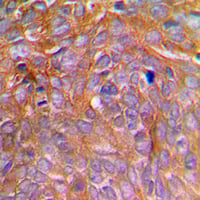 IHC (Immunohiostchemistry)
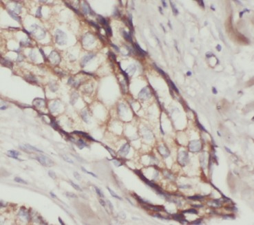
(Immunohistochemical analysis of Caspase 8 (pS347) staining in human breast cancer formalin fixed paraffin embedded tissue section. The section was pre-treated using heat mediated antigen retrieval with sodium citrate buffer (pH 6.0). The section was then incubated with the antibody at room temperature and detected using an HRP conjugated compact polymer system. DAB was used as the chromogen. The section was then counterstained with haematoxylin and mounted with DPX.)
IHC (Immunohiostchemistry)
(Immunohistochemical analysis of Caspase 8 (pS347) staining in human breast cancer formalin fixed paraffin embedded tissue section. The section was pre-treated using heat mediated antigen retrieval with sodium citrate buffer (pH 6.0). The section was then incubated with the antibody at room temperature and detected using an HRP conjugated compact polymer system. DAB was used as the chromogen. The section was then counterstained with haematoxylin and mounted with DPX.)
Caspase 8 (pS347), Polyclonal Antibody (Cat# AAA105209)
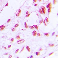
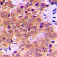
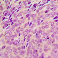 IHC (Immunohiostchemistry)
(Immunohistochemical analysis of SYK (pY348) staining in human breast cancer formalin fixed paraffin embedded tissue section. The section was pre-treated using heat mediated antigen retrieval with sodium citrate buffer (pH 6.0). The section was then incubated with the antibody at room temperature and detected using an HRP conjugated compact polymer system. DAB was used as the chromogen. The section was then counterstained with haematoxylin and mounted with DPX.)
IHC (Immunohiostchemistry)
(Immunohistochemical analysis of SYK (pY348) staining in human breast cancer formalin fixed paraffin embedded tissue section. The section was pre-treated using heat mediated antigen retrieval with sodium citrate buffer (pH 6.0). The section was then incubated with the antibody at room temperature and detected using an HRP conjugated compact polymer system. DAB was used as the chromogen. The section was then counterstained with haematoxylin and mounted with DPX.)
SYK (pY348), Polyclonal Antibody (Cat# AAA105218)

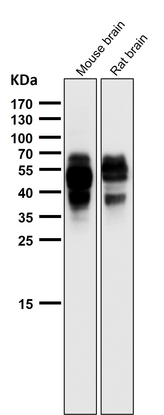
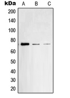
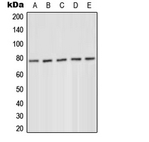
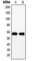
 WB (Western Blot)
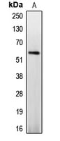
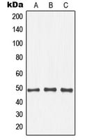
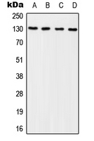
(Western blot analysis of GLUR4 (pS862) expression in HEK293T (A), rat brain (B), mouse brain (C), MCF7 (0) whole cell lysates.)
WB (Western Blot)
(Western blot analysis of GLUR4 (pS862) expression in HEK293T (A), rat brain (B), mouse brain (C), MCF7 (0) whole cell lysates.)
GLUR4 (pS862), Polyclonal Antibody (Cat# AAA105227)
 ELISA (Direct ELISA)
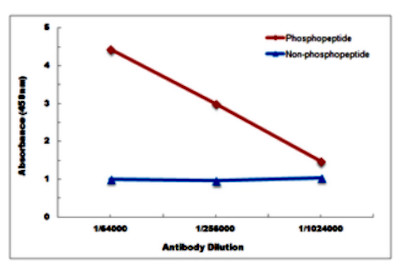
(Direct ELISA antibody dose-response curve using Anti-JNK1/2/3 (pT183) Antibody. Antigen (phosphopeptide and non-phosphopeptide) concentration is 5 ug/ml. Goat Anti-Rabbit IgG (H&L) - HRP was used as the secondary antibody, and signal was developed by TMB substrate.)
ELISA (Direct ELISA)
(Direct ELISA antibody dose-response curve using Anti-JNK1/2/3 (pT183) Antibody. Antigen (phosphopeptide and non-phosphopeptide) concentration is 5 ug/ml. Goat Anti-Rabbit IgG (H&L) - HRP was used as the secondary antibody, and signal was developed by TMB substrate.)
JNK1/2/3 (pT183), Polyclonal Antibody (Cat# AAA104862)
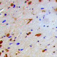 IHC (Immunohiostchemistry)
(Immunohistochemical analysis of IKB beta (pT19) staining in human brain formalin fixed paraffin embedded tissue section. The section was pre-treated using heat mediated antigen retrieval with sodium citrate buffer (pH 6.0). The section was then incubated with the antibody at room temperature and detected using an HRP conjugated compact polymer system. DAB was used as the chromogen. The section was then counterstained with haematoxylin and mounted with DPX.)
IHC (Immunohiostchemistry)
(Immunohistochemical analysis of IKB beta (pT19) staining in human brain formalin fixed paraffin embedded tissue section. The section was pre-treated using heat mediated antigen retrieval with sodium citrate buffer (pH 6.0). The section was then incubated with the antibody at room temperature and detected using an HRP conjugated compact polymer system. DAB was used as the chromogen. The section was then counterstained with haematoxylin and mounted with DPX.)
IKK beta (pT19), Polyclonal Antibody (Cat# AAA104628)
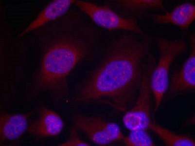
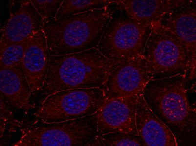
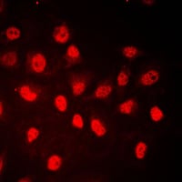
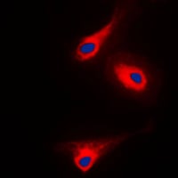 IF (Immunofluorescence)
(Immunofluorescent analysis of PLC gamma 1 (pY1253) staining in A431 cells. Formalin-fixed cells were permeabilized with 0.1% Triton X-100 in TBS for 5-10 minutes and blocked with 3% BSA-PBS for 30 minutes at room temperature. Cells were probed with the primary antibody in 3% BSA-PBS and incubated overnight at 4 °C in a humidified chamber. Cells were washed with PBST and incubated with a DyLight 594-conjugated secondary antibody (red) in PBS at room temperature in the dark. DAPI was used to stain the cell nuclei (blue).)
IF (Immunofluorescence)
(Immunofluorescent analysis of PLC gamma 1 (pY1253) staining in A431 cells. Formalin-fixed cells were permeabilized with 0.1% Triton X-100 in TBS for 5-10 minutes and blocked with 3% BSA-PBS for 30 minutes at room temperature. Cells were probed with the primary antibody in 3% BSA-PBS and incubated overnight at 4 °C in a humidified chamber. Cells were washed with PBST and incubated with a DyLight 594-conjugated secondary antibody (red) in PBS at room temperature in the dark. DAPI was used to stain the cell nuclei (blue).)
PLC gamma 1 (pY1253), Polyclonal Antibody (Cat# AAA104686)
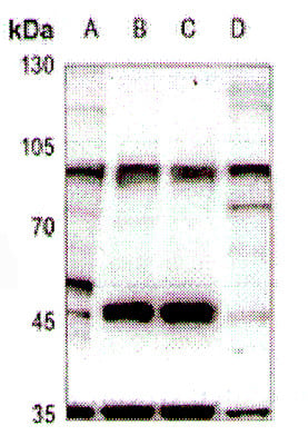
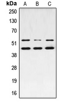
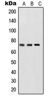
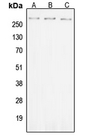 WB (Western Blot)
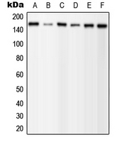
(Western blot analysis of ROS (pY2114) expression in SKOVCAR (A), Raw264.7 (B), PC12 (C) whole cell lysates.)
WB (Western Blot)
(Western blot analysis of ROS (pY2114) expression in SKOVCAR (A), Raw264.7 (B), PC12 (C) whole cell lysates.)
ROS (pY2114), Polyclonal Antibody (Cat# AAA104702)
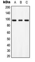 WB (Western Blot)
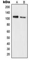
(Western blot analysis of AXL (pY697) expression in HEK293T (A), NIH3T3 (B), PC12 (C) whole cell lysates.)
WB (Western Blot)
(Western blot analysis of AXL (pY697) expression in HEK293T (A), NIH3T3 (B), PC12 (C) whole cell lysates.)
AXL (pY697), Polyclonal Antibody (Cat# AAA105077)
 IHC (Immunohiostchemistry)
(Immunohistochemical analysis of PDPK1 (pS241) staining in human breast cancer formalin fixed paraffin embedded tissue section. The section was pre-treated using heat mediated antigen retrieval with sodium citrate buffer (pH 6.0). The section was then incubated with the antibody at room temperature and detected using an HRP conjugated compact polymer system. DAB was used as the chromogen. The section was then counterstained with haematoxylin and mounted with DPX.)
IHC (Immunohiostchemistry)
(Immunohistochemical analysis of PDPK1 (pS241) staining in human breast cancer formalin fixed paraffin embedded tissue section. The section was pre-treated using heat mediated antigen retrieval with sodium citrate buffer (pH 6.0). The section was then incubated with the antibody at room temperature and detected using an HRP conjugated compact polymer system. DAB was used as the chromogen. The section was then counterstained with haematoxylin and mounted with DPX.)
PDPK1 (pS241), Polyclonal Antibody (Cat# AAA105097)
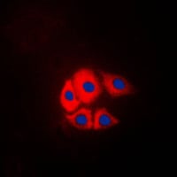
 IF (Immunofluorescence)
(Immunofluorescent analysis of Nucleophosmin (pT199) staining in HeLa cells. Formalin-fixed cells were permeabilized with 0.1% Triton X-100 in TBS for 5-10 minutes and blocked with 3% BSA-PBS for 30 minutes at room temperature. Cells were probed with the primary antibody in 3% BSA-PBS and incubated overnight at 4 °C in a humidified chamber. Cells were washed with PBST and incubated with a DyLight 594-conjugated secondary antibody (red) in PBS at room temperature in the dark.)
IF (Immunofluorescence)
(Immunofluorescent analysis of Nucleophosmin (pT199) staining in HeLa cells. Formalin-fixed cells were permeabilized with 0.1% Triton X-100 in TBS for 5-10 minutes and blocked with 3% BSA-PBS for 30 minutes at room temperature. Cells were probed with the primary antibody in 3% BSA-PBS and incubated overnight at 4 °C in a humidified chamber. Cells were washed with PBST and incubated with a DyLight 594-conjugated secondary antibody (red) in PBS at room temperature in the dark.)
Nucleophosmin (pT199), Polyclonal Antibody (Cat# AAA105144)
 IF (Immunofluorescence)
(Immunofluorescent analysis of p130 Cas (pY249) staining in NIH3T3 cells. Formalin-fixed cells were permeabilized with 0.1% Triton X-100 in TBS for 5-10 minutes and blocked with 3% BSA-PBS for 30 minutes at room temperature. Cells were probed with the primary antibody in 3% BSA-PBS and incubated overnight at 4 °C in a humidified chamber. Cells were washed with PBST and incubated with a DyLight 594-conjugated secondary antibody (red) in PBS at room temperature in the dark. DAPI was used to stain the cell nuclei (blue).)
IF (Immunofluorescence)
(Immunofluorescent analysis of p130 Cas (pY249) staining in NIH3T3 cells. Formalin-fixed cells were permeabilized with 0.1% Triton X-100 in TBS for 5-10 minutes and blocked with 3% BSA-PBS for 30 minutes at room temperature. Cells were probed with the primary antibody in 3% BSA-PBS and incubated overnight at 4 °C in a humidified chamber. Cells were washed with PBST and incubated with a DyLight 594-conjugated secondary antibody (red) in PBS at room temperature in the dark. DAPI was used to stain the cell nuclei (blue).)
p130 Cas (pY249), Polyclonal Antibody (Cat# AAA105369)
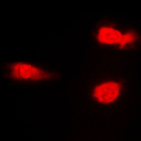
 IF (Immunofluorescence)
(Immunofluorescent analysis of RB1 (pS780) staining in HeLa cells. Formalin-fixed cells were permeabilized with 0.1% Triton X-100 in TBS for 5-10 minutes and blocked with 3% BSA-PBS for 30 minutes at room temperature. Cells were probed with the primary antibody in 3% BSA-PBS and incubated overnight at 4 °C in a humidified chamber. Cells were washed with PBST and incubated with a DyLight 594-conjugated secondary antibody (red) in PBS at room temperature in the dark.)
IF (Immunofluorescence)
(Immunofluorescent analysis of RB1 (pS780) staining in HeLa cells. Formalin-fixed cells were permeabilized with 0.1% Triton X-100 in TBS for 5-10 minutes and blocked with 3% BSA-PBS for 30 minutes at room temperature. Cells were probed with the primary antibody in 3% BSA-PBS and incubated overnight at 4 °C in a humidified chamber. Cells were washed with PBST and incubated with a DyLight 594-conjugated secondary antibody (red) in PBS at room temperature in the dark.)
RB1 (pS780), Polyclonal Antibody (Cat# AAA105310)
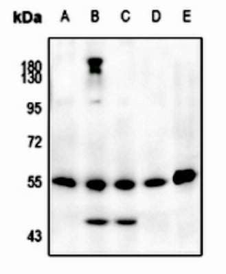
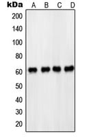
 WB (Western Blot)
(Western blot analysis of PKC delta (pY52) expression in MCF7 serum starvation-treated (A), HeLa PMA-treated (B), Jurkat (C), Raw264.7 serum starvation-treated (D), H9C2 serum starvation-treated (E) whole cell lysates.)
WB (Western Blot)
(Western blot analysis of PKC delta (pY52) expression in MCF7 serum starvation-treated (A), HeLa PMA-treated (B), Jurkat (C), Raw264.7 serum starvation-treated (D), H9C2 serum starvation-treated (E) whole cell lysates.)
PKC delta (pY52), Polyclonal Antibody (Cat# AAA104749)
 IHC (Immunohiostchemistry)
(Immunohistochemical analysis of NF-kappaB p65 (pS468) staining in human breast cancer formalin fixed paraffin embedded tissue section. The section was pre-treated using heat mediated antigen retrieval with sodium citrate buffer (pH 6.0). The section was then incubated with the antibody at room temperature and detected using an HRP conjugated compact polymer system. DAB was used as the chromogen. The section was then counterstained with haematoxylin and mounted with DPX.)
IHC (Immunohiostchemistry)
(Immunohistochemical analysis of NF-kappaB p65 (pS468) staining in human breast cancer formalin fixed paraffin embedded tissue section. The section was pre-treated using heat mediated antigen retrieval with sodium citrate buffer (pH 6.0). The section was then incubated with the antibody at room temperature and detected using an HRP conjugated compact polymer system. DAB was used as the chromogen. The section was then counterstained with haematoxylin and mounted with DPX.)
NF-kappaB p65 (pS468), Polyclonal Antibody (Cat# AAA104769)
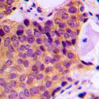
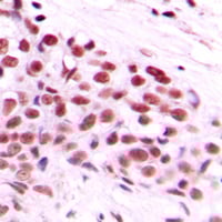 IHC (Immunohiostchemistry)
(Immunohistochemical analysis of c-Myc (pT58) staining in human breast cancer formalin fixed paraffin embedded tissue section. The section was pre-treated using heat mediated antigen retrieval with sodium citrate buffer (pH 6.0). The section was then incubated with the antibody at room temperature and detected using an HRP conjugated compact polymer system. DAB was used as the chromogen. The section was then counterstained with haematoxylin and mounted with DPX.)
IHC (Immunohiostchemistry)
(Immunohistochemical analysis of c-Myc (pT58) staining in human breast cancer formalin fixed paraffin embedded tissue section. The section was pre-treated using heat mediated antigen retrieval with sodium citrate buffer (pH 6.0). The section was then incubated with the antibody at room temperature and detected using an HRP conjugated compact polymer system. DAB was used as the chromogen. The section was then counterstained with haematoxylin and mounted with DPX.)
c-Myc (pT58), Polyclonal Antibody (Cat# AAA104779)
What Are Phospho Antibodies?
Protein phosphorylation is a process where a phosphate group is added to certain amino acid residues of a protein – usually serine (S), threonine (T), or tyrosine (Y) - by enzymes called kinases. This process is integral in controlling cellular signaling, cellular growth, and other biological functions.
Our catalog includes a wide range of phospho-specific antibodies that can accurately detect this important marker. They perform strongly in widely-used laboratory applications such as Western blot, flow cytometry, immunohistochemistry, and immunofluorescence microscopy. We value your trust in us and are committed to providing top-quality products and services. All of our antibodies are guaranteed to work for the applications and species indicated on our website & associated product pages.
What Are The Key Applications of Phospho Antibodies?
1. Western Blotting
One of the first steps a researcher can take in utilizing these phospho-specific antibodies, is to check if the antibody works using a technique referred to as “Western blot”. For those unfamiliar, Western Blot aids in showing whether the protein that the antibody recognizes is appearing at the correct/expected size. These phospho-specific antibodies should also be able to detect changes in the target protein’s phosphorylation (on/off state) when cells are stimulated in certain ways.
2. Staining of Fixed Cells (Immunocytochemistry)
Another routine use of these phospho-specific antibodies, is to test if the antibody is able to demonstrate similar performance when used on fixed cells (intact cells that have been preserved) as it did in the Western blot tests. It is an important aspect in many cases to confirm that the antibody works in actual intact cell samples. Ideally, the method used for cellular fixation should be the same as what is used in pathology labs (like using 10% formalin). To check if the antibody works well in tissue sections (FFPE), researchers will often test it on fixed cells that are processed similar to tissue samples.
3. Specificity Tests Using Peptides
In order to make sure that the antibody is only binding to the right target:
- Laboratory technicians will mix the antibody with phospho-peptides (short segments of the protein containing the phosphate group modification).
- If the antibody signal disappears, it is confirmation that it is binding to the correct phosphorylated location.
- A more robust test is to use both the phosphorylated and non-phosphorylated (dephosphorylated) versions of the protein. The antibody should react only with the phosphorylated one.
- Another method sometimes utilized is to treat the sample with an enzyme, such as alkaline phosphatase, that specifically removes phosphate groups. If the antibody signal disappears after this, it also confirms specificity.
4. Genetic Confirmation
As a final step, scientists can genetically manipulate the nucleotide sequence and alter the target protein by removing the exact site where phosphorylation happens. If the antibody no longer appears to detect the modified protein, it is strong evidence supporting the antibody being specific for that phosphorylated site.
Why Buy Phospho Antibodies Through Us?
- The production laboratory adheres to strict and consistent protocols prior to releasing any of these phospho-specific antibodies:
- Standard methods and proper controls in all tests to ensure high quality.
- These antibodies are tested and validated in different cell types and species.
- High quality control criterion to ensure each batch is consistent, so you will obtain reliable results every time.
FAQ
1. What Are Phospho-Specific Antibodies?
Phospho-specific antibodies are made to detect proteins only when they have a phosphate group linked to a specific amino acid residue. This empowers scientists understand if a protein is "turned on" or active, based on its phosphorylation state.
2. How to Detect Phosphorylated Proteins in a Western Blot?
To find out if a protein is phosphorylated using Western blot:
- Use a phospho-specific antibody that binds only to the phosphorylated form of the protein.
- You can also use a “regular” antibody for the same amino acid sequence of the protein that the phospho-specific antibody is binding to (but in this case, this antibody will not bind if there is a phosphate group present) in order to compare how much of it is phosphorylated versus how much is non-phosphorylated (or “total” protein, if the “normal” antibody’s epitopes are non-phospho-site-specific).
3. How to Choose the Best Antibody?
Here are some simple tips to help you pick the right antibody:
- Know your target
- Match your sample characteristics
- Confirm the intended use is appropriate
- Check “host” and “type”
- Check the “quality” of the presented data/images
- Appraise whether the available validation meets your needs