Filters
Clonality
Type
Reactivity
Gene Name
Isotype
Host
Application
Clone
1192 results for " A G G" - showing 400-450


 Application Data
(Staining of mouse peripheral blood platelets with HAMSTER ANTI MOUSE CD61:RPE)
Application Data
(Staining of mouse peripheral blood platelets with HAMSTER ANTI MOUSE CD61:RPE)
Integrin, beta3, Monoclonal Antibody (Cat# AAA14696)
Full Name
Integrin, beta3 (ITGB3, CD61, GP3A, GPIIIa, HPA-1, NAIT, Platelet Fibrinogen Receptor beta Subunit, Platelet Glycoprotein IIIa, Platelet Membrane Glycoprotein IIIa) (PE)
Reactivity
Mouse, Rat
Applications
Flow Cytometry
Purity
Affinity Purified
Purified by Protein G affinity chromatography.
Purified by Protein G affinity chromatography.
Pricing


 Application Data
(Staining of NALM6 cells with MOUSE ANTI HUMAN CD10:ALEXA 488)
Application Data
(Staining of NALM6 cells with MOUSE ANTI HUMAN CD10:ALEXA 488)
CD10, Monoclonal Antibody (Cat# AAA26765)
Full Name
CD10 (CALLA, CD 10, Common Acute Lymphocytic Leukemia Antigen, Enkephalinase, gp100, Membrane Metalloendopeptidase, MME, Neprilysin, Neutral Endopeptidase, Pmel17) (PE)
Gene Names
MME; NEP; SFE; CD10; CALLA
Reactivity
Human
Applications
Flow Cytometry, Immunohistochemistry, Immunoprecipitation, Western Blot
Purity
Purified by protein G affinity chromatography from tissue culture supernatant.
Pricing


 Application Data
(Figure A. Alexa Fluor 488 conjugated Rat anti Mouse CD4 . Figure B. Alexa Fluor 488 conjugated Rat anti Mouse CD4 and StarBright Violet 610 conjugated Rat anti Mouse CD3 . All experiments performed on red blood lys)
Application Data
(Figure A. Alexa Fluor 488 conjugated Rat anti Mouse CD4 . Figure B. Alexa Fluor 488 conjugated Rat anti Mouse CD4 and StarBright Violet 610 conjugated Rat anti Mouse CD3 . All experiments performed on red blood lys)
CD4, Monoclonal Antibody (Cat# AAA12287)
Full Name
Rat anti Mouse CD4:Amethyst Orange
Gene Names
Cd4; L3T4; Ly-4
Reactivity
Mouse
Applications
ELISA
Purity
Purified IgG prepared by affinity chromatography on Protein G from tissue culture supernatant
Pricing


 IF (Immunofluorescence)
(Immunofluorescence of TET3 in human brain tissue with TET3 antibody at 20 μg/mL.)
IF (Immunofluorescence)
(Immunofluorescence of TET3 in human brain tissue with TET3 antibody at 20 μg/mL.)
TET3, Polyclonal Antibody (Cat# AAA10939)
Full Name
TET3 Antibody
Gene Names
TET3; hCG_40738
Reactivity
Human, Mouse, Rat
Applications
Immunofluorescence, Immunohistochemistry, Western Blot
Purity
TET3 Antibody is affinity chromatography purified via peptide column.
Pricing

 IF (Immunofluorescence)
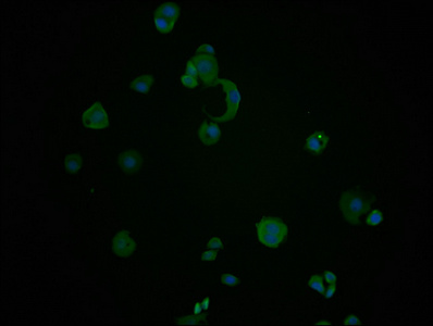
(ICC/IF analysis of ISG15 in HeLa cells line, stained with DAPI (Blue) for nucleus staining and monoclonal anti-human ISG15 antibody (1:100) with goat anti-mouse IgG-Alexa fluor 488 conjugate (Green).)
IF (Immunofluorescence)
(ICC/IF analysis of ISG15 in HeLa cells line, stained with DAPI (Blue) for nucleus staining and monoclonal anti-human ISG15 antibody (1:100) with goat anti-mouse IgG-Alexa fluor 488 conjugate (Green).)
ISG15, Monoclonal Antibody (Cat# AAA11725)
Full Name
ISG15 antibody
Gene Names
ISG15; G1P2; IP17; UCRP; IFI15; IMD38; hUCRP
Reactivity
Human
Applications
Western Blot, Immunocytochemistry, Immunofluorescence, Flow Cytometry
Purity
By protein-A affinity chromatography
Pricing


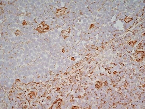
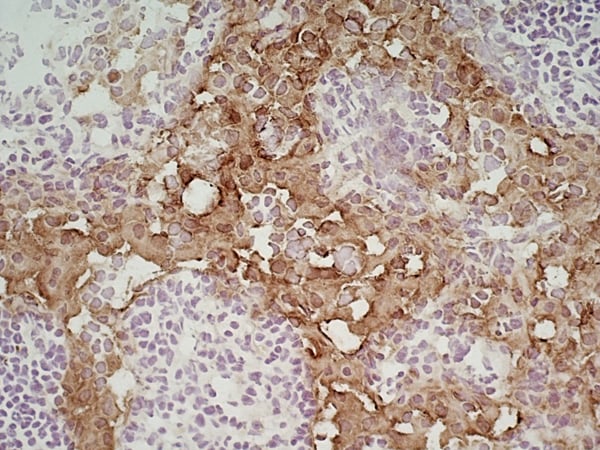
 IHC (Immunohistochemistry)
(Immunohistochemistry analysis of human spleen tissue stained with KDEL, mAb (10C3) at 10µg/ml.)
IHC (Immunohistochemistry)
(Immunohistochemistry analysis of human spleen tissue stained with KDEL, mAb (10C3) at 10µg/ml.)
KDEL, Monoclonal Antibody (Cat# AAA14517)
Full Name
KDEL monoclonal antibody (10C3)
Gene Names
Hspa5; BIP; GRP78
Reactivity
Avian, Insect, Mammalian, Plant, Yeast
Applications
Flow Cytometry, Immunocytochemistry, Immunofluorescence, Immunohistochemistry, Immunoprecipitation, Western Blot, Immunoelectron Microscopy
Purity
Protein G affinity purified.
Pricing


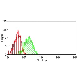
 FCM (Flow Cytometry)
(Overlay histogram showing MCF-7 cells stained with (red line). The cells were fixed with 70% Ethylalcohol (18h) and then incubated in 10% normal goat serum to block non-specific protein-protein interactions followed by the primary antibody at 1/200 for 1 h at 4 degree C. The secondary antibody used was FITC goat anti-mouse IgG(H+L) at 1/100 dilution for 30min at 4 degree C. Isotype control antibody (green line) was mouse IgG1 used under the same conditions. Acquisition of >10,000 events was performed.)
FCM (Flow Cytometry)
(Overlay histogram showing MCF-7 cells stained with (red line). The cells were fixed with 70% Ethylalcohol (18h) and then incubated in 10% normal goat serum to block non-specific protein-protein interactions followed by the primary antibody at 1/200 for 1 h at 4 degree C. The secondary antibody used was FITC goat anti-mouse IgG(H+L) at 1/100 dilution for 30min at 4 degree C. Isotype control antibody (green line) was mouse IgG1 used under the same conditions. Acquisition of >10,000 events was performed.)
HSPA8, Monoclonal Antibody (Cat# AAA27046)
Full Name
HSPA8 Monoclonal Antibody
Gene Names
HSPA8; LAP1; HSC54; HSC70; HSC71; HSP71; HSP73; NIP71; HSPA10
Reactivity
Human
Applications
Western Blot, Immunohistochemistry, Immunofluorescence, Flow Cytometry
Purity
>95%, Protein G purified
Pricing


 FCM (Flow Cytometry)
(Figure 9. Flow Cytometry analysis of SiHa cells using anti-DCTN1/p150-glued antibody (AAA19253).Overlay histogram showing SiHa cells stained with AAA19253 (Blue line). The cells were blocked with 10% normal goat serum. And then incubated with rabbit anti-DCTN1/p150-glued Antibody (AAA19253, 1μg/1x106 cells) for 30 min at 20 degree C. DyLight®488 conjugated goat anti-rabbit IgG (5-10μg/1x106 cells) was used as secondary antibody for 30 minutes at 20 degree C. Isotype control antibody (Green line) was rabbit IgG (1μg/1x106) used under the same conditions. Unlabelled sample (Red line) was also used as a control.)
FCM (Flow Cytometry)
(Figure 9. Flow Cytometry analysis of SiHa cells using anti-DCTN1/p150-glued antibody (AAA19253).Overlay histogram showing SiHa cells stained with AAA19253 (Blue line). The cells were blocked with 10% normal goat serum. And then incubated with rabbit anti-DCTN1/p150-glued Antibody (AAA19253, 1μg/1x106 cells) for 30 min at 20 degree C. DyLight®488 conjugated goat anti-rabbit IgG (5-10μg/1x106 cells) was used as secondary antibody for 30 minutes at 20 degree C. Isotype control antibody (Green line) was rabbit IgG (1μg/1x106) used under the same conditions. Unlabelled sample (Red line) was also used as a control.)
DCTN1/p150-glued, Polyclonal Antibody (Cat# AAA19253)
Full Name
Anti-DCTN1/p150-glued Antibody
Gene Names
DCTN1; P135; DP-150; DAP-150
Reactivity
Human, Mouse, Rat
Applications
Western Blot, Immunohistochemistry, Immunocytochemistry, Immunofluorescence, Flow Cytometry, Direct ELISA
Purity
Immunogen affinity purified.
Pricing


 Application Data
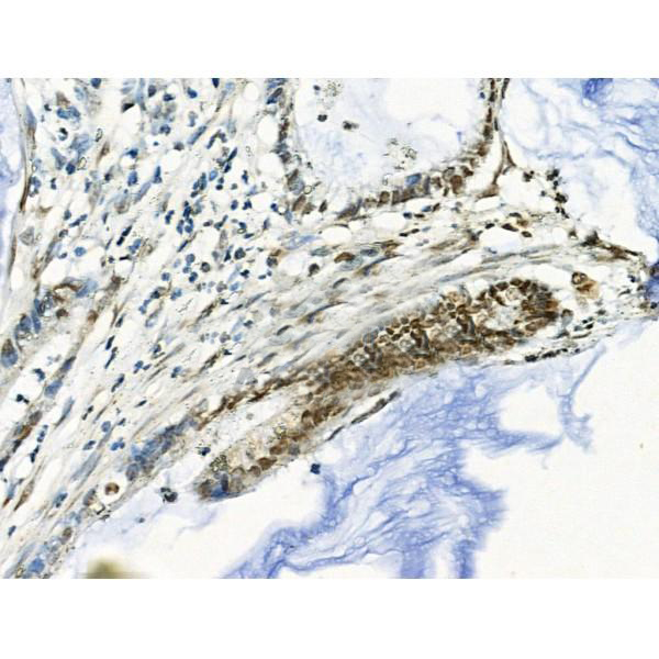
(Published customer image: Leukocyte infiltration in COX-2-M/-M and COX-2+/+ mice. MPO enzymatic activity (panel A) was statistically similar in COX-2-M/-M and COX-2+/+ livers at 6 h and 24 h post-IRI. Ly-6G+ neutrophil (panel B) and granulocyte (panel C) infiltration were also comparable in COX-2-M/-M and COX-2+/+ livers after IRI. Mac-1+ (panel D) and CD68 (panel E) infiltrating macrophages were significantly reduced in COX-2-M/-M livers at 24 h post-reperfusion, but were statistically indistinguishable in COX-2-M/-M and COX-2+/+ livers at 6 h after IRI. No statistical differences in MMP-9 expression (panel F) could be demonstrated in livers of COX-2-M/-M and COX-2+/+ mice post-IRI. Representative immunostaining (panel G) of infiltrating Ly-6G+ (a,b,e,f) and Mac-1+ (c,d,g,h) leukocytes in livers of COX-2+/+ (a,c,e,g) and COX-2-M/-M (b,d,f,h) mice at 6 h (a to d) and 24 h (e to h) post IRI; (n = 5 -6/group; * indicates p)
Application Data
(Published customer image: Leukocyte infiltration in COX-2-M/-M and COX-2+/+ mice. MPO enzymatic activity (panel A) was statistically similar in COX-2-M/-M and COX-2+/+ livers at 6 h and 24 h post-IRI. Ly-6G+ neutrophil (panel B) and granulocyte (panel C) infiltration were also comparable in COX-2-M/-M and COX-2+/+ livers after IRI. Mac-1+ (panel D) and CD68 (panel E) infiltrating macrophages were significantly reduced in COX-2-M/-M livers at 24 h post-reperfusion, but were statistically indistinguishable in COX-2-M/-M and COX-2+/+ livers at 6 h after IRI. No statistical differences in MMP-9 expression (panel F) could be demonstrated in livers of COX-2-M/-M and COX-2+/+ mice post-IRI. Representative immunostaining (panel G) of infiltrating Ly-6G+ (a,b,e,f) and Mac-1+ (c,d,g,h) leukocytes in livers of COX-2+/+ (a,c,e,g) and COX-2-M/-M (b,d,f,h) mice at 6 h (a to d) and 24 h (e to h) post IRI; (n = 5 -6/group; * indicates p)
CD68, Monoclonal Antibody (Cat# AAA12107)
Full Name
RAT ANTI MOUSE CD68
Gene Names
Cd68; Lamp4; gp110; Scard1
Applications
Immunohistochemistry, Flow Cytometry, Immunofluorescence, Immunoprecipitation, Immunohistochemistry, Western Blot
Purity
Purified
Purified IgG - liquid
Purified IgG - liquid
Pricing


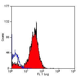
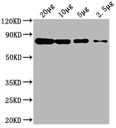
 WB (Western Blot)
(Western blot analysis of MUTYH over-expressed 293 cell line, cotransfected with MUTYH Validated Chimera RNAi (Lane 2) or non-transfected control (Lane 1). Blot probed with MUTYH monoclonal antibody GAPDH (36.1kD) used as specificity and loading control.)
WB (Western Blot)
(Western blot analysis of MUTYH over-expressed 293 cell line, cotransfected with MUTYH Validated Chimera RNAi (Lane 2) or non-transfected control (Lane 1). Blot probed with MUTYH monoclonal antibody GAPDH (36.1kD) used as specificity and loading control.)
MUTYH, Monoclonal Antibody (Cat# AAA25759)
Full Name
MUTYH (A/G-specific Adenine DNA Glycosylase, MutY Homolog, hMYH, MYH) (PE)
Gene Names
MUTYH; MYH
Reactivity
Human
Applications
Immunoprecipitation, Western Blot
Purity
Purified by Protein A Affinity Chromatography.
Pricing


 IF (Immunofluorescence)
(Immunofluorescence of PPARGC1A in rat heart tissue with PPARGC1A antibody at 20 μg/mL.)
IF (Immunofluorescence)
(Immunofluorescence of PPARGC1A in rat heart tissue with PPARGC1A antibody at 20 μg/mL.)
PPARGC1A, Polyclonal Antibody (Cat# AAA10967)
Full Name
PPARGC1A Antibody
Gene Names
PPARGC1A; LEM6; PGC1; PGC1A; PGC-1v; PPARGC1; PGC-1(alpha)
Reactivity
Human, Mouse, Rat
Applications
Western Blot, Immunohistochemistry, Immunofluorescence
Purity
PPARGC1A antibody is affinity chromatography purified via peptide column.
Pricing


 Application Data
(Staining of mouse peritoneal macrophages with Rat anti Mouse CD204: Alexa Fluor 488 (AAA12072A488))
Application Data
(Staining of mouse peritoneal macrophages with Rat anti Mouse CD204: Alexa Fluor 488 (AAA12072A488))
CD204, Monoclonal Antibody (Cat# AAA12072)
Full Name
RAT ANTI MOUSE CD204
Gene Names
Msr1; MSR; Scvr; MRS-A; MSR-A; SR-AI; SR-AII; Scara1
Reactivity
Channel catfish, Pig
Applications
Immunohistochemistry, Flow Cytometry, Immunoprecipitation, Western Blot
Pricing


 IP (Immunoprecipitation)
(LAMP1 was immunoprecipitated using:Lane A:0.5 mg Jurkat Whole Cell Lysate1 uL anti-LAMP1 rabbit monoclonal antibody and 15 ul of 50 % Protein G agarose.Primary antibody:Anti-LAMP1 rabbit monoclonal antibody,at 1:500 dilution Secondary antibody:Dylight 800-labeled antibody to rabbit IgG (H+L), at 1:5000 dilution Developed using the odssey technique.Performed under reducing conditions.Predicted band size: 45 kDaObserved band size: 113 kDa)
IP (Immunoprecipitation)
(LAMP1 was immunoprecipitated using:Lane A:0.5 mg Jurkat Whole Cell Lysate1 uL anti-LAMP1 rabbit monoclonal antibody and 15 ul of 50 % Protein G agarose.Primary antibody:Anti-LAMP1 rabbit monoclonal antibody,at 1:500 dilution Secondary antibody:Dylight 800-labeled antibody to rabbit IgG (H+L), at 1:5000 dilution Developed using the odssey technique.Performed under reducing conditions.Predicted band size: 45 kDaObserved band size: 113 kDa)
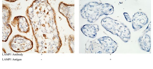
LAMP1, Monoclonal Antibody (Cat# AAA27745)
Full Name
Recombinant Anti-LAMP1 Antibody, Rabbit Monoclonal
Gene Names
LAMP1; LAMPA; CD107a; LGP120
Reactivity
Human
Applications
Western Blot, Immunohistochemistry, Flow Cytometry, Immunocytochemistry, Immunofluorescence, Immunoprecipitation
Purity
Protein A
Pricing


 IP (Immunoprecipitation)
(Immunoprecipitating ENO1 in HepG2 whole cell lysate Lane 1: Mouse control IgG (1ug) instead of in HepG2 whole cell lysate. For western blotting, a HRP-conjugated Protein G antibody was used as the secondary antibody (1/5000) Lane 2: (1ul) + HepG2 whole cell lysate (500ug) Lane 3: HepG2 whole cell lysate (10ug))
IP (Immunoprecipitation)
(Immunoprecipitating ENO1 in HepG2 whole cell lysate Lane 1: Mouse control IgG (1ug) instead of in HepG2 whole cell lysate. For western blotting, a HRP-conjugated Protein G antibody was used as the secondary antibody (1/5000) Lane 2: (1ul) + HepG2 whole cell lysate (500ug) Lane 3: HepG2 whole cell lysate (10ug))
ENO1, Monoclonal Antibody (Cat# AAA27044)
Full Name
ENO1 Monoclonal Antibody
Gene Names
ENO1; NNE; PPH; MPB1; ENO1L1
Reactivity
Human, Mouse, Rat, Rabbit
Applications
Western Blot, Immunofluorescence, Flow Cytometry, Immunoprecipitation
Purity
>95%, Protein G purified
Pricing


 FCM (Flow Cytometry)
(Figure 8. Flow Cytometry analysis of U251 cells using anti-GABARAP antibody (AAA19250).Overlay histogram showing U251 cells stained with AAA19250 (Blue line). The cells were blocked with 10% normal goat serum. And then incubated with rabbit anti-GABARAP Antibody (AAA19250, 1μg/1x106 cells) for 30 min at 20 degree C. DyLight®488 conjugated goat anti-rabbit IgG (5-10μg/1x106 cells) was used as secondary antibody for 30 minutes at 20 degree C. Isotype control antibody (Green line) was rabbit IgG (1μg/1x106) used under the same conditions. Unlabelled sample (Red line) was also used as a control.)
FCM (Flow Cytometry)
(Figure 8. Flow Cytometry analysis of U251 cells using anti-GABARAP antibody (AAA19250).Overlay histogram showing U251 cells stained with AAA19250 (Blue line). The cells were blocked with 10% normal goat serum. And then incubated with rabbit anti-GABARAP Antibody (AAA19250, 1μg/1x106 cells) for 30 min at 20 degree C. DyLight®488 conjugated goat anti-rabbit IgG (5-10μg/1x106 cells) was used as secondary antibody for 30 minutes at 20 degree C. Isotype control antibody (Green line) was rabbit IgG (1μg/1x106) used under the same conditions. Unlabelled sample (Red line) was also used as a control.)
GABARAP, Polyclonal Antibody (Cat# AAA19250)
Full Name
Anti-GABARAP Antibody
Gene Names
GABARAP; MM46; ATG8A; GABARAP-a
Reactivity
Human, Mouse, Rat
Applications
Western Blot, Immunohistochemistry, Flow Cytometry
Purity
Immunogen affinity purified.
Pricing


 Application Data
(Published customer image: Increased accumulation of repair-associated macrophages surrounding collaterals in ischemic hind limbs is PAR2-dependent. (A) Stainings of CD206-positive macrophages (green) and SMA-positive vessels (red) in non-ischemic (control) and ischemic (ligated) hind limbs of WT, PAR1-/- and PAR2-/- mice are shown. Nuclei were visualized with DAPI (blue). Arrows indicate single macrophages in the non-ischemic adductor. Quantification of the average number of repair-associated macrophages per vessel is indicated on the right. (B) Correlation between the number of CD206-positive macrophages in the ischemic tissues and the expression of CD11b and (C) CD115 on monocytes. ** p)
Application Data
(Published customer image: Increased accumulation of repair-associated macrophages surrounding collaterals in ischemic hind limbs is PAR2-dependent. (A) Stainings of CD206-positive macrophages (green) and SMA-positive vessels (red) in non-ischemic (control) and ischemic (ligated) hind limbs of WT, PAR1-/- and PAR2-/- mice are shown. Nuclei were visualized with DAPI (blue). Arrows indicate single macrophages in the non-ischemic adductor. Quantification of the average number of repair-associated macrophages per vessel is indicated on the right. (B) Correlation between the number of CD206-positive macrophages in the ischemic tissues and the expression of CD11b and (C) CD115 on monocytes. ** p)
CD206, Monoclonal Antibody (Cat# AAA12124)
Full Name
RAT ANTI MOUSE CD206:RPE
Gene Names
Mrc1; MR; CD206; AW259686
Applications
Flow Cytometry
Pricing
 WB (Western Blot)
(Sample: Recombinant IL8, Rabbit.)
WB (Western Blot)
(Sample: Recombinant IL8, Rabbit.)
Interleukin 8, Monoclonal Antibody (Cat# AAA20823)
Full Name
Monoclonal Antibody to Interleukin 8 (IL8)
Gene Names
CXCL8; IL8
Reactivity
Rabbit, Rat
Applications
Westen Blot, Immunohistochemistry, Immunocytochemistry, Immunoprecipitation
Purity
Protein A + Protein G affinity chromatography
Pricing


 Application Data
(C:FGFR2/isolectinB4 (C) and FGFR1/isolectinB4 (D) staining of apparent mesenchymal cells and the subpopulation of endothelial cells. Virtually all other dispersed apparent mesenchymal cells express FGFR1 and FGFR2 (merged image in E). F: FGFR2 (F) and FGFR1 (G) staining in clustered cells of epithelial origin (inferred by morphology here) demonstrating that epithelial cells express both FGFR1 and FGFR2 (merged image with DAPI staining in H).)
Application Data
(C:FGFR2/isolectinB4 (C) and FGFR1/isolectinB4 (D) staining of apparent mesenchymal cells and the subpopulation of endothelial cells. Virtually all other dispersed apparent mesenchymal cells express FGFR1 and FGFR2 (merged image in E). F: FGFR2 (F) and FGFR1 (G) staining in clustered cells of epithelial origin (inferred by morphology here) demonstrating that epithelial cells express both FGFR1 and FGFR2 (merged image with DAPI staining in H).)
FGFR2, Polyclonal Antibody (Cat# AAA26853)
Full Name
FGFR2, NT (FGFR2, BEK, KGFR, KSAM, Fibroblast growth factor receptor 2, K-sam, Keratinocyte growth factor receptor, CD332) (Biotin)
Gene Names
FGFR2; BEK; JWS; BBDS; CEK3; CFD1; ECT1; KGFR; TK14; TK25; BFR-1; CD332; K-SAM
Reactivity
Human, Monkey, Mouse, Rat
Applications
FC/FACS, EIA, IF, IHC, WB
Purity
Purified by Protein G Affinity Chromatography.
Pricing


 Application Data
(Staining of Rat peripheral blood lymphocytes with Mouse anti Rat CD49d:RPE (MCA2872PE))
Application Data
(Staining of Rat peripheral blood lymphocytes with Mouse anti Rat CD49d:RPE (MCA2872PE))
CD49d, Monoclonal Antibody (Cat# AAA26798)
Full Name
CD49d (Antigen CD49d, CD49d Antigen, CDw49d, Alpha 4 Subunit of VLA-4 Receptor, Integrin alpha IV, Integrin alpha 4, IA4, ITGA4, LPAM23, MGC90518, Very Late Activation Protein 4 Receptor Alpha 4 Subunit, VLA4, VLA-4) (MaxLight 490)
Reactivity
Rat
Applications
Flow Cytometry, Immunoprecipitation
Purity
Purified by Protein G Affinity Chromatography
Pricing


 FCM (Flow Cytometry)
(Overlay histogram showing HepG2 cells stained with AAA28062 (red line) at 1:100. The cells were fixed in 4% formaldehyde and permeated by 0.2% TritonX-100. Then 10% normal goat serum was Incubated to block non-specific protein-protein interactions followed by the antibody (1ug/1*106cells) for 1 h at 4 degree C. The secondary antibody used was FITC-conjugated Goat Anti-Mouse IgG(H+L) at 1/100 dilution for 30min at 4 degree C. Isotype control antibody (green line) was mouse IgG2b (1ug/1*106cells) used under the same conditions. Acquisition of >10,000 events was performed.)
FCM (Flow Cytometry)
(Overlay histogram showing HepG2 cells stained with AAA28062 (red line) at 1:100. The cells were fixed in 4% formaldehyde and permeated by 0.2% TritonX-100. Then 10% normal goat serum was Incubated to block non-specific protein-protein interactions followed by the antibody (1ug/1*106cells) for 1 h at 4 degree C. The secondary antibody used was FITC-conjugated Goat Anti-Mouse IgG(H+L) at 1/100 dilution for 30min at 4 degree C. Isotype control antibody (green line) was mouse IgG2b (1ug/1*106cells) used under the same conditions. Acquisition of >10,000 events was performed.)
CD63, Monoclonal Antibody (Cat# AAA28062)
Full Name
CD63 Monoclonal Antibody
Gene Names
Cd63; ME491; C75951; Tspan30
Reactivity
Human, Rabbit
Applications
Western Blot, Immunohistochemistry, Immunofluorescence, Flow Cytometry
Purity
>95%, Protein G purified
Pricing


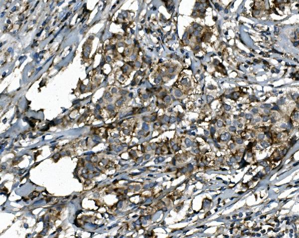
 Application Data
(Published customer image:Phycoerythrin conjugated Mouse anti Human CD83 antibody, clone HB15e used for the evaluation of CD83 expression on monocyte derived dendritic cells by flow cytometry.Phenotypic characterization of immunogenic and tolerogenic moDC populations by flow cytometry. Monocytes were negatively selected from PBMC using magnetic beads. Immature moDC were generated with IL-4 and GM-CSF for 6 days. 15d-PGJ2 (PGJ2 DC) and dexamethasone plus 1alpha,25-dihydroxyvitamin were added to generate tolerogenic moDC, respectively (PGJ2 DC and Dex/VD3 DC). To generate immunogenic moDC, immature moDC were stimulated for 24 h with LPS, polyI:C and a cytokine cocktail containing TNF-alpha, IL-1beta, IL-6 and PGE2, respectively. The phenotypes of the cells were analyzed by flow cytometry. Live cells were gated according to FSC/SSC. One representative experiment out of three is shown.From: Sprater F, Hovden A-O, Appel S (2012)Expression of ESE-3 Isoforms in Immunogenic and Tolerogenic Human Monocyte-Derived Dendritic Cells.PLoS ONE 7(11): e49577.)
Application Data
(Published customer image:Phycoerythrin conjugated Mouse anti Human CD83 antibody, clone HB15e used for the evaluation of CD83 expression on monocyte derived dendritic cells by flow cytometry.Phenotypic characterization of immunogenic and tolerogenic moDC populations by flow cytometry. Monocytes were negatively selected from PBMC using magnetic beads. Immature moDC were generated with IL-4 and GM-CSF for 6 days. 15d-PGJ2 (PGJ2 DC) and dexamethasone plus 1alpha,25-dihydroxyvitamin were added to generate tolerogenic moDC, respectively (PGJ2 DC and Dex/VD3 DC). To generate immunogenic moDC, immature moDC were stimulated for 24 h with LPS, polyI:C and a cytokine cocktail containing TNF-alpha, IL-1beta, IL-6 and PGE2, respectively. The phenotypes of the cells were analyzed by flow cytometry. Live cells were gated according to FSC/SSC. One representative experiment out of three is shown.From: Sprater F, Hovden A-O, Appel S (2012)Expression of ESE-3 Isoforms in Immunogenic and Tolerogenic Human Monocyte-Derived Dendritic Cells.PLoS ONE 7(11): e49577.)
CD83, Monoclonal Antibody (Cat# AAA12280)
Full Name
MOUSE ANTI HUMAN CD83
Reactivity
Human
Applications
Flow Cytometry, Immunohistochemistry, Immunohistochemistry, Immunoprecipitation, Immunofluorescence
Pricing


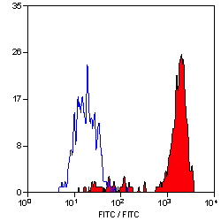
 FCM (Flow Cytometry)
(Flow cytometry analysis of CD80 overexpressing HEK293 cells using CD80 antibody and control mouse IgG antibody at 10 μg/ml. Blue: Untransfected HEK293 cells. Yellow: CD80 overexpressing HEK293 cells.)
FCM (Flow Cytometry)
(Flow cytometry analysis of CD80 overexpressing HEK293 cells using CD80 antibody and control mouse IgG antibody at 10 μg/ml. Blue: Untransfected HEK293 cells. Yellow: CD80 overexpressing HEK293 cells.)
CD80, Monoclonal Antibody (Cat# AAA10997)
Full Name
CD80 Antibody [10A1]
Gene Names
CD80; B7; BB1; B7-1; B7.1; LAB7; CD28LG; CD28LG1
Reactivity
Human
Applications
Immunohistochemistry, Immunocytochemistry, Immunofluorescence, Flow Cytometry
Purity
Protein A purified
Pricing


 ELISA
(A sandwich ELISA was performed using the anti-LAG3 mAbs as the capture antibodies for the LAG3 extracellular domain antigen with biotin-labeled Risk-Free anti-LAG3 mAbs as the detection antibodies.)
ELISA
(A sandwich ELISA was performed using the anti-LAG3 mAbs as the capture antibodies for the LAG3 extracellular domain antigen with biotin-labeled Risk-Free anti-LAG3 mAbs as the detection antibodies.)
LAG3, Monoclonal Antibody (Cat# AAA11017)
Full Name
LAG3 Antibody [9F9]
Gene Names
LAG3; CD223
Reactivity
Human
Applications
Immunohistochemistry, Immunocytochemistry, Immunofluorescence, Flow Cytometry
Purity
Protein A purified
Pricing


 ELISA
(Titration curve analysis of VISTA antibody to detect recombinant VISTA in ELISA at decreasing concentrations.)
ELISA
(Titration curve analysis of VISTA antibody to detect recombinant VISTA in ELISA at decreasing concentrations.)
VISTA, Monoclonal Antibody (Cat# AAA11013)
Full Name
VISTA Antibody [6D2]
Gene Names
VSIR; B7H5; GI24; B7-H5; PD-1H; SISP1; VISTA; PP2135; C10orf54; DD1alpha
Reactivity
Human
Applications
Immunohistochemistry, Immunocytochemistry, Immunofluorescence, Flow Cytometry
Purity
Protein A purified
Pricing


 Application Data
(Staining of mouse spleen with Rat anti Mouse CD4:RPE)
Application Data
(Staining of mouse spleen with Rat anti Mouse CD4:RPE)
CD4, Monoclonal Antibody (Cat# AAA12233)
Full Name
RAT ANTI MOUSE CD4
Gene Names
Cd4; L3T4; Ly-4
Applications
Immunohistochemistry, Flow Cytometry, Immunofluorescence, Immunoprecipitation, Western Blot
Pricing


 Application Data
(Staining of mouse thymus with RAT ANTI MOUSE CD150: RPE)
Application Data
(Staining of mouse thymus with RAT ANTI MOUSE CD150: RPE)
CD150, Monoclonal Antibody (Cat# AAA26776)
Full Name
CD150 (CD150 Antigen, Cdw150, 4933415F16, Estm51, Ipo 3, Ipo-3, OTTHUMP00000060252, Signaling Lymphocytic Activation Molecule, SLAM, Signaling Lymphocytic Activation Molecule Family Member 1, SLAMF1 Protein) (MaxLight 405)
Reactivity
Mouse
Applications
Flow Cytometry
Purity
Purified by Protein G Affinity Chromatography
Pricing


 Application Data
CD274, Monoclonal Antibody (Cat# AAA26754)
Full Name
CD274 (CD274 Antigen, B7 Homolog 1, B7H1, B7-H1, B7-H, MGC142294, MGC142296, OTTHUMP00000021029, Programmed Cell Death 1 Ligand 1, PDCD1 Ligand 1, PDCD1L1, PDCD1LG1, Programmed Death Ligand 1, PDL1, PD-L1, RGD1566211) (HRP)
Gene Names
CD274; B7-H; B7H1; PDL1; PD-L1; PDCD1L1; PDCD1LG1
Reactivity
Human
Applications
ELISA
Purity
Purified by protein G affinity chromatography from tissue culture supernatant.
Pricing


 Application Data
(Staining of mouse peritoneal macrophages with RAT ANTI MOUSE F4/80 ANTIGEN:FITC)
Application Data
(Staining of mouse peritoneal macrophages with RAT ANTI MOUSE F4/80 ANTIGEN:FITC)
EMR1, Monoclonal Antibody (Cat# AAA26772)
Full Name
EMR1 (EGF-like Module Containing Mucin-like Hormone Receptor-like 1, EMR1 Hormone Receptor, Cell Surface Glycoprotein EMR1, Cell Surface Glycoprotein F4/80, DD7A5-7, EGF-TM7, F4/80, Gpf480, Lymphocyte Antigen 71, Ly71, TM7LN3) (PE)
Reactivity
Mouse
Applications
Flow Cytometry, Immunohistochemistry, Immunoprecipitation, Radioimmunoassay, Western Blot
Purity
Purified by protein G affinity chromatography from tissue culture supernatant.
Pricing


 Application Data
(Staining of mouse peritoneal macrophages with RAT ANTI MOUSE F4/80 ANTIGEN:FITC)
Application Data
(Staining of mouse peritoneal macrophages with RAT ANTI MOUSE F4/80 ANTIGEN:FITC)
EMR1, Monoclonal Antibody (Cat# AAA26749)
Full Name
EMR1 (EGF-like Module Containing Mucin-like Hormone Receptor-like 1, EMR1 Hormone Receptor, Cell Surface Glycoprotein EMR1, Cell Surface Glycoprotein F4/80, DD7A5-7, EGF-TM7, F4/80, Gpf480, Lymphocyte Antigen 71, Ly71, TM7LN3) (FITC)
Reactivity
Mouse
Applications
Flow Cytometry, Immunohistochemistry, Immunoprecipitation, Radioimmunoassay, Western Blot
Purity
Purified by protein G affinity chromatography from tissue culture supernatant.
Pricing


 Application Data
(Published customer image: Leukocyte infiltration in COX-2-M/-M and COX-2+/+ mice. MPO enzymatic activity (panel A) was statistically similar in COX-2-M/-M and COX-2+/+ livers at 6 h and 24 h post-IRI. Ly-6G+ neutrophil (panel B) and granulocyte (panel C) infiltration were also comparable in COX-2-M/-M and COX-2+/+ livers after IRI. Mac-1+ (panel D) and CD68 (panel E) infiltrating macrophages were significantly reduced in COX-2-M/-M livers at 24 h post-reperfusion, but were statistically indistinguishable in COX-2-M/-M and COX-2+/+ livers at 6 h after IRI. No statistical differences in MMP-9 expression (panel F) could be demonstrated in livers of COX-2-M/-M and COX-2+/+ mice post-IRI. Representative immunostaining (panel G) of infiltrating Ly-6G+ (a,b,e,f) and Mac-1+ (c,d,g,h) leukocytes in livers of COX-2+/+ (a,c,e,g) and COX-2-M/-M (b,d,f,h) mice at 6 h (a to d) and 24 h (e to h) post IRI; (n = 5 -6/group; * indicates p)
Application Data
(Published customer image: Leukocyte infiltration in COX-2-M/-M and COX-2+/+ mice. MPO enzymatic activity (panel A) was statistically similar in COX-2-M/-M and COX-2+/+ livers at 6 h and 24 h post-IRI. Ly-6G+ neutrophil (panel B) and granulocyte (panel C) infiltration were also comparable in COX-2-M/-M and COX-2+/+ livers after IRI. Mac-1+ (panel D) and CD68 (panel E) infiltrating macrophages were significantly reduced in COX-2-M/-M livers at 24 h post-reperfusion, but were statistically indistinguishable in COX-2-M/-M and COX-2+/+ livers at 6 h after IRI. No statistical differences in MMP-9 expression (panel F) could be demonstrated in livers of COX-2-M/-M and COX-2+/+ mice post-IRI. Representative immunostaining (panel G) of infiltrating Ly-6G+ (a,b,e,f) and Mac-1+ (c,d,g,h) leukocytes in livers of COX-2+/+ (a,c,e,g) and COX-2-M/-M (b,d,f,h) mice at 6 h (a to d) and 24 h (e to h) post IRI; (n = 5 -6/group; * indicates p)
CD68, Monoclonal Antibody (Cat# AAA12108)
Full Name
RAT ANTI MOUSE CD68:RPE
Gene Names
Cd68; Lamp4; gp110; Scard1
Applications
Flow Cytometry
Pricing


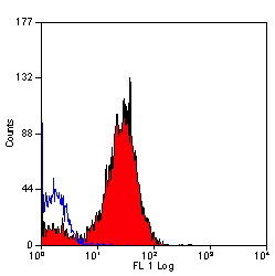
 FCM (Flow Cytometry)
(Flow cytometric analysis of Hela cells with Histone H4 (acetyl K16) antibody at 1/100 dilution (red) compared with an unlabelled control (cells without incubation with primary antibody; black). Alexa Fluor 488-conjugated goat anti-rabbit IgG was used as the secondary antibody.)
FCM (Flow Cytometry)
(Flow cytometric analysis of Hela cells with Histone H4 (acetyl K16) antibody at 1/100 dilution (red) compared with an unlabelled control (cells without incubation with primary antibody; black). Alexa Fluor 488-conjugated goat anti-rabbit IgG was used as the secondary antibody.)
Histone H4, Monoclonal Antibody (Cat# AAA30539)
Full Name
Histone H4 (Acetyl K16) Antibody
Gene Names
HIST2H4B; H4/o
Reactivity
Human, Mouse, Rat
Applications
Western Blot, Immunocytochemistry, Immunofluorescence, Immunohistochemistry, Flow Cytometry
Purity
ProA affinity purified
Pricing
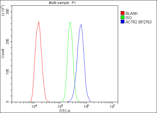


 FCM (Flow Cytometry)
(Figure 6. Flow Cytometry analysis of C6 cells using anti-Arp2/ACTR2 antibody (AAA19284).Overlay histogram showing C6 cells stained with AAA19284 (Blue line). The cells were blocked with 10% normal goat serum. And then incubated with rabbit anti-Arp2/ACTR2 Antibody (AAA19284,1μg/1x106 cells) for 30 min at 20 degree C. DyLight®488 conjugated goat anti-rabbit IgG (5-10μg/1x106 cells) was used as secondary antibody for 30 minutes at 20 degree C. Isotype control antibody (Green line) was rabbit IgG (1μg/1x106) used under the same conditions. Unlabelled sample (Red line) was also used as a control.)
FCM (Flow Cytometry)
(Figure 6. Flow Cytometry analysis of C6 cells using anti-Arp2/ACTR2 antibody (AAA19284).Overlay histogram showing C6 cells stained with AAA19284 (Blue line). The cells were blocked with 10% normal goat serum. And then incubated with rabbit anti-Arp2/ACTR2 Antibody (AAA19284,1μg/1x106 cells) for 30 min at 20 degree C. DyLight®488 conjugated goat anti-rabbit IgG (5-10μg/1x106 cells) was used as secondary antibody for 30 minutes at 20 degree C. Isotype control antibody (Green line) was rabbit IgG (1μg/1x106) used under the same conditions. Unlabelled sample (Red line) was also used as a control.)
Arp2/ACTR2, Polyclonal Antibody (Cat# AAA19284)
Full Name
Anti-Arp2/ACTR2 Antibody
Gene Names
ACTR2; ARP2
Reactivity
Human, Mouse, Monkey, Rat
Applications
WB, IHC-P, ICC, IF, FC/FACS/FCM, EIA
Purity
Immunogen affinity purified.
Pricing
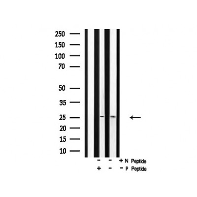
 WB (Western Blot)
(Western blot analysis of ABIN3 in human spleen tissue lysate with ABIN3 antibody at 1 μg/mL in (A) the absence and (B) the presence of blocking peptide.)
WB (Western Blot)
(Western blot analysis of ABIN3 in human spleen tissue lysate with ABIN3 antibody at 1 μg/mL in (A) the absence and (B) the presence of blocking peptide.)
ABIN3, Blocking Peptide (Cat# AAA10964)
Full Name
ABIN3 Peptide
Gene Names
TNIP3; LIND; ABIN-3
Applications
Blocking
Pricing


 Application Data
(Staining of NALM6 cells with MOUSE ANTI HUMAN CD10:ALEXA 488)
Application Data
(Staining of NALM6 cells with MOUSE ANTI HUMAN CD10:ALEXA 488)
CD10, Monoclonal Antibody (Cat# AAA26789)
Full Name
CD10 (CALLA, CD 10, Common Acute Lymphocytic Leukemia Antigen, Enkephalinase, gp100, Membrane Metalloendopeptidase, MME, Neprilysin, Neutral Endopeptidase, Pmel17) (MaxLight 490)
Gene Names
MME; NEP; SFE; CD10; CALLA
Reactivity
Human
Applications
Flow Cytometry, Immunohistochemistry, Immunoprecipitation, Western Blot
Purity
Purified by protein G affinity chromatography from tissue culture supernatant.
Pricing


 Application Data
(Staining of human peripheral blood lymphocytes with Mouse anti Human CD20: Pacific Blue)
Application Data
(Staining of human peripheral blood lymphocytes with Mouse anti Human CD20: Pacific Blue)
CD20, Monoclonal Antibody (Cat# AAA11837)
Full Name
MOUSE ANTI HUMAN CD20:APC
Gene Names
MS4A1; B1; S7; Bp35; CD20; CVID5; MS4A2; LEU-16
Applications
Flow Cytometry
Pricing


 WB (Western Blot)
(ACBD3 monoclonal antibody. Western Blot analysis of ACBD3 expression in PC-12.)
WB (Western Blot)
(ACBD3 monoclonal antibody. Western Blot analysis of ACBD3 expression in PC-12.)
ACBD3, Monoclonal Antibody (Cat# AAA24412)
Full Name
ACBD3 (Golgi Resident Protein GCP60, Acyl-CoA-binding Domain-containing Protein 3, Golgi Complex-associated Protein 1, GOCAP1, Golgi Phosphoprotein 1, GOLPH1, PBR- and PKA-associated Protein 7, Peripheral Benzodiazepine Receptor-associated Protein PAP7, G
Gene Names
ACBD3; PAP7; GCP60; GOCAP1; GOLPH1
Reactivity
Human, Mouse, Rat
Applications
Immunofluorescence, Immunohistochemistry, Western Blot
Purity
Purified by Protein A Affinity Chromatography.
Pricing


 IF (Immunofluorescence)
(Immunofluorescence Analysis of T98G cells labeling Pgp9.5 with Pgp9.5 / UchL1 Mouse Monoclonal Antibody (31A3) followed by Goat anti-Mouse IgG-CF488 (Green). The nuclear counterstain is Nucspot (Red))
IF (Immunofluorescence)
(Immunofluorescence Analysis of T98G cells labeling Pgp9.5 with Pgp9.5 / UchL1 Mouse Monoclonal Antibody (31A3) followed by Goat anti-Mouse IgG-CF488 (Green). The nuclear counterstain is Nucspot (Red))
PGP9.5 / UchL1, Monoclonal Antibody (Cat# AAA13809)
Full Name
PGP9.5 / UchL1 (pan-Neuronal Marker) Mouse Monoclonal Antibody
Gene Names
UCHL1; NDGOA; PARK5; PGP95; PGP9.5; Uch-L1; HEL-117; PGP 9.5; HEL-S-53
Reactivity
Human, Mouse, Rat, Cow, Pig, Dog
Applications
Western Blot, Immunohistochemistry
Pricing


 IHC (Immunohistochemistry)
(Formalin-fixed, paraffin-embedded Rat Skeletal Muscle stained with Muscle Specific Actin Monoclonal Antibody (MSA/953))
IHC (Immunohistochemistry)
(Formalin-fixed, paraffin-embedded Rat Skeletal Muscle stained with Muscle Specific Actin Monoclonal Antibody (MSA/953))
Actin, Monoclonal Antibody (Cat# AAA13822)
Full Name
Actin, Muscle Specific (Muscle Cell Marker) Mouse Monoclonal Antibody
Gene Names
ACTA2; AAT6; ACTSA; MYMY5
Reactivity
Human, Rabbit, Rat
Applications
Flow Cytometry, Immunofluorescence, Western Blot, Immunohistochemistry
Pricing


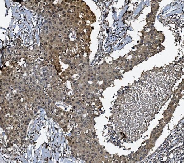
 IHC (Immunohistchemistry)
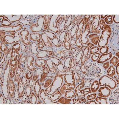
(At 1/100 staining Human prostate cancer and adjacent normal tissues by IHC-P. The sample was formaldehyde fixed and a heat mediated antigen retrieval step in citrate buffer was performed. The sample was then blocked and incubated with the primary antibody at 4 degree C overnight. An HRP conjugated anti-Rabbit antibody was used as the secondary antibody.)
IHC (Immunohistchemistry)
(At 1/100 staining Human prostate cancer and adjacent normal tissues by IHC-P. The sample was formaldehyde fixed and a heat mediated antigen retrieval step in citrate buffer was performed. The sample was then blocked and incubated with the primary antibody at 4 degree C overnight. An HRP conjugated anti-Rabbit antibody was used as the secondary antibody.)
TOPBP1, Polyclonal Antibody (Cat# AAA31467)
Full Name
Phospho-TOPBP1 (Ser1159) Antibody
Gene Names
TOPBP1; TOP2BP1
Reactivity
Human, Mouse, Rat
Predicted Reactivity: Pig (100%), Zebrafish (80%), Bovine (100%), Horse (100%), Sheep (100%), Rabbit (100%), Dog (100%), Chicken (80%), Xenopus (80%)
Predicted Reactivity: Pig (100%), Zebrafish (80%), Bovine (100%), Horse (100%), Sheep (100%), Rabbit (100%), Dog (100%), Chicken (80%), Xenopus (80%)
Applications
Western Blot, Immunohistochemistry, Peptide ELISA
Purity
The antibody is from purified rabbit serum by affinity purification via sequential chromatography on phospho-peptide and non-phospho-peptide affinity columns.
Pricing


 IF (Immunofluorescence)
(Figure 6. IF analysis of SGK1 using anti- SGK1 antibody (AAA19225).SGK1 was detected in immunocytochemical section of A549 cells. Enzyme antigen retrieval was performed using IHC enzyme antigen retrieval reagent for 15 mins. The cells were blocked with 10% goat serum. And then incubated with 4μg/mL rabbit anti- SGK1 Antibody (AAA19225) overnight at 4 degree C. DyLight®488 Conjugated Goat Anti-Rabbit IgG was used as secondary antibody at 1:100 dilution and incubated for 30 minutes at 37 degree C. The section was counterstained with DAPI. Visualize using a fluorescence microscope and filter sets appropriate for the label used.)
IF (Immunofluorescence)
(Figure 6. IF analysis of SGK1 using anti- SGK1 antibody (AAA19225).SGK1 was detected in immunocytochemical section of A549 cells. Enzyme antigen retrieval was performed using IHC enzyme antigen retrieval reagent for 15 mins. The cells were blocked with 10% goat serum. And then incubated with 4μg/mL rabbit anti- SGK1 Antibody (AAA19225) overnight at 4 degree C. DyLight®488 Conjugated Goat Anti-Rabbit IgG was used as secondary antibody at 1:100 dilution and incubated for 30 minutes at 37 degree C. The section was counterstained with DAPI. Visualize using a fluorescence microscope and filter sets appropriate for the label used.)
SGK1, Polyclonal Antibody (Cat# AAA19225)
Full Name
Anti-SGK1 Antibody
Gene Names
SGK1; SGK
Reactivity
Human
Applications
Western Blot, Immunohistochemistry, Immunocytochemistry, Immunofluorescence, Flow Cytometry
Purity
Immunogen affinity purified.
Pricing


 IF (Immunofluorescence)
(Fluorescent confocal image of MCF-7 cell stained with BHLH3 Antibody (N-term). MCF-7 cells were fixed with 4% PFA (20 min), permeabilized with Triton X-100 (0.1%, 10 min), then incubated with BHLH3 primary antibody (1:25, 1 h at 37 degree). For secondary antibody, Alexa Fluor 488 conjugated donkey anti-rabbit antibody (green) was used (1:400, 50 min at 37 degree).Cytoplasmic actin was counterstained with Alexa Fluor 555 (red) conjugated Phalloidin (7units/ml, 1 h at 37 degree). Nuclei were counterstained with DAPI (blue) (10 ug/ml, 10 min).BHLH3 immunoreactivity is localized to nucleus significantly.)
IF (Immunofluorescence)
(Fluorescent confocal image of MCF-7 cell stained with BHLH3 Antibody (N-term). MCF-7 cells were fixed with 4% PFA (20 min), permeabilized with Triton X-100 (0.1%, 10 min), then incubated with BHLH3 primary antibody (1:25, 1 h at 37 degree). For secondary antibody, Alexa Fluor 488 conjugated donkey anti-rabbit antibody (green) was used (1:400, 50 min at 37 degree).Cytoplasmic actin was counterstained with Alexa Fluor 555 (red) conjugated Phalloidin (7units/ml, 1 h at 37 degree). Nuclei were counterstained with DAPI (blue) (10 ug/ml, 10 min).BHLH3 immunoreactivity is localized to nucleus significantly.)
BHLH3, Polyclonal Antibody (Cat# AAA28734)
Full Name
BHLH3 Antibody (N-term)
Gene Names
BHLHE41; DEC2; hDEC2; BHLHB3; SHARP1
Reactivity
Human, Mouse
Applications
Flow Cytometry, Immunofluorescence, Western Blot
Purity
This antibody is purified through a protein A column, followed by peptide affinity purification.
Pricing


 FCM (Flow Cytometry)
(Figure 8. Flow Cytometry analysis of HEPA1-6 cells using anti-PI-16/PI16 antibody (AAA19330).Overlay histogram showing HEPA1-6 cells stained with AAA19330 (Blue line). The cells were blocked with 10% normal goat serum. And then incubated with rabbit anti-PI-16/PI16 Antibody (AAA19330, 1μg/1x106 cells) for 30 min at 20 degree C. DyLight®488 conjugated goat anti-rabbit IgG (5-10μg/1x106 cells) was used as secondary antibody for 30 minutes at 20 degree C. Isotype control antibody (Green line) was rabbit IgG (1μg/1x106) used under the same conditions. Unlabelled sample (Red line) was also used as a control.)
FCM (Flow Cytometry)
(Figure 8. Flow Cytometry analysis of HEPA1-6 cells using anti-PI-16/PI16 antibody (AAA19330).Overlay histogram showing HEPA1-6 cells stained with AAA19330 (Blue line). The cells were blocked with 10% normal goat serum. And then incubated with rabbit anti-PI-16/PI16 Antibody (AAA19330, 1μg/1x106 cells) for 30 min at 20 degree C. DyLight®488 conjugated goat anti-rabbit IgG (5-10μg/1x106 cells) was used as secondary antibody for 30 minutes at 20 degree C. Isotype control antibody (Green line) was rabbit IgG (1μg/1x106) used under the same conditions. Unlabelled sample (Red line) was also used as a control.)
PI-16/PI16, Polyclonal Antibody (Cat# AAA19330)
Full Name
Anti-PI-16/PI16 Antibody
Gene Names
PI16; PSPBP; CRISP9; MSMBBP
Reactivity
Human, Mouse, Rat, Monkey
Applications
WB, IHC-P, FC/FACS/FCM, EIA
Purity
Immunogen affinity purified.
Pricing


 Application Data
(At 25 degree C. The primary antibody was diluted at 1/200 and incubated with the sample for 1 hour at 37 degree C. An Alexa Fluor 594 conjugated goat anti-rabbit IgG (H+L) Ab, diluted at 1/600, was used as the secondary antibody.)
Application Data
(At 25 degree C. The primary antibody was diluted at 1/200 and incubated with the sample for 1 hour at 37 degree C. An Alexa Fluor 594 conjugated goat anti-rabbit IgG (H+L) Ab, diluted at 1/600, was used as the secondary antibody.)
p27 Kip1, Polyclonal Antibody (Cat# AAA31387)
Full Name
Phospho-p27 Kip1 (Thr157) Antibody
Gene Names
CDKN1B; KIP1; MEN4; CDKN4; MEN1B; P27KIP1
Reactivity
Human, Mouse, Rat
Predicted Reactivity: Bovine (89%), Sheep (89%), Rabbit (100%), Dog (100%), Chicken (89%)
Predicted Reactivity: Bovine (89%), Sheep (89%), Rabbit (100%), Dog (100%), Chicken (89%)
Applications
Western Blot, Immunohistochemistry, Immunofluorescence, Immunocytochemistry, Peptide ELISA
Purity
The antibody is from purified rabbit serum by affinity purification via sequential chromatography on phospho-peptide and non-phospho-peptide affinity columns.
Pricing


 Application Data
(Staining of mouse peripheral blood platelets with HAMSTER ANTI MOUSE CD61:RPE)
Application Data
(Staining of mouse peripheral blood platelets with HAMSTER ANTI MOUSE CD61:RPE)
Integrin, beta3, Monoclonal Antibody (Cat# AAA26788)
Full Name
Integrin, beta3 (ITGB3, CD61, GP3A, GPIIIa, HPA-1, NAIT, Platelet Fibrinogen Receptor beta Subunit, Platelet Glycoprotein IIIa, Platelet Membrane Glycoprotein IIIa) (MaxLight 405)
Reactivity
Mouse, Rat
Applications
Flow Cytometry, Immunoprecipitation
Purity
Purified by Protein G Affinity Chromatography
Pricing


 Application Data
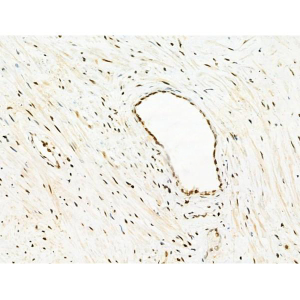
(Immunoperoxidase staining of a mouse lymph node cryosection with Rat anti Mouse MARCO antibody, clone ED31 followed by horseradish peroxidase conjugated Goat anti Rat IgG antibody . Low power)
Application Data
(Immunoperoxidase staining of a mouse lymph node cryosection with Rat anti Mouse MARCO antibody, clone ED31 followed by horseradish peroxidase conjugated Goat anti Rat IgG antibody . Low power)
MARCO, Monoclonal Antibody (Cat# AAA12098)
Full Name
RAT ANTI MOUSE MARCO
Gene Names
Marco; Ly112; Scara2; AI323439
Applications
Immunohistochemistry, Immunofluorescence, Western Blot
Pricing


 ICC (Immunocytochemistry)
(Immunostaining analysis in HeLa cells. HeLa cells were fixed with 4% paraformaldehyde and permeabilized with 0.1% Triton X-100 in PBS. The cells were immunostained with anti-CDC5L mAb. [Lot No. 2136C1a-1])
ICC (Immunocytochemistry)
(Immunostaining analysis in HeLa cells. HeLa cells were fixed with 4% paraformaldehyde and permeabilized with 0.1% Triton X-100 in PBS. The cells were immunostained with anti-CDC5L mAb. [Lot No. 2136C1a-1])
CDC5L, Monoclonal Antibody (Cat# AAA10605)
Full Name
Mouse monoclonal antibody Anti-Human CDC5L
Gene Names
CDC5L; CDC5; CEF1; PCDC5RP; CDC5-LIKE; dJ319D22.1
Reactivity
Human
Applications
Dot Blot, Immunocytochemistry, Immunoprecipitation, Western Blot, Flow Cytometry
Pricing


 Application Data
(Staining of human peripheral blood granulocytes with Mouse anti Human CD13: Alexa Fluor 647 (AAA11927A647))
Application Data
(Staining of human peripheral blood granulocytes with Mouse anti Human CD13: Alexa Fluor 647 (AAA11927A647))
CD13, Monoclonal Antibody (Cat# AAA11927)
Full Name
MOUSE ANTI HUMAN CD13
Gene Names
ANPEP; APN; CD13; LAP1; P150; PEPN; GP150
Reactivity
Rhesus Monkey
Applications
Immunohistochemistry, Flow Cytometry, Immunoprecipitation
Pricing


 IF (Immunofluorescence)
(ICC/IF analysis of FABP7 in PC3 cells. The cell was stained with AAA11731 (1:100). The secondary antibody (green) was used Alexa Fluor 488. DAPI was stained the cell nucleus (blue).)
IF (Immunofluorescence)
(ICC/IF analysis of FABP7 in PC3 cells. The cell was stained with AAA11731 (1:100). The secondary antibody (green) was used Alexa Fluor 488. DAPI was stained the cell nucleus (blue).)
FABP7, Monoclonal Antibody (Cat# AAA11731)
Full Name
FABP7 antibody
Gene Names
FABP7; MRG; BLBP; FABPB; B-FABP
Reactivity
Human, Mouse
Applications
Western Blot, Immunocytochemistry, Immunofluorescence
Purity
By protein-G affinity chromatography
Pricing


 IF (Immunofluorescence)
Cytokeratin 18 (KRT18), Monoclonal Antibody (Cat# AAA13836)
Full Name
Cytokeratin 18 (KRT18) Mouse Monoclonal Antibody
Gene Names
KRT18; K18; CK-18; CYK18
Reactivity
Human.
Does not react with Mouse, Rat, Sheep, Hamster, Cow, Dog and Pig
Does not react with Mouse, Rat, Sheep, Hamster, Cow, Dog and Pig
Applications
Flow Cytometry, Immunofluorescence, Immunohistochemistry
Pricing


 IF (Immunofluorescence)
(Confocal Immunofluorescent analysis of SK-OV-3 cells using AF488-labeled Isotype Control Monoclonal Antibody (IgG1) (Green).DAPI was used to stain the cell nuclei (blue). (Negative Control))
IF (Immunofluorescence)
(Confocal Immunofluorescent analysis of SK-OV-3 cells using AF488-labeled Isotype Control Monoclonal Antibody (IgG1) (Green).DAPI was used to stain the cell nuclei (blue). (Negative Control))
Ep-CAM /CD326, Monoclonal Antibody (Cat# AAA13838)
Full Name
Ep-CAM /CD326 (Epithelial Marker) Mouse Monoclonal Antibody
Gene Names
EPCAM; ESA; KSA; M4S1; MK-1; DIAR5; EGP-2; EGP40; KS1/4; MIC18; TROP1; EGP314; HNPCC8; TACSTD1
Reactivity
Human.
Does not react with Mouse and Rat
Does not react with Mouse and Rat
Applications
Flow Cytometry, Immunofluorescence, Western Blot, Immunohistochemistry
Pricing