Filters
Clonality
Type
Reactivity
Gene Name
Isotype
Host
Application
Clone
1192 results for " A G G" - showing 300-350


 Application Data
(Staining of mouse spleen with Rat anti Mouse CD4:RPE)
Application Data
(Staining of mouse spleen with Rat anti Mouse CD4:RPE)
CD4, Monoclonal Antibody (Cat# AAA12230)
Full Name
RAT ANTI MOUSE CD4
Gene Names
Cd4; L3T4; Ly-4
Applications
Immunohistochemistry, Flow Cytometry, Immunofluorescence, Immunoprecipitation, Western Blot
Pricing


 Application Data
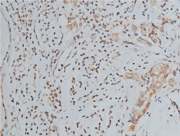
(Formalin fixed, paraffin embedded human breast cancer biopsy stained with Mouse anti Human estrogen receptor beta5 antibody followed by HRP polymer detection and DAB substrate development following heat mediated antigen retrieval using citrate buffer at pH6.2 (low power))
Application Data
(Formalin fixed, paraffin embedded human breast cancer biopsy stained with Mouse anti Human estrogen receptor beta5 antibody followed by HRP polymer detection and DAB substrate development following heat mediated antigen retrieval using citrate buffer at pH6.2 (low power))
ESTROGEN RECEPTOR BETA 5, Monoclonal Antibody (Cat# AAA12216)
Full Name
MOUSE ANTI HUMAN ESTROGEN RECEPTOR BETA 5
Gene Names
ESR2; Erb; ESRB; ESTRB; NR3A2; ER-BETA; ESR-BETA
Applications
Immunohistochemistry, Western Blot
Pricing


 Application Data
(Published customer image Infiltration of GFP+ BM-cells in infarct and peri-infarct regions. (A-B) Dot plots of viable macrophages/granulocytes (CD11b+CD45high, top right quadrants) and microglia (CD11b+CD45dim, bottom right quadrants) in cortex from BM-chimeric unmanipulated mice and mice exposed to pMCAO. (C) Bar graph showing mean numbers of CD11b+CD45dim microglia and CD11b+CD45high macrophages/granulocytes in BM-chimeric mice 24 hours after pMCAO, subdivided based on expression of GFP (n = 5). Approximately 92% of of the CD45high population were GFP+. (D) Estimation and comparison of mean numbers of CD11b+CD45dim microglia in non-chimeric (n = 10) versus BM-chimeric mice (n = 5) 24 hours after of pMCAO shows significantly fewer CD11b+CD45dim microglial cells in irradiated mice. (E) Overview, showing distribution of infiltrating GFP+ BM-derived cells into infarct (IF) and peri-infarct (P-IF) regions 24 hours after pMCAO. (E-G) By 24 hours, GFP+ single cells (F) and vessel-associated aggregates of GFP+ cells (arrows in G) were observed in infarct and peri-infarct regions. Some of the vessel-associated cells were round, leukocyte-like cells (arrows) while others were elongated cells lining the vasculature (arrow heads in G and in insert). (H) Bar graph showing mean numbers of single GFP+ cells and vessel-associated aggregates of GFP+ cells in ipsi- and contralateral cortex 24 hours after surgery (n = 10). (I-P) Immunohistochemical staining of CD45.1 (I, K), CD45.2 (J, L), IgG2a (M, O) and CD45 (N, P) in ischemic tissue in BM-chimeric (I, J, M, N) and non-chimeric mice (K, L, O, P) 24 hours after pMCAO. N.D, none detected. Scale bars: 200 um (A), 10 um (B, C). 50 um (I-P) *P < 0.05, **P < 0.01, and ***P < 0.001.From: Clausen BH, Lambertsen KL, Babcock AA, Holm TH, Dagnaes-Hansen F, Finsen B. Interleukin-1beta and tumor necrosis factor-alpha are expressed by different subsets of microglia and macrophages after ischemic stroke in mice. J Neuroinflammation. 2008 Oct 23;5:46.)
Application Data
(Published customer image Infiltration of GFP+ BM-cells in infarct and peri-infarct regions. (A-B) Dot plots of viable macrophages/granulocytes (CD11b+CD45high, top right quadrants) and microglia (CD11b+CD45dim, bottom right quadrants) in cortex from BM-chimeric unmanipulated mice and mice exposed to pMCAO. (C) Bar graph showing mean numbers of CD11b+CD45dim microglia and CD11b+CD45high macrophages/granulocytes in BM-chimeric mice 24 hours after pMCAO, subdivided based on expression of GFP (n = 5). Approximately 92% of of the CD45high population were GFP+. (D) Estimation and comparison of mean numbers of CD11b+CD45dim microglia in non-chimeric (n = 10) versus BM-chimeric mice (n = 5) 24 hours after of pMCAO shows significantly fewer CD11b+CD45dim microglial cells in irradiated mice. (E) Overview, showing distribution of infiltrating GFP+ BM-derived cells into infarct (IF) and peri-infarct (P-IF) regions 24 hours after pMCAO. (E-G) By 24 hours, GFP+ single cells (F) and vessel-associated aggregates of GFP+ cells (arrows in G) were observed in infarct and peri-infarct regions. Some of the vessel-associated cells were round, leukocyte-like cells (arrows) while others were elongated cells lining the vasculature (arrow heads in G and in insert). (H) Bar graph showing mean numbers of single GFP+ cells and vessel-associated aggregates of GFP+ cells in ipsi- and contralateral cortex 24 hours after surgery (n = 10). (I-P) Immunohistochemical staining of CD45.1 (I, K), CD45.2 (J, L), IgG2a (M, O) and CD45 (N, P) in ischemic tissue in BM-chimeric (I, J, M, N) and non-chimeric mice (K, L, O, P) 24 hours after pMCAO. N.D, none detected. Scale bars: 200 um (A), 10 um (B, C). 50 um (I-P) *P < 0.05, **P < 0.01, and ***P < 0.001.From: Clausen BH, Lambertsen KL, Babcock AA, Holm TH, Dagnaes-Hansen F, Finsen B. Interleukin-1beta and tumor necrosis factor-alpha are expressed by different subsets of microglia and macrophages after ischemic stroke in mice. J Neuroinflammation. 2008 Oct 23;5:46.)
CD11b, Monoclonal Antibody (Cat# AAA12185)
Full Name
RAT ANTI MOUSE CD11b
Gene Names
Itgam; CR3; CR3A; MAC1; Cd11b; Ly-40; Mac-1; Mac-1a; CD11b/CD18; F730045J24Rik
Applications
Immunohistochemistry, Flow Cytometry, Immunofluorescence, Immunoprecipitation
Pricing


 Application Data
(Published customer image Infiltration of GFP+ BM-cells in infarct and peri-infarct regions. (A-B) Dot plots of viable macrophages/granulocytes (CD11b+CD45high, top right quadrants) and microglia (CD11b+CD45dim, bottom right quadrants) in cortex from BM-chimeric unmanipulated mice and mice exposed to pMCAO. (C) Bar graph showing mean numbers of CD11b+CD45dim microglia and CD11b+CD45high macrophages/granulocytes in BM-chimeric mice 24 hours after pMCAO, subdivided based on expression of GFP (n = 5). Approximately 92% of of the CD45high population were GFP+. (D) Estimation and comparison of mean numbers of CD11b+CD45dim microglia in non-chimeric (n = 10) versus BM-chimeric mice (n = 5) 24 hours after of pMCAO shows significantly fewer CD11b+CD45dim microglial cells in irradiated mice. (E) Overview, showing distribution of infiltrating GFP+ BM-derived cells into infarct (IF) and peri-infarct (P-IF) regions 24 hours after pMCAO. (E-G) By 24 hours, GFP+ single cells (F) and vessel-associated aggregates of GFP+ cells (arrows in G) were observed in infarct and peri-infarct regions. Some of the vessel-associated cells were round, leukocyte-like cells (arrows) while others were elongated cells lining the vasculature (arrow heads in G and in insert). (H) Bar graph showing mean numbers of single GFP+ cells and vessel-associated aggregates of GFP+ cells in ipsi- and contralateral cortex 24 hours after surgery (n = 10). (I-P) Immunohistochemical staining of CD45.1 (I, K), CD45.2 (J, L), IgG2a (M, O) and CD45 (N, P) in ischemic tissue in BM-chimeric (I, J, M, N) and non-chimeric mice (K, L, O, P) 24 hours after pMCAO. N.D, none detected. Scale bars: 200 um (A), 10 um (B, C). 50 um (I-P) *P < 0.05, **P < 0.01, and ***P < 0.001.From: Clausen BH, Lambertsen KL, Babcock AA, Holm TH, Dagnaes-Hansen F, Finsen B. Interleukin-1beta and tumor necrosis factor-alpha are expressed by different subsets of microglia and macrophages after ischemic stroke in mice. J Neuroinflammation. 2008 Oct 23;5:46.)
Application Data
(Published customer image Infiltration of GFP+ BM-cells in infarct and peri-infarct regions. (A-B) Dot plots of viable macrophages/granulocytes (CD11b+CD45high, top right quadrants) and microglia (CD11b+CD45dim, bottom right quadrants) in cortex from BM-chimeric unmanipulated mice and mice exposed to pMCAO. (C) Bar graph showing mean numbers of CD11b+CD45dim microglia and CD11b+CD45high macrophages/granulocytes in BM-chimeric mice 24 hours after pMCAO, subdivided based on expression of GFP (n = 5). Approximately 92% of of the CD45high population were GFP+. (D) Estimation and comparison of mean numbers of CD11b+CD45dim microglia in non-chimeric (n = 10) versus BM-chimeric mice (n = 5) 24 hours after of pMCAO shows significantly fewer CD11b+CD45dim microglial cells in irradiated mice. (E) Overview, showing distribution of infiltrating GFP+ BM-derived cells into infarct (IF) and peri-infarct (P-IF) regions 24 hours after pMCAO. (E-G) By 24 hours, GFP+ single cells (F) and vessel-associated aggregates of GFP+ cells (arrows in G) were observed in infarct and peri-infarct regions. Some of the vessel-associated cells were round, leukocyte-like cells (arrows) while others were elongated cells lining the vasculature (arrow heads in G and in insert). (H) Bar graph showing mean numbers of single GFP+ cells and vessel-associated aggregates of GFP+ cells in ipsi- and contralateral cortex 24 hours after surgery (n = 10). (I-P) Immunohistochemical staining of CD45.1 (I, K), CD45.2 (J, L), IgG2a (M, O) and CD45 (N, P) in ischemic tissue in BM-chimeric (I, J, M, N) and non-chimeric mice (K, L, O, P) 24 hours after pMCAO. N.D, none detected. Scale bars: 200 um (A), 10 um (B, C). 50 um (I-P) *P < 0.05, **P < 0.01, and ***P < 0.001.From: Clausen BH, Lambertsen KL, Babcock AA, Holm TH, Dagnaes-Hansen F, Finsen B. Interleukin-1beta and tumor necrosis factor-alpha are expressed by different subsets of microglia and macrophages after ischemic stroke in mice. J Neuroinflammation. 2008 Oct 23;5:46.)
CD11b, Monoclonal Antibody (Cat# AAA12183)
Full Name
RAT ANTI MOUSE CD11b:FITC
Gene Names
Itgam; CR3; CR3A; MAC1; Cd11b; Ly-40; Mac-1; Mac-1a; CD11b/CD18; F730045J24Rik
Applications
Flow Cytometry
Pricing


 Application Data
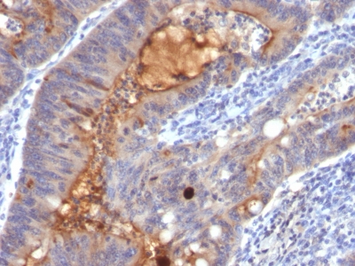
(Immunoperoxidase staining of a human tonsil cryosection with Mouse anti Human CD163 antibody, clone EDHu-1 followed by the Histar detection system . Low power)
Application Data
(Immunoperoxidase staining of a human tonsil cryosection with Mouse anti Human CD163 antibody, clone EDHu-1 followed by the Histar detection system . Low power)
CD163, Monoclonal Antibody (Cat# AAA12100)
Full Name
MOUSE ANTI HUMAN CD163
Gene Names
CD163; M130; MM130
Applications
Immunohistochemistry, Flow Cytometry, Immunofluorescence, Immunoassay, Immunohistochemistry, Western Blot
Pricing


 Application Data
(Staining of human peripheral blood lymphocytes with MOUSE ANTI HUMAN CD45RA:FITC (MCA88F))
Application Data
(Staining of human peripheral blood lymphocytes with MOUSE ANTI HUMAN CD45RA:FITC (MCA88F))
CD45RA, Monoclonal Antibody (Cat# AAA26734)
Full Name
CD45RA (CD45 Antigen, B220, GP180, Leukocyte Common Antigen, LCA, L-CA, LY5, LY-5, Protein Tyrosine Phosphatase Receptor Type C Polypeptide, PTPRC, T200, T200 Glycoprotein) (Biotin)
Reactivity
Human, Monkey
Applications
Flow Cytometry, Immunohistochemistry
Purity
Purified by protein G affinity chromatography from tissue culture supernatant.
Pricing


 IF (Immunofluorescence)
(Immunofluorescence of Clusterin in mouse brain tissue with Clusterin Antibody at 20 μg/mL.Green: Clusterin Antibody (3856)Red: Phylloidin stainingBlue: DAPI staining)
IF (Immunofluorescence)
(Immunofluorescence of Clusterin in mouse brain tissue with Clusterin Antibody at 20 μg/mL.Green: Clusterin Antibody (3856)Red: Phylloidin stainingBlue: DAPI staining)
Clusterin, Polyclonal Antibody (Cat# AAA10914)
Full Name
Clusterin Antibody
Gene Names
CLU; CLI; AAG4; APOJ; CLU1; CLU2; KUB1; SGP2; APO-J; SGP-2; SP-40; TRPM2; TRPM-2; NA1/NA2
Reactivity
Human
Applications
Western Blot, Immunohistochemistry
Purity
Clusterin Antibody is affinity chromatography purified via peptide column.
Pricing


 Application Data
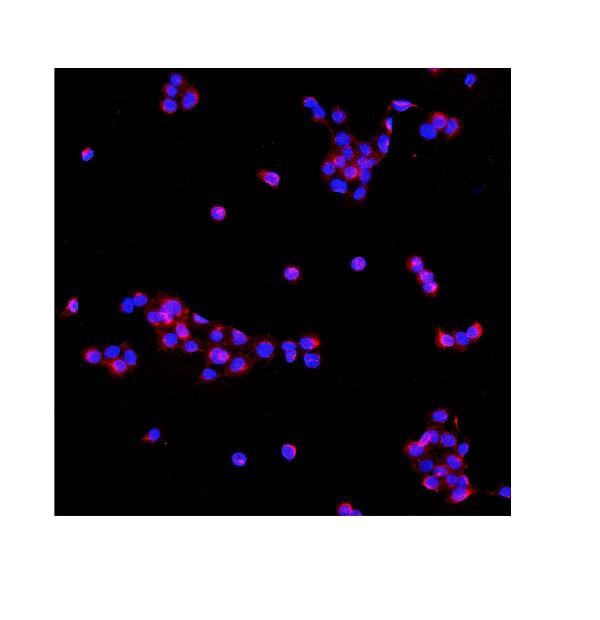
(At 25 degree C. Samples were then incubated with primary Ab(At 37 degree C. An AlexaFluor594 conjugated goat anti-rabbit IgG(H+L) Ab(Red) and an AlexaFluor488 conjugated goat anti-mouse IgG(H+L) Ab(Green) were used as the secondary antibody.The nuclear counter stain is DAPI (blue).)
Application Data
(At 25 degree C. Samples were then incubated with primary Ab(At 37 degree C. An AlexaFluor594 conjugated goat anti-rabbit IgG(H+L) Ab(Red) and an AlexaFluor488 conjugated goat anti-mouse IgG(H+L) Ab(Green) were used as the secondary antibody.The nuclear counter stain is DAPI (blue).)
TOPBP1, Polyclonal Antibody (Cat# AAA31285)
Full Name
Phospho-TOPBP1 (Ser1138) Antibody
Gene Names
TOPBP1; TOP2BP1
Reactivity
Human, Mouse, Rat
Applications
Immunohistochemistry, Immunofluorescence, Immunocytochemistry, Peptide ELISA
Purity
The antibody is from purified rabbit serum by affinity purification via sequential chromatography on phospho-peptide and non-phospho-peptide affinity columns.
Pricing


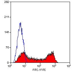
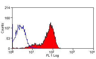
 FCM (Flow Cytometry)
(Figure 7. Flow Cytometry analysis of U251 cells using anti-Serum Response Factor/SRF antibody (AAA19221).Overlay histogram showing U251 cells stained with AAA19221 (Blue line). The cells were blocked with 10% normal goat serum. And then incubated with rabbit anti-Serum Response Factor/SRF Antibody (AAA19221, 1μg/1x106 cells) for 30 min at 20 degree C. DyLight®488 conjugated goat anti-rabbit IgG (5-10μg/1x106 cells) was used as secondary antibody for 30 minutes at 20 degree C. Isotype control antibody (Green line) was rabbit IgG (1μg/1x106) used under the same conditions. Unlabelled sample (Red line) was also used as a control.)
FCM (Flow Cytometry)
(Figure 7. Flow Cytometry analysis of U251 cells using anti-Serum Response Factor/SRF antibody (AAA19221).Overlay histogram showing U251 cells stained with AAA19221 (Blue line). The cells were blocked with 10% normal goat serum. And then incubated with rabbit anti-Serum Response Factor/SRF Antibody (AAA19221, 1μg/1x106 cells) for 30 min at 20 degree C. DyLight®488 conjugated goat anti-rabbit IgG (5-10μg/1x106 cells) was used as secondary antibody for 30 minutes at 20 degree C. Isotype control antibody (Green line) was rabbit IgG (1μg/1x106) used under the same conditions. Unlabelled sample (Red line) was also used as a control.)
Serum Response Factor/SRF, Polyclonal Antibody (Cat# AAA19221)
Full Name
Anti-Serum Response Factor/SRF Antibody
Gene Names
SRF; MCM1
Reactivity
Human, Mouse, Rat
Applications
Western Blot, Immunohistochemistry, Immunocytochemistry, Immunofluorescence, Flow Cytometry, Direct ELISA
Purity
Immunogen affinity purified.
Pricing


 Application Data
(Staining of human peripheral blood lymphocytes with MOUSE ANTI HUMAN CD3:ALEXA 647)
Application Data
(Staining of human peripheral blood lymphocytes with MOUSE ANTI HUMAN CD3:ALEXA 647)
CD3, Monoclonal Antibody (Cat# AAA26745)
Full Name
CD3 (FITC)
Reactivity
Human
Applications
Flow Cytometry, Immunohistochemistry
Purity
Purified by Protein G Affinity Chromatography
Pricing


 Application Data
(Staining of mouse peripheral blood platelets with HAMSTER ANTI MOUSE CD61:RPE)
Application Data
(Staining of mouse peripheral blood platelets with HAMSTER ANTI MOUSE CD61:RPE)
Integrin, beta3, Monoclonal Antibody (Cat# AAA26764)
Full Name
Integrin, beta3 (ITGB3, CD61, GP3A, GPIIIa, HPA-1, NAIT, Platelet Fibrinogen Receptor beta Subunit, Platelet Glycoprotein IIIa, Platelet Membrane Glycoprotein IIIa) (HRP)
Reactivity
Mouse, Rat
Applications
Flow Cytometry, Immunoprecipitation
Purity
Purified by Protein G Affinity Chromatography
Pricing


 Application Data
(Staining of human peripheral blood lymphocytes with MOUSE ANTI HUMAN CD45RA:FITC (MCA88F))
Application Data
(Staining of human peripheral blood lymphocytes with MOUSE ANTI HUMAN CD45RA:FITC (MCA88F))
CD45RA, Monoclonal Antibody (Cat# AAA26747)
Full Name
CD45RA (CD45 Antigen, B220, GP180, Leukocyte Common Antigen, LCA, L-CA, LY5, LY-5, Protein Tyrosine Phosphatase Receptor Type C Polypeptide, PTPRC, T200, T200 Glycoprotein) (FITC)
Reactivity
Human, Monkey
Applications
Flow Cytometry, Immunohistochemistry
Purity
Purified by protein G affinity chromatography from tissue culture supernatant.
Pricing


 Application Data
(Staining of mouse peritoneal macrophages with RAT ANTI MOUSE F4/80 ANTIGEN:FITC)
Application Data
(Staining of mouse peritoneal macrophages with RAT ANTI MOUSE F4/80 ANTIGEN:FITC)
EMR1, Monoclonal Antibody (Cat# AAA26786)
Full Name
EMR1 (EGF-like Module Containing Mucin-like Hormone Receptor-like 1, EMR1 Hormone Receptor, Cell Surface Glycoprotein EMR1, Cell Surface Glycoprotein F4/80, DD7A5-7, EGF-TM7, F4/80, Gpf480, Lymphocyte Antigen 71, Ly71, TM7LN3) (MaxLight 405)
Reactivity
Mouse
Applications
Flow Cytometry, Immunohistochemistry, Immunoprecipitation, Radioimmunoassay, Western Blot
Purity
Purified by protein G affinity chromatography from tissue culture supernatant.
Pricing


 Transfection Control
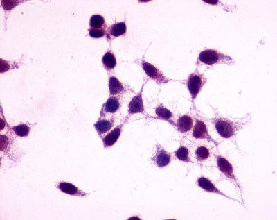
(Anti-GPR27 antibody immunocytochemistry (ICC) staining of untransfected HEK293 human embryonic kidney cells.)
Transfection Control
(Anti-GPR27 antibody immunocytochemistry (ICC) staining of untransfected HEK293 human embryonic kidney cells.)
GPR27, Polyclonal Antibody (Cat# AAA12362)
Full Name
Rabbit Polyclonal to Human GPR27
Gene Names
GPR27; SREB1
Reactivity
Gorilla, Human, Monkey
Applications
Immunohistochemistry, Immunocytochemistry
Purity
Immunoaffinity Purified
Pricing


 ELISA
(A sandwich ELISA was performed using the anti-PD-L1 mAb (5 μg/ml) as the capture antibody. Biotin-labeled anti-PD-L1 mAb (1 μg/ml) and streptavidin-HRP (0.1 μg/ml) were used for detection. Detection range is from 10 ng to 20 pg.)
ELISA
(A sandwich ELISA was performed using the anti-PD-L1 mAb (5 μg/ml) as the capture antibody. Biotin-labeled anti-PD-L1 mAb (1 μg/ml) and streptavidin-HRP (0.1 μg/ml) were used for detection. Detection range is from 10 ng to 20 pg.)
PDL1, Monoclonal Antibody (Cat# AAA10995)
Full Name
PDL1 Antibody [1D7]
Gene Names
CD274; B7-H; B7H1; PDL1; PD-L1; PDCD1L1; PDCD1LG1
Reactivity
Human
Applications
Western Blot, Immunohistochemistry, Immunocytochemistry, Immunofluorescence
Purity
Protein A purified
Pricing


 IHC (Immunohistchemistry)
(Immunochemical staining CD21 in cynomolgus lymph node with rabbit polyclonal antibody at 1:300 dilution, formalin-fixed paraffin embedded sections.)
IHC (Immunohistchemistry)
(Immunochemical staining CD21 in cynomolgus lymph node with rabbit polyclonal antibody at 1:300 dilution, formalin-fixed paraffin embedded sections.)
CD21, Polyclonal Antibody (Cat# AAA27739)
Full Name
Anti-CD21 Antibody, Rabbit Polyclonal
Gene Names
CR2; CR; C3DR; CD21; CVID7; SLEB9
Reactivity
Human, Cynomolgus
Applications
Western Blot, Immunohistochemistry, Immunoprecipitation
Purity
Protein A & Antigen Affinity
Pricing


 WB (Western Blot)
(Anti-PARP1 mouse monoclonal antibody at 1:500 dilutionLane A: Jurkat Whole Cell LysateLysates/proteins at 30 ug per lane.SecondaryGoat Anti-Mouse IgG H&L (Dylight800) at 1/15000 dilution.Developed using the Odyssey technique. Performed under reducing conditions.Predicted band size:113 kDaObserved band size:113 kDa)
WB (Western Blot)
(Anti-PARP1 mouse monoclonal antibody at 1:500 dilutionLane A: Jurkat Whole Cell LysateLysates/proteins at 30 ug per lane.SecondaryGoat Anti-Mouse IgG H&L (Dylight800) at 1/15000 dilution.Developed using the Odyssey technique. Performed under reducing conditions.Predicted band size:113 kDaObserved band size:113 kDa)
PARP, Monoclonal Antibody (Cat# AAA27741)
Full Name
Anti-PARP Antibody, Mouse Monoclonal
Gene Names
PARP1; PARP; PPOL; ADPRT; ARTD1; ADPRT1; PARP-1; ADPRT 1; pADPRT-1
Reactivity
Human
Applications
Western Blot, Immunohistochemistry, Immunoprecipitation
Purity
Protein A
Pricing
 SDS-PAGE
(SDS-PAGE Analysis Purified Secretory Component Mouse Monoclonal Antibody (SPM217). Confirmation of Purity and Integrity of Antibody.)
SDS-PAGE
(SDS-PAGE Analysis Purified Secretory Component Mouse Monoclonal Antibody (SPM217). Confirmation of Purity and Integrity of Antibody.)
IgA Secretory Component / ECM1, Monoclonal Antibody (Cat# AAA13802)
Full Name
IgA Secretory Component / ECM1 Mouse Monoclonal Antibody
Gene Names
ECM1; URBWD
Reactivity
Human, Rat
Applications
Flow Cytometry, Immunofluorescence, Immunohistochemistry
Pricing


 Application Data
(Staining of mouse peritoneal macrophages with Rat anti Mouse CD11b)
Application Data
(Staining of mouse peritoneal macrophages with Rat anti Mouse CD11b)
CD11b, Monoclonal Antibody (Cat# AAA12187)
Full Name
RAT ANTI MOUSE CD11b:Biotin
Gene Names
Itgam; CR3; CR3A; MAC1; Cd11b; Ly-40; Mac-1; Mac-1a; CD11b/CD18; F730045J24Rik
Applications
Flow Cytometry
Pricing


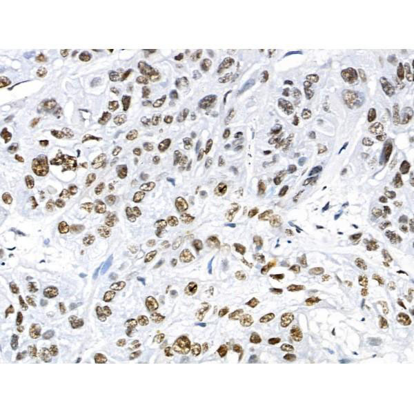
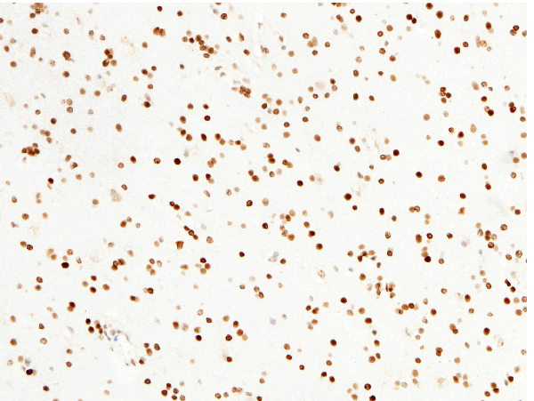
 IHC (Immunohistochemistry)
(AAA30929 at 1/200 staining human colon tissue sections by IHC-P. The tissue was formaldehyde fixed and a heat mediated antigen retrieval step in citrate buffer was performed. The tissue was then blocked and incubated with the antibody for 1.5 hours at 22 degree C. An HRP conjugated goat anti-rabbit antibody was used as the secondary.)
IHC (Immunohistochemistry)
(AAA30929 at 1/200 staining human colon tissue sections by IHC-P. The tissue was formaldehyde fixed and a heat mediated antigen retrieval step in citrate buffer was performed. The tissue was then blocked and incubated with the antibody for 1.5 hours at 22 degree C. An HRP conjugated goat anti-rabbit antibody was used as the secondary.)
Histone H3, Polyclonal Antibody (Cat# AAA30929)
Full Name
Histone H3 Antibody
Gene Names
HIST1H3A; H3/A; H3FA
Reactivity
Human, Mouse, Rat
Applications
Western Blot, Immunohistochemistry, Immunofluorescence, Immunocytochemistry
Purity
The antiserum was purified by peptide affinity chromatography using SulfoLink Coupling Resin.
Pricing


 Application Data
(Published customer image: Representative images of the inflammatory changes in the facial nucleus during axonal regeneration, one week following facial nerve transaction. a, b: CD11b immunoreactivity for microglia is increased in the axotomized facial nucleus, and microglia enwrap the facial motor neurons, e.g. at arrows. The regenerating neurons were retrogradely labelled with fluorogold. c, d: CD6- positive T-cells accumulated in the injured motor nucleus (arrows). They had little cytoplasm but dense nuclei (c) and were sometimes clustered around neurons retrogradely labelled with fluorogold (d). The scale bar in (a) also applies to (b) and that in (c) also applies to (d).From: Shokouhi et al. BMC Neuroscience 2010 11:13.)
Application Data
(Published customer image: Representative images of the inflammatory changes in the facial nucleus during axonal regeneration, one week following facial nerve transaction. a, b: CD11b immunoreactivity for microglia is increased in the axotomized facial nucleus, and microglia enwrap the facial motor neurons, e.g. at arrows. The regenerating neurons were retrogradely labelled with fluorogold. c, d: CD6- positive T-cells accumulated in the injured motor nucleus (arrows). They had little cytoplasm but dense nuclei (c) and were sometimes clustered around neurons retrogradely labelled with fluorogold (d). The scale bar in (a) also applies to (b) and that in (c) also applies to (d).From: Shokouhi et al. BMC Neuroscience 2010 11:13.)
CD11b, Monoclonal Antibody (Cat# AAA11876)
Full Name
MOUSE ANTI RAT CD11b:FITC
Gene Names
ITGAM; CD11B
Applications
Flow Cytometry
Pricing


 Application Data
(Analysis of Protein Array containing more than 19, 000 full-length human proteins using Oct-2 Mouse Monoclonal Antibody (OCT2/2137) Z- and S- Score: The Z-score represents the strength of a signal that a monoclonal antibody (MAb) (in combination with a fluorescently-tagged anti-IgG secondary antibody) produces when binding to a particular protein on the HuProtTM array. Z-scores are described in units of standard deviations (SD's) above the mean value of all signals generated on that array. If targets on HuProtTM are arranged in descending order of the Z-score, the S-score is the difference (also in units of SD's) between the Z-score. S-score therefore represents the relative target specificity of a MAb to its intended target. A MAb is considered to specific to its intended target, if the MAb has an S-score of at least 2.5. For example, if a MAb binds to protein X with a Z-score of 43 and to protein Y with a Z-score of 14, then the S-score for the binding of that MAb to protein X is equal to 29.)
Application Data
(Analysis of Protein Array containing more than 19, 000 full-length human proteins using Oct-2 Mouse Monoclonal Antibody (OCT2/2137) Z- and S- Score: The Z-score represents the strength of a signal that a monoclonal antibody (MAb) (in combination with a fluorescently-tagged anti-IgG secondary antibody) produces when binding to a particular protein on the HuProtTM array. Z-scores are described in units of standard deviations (SD's) above the mean value of all signals generated on that array. If targets on HuProtTM are arranged in descending order of the Z-score, the S-score is the difference (also in units of SD's) between the Z-score. S-score therefore represents the relative target specificity of a MAb to its intended target. A MAb is considered to specific to its intended target, if the MAb has an S-score of at least 2.5. For example, if a MAb binds to protein X with a Z-score of 43 and to protein Y with a Z-score of 14, then the S-score for the binding of that MAb to protein X is equal to 29.)
OCT-2 (POU2F2), Monoclonal Antibody (Cat# AAA23902)
Full Name
OCT-2 (POU2F2) (B-Cell Marker)
Gene Names
POU2F2; OCT2; OTF2; Oct-2
Reactivity
Human. Others not known.
Applications
Western Blot, Immunohistochemistry
Pricing


 Application Data
(Staining of J774 cells with Rat anti Mouse F4/80 antigen Biotin)
Application Data
(Staining of J774 cells with Rat anti Mouse F4/80 antigen Biotin)
F4/80, Monoclonal Antibody (Cat# AAA12169)
Full Name
RAT ANTI MOUSE F4/80
Gene Names
Emr1; Ly71; F4/80; Gpf480; TM7LN3; DD7A5-7; EGF-TM7
Applications
Immunohistochemistry, Fluorescence Microscopy, Flow Cytometry, Immunofluorescence, Immunoprecipitation, Immunohistochemistry, Radioimmunoassay, Immunohistochemistry, Western Blot
Pricing


 Application Data
(Staining of Rat peripheral blood lymphocytes with Mouse anti Rat CD49d:RPE (MCA2872PE))
Application Data
(Staining of Rat peripheral blood lymphocytes with Mouse anti Rat CD49d:RPE (MCA2872PE))
CD49d, Monoclonal Antibody (Cat# AAA26760)
Full Name
CD49d (Antigen CD49d, CD49d Antigen, CDw49d, Alpha 4 Subunit of VLA-4 Receptor, Integrin alpha IV, Integrin alpha 4, IA4, ITGA4, LPAM23, MGC90518, Very Late Activation Protein 4 Receptor Alpha 4 Subunit, VLA4, VLA-4) (HRP)
Reactivity
Rat
Applications
Flow Cytometry, Immunoprecipitation
Purity
Purified by Protein G Affinity Chromatography
Pricing


 ELISA
(Titration curve analysis of PD-1 mAbs to detect recombinant PD-1 in ELISA with abs at decreasing concentrations.)
ELISA
(Titration curve analysis of PD-1 mAbs to detect recombinant PD-1 in ELISA with abs at decreasing concentrations.)
PD-1, Monoclonal Antibody (Cat# AAA10981)
Full Name
PD-1 Antibody [10B3]
Gene Names
PDCD1; PD1; PD-1; CD279; SLEB2; hPD-1; hPD-l; hSLE1
Reactivity
Human
Applications
Western Blot, Immunohistochemistry, Immunocytochemistry, Immunofluorescence, Flow Cytometry
Purity
Protein A purified IgG1.
Pricing


 WB (Western Blot)
(Western Blot analysis of ATP6V1G2 expression in transfected 293T cell line by ATP6V1G2 monoclonal antibody. Lane 1: ATP6V1G2 transfected lysate (13.6kD). Lane 2: Non-transfected lysate.)
WB (Western Blot)
(Western Blot analysis of ATP6V1G2 expression in transfected 293T cell line by ATP6V1G2 monoclonal antibody. Lane 1: ATP6V1G2 transfected lysate (13.6kD). Lane 2: Non-transfected lysate.)
ATP6V1G2, Monoclonal Antibody (Cat# AAA25621)
Full Name
ATP6V1G2 (V-type Proton ATPase Subunit G 2, V-ATPase Subunit G 2, V-ATPase 13kD Subunit 2, Vacuolar Proton Pump Subunit G 2, ATP6G, ATP6G2, NG38) (PE)
Gene Names
ATP6V1G2; NG38; ATP6G; VMA10; ATP6G2
Reactivity
Human, Mouse, Rat
Applications
Western Blot
Purity
Purified by Protein A Affinity Chromatography.
Pricing


 Application Data
(Figure A. FITC conjugated Rat anti Mouse CD19 . Figure B. FITC conjugated Rat anti Mouse CD19 and SBV440 conjugated Rat anti Mouse CD45R . All experiments performed on mouse splenocytes in the presence of 10% mouse seru)
Application Data
(Figure A. FITC conjugated Rat anti Mouse CD19 . Figure B. FITC conjugated Rat anti Mouse CD19 and SBV440 conjugated Rat anti Mouse CD45R . All experiments performed on mouse splenocytes in the presence of 10% mouse seru)
CD19, Monoclonal Antibody (Cat# AAA12286)
Full Name
Rat anti Mouse CD19:Amethyst Orange
Gene Names
Cd19; AW495831
Reactivity
Mouse
Applications
Flow Cytometry
Purity
Purified IgG prepared by affinity chromatography on Protein G from tissue culture supernatant
Pricing


 IP (Immunoprecipitation)
(HDAC2 was immunoprecipitated using:Lane A:0.5 mg Jurkat Whole Cell LysateLane B:0.5 mg NIH-3T3 Whole Cell LysateLane C:0.5 mg Hela Whole Cell Lysate4 uL anti-HDAC2 rabbit polyclonal antibody and 15 ul of 50 % Protein G agarose.Primary antibody:Anti-HDAC2 rabbit polyclonal antibody,at 1:100 dilution Secondary antibody:Dylight 800-labeled antibody to rabbit IgG (H+L), at 1:5000 dilution Developed using the odssey technique.Performed under reducing conditions.Predicted band size: 60 kDaObserved band size: 60 kDa)
IP (Immunoprecipitation)
(HDAC2 was immunoprecipitated using:Lane A:0.5 mg Jurkat Whole Cell LysateLane B:0.5 mg NIH-3T3 Whole Cell LysateLane C:0.5 mg Hela Whole Cell Lysate4 uL anti-HDAC2 rabbit polyclonal antibody and 15 ul of 50 % Protein G agarose.Primary antibody:Anti-HDAC2 rabbit polyclonal antibody,at 1:100 dilution Secondary antibody:Dylight 800-labeled antibody to rabbit IgG (H+L), at 1:5000 dilution Developed using the odssey technique.Performed under reducing conditions.Predicted band size: 60 kDaObserved band size: 60 kDa)
HDAC2, Polyclonal Antibody (Cat# AAA27718)
Full Name
Anti-HDAC2 Antibody, Rabbit Polyclonal
Gene Names
HDAC2; HD2; RPD3; YAF1
Reactivity
Human
Applications
Western Blot, Immunohistochemistry, Immunocytochemistry, Immunofluorescence, Immunoprecipitation
Purity
Protein A & Antigen Affinity
Pricing


 Application Data
(Immunoperoxidase staining of human tonsil cryosection using Mouse anti Human CD3 antibody, clone UCHT1 followed by horseradish peroxidase Goat anti Mouse IgG2a antibody as a detection reagent. Medium power)
Application Data
(Immunoperoxidase staining of human tonsil cryosection using Mouse anti Human CD3 antibody, clone UCHT1 followed by horseradish peroxidase Goat anti Mouse IgG2a antibody as a detection reagent. Medium power)
CD3, Monoclonal Antibody (Cat# AAA12156)
Full Name
MOUSE ANTI HUMAN CD3
Gene Names
CD3G; T3G; IMD17; CD3-GAMMA
Applications
Immunohistochemistry, Flow Cytometry
Pricing


 Application Data
(Staining of J774 cells with Rat anti Mouse F4/80 antigen Biotin)
Application Data
(Staining of J774 cells with Rat anti Mouse F4/80 antigen Biotin)
F4/80, Monoclonal Antibody (Cat# AAA12163)
Full Name
RAT ANTI MOUSE F4/80:Biotin
Gene Names
Emr1; Ly71; F4/80; Gpf480; TM7LN3; DD7A5-7; EGF-TM7
Applications
Flow Cytometry
Pricing


 IP (Immunoprecipitation)
(Immunoprecipitating GAPDH in Hela whole cell lysate Lane 1: Mouse control IgG instead of AAA27043 in Hela whole cell lysate. Lane 2: AAA27043 (5ul) + Hela whole cell lysate (500ug) Lane 3: Hela whole cell lysate (10ug) For western blotting, the blot was detected with AAA27043 at 1:5000, and a HRPconjugated Protein G antibody was used as the secondary antibody at 1:2000)
IP (Immunoprecipitation)
(Immunoprecipitating GAPDH in Hela whole cell lysate Lane 1: Mouse control IgG instead of AAA27043 in Hela whole cell lysate. Lane 2: AAA27043 (5ul) + Hela whole cell lysate (500ug) Lane 3: Hela whole cell lysate (10ug) For western blotting, the blot was detected with AAA27043 at 1:5000, and a HRPconjugated Protein G antibody was used as the secondary antibody at 1:2000)
GAPDH, Monoclonal Antibody (Cat# AAA27043)
Full Name
GAPDH Monoclonal Antibody
Reactivity
Human, Rat, Rabbit, Mouse
Applications
Western Blot, Immunohistochemistry, Immunoprecipitation, Immunofluorescence
Purity
>95%, Protein G purified
Pricing


 FCM (Flow Cytometry)
(Figure 7. Flow Cytometry analysis of U87 cells using anti-PDE6 beta/PDE6B antibody (AAA19261).Overlay histogram showing U87 cells stained with AAA19261 (Blue line). The cells were blocked with 10% normal goat serum. And then incubated with rabbit anti-PDE6 beta/PDE6B Antibody (AAA19261,1μg/1x106 cells) for 30 min at 20 degree C. DyLight®488 conjugated goat anti-rabbit IgG (5-10μg/1x106 cells) was used as secondary antibody for 30 minutes at 20 degree C. Isotype control antibody (Green line) was rabbit IgG (1μg/1x106) used under the same conditions. Unlabelled sample (Red line) was also used as a control.)
FCM (Flow Cytometry)
(Figure 7. Flow Cytometry analysis of U87 cells using anti-PDE6 beta/PDE6B antibody (AAA19261).Overlay histogram showing U87 cells stained with AAA19261 (Blue line). The cells were blocked with 10% normal goat serum. And then incubated with rabbit anti-PDE6 beta/PDE6B Antibody (AAA19261,1μg/1x106 cells) for 30 min at 20 degree C. DyLight®488 conjugated goat anti-rabbit IgG (5-10μg/1x106 cells) was used as secondary antibody for 30 minutes at 20 degree C. Isotype control antibody (Green line) was rabbit IgG (1μg/1x106) used under the same conditions. Unlabelled sample (Red line) was also used as a control.)
PDE6 beta/PDE6B, Polyclonal Antibody (Cat# AAA19261)
Full Name
Anti-PDE6 beta/PDE6B Antibody
Reactivity
Human, Mouse, Rat
Applications
Western Blot, Immunohistochemistry, Immunocytochemistry, Immunofluorescence, Flow Cytometry, Direct ELISA
Purity
Immunogen affinity purified.
Pricing


 Application Data
(Staining of mouse thymus with RAT ANTI MOUSE CD150: RPE)
Application Data
(Staining of mouse thymus with RAT ANTI MOUSE CD150: RPE)
CD150, Monoclonal Antibody (Cat# AAA26752)
Full Name
CD150 (CD150 Antigen, Cdw150, 4933415F16, Estm51, Ipo 3, Ipo-3, OTTHUMP00000060252, Signaling Lymphocytic Activation Molecule, SLAM, Signaling Lymphocytic Activation Molecule Family Member 1, SLAMF1 Protein) (HRP)
Reactivity
Mouse
Applications
Flow Cytometry
Purity
Purified by Protein G Affinity Chromatography
Pricing


 Application Data
(Staining of mouse thymus with RAT ANTI MOUSE CD150: RPE)
Application Data
(Staining of mouse thymus with RAT ANTI MOUSE CD150: RPE)
CD150, Monoclonal Antibody (Cat# AAA26741)
Full Name
CD150 (CD150 Antigen, Cdw150, 4933415F16, Estm51, Ipo 3, Ipo-3, OTTHUMP00000060252, Signaling Lymphocytic Activation Molecule, SLAM, Signaling Lymphocytic Activation Molecule Family Member 1, SLAMF1 Protein) (FITC)
Reactivity
Mouse
Applications
Flow Cytometry
Purity
Purified by Protein G Affinity Chromatography
Pricing


 Application Data
(Staining of human peripheral blood lymphocytes with Mouse anti Human CD4: Pacific Blue)
Application Data
(Staining of human peripheral blood lymphocytes with Mouse anti Human CD4: Pacific Blue)
CD4, Monoclonal Antibody (Cat# AAA11860)
Full Name
MOUSE ANTI HUMAN CD4:FITC
Gene Names
CD4; CD4mut
Applications
Flow Cytometry
Pricing


 WB (Western Blot)
(Anti-Beta-Tubulin mouse monoclonal antibody at 1:2000 dilution Lane A: HepG2 Whole Cell Lysate Lane B: Daudi Whole Cell Lysate Lane C: MOLT-4 Whole Cell Lysate Lane D: A549 Whole Cell Lysate Lane E: 293T Whole Cell Lysate Lane F: HelaS3 Whole Cell Lysate Lysates/proteins at 30 ug per lane. Secondary Goat Anti-Mouse IgG H&L (Dylight800) at 1/15000 dilution. Developed using the Odyssey technique. Performed under reducing conditions. Predicted band size:50 kDa Observed band size:54 kDa)
WB (Western Blot)
(Anti-Beta-Tubulin mouse monoclonal antibody at 1:2000 dilution Lane A: HepG2 Whole Cell Lysate Lane B: Daudi Whole Cell Lysate Lane C: MOLT-4 Whole Cell Lysate Lane D: A549 Whole Cell Lysate Lane E: 293T Whole Cell Lysate Lane F: HelaS3 Whole Cell Lysate Lysates/proteins at 30 ug per lane. Secondary Goat Anti-Mouse IgG H&L (Dylight800) at 1/15000 dilution. Developed using the Odyssey technique. Performed under reducing conditions. Predicted band size:50 kDa Observed band size:54 kDa)
Beta-Tubulin, Monoclonal Antibody (Cat# AAA27713)
Full Name
Beta-Tubulin Loading Control Antibody, Mouse MAb
Reactivity
Human
Applications
Western Blot, Immunohistochemistry, Immunocytochemistry, Immunofluorescence, Immunoprecipitation
Purity
Protein A
Pricing


 SDS-PAGE
(SDS-PAGE Analysis Purified HSP60 Mouse Monoclonal Antibody (CPTC-HSPD1-1). Confirmation of Purity and Integrity of Antibody.)
SDS-PAGE
(SDS-PAGE Analysis Purified HSP60 Mouse Monoclonal Antibody (CPTC-HSPD1-1). Confirmation of Purity and Integrity of Antibody.)
HSP60 (Heat Shock Protein 60), Monoclonal Antibody (Cat# AAA23919)
Full Name
HSP60 (Heat Shock Protein 60) (Mitochondrial Marker)
Gene Names
HSPD1; HLD4; CPN60; GROEL; HSP60; HSP65; SPG13; HSP-60; HuCHA60
Reactivity
Human
Applications
Flow Cytometry, Immunofluorescence, Western Blot, Immunohistochemistry
Purity
Purified Ab with BSA and Azide at 200ug/ml OR Purified Ab WITHOUT BSA and Azide at 1.0mg/ml
Pricing


 Application Data
(Staining of human peripheral blood lymphocytes with MOUSE ANTI HUMAN CD3:ALEXA 647)
Application Data
(Staining of human peripheral blood lymphocytes with MOUSE ANTI HUMAN CD3:ALEXA 647)
CD3, Monoclonal Antibody (Cat# AAA26756)
Full Name
CD3 (HRP)
Reactivity
Human
Applications
Flow Cytometry, Immunohistochemistry
Purity
Purified by Protein G Affinity Chromatography
Pricing


 Application Data
(Staining of human peripheral blood monocytes with MOUSE ANTI HUMAN CD274:RPE)
Application Data
(Staining of human peripheral blood monocytes with MOUSE ANTI HUMAN CD274:RPE)
CD274, Monoclonal Antibody (Cat# AAA26792)
Full Name
CD274 (CD274 Antigen, B7 Homolog 1, B7H1, B7-H1, B7-H, MGC142294, MGC142296, OTTHUMP00000021029, Programmed Cell Death 1 Ligand 1, PDCD1 Ligand 1, PDCD1L1, PDCD1LG1, Programmed Death Ligand 1, PDL1, PD-L1, RGD1566211) (MaxLight 490)
Gene Names
CD274; B7-H; B7H1; PDL1; PD-L1; PDCD1L1; PDCD1LG1
Reactivity
Human
Applications
Flow Cytometry
Purity
Purified by protein G affinity chromatography from tissue culture supernatant.
Pricing


 IHC (Immunohistchemistry)
(Immunohistochemistry of TFF3 in human small intestine tissue with TFF3 antibody at 2 μg/ml.)
IHC (Immunohistchemistry)
(Immunohistochemistry of TFF3 in human small intestine tissue with TFF3 antibody at 2 μg/ml.)
TFF3, Polyclonal Antibody (Cat# AAA10911)
Full Name
TFF3 Antibody
Gene Names
TFF3; ITF; P1B; TFI
Reactivity
Human, Mouse, Rat
Applications
Western Blot, Immunohistochemistry, Immunofluorescence
Purity
TFF3 Antibody is affinity chromatography purified via peptide column.
Pricing


 ELISA
(Titration curve analysis of VISTA antibody to detect recombinant VISTA in ELISA at decreasing concentrations.)
ELISA
(Titration curve analysis of VISTA antibody to detect recombinant VISTA in ELISA at decreasing concentrations.)
VISTA, Monoclonal Antibody (Cat# AAA11014)
Full Name
VISTA Antibody [8E11]
Gene Names
VSIR; B7H5; GI24; B7-H5; PD-1H; SISP1; VISTA; PP2135; C10orf54; DD1alpha
Reactivity
Human
Applications
Immunohistochemistry, Immunocytochemistry, Immunofluorescence, Flow Cytometry
Purity
Protein A purified
Pricing


 WB (Western Blot)
(Western blot analysis of GPR3 over-expressed 293 cell line, cotransfected with GPR3 Validated Chimera RNAi (Lane 2) or non-transfected control (Lane 1). Blot probed with GPR3 monoclonal antibody. GAPDH (36.1kD) used as specificity and loading control.)
WB (Western Blot)
(Western blot analysis of GPR3 over-expressed 293 cell line, cotransfected with GPR3 Validated Chimera RNAi (Lane 2) or non-transfected control (Lane 1). Blot probed with GPR3 monoclonal antibody. GAPDH (36.1kD) used as specificity and loading control.)
GPR3, Monoclonal Antibody (Cat# AAA25115)
Full Name
GPR3 (G-protein Coupled Receptor 3, ACCA Orphan Receptor, ACCA) (FITC)
Gene Names
GPR3; ACCA
Reactivity
Human
Applications
Immunohistochemistry, Western Blot
Purity
Purified by Protein A Affinity Chromatography.
Pricing


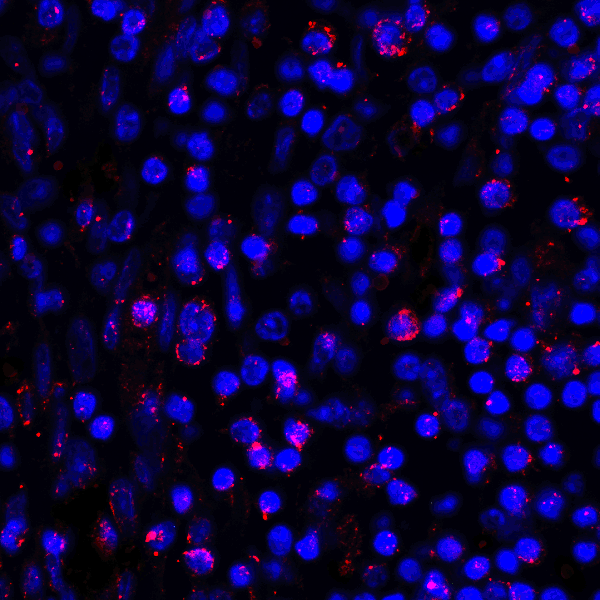
 IF (Immunofluorescence)
(Immunofluorescence analysis of A549 cells using MSH6 antibody.)
IF (Immunofluorescence)
(Immunofluorescence analysis of A549 cells using MSH6 antibody.)
MSH6, Polyclonal Antibody (Cat# AAA10686)
Full Name
MSH6 Polyclonal Antibody
Gene Names
MSH6; GTBP; HSAP; p160; GTMBP; HNPCC5
Reactivity
Human, Mouse, Monkey
Applications
Western Blot, Immunohistochemistry, Immunofluorescence
Purity
Affinity Purification
Pricing


 Application Data
(Immunofluorescesence staining of rat lymph node cryosection with Mouse anti Rat CD8beta, clone 341 , red in A and Mouse anti Rat anti Mouse CD4 antibody, clone W3/25 , green in B. C is the merged picture C with nuclei counterstained blue using DAPI. Low power)
Application Data
(Immunofluorescesence staining of rat lymph node cryosection with Mouse anti Rat CD8beta, clone 341 , red in A and Mouse anti Rat anti Mouse CD4 antibody, clone W3/25 , green in B. C is the merged picture C with nuclei counterstained blue using DAPI. Low power)
CD8 BETA, Monoclonal Antibody (Cat# AAA11894)
Full Name
MOUSE ANTI RAT CD8 BETA:FITC
Gene Names
Cd8b; Cd8b1
Applications
Flow Cytometry
Pricing


 ICC (Immunocytochemistry)
(Immunostaining analysis in HeLa cells. HeLa cells were fixed with 4% paraformaldehyde and permeabilized with 0.1% Triton X-100 in PBS. The cells were immunostained with anti-HARS mAb. [Lot No. HARSA6-1])
ICC (Immunocytochemistry)
(Immunostaining analysis in HeLa cells. HeLa cells were fixed with 4% paraformaldehyde and permeabilized with 0.1% Triton X-100 in PBS. The cells were immunostained with anti-HARS mAb. [Lot No. HARSA6-1])
HARS, Monoclonal Antibody (Cat# AAA10601)
Full Name
Mouse monoclonal antibody Anti-Human HARS
Gene Names
HARS; HRS; USH3B
Reactivity
Human
Applications
Dot Blot, Immunocytochemistry, Immunoprecipitation, Western Blot, Flow Cytometry
Pricing


 IP (Immunoprecipitation)
(MAP2K3 was immunoprecipitated using: Lane A:0.5 mg Jurkat Whole Cell Lysate Lane B:0.5 mg HepG2 Whole Cell Lysate Lane C:0.5 mg HeLa Whole Cell Lysate 2 uL anti-MAP2K3 rabbit polyclonal antibody and 60 ug of Immunomagnetic beads Protein A/G. Primary antibody: Anti-MAP2K3 rabbit polyclonal antibody,at 1:100 dilution Secondary antibody: Clean-Blot IP Detection Reagent (HRP) at 1:1000dilution Developed using the ECL technique. Performed under reducing conditions. Predicted band size: 39 kDa Observed band size :39 kDa)
IP (Immunoprecipitation)
(MAP2K3 was immunoprecipitated using: Lane A:0.5 mg Jurkat Whole Cell Lysate Lane B:0.5 mg HepG2 Whole Cell Lysate Lane C:0.5 mg HeLa Whole Cell Lysate 2 uL anti-MAP2K3 rabbit polyclonal antibody and 60 ug of Immunomagnetic beads Protein A/G. Primary antibody: Anti-MAP2K3 rabbit polyclonal antibody,at 1:100 dilution Secondary antibody: Clean-Blot IP Detection Reagent (HRP) at 1:1000dilution Developed using the ECL technique. Performed under reducing conditions. Predicted band size: 39 kDa Observed band size :39 kDa)
MEK3/MKK3, Polyclonal Antibody (Cat# AAA27720)
Full Name
Anti-MEK3/MKK3 Antibody, Rabbit Polyclonal
Gene Names
MAP2K3; MEK3; MKK3; MAPKK3; PRKMK3; SAPKK2; SAPKK-2
Reactivity
Human, Mouse
Applications
Western Blot, Immunohistochemistry, Immunocytochemistry, Immunofluorescence, Immunoprecipitation
Purity
Protein A & Antigen Affinity
Pricing


 Application Data
(Staining of J774 cells with Rat anti Mouse F4/80 antigen Biotin)
Application Data
(Staining of J774 cells with Rat anti Mouse F4/80 antigen Biotin)
F4/80, Monoclonal Antibody (Cat# AAA12174)
Full Name
RAT ANTI MOUSE F4/80
Gene Names
Emr1; Ly71; F4/80; Gpf480; TM7LN3; DD7A5-7; EGF-TM7
Applications
Immunohistochemistry, Fluorescence Microscopy, Flow Cytometry, Immunofluorescence, Immunoprecipitation, Immunohistochemistry, Radioimmunoassay, Immunohistochemistry, Western Blot
Pricing


 IP (Immunoprecipitation)
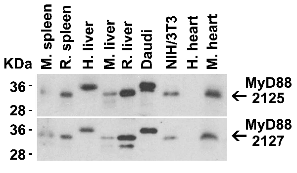
(Figure 13 Immunoprecipitation Validation in HEK293 cells (Kawai et al., 2004)HEK293 cells were transiently transfected with DYKDDDDK-IRF7. Ccell lysates were immunoprecipitated with control rabbit anti-mouse immunoglobulin serum (IgG) or anti-MyD88 (Ab1 and Ab2), followed by immunoblotting with anti-DYKDDDDK.)
IP (Immunoprecipitation)
(Figure 13 Immunoprecipitation Validation in HEK293 cells (Kawai et al., 2004)HEK293 cells were transiently transfected with DYKDDDDK-IRF7. Ccell lysates were immunoprecipitated with control rabbit anti-mouse immunoglobulin serum (IgG) or anti-MyD88 (Ab1 and Ab2), followed by immunoblotting with anti-DYKDDDDK.)
MYD88, Polyclonal Antibody (Cat# AAA10930)
Full Name
MYD88 Antibody
Gene Names
MYD88; MYD88D
Reactivity
Human, Mouse
Applications
Western Blot, Immunohistochemistry, Immunofluorescence
Purity
MYD88 Antibody is affinity chromatography purified via peptide column.
Pricing


 FCM (Flow Cytometry)
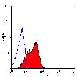
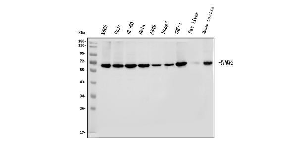
(Figure 8. Flow Cytometry analysis of THP-1 cells using anti-YTHDF2 antibody (AAA19260).Overlay histogram showing THP-1 cells stained with AAA19260 (Blue line). The cells were blocked with 10% normal goat serum. And then incubated with rabbit anti-YTHDF2 Antibody (AAA19260, 1μg/1x106 cells) for 30 min at 20 degree C. DyLight®488 conjugated goat anti-rabbit IgG (5-10μg/1x106 cells) was used as secondary antibody for 30 minutes at 20 degree C. Isotype control antibody (Green line) was rabbit IgG (1μg/1x106) used under the same conditions. Unlabelled sample (Red line) was also used as a control.)
FCM (Flow Cytometry)
(Figure 8. Flow Cytometry analysis of THP-1 cells using anti-YTHDF2 antibody (AAA19260).Overlay histogram showing THP-1 cells stained with AAA19260 (Blue line). The cells were blocked with 10% normal goat serum. And then incubated with rabbit anti-YTHDF2 Antibody (AAA19260, 1μg/1x106 cells) for 30 min at 20 degree C. DyLight®488 conjugated goat anti-rabbit IgG (5-10μg/1x106 cells) was used as secondary antibody for 30 minutes at 20 degree C. Isotype control antibody (Green line) was rabbit IgG (1μg/1x106) used under the same conditions. Unlabelled sample (Red line) was also used as a control.)
YTHDF2, Polyclonal Antibody (Cat# AAA19260)
Full Name
Anti-YTHDF2 Antibody
Gene Names
YTHDF2; HGRG8; NY-REN-2
Reactivity
Human, Mouse, Rat
Applications
Western Blot, Immunohistochemistry, Immunocytochemistry, Immunofluorescence, Flow Cytometry
Purity
Immunogen affinity purified.
Pricing


 Application Data
(At 25 degree C. The primary antibody was diluted at 1/200 and incubated with the sample for 1 hour at 37 degree C. An Alexa Fluor 594 conjugated goat anti-rabbit IgG (H+L) Ab, diluted at 1/600, was used as the secondary antibody.)
Application Data
(At 25 degree C. The primary antibody was diluted at 1/200 and incubated with the sample for 1 hour at 37 degree C. An Alexa Fluor 594 conjugated goat anti-rabbit IgG (H+L) Ab, diluted at 1/600, was used as the secondary antibody.)
p27 Kip1, Polyclonal Antibody (Cat# AAA31388)
Full Name
Phospho-p27 Kip1 (Ser178) Antibody
Gene Names
CDKN1B; KIP1; MEN4; CDKN4; MEN1B; P27KIP1
Reactivity
Human, Mouse, Rat
Predicted Reactivity: Bovine (100%), Sheep (100%), Rabbit (100%), Dog (100%)
Predicted Reactivity: Bovine (100%), Sheep (100%), Rabbit (100%), Dog (100%)
Applications
Western Blot, Immunohistochemistry, Immunofluorescence, Immunocytochemistry, Peptide ELISA
Purity
The antibody is from purified rabbit serum by affinity purification via sequential chromatography on phospho-peptide and non-phospho-peptide affinity columns.
Pricing