Filters
Clonality
Type
Reactivity
Gene Name
Isotype
Host
Application
Clone
1192 results for " A G G" - showing 450-500
 FCM (Flow Cytometry)
(Figure 2. Flow Cytometry analysis of A549 cells using anti-WEE1 antibody (AAA19240).Overlay histogram showing A549 cells stained with AAA19240 (Blue line). The cells were blocked with 10% normal goat serum. And then incubated with rabbit anti-WEE1 Antibody (AAA19240, 1μg/1x106 cells) for 30 min at 20 degree C. DyLight®488 conjugated goat anti-rabbit IgG (5-10μg/1x106 cells) was used as secondary antibody for 30 minutes at 20 degree C. Isotype control antibody (Green line) was rabbit IgG (1μg/1x106) used under the same conditions. Unlabelled sample (Red line) was also used as a control.)
FCM (Flow Cytometry)
(Figure 2. Flow Cytometry analysis of A549 cells using anti-WEE1 antibody (AAA19240).Overlay histogram showing A549 cells stained with AAA19240 (Blue line). The cells were blocked with 10% normal goat serum. And then incubated with rabbit anti-WEE1 Antibody (AAA19240, 1μg/1x106 cells) for 30 min at 20 degree C. DyLight®488 conjugated goat anti-rabbit IgG (5-10μg/1x106 cells) was used as secondary antibody for 30 minutes at 20 degree C. Isotype control antibody (Green line) was rabbit IgG (1μg/1x106) used under the same conditions. Unlabelled sample (Red line) was also used as a control.)
WEE1, Polyclonal Antibody (Cat# AAA19240)
Full Name
Anti-WEE1 Antibody
Gene Names
WEE1; WEE1A; WEE1hu
Reactivity
Human, Mouse
Applications
Western Blot, Flow Cytometry, Direct ELISA
Purity
Immunogen affinity purified.
Pricing


 Application Data
(Staining of mouse spleen with Hamster anti Mouse CD81: Alexa Fluor 488 (AAA11941A488))
Application Data
(Staining of mouse spleen with Hamster anti Mouse CD81: Alexa Fluor 488 (AAA11941A488))
CD81, Monoclonal Antibody (Cat# AAA11941)
Full Name
HAMSTER ANTI MOUSE CD81
Gene Names
Cd81; Tapa1; Tapa-1; Tspan28
Reactivity
Rat
Applications
Immunohistochemistry, Flow Cytometry, Immunoprecipitation, Western Blot
Pricing


 Application Data
(Staining of mouse thymus with RAT ANTI MOUSE CD150: RPE)
Application Data
(Staining of mouse thymus with RAT ANTI MOUSE CD150: RPE)
CD150, Monoclonal Antibody (Cat# AAA26727)
Full Name
CD150 (CD150 Antigen, Cdw150, 4933415F16, Estm51, Ipo 3, Ipo-3, OTTHUMP00000060252, Signaling Lymphocytic Activation Molecule, SLAM, Signaling Lymphocytic Activation Molecule Family Member 1, SLAMF1 Protein) (Biotin)
Reactivity
Mouse
Applications
Flow Cytometry
Purity
Purified by Protein G Affinity Chromatography
Pricing


 Application Data
(Staining of mouse peritoneal macrophages with RAT ANTI MOUSE F4/80 ANTIGEN:FITC)
Application Data
(Staining of mouse peritoneal macrophages with RAT ANTI MOUSE F4/80 ANTIGEN:FITC)
EMR1, Monoclonal Antibody (Cat# AAA26800)
Full Name
EMR1 (EGF-like Module Containing Mucin-like Hormone Receptor-like 1, EMR1 Hormone Receptor, Cell Surface Glycoprotein EMR1, Cell Surface Glycoprotein F4/80, DD7A5-7, EGF-TM7, F4/80, Gpf480, Lymphocyte Antigen 71, Ly71, TM7LN3) (MaxLight 490)
Reactivity
Mouse
Applications
Flow Cytometry, Immunohistochemistry, Immunoprecipitation, Radioimmunoassay, Western Blot
Purity
Purified by protein G affinity chromatography from tissue culture supernatant.
Pricing


 Application Data
(Published clone specific image Alloimmunity-associated cytotoxicity is mediated through the NKG2D receptor. (A) Liver expression of nkg2d on day ten after liver transplantation. (B) Representative NKG2D expression levels in blood NK cells (left) and monocytes (right) of allogeneic (black) and syngeneic (grey) recipients. Isotype was used as control (dashed lines). (C) Sorted blood NK cell cytotoxicity inhibition with anti-NKG2D antibody or with anti-NKp30 antibody. (D) Levels of NKG2D ligand (rae1l, rrlt and irp94) expression in the liver on day ten after transplantation. (E) Levels of NKG2D ligand (rae1l, rrlt and irp94) expression in rat HCC cell lines. (F) Representative level of recombinant NKG2D-Fc binding to rat HCC cells lines. *p)
Application Data
(Published clone specific image Alloimmunity-associated cytotoxicity is mediated through the NKG2D receptor. (A) Liver expression of nkg2d on day ten after liver transplantation. (B) Representative NKG2D expression levels in blood NK cells (left) and monocytes (right) of allogeneic (black) and syngeneic (grey) recipients. Isotype was used as control (dashed lines). (C) Sorted blood NK cell cytotoxicity inhibition with anti-NKG2D antibody or with anti-NKp30 antibody. (D) Levels of NKG2D ligand (rae1l, rrlt and irp94) expression in the liver on day ten after transplantation. (E) Levels of NKG2D ligand (rae1l, rrlt and irp94) expression in rat HCC cell lines. (F) Representative level of recombinant NKG2D-Fc binding to rat HCC cells lines. *p)
CD172a, Monoclonal Antibody (Cat# AAA11875)
Full Name
MOUSE ANTI RAT CD172a:FITC
Gene Names
Sirpa; Bit; Ptpns1; SHPS-1
Applications
Flow Cytometry
Pricing


 Application Data
(Analysis of Protein Array containing more than 19, 000 full-length human proteins using PU.1 Mouse Monoclonal Antibody (PU1/2446). Z- and S- Score: The Z-score represents the strength of a signal that a monoclonal antibody (MAb) (in combination with a fluorescently-tagged anti-IgG secondary antibody) produces when binding to a particular protein on the HuProtTM array. Z-scores are described in units of standard deviations (SD's) above the mean value of all signals generated on that array. If targets on HuProtTM are arranged in descending order of the Z-score, the S-score is the difference (also in units of SD's) between the Z-score. S-score therefore represents the relative target specificity of a MAb to its intended target. A MAb is considered to specific to its intended target, if the MAb has an S-score of at least 2.5. For example, if a MAb binds to protein X with a Z-score of 43 and to protein Y with a Z-score of 14, then the S-score for the binding of that MAb to protein X is equal to 29.)
Application Data
(Analysis of Protein Array containing more than 19, 000 full-length human proteins using PU.1 Mouse Monoclonal Antibody (PU1/2446). Z- and S- Score: The Z-score represents the strength of a signal that a monoclonal antibody (MAb) (in combination with a fluorescently-tagged anti-IgG secondary antibody) produces when binding to a particular protein on the HuProtTM array. Z-scores are described in units of standard deviations (SD's) above the mean value of all signals generated on that array. If targets on HuProtTM are arranged in descending order of the Z-score, the S-score is the difference (also in units of SD's) between the Z-score. S-score therefore represents the relative target specificity of a MAb to its intended target. A MAb is considered to specific to its intended target, if the MAb has an S-score of at least 2.5. For example, if a MAb binds to protein X with a Z-score of 43 and to protein Y with a Z-score of 14, then the S-score for the binding of that MAb to protein X is equal to 29.)
PU.1 (SPI-1), Monoclonal Antibody (Cat# AAA23903)
Full Name
PU.1 (SPI-1) (B-Cell Marker)
Gene Names
SPI1; OF; PU.1; SFPI1; SPI-1; SPI-A
Reactivity
Human. Others not known.
Applications
Western Blot, Immunohistochemistry
Pricing


 FCM (Flow Cytometry)
(Figure 10. Flow Cytometry analysis of K562 cells using anti-RCC1 antibody (AAA19263).Overlay histogram showing K562 cells stained with AAA19263 (Blue line). The cells were blocked with 10% normal goat serum. And then incubated with rabbit anti-RCC1 Antibody (AAA19263,1μg/1x106 cells) for 30 min at 20 degree C. DyLight®488 conjugated goat anti-rabbit IgG (5-10μg/1x106 cells) was used as secondary antibody for 30 minutes at 20 degree C. Isotype control antibody (Green line) was rabbit IgG (1μg/1x106) used under the same conditions. Unlabelled sample (Red line) was also used as a control.)
FCM (Flow Cytometry)
(Figure 10. Flow Cytometry analysis of K562 cells using anti-RCC1 antibody (AAA19263).Overlay histogram showing K562 cells stained with AAA19263 (Blue line). The cells were blocked with 10% normal goat serum. And then incubated with rabbit anti-RCC1 Antibody (AAA19263,1μg/1x106 cells) for 30 min at 20 degree C. DyLight®488 conjugated goat anti-rabbit IgG (5-10μg/1x106 cells) was used as secondary antibody for 30 minutes at 20 degree C. Isotype control antibody (Green line) was rabbit IgG (1μg/1x106) used under the same conditions. Unlabelled sample (Red line) was also used as a control.)
RCC1, Polyclonal Antibody (Cat# AAA19263)
Full Name
Anti-RCC1 Antibody
Gene Names
RCC1; CHC1; RCC1-I; SNHG3-RCC1
Reactivity
Human, Mouse, Rat
Applications
Western Blot, Immunohistochemistry, Immunohistochemistry, Immunocytochemistry, Immunofluorescence, Flow Cytometry, Direct ELISA
Purity
Immunogen affinity purified.
Pricing


 Application Data
(Staining of mouse spleen cells with HAMSTER ANTI MOUSE CD29: Alexa Fluor® 647)
Application Data
(Staining of mouse spleen cells with HAMSTER ANTI MOUSE CD29: Alexa Fluor® 647)
Integrin, beta1, Monoclonal Antibody (Cat# AAA26829)
Full Name
Integrin, beta1 (CD29, Very Late Antigen, VLAb1, Platelet gplla, ITGB1, Fibrinogen Receptor beta Subunit, FNRB, Integrin VLA4 Subunit beta, MDF2, MSK12, Very Late Activation Protein beta Polypeptide, VLAB, VLAbeta) (MaxLight 650)
Reactivity
Mouse, Rat
Applications
FC/FACS, IP
Purity
Purified by Protein G Affinity Chromatography
Pricing


 Application Data
(Staining of mouse spleen cells with HAMSTER ANTI MOUSE CD29: Alexa Fluor® 647)
Application Data
(Staining of mouse spleen cells with HAMSTER ANTI MOUSE CD29: Alexa Fluor® 647)
Integrin, beta1, Monoclonal Antibody (Cat# AAA26815)
Full Name
Integrin, beta1 (CD29, Very Late Antigen, VLAb1, Platelet gplla, ITGB1, Fibrinogen Receptor beta Subunit, FNRB, Integrin VLA4 Subunit beta, MDF2, MSK12, Very Late Activation Protein beta Polypeptide, VLAB, VLAbeta) (MaxLight 550)
Reactivity
Mouse, Rat
Applications
FC/FACS, IP
Purity
Purified by Protein G Affinity Chromatography
Pricing


 IHC (Immunohistochemistry)
(DAB staining on IHC-P Samples: Human Skin cancer TissuePrimary Ab: 30 ug/ml Mouse Anti-Human ADAM17 AntibodySecond Ab: 2 ug/mL HRP-Linked CaprineAnti-Mouse IgG PolyclonalAntibody (Catalog:))
IHC (Immunohistochemistry)
(DAB staining on IHC-P Samples: Human Skin cancer TissuePrimary Ab: 30 ug/ml Mouse Anti-Human ADAM17 AntibodySecond Ab: 2 ug/mL HRP-Linked CaprineAnti-Mouse IgG PolyclonalAntibody (Catalog:))
A Disintegrin And Metalloprotease 17, Monoclonal Antibody (Cat# AAA20829)
Full Name
Monoclonal Antibody to A Disintegrin And Metalloprotease 17 (ADAM17)
Gene Names
ADAM17; CSVP; TACE; NISBD; ADAM18; CD156B; NISBD1
Reactivity
Human, Mouse, Rat
Applications
WB, IHC, ICC, IP
Purity
Protein A + Protein G affinity chromatography
Pricing


 Application Data
(Staining of human peripheral blood lymphocytes with Mouse anti Human CD20: Pacific Blue)
Application Data
(Staining of human peripheral blood lymphocytes with Mouse anti Human CD20: Pacific Blue)
CD20, Monoclonal Antibody (Cat# AAA11868)
Full Name
MOUSE ANTI HUMAN CD20:FITC
Gene Names
MS4A1; B1; S7; Bp35; CD20; CVID5; MS4A2; LEU-16
Applications
Flow Cytometry
Pricing


 FCM (Flow Cytometry)
(Figure 8. Flow Cytometry analysis of SiHa cells using anti- ASS1 antibody (AAA19369).Overlay histogram showing SiHa cells stained with AAA19369 (Blue line). The cells were blocked with 10% normal goat serum. And then incubated with mouse anti-ASS1 Antibody (AAA19369, 1μg/1x106 cells) for 30 min at 20 degree C. DyLight®488 conjugated goat anti-mouse IgG (BA1126, 5-10μg/1x106 cells) was used as secondary antibody for 30 minutes at 20 degree C. Isotype control antibody (Green line) was mouse IgG (1μg/1x106) used under the same conditions. Unlabelled sample (Red line) was also used as a control.)
FCM (Flow Cytometry)
(Figure 8. Flow Cytometry analysis of SiHa cells using anti- ASS1 antibody (AAA19369).Overlay histogram showing SiHa cells stained with AAA19369 (Blue line). The cells were blocked with 10% normal goat serum. And then incubated with mouse anti-ASS1 Antibody (AAA19369, 1μg/1x106 cells) for 30 min at 20 degree C. DyLight®488 conjugated goat anti-mouse IgG (BA1126, 5-10μg/1x106 cells) was used as secondary antibody for 30 minutes at 20 degree C. Isotype control antibody (Green line) was mouse IgG (1μg/1x106) used under the same conditions. Unlabelled sample (Red line) was also used as a control.)
ASS1, Monoclonal Antibody (Cat# AAA19369)
Full Name
Anti-ASS1 Antibody (monoclonal, 5I5)
Gene Names
ASS1; ASS; CTLN1
Reactivity
Human, Mouse, Rat, Monkey
Applications
WB, IHC-P, ICC, IF, FC/FACS/FCM
Purity
Immunogen affinity purified.
Pricing


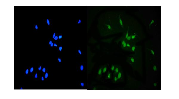
 IF (Immunofluorescence)
(Immunofluorescence of PD-L2 in human colon carcinoma tissue with PD-L2antibody at 20 ?g/mL. Green: PDL2 Antibody [4E10] (AAA10985)Blue: DAPI staining)
IF (Immunofluorescence)
(Immunofluorescence of PD-L2 in human colon carcinoma tissue with PD-L2antibody at 20 ?g/mL. Green: PDL2 Antibody [4E10] (AAA10985)Blue: DAPI staining)
PDL2, Monoclonal Antibody (Cat# AAA10985)
Full Name
PDL2 Antibody [4E10]
Gene Names
PDCD1LG2; B7DC; Btdc; PDL2; CD273; PD-L2; PDCD1L2; bA574F11.2
Reactivity
Human
Applications
Western Blot, Immunohistochemistry, Immunocytochemistry, Immunofluorescence, Flow Cytometry
Purity
PD-L2 Antibody is supplied as protein A purified IgG1.
Pricing


 Application Data
(Published customer image: Leukocyte infiltration in COX-2-M/-M and COX-2+/+ mice. MPO enzymatic activity (panel A) was statistically similar in COX-2-M/-M and COX-2+/+ livers at 6 h and 24 h post-IRI. Ly-6G+ neutrophil (panel B) and granulocyte (panel C) infiltration were also comparable in COX-2-M/-M and COX-2+/+ livers after IRI. Mac-1+ (panel D) and CD68 (panel E) infiltrating macrophages were significantly reduced in COX-2-M/-M livers at 24 h post-reperfusion, but were statistically indistinguishable in COX-2-M/-M and COX-2+/+ livers at 6 h after IRI. No statistical differences in MMP-9 expression (panel F) could be demonstrated in livers of COX-2-M/-M and COX-2+/+ mice post-IRI. Representative immunostaining (panel G) of infiltrating Ly-6G+ (a,b,e,f) and Mac-1+ (c,d,g,h) leukocytes in livers of COX-2+/+ (a,c,e,g) and COX-2-M/-M (b,d,f,h) mice at 6 h (a to d) and 24 h (e to h) post IRI; (n = 5 -6/group; * indicates p)
Application Data
(Published customer image: Leukocyte infiltration in COX-2-M/-M and COX-2+/+ mice. MPO enzymatic activity (panel A) was statistically similar in COX-2-M/-M and COX-2+/+ livers at 6 h and 24 h post-IRI. Ly-6G+ neutrophil (panel B) and granulocyte (panel C) infiltration were also comparable in COX-2-M/-M and COX-2+/+ livers after IRI. Mac-1+ (panel D) and CD68 (panel E) infiltrating macrophages were significantly reduced in COX-2-M/-M livers at 24 h post-reperfusion, but were statistically indistinguishable in COX-2-M/-M and COX-2+/+ livers at 6 h after IRI. No statistical differences in MMP-9 expression (panel F) could be demonstrated in livers of COX-2-M/-M and COX-2+/+ mice post-IRI. Representative immunostaining (panel G) of infiltrating Ly-6G+ (a,b,e,f) and Mac-1+ (c,d,g,h) leukocytes in livers of COX-2+/+ (a,c,e,g) and COX-2-M/-M (b,d,f,h) mice at 6 h (a to d) and 24 h (e to h) post IRI; (n = 5 -6/group; * indicates p)
CD68, Monoclonal Antibody (Cat# AAA12102)
Full Name
RAT ANTI MOUSE CD68
Gene Names
Cd68; Lamp4; gp110; Scard1
Applications
Immunohistochemistry, Flow Cytometry, Immunofluorescence, Immunoprecipitation, Immunohistochemistry, Western Blot
Pricing


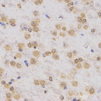
 IHC (Immunohistchemistry)
(Immunohistochemistry of paraffin-embedded mouse kidney using DiMethyl-Histone H3-K9 antibody at dilution of 1:200 (40x lens).)
IHC (Immunohistchemistry)
(Immunohistochemistry of paraffin-embedded mouse kidney using DiMethyl-Histone H3-K9 antibody at dilution of 1:200 (40x lens).)
H3K9me2, Antibody (Cat# AAA10665)
Full Name
Histone H3K9me2 Polyclonal Antibody
Gene Names
HIST3H3; H3t; H3.4; H3/g; H3FT
Reactivity
Human, Mouse, Rat, Other (Wide Range)
Applications
Western Blot, Immunohistochemistry, Immunofluorescence, Immunoprecipitation, Chromatin Immunoprecipitation, Chromatin Immunoprecipitation
Purity
Affinity Purification
Pricing


 Application Data
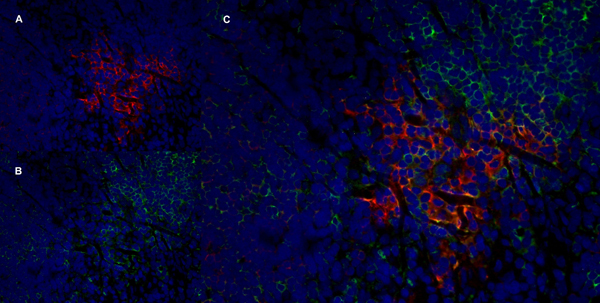
(Immunofluorescence staining of a mouse lymph node cryosection with Rat anti MouseCD19 antibody, clone 6D5 , green in A and Rat anti Mlouse CD8 antibody, clone YTS105.18 , red in B. Merged image in C with nuclei counterstained blue using DAPI. Low power)
Application Data
(Immunofluorescence staining of a mouse lymph node cryosection with Rat anti MouseCD19 antibody, clone 6D5 , green in A and Rat anti Mlouse CD8 antibody, clone YTS105.18 , red in B. Merged image in C with nuclei counterstained blue using DAPI. Low power)
CD19, Monoclonal Antibody (Cat# AAA11932)
Full Name
RAT ANTI MOUSE CD19
Gene Names
Cd19; AW495831
Applications
Immunohistochemistry, Flow Cytometry, Immunoprecipitation
Pricing


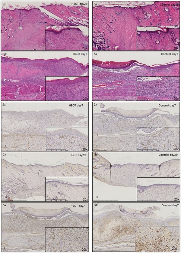
 Application Data
(Published customer image: Histological staining of control and HBOT wounds at post-wounding days 7 and 29. A -D) H&E staining. E -H) CD34 immunohistochemistry. I+J) CD68 immunohistochemistry.From: uk B, Tong M, Fijneman EMG, van Neck JW (2014) Hyperbaric Oxygen Therapy to Treat Diabetes Impaired Wound Healing in Rats. PLoS ONE 9(10): e108533.)
Application Data
(Published customer image: Histological staining of control and HBOT wounds at post-wounding days 7 and 29. A -D) H&E staining. E -H) CD34 immunohistochemistry. I+J) CD68 immunohistochemistry.From: uk B, Tong M, Fijneman EMG, van Neck JW (2014) Hyperbaric Oxygen Therapy to Treat Diabetes Impaired Wound Healing in Rats. PLoS ONE 9(10): e108533.)
CD68, Monoclonal Antibody (Cat# AAA12151)
Full Name
MOUSE ANTI RAT CD68
Applications
Immunohistochemistry, Flow Cytometry, Immunofluorescence, Immunoprecipitation, Immunohistochemistry, Radioimmunoassay, Western Blot
Pricing


 Application Data
(Staining of mouse thymus with RAT ANTI MOUSE CD150: RPE)
Application Data
(Staining of mouse thymus with RAT ANTI MOUSE CD150: RPE)
CD150, Monoclonal Antibody (Cat# AAA26804)
Full Name
CD150 (CD150 Antigen, Cdw150, 4933415F16, Estm51, Ipo 3, Ipo-3, OTTHUMP00000060252, Signaling Lymphocytic Activation Molecule, SLAM, Signaling Lymphocytic Activation Molecule Family Member 1, SLAMF1 Protein) (MaxLight 550)
Reactivity
Mouse
Applications
Flow Cytometry
Purity
Purified by Protein G Affinity Chromatography
Pricing


 Application Data
(Staining of mouse peripheral blood platelets with HAMSTER ANTI MOUSE CD61:RPE)
Application Data
(Staining of mouse peripheral blood platelets with HAMSTER ANTI MOUSE CD61:RPE)
Integrin, beta3, Monoclonal Antibody (Cat# AAA26816)
Full Name
Integrin, beta3 (ITGB3, CD61, GP3A, GPIIIa, HPA-1, NAIT, Platelet Fibrinogen Receptor beta Subunit, Platelet Glycoprotein IIIa, Platelet Membrane Glycoprotein IIIa) (MaxLight 550)
Reactivity
Mouse, Rat
Applications
FC/FACS, IP
Purity
Purified by Protein G Affinity Chromatography
Pricing
 WB (Western Blot)
(Western Blot under reducing conditions of human ovarian cancer tissue lysates using AAA14734 2ug/ml. Followed by IgG (HRP) Anti-Mouse. A specific band was detected for Mesothelin at approximately 50kD.)
WB (Western Blot)
(Western Blot under reducing conditions of human ovarian cancer tissue lysates using AAA14734 2ug/ml. Followed by IgG (HRP) Anti-Mouse. A specific band was detected for Mesothelin at approximately 50kD.)
MSLN, Monoclonal Antibody (Cat# AAA14734)
Full Name
MSLN (Mesothelin, CAK1 Antigen, MPF, Pre-pro-megakaryocyte-potentiating Factor, SMR)
Gene Names
MSLN; MPF; SMRP
Reactivity
Human
Applications
EL/EIA, WB, SW
Purity
Affinity Purified
Purified by Protein G affinity chromatography.
Purified by Protein G affinity chromatography.
Pricing


 Application Data
(C:FGFR2/isolectinB4 (C) and FGFR1/isolectinB4 (D) staining of apparent mesenchymal cells and the subpopulation of endothelial cells. Virtually all other dispersed apparent mesenchymal cells express FGFR1 and FGFR2 (merged image in E). F: FGFR2 (F) and FGFR1 (G) staining in clustered cells of epithelial origin (inferred by morphology here) demonstrating that epithelial cells express both FGFR1 and FGFR2 (merged image with DAPI staining in H).)
Application Data
(C:FGFR2/isolectinB4 (C) and FGFR1/isolectinB4 (D) staining of apparent mesenchymal cells and the subpopulation of endothelial cells. Virtually all other dispersed apparent mesenchymal cells express FGFR1 and FGFR2 (merged image in E). F: FGFR2 (F) and FGFR1 (G) staining in clustered cells of epithelial origin (inferred by morphology here) demonstrating that epithelial cells express both FGFR1 and FGFR2 (merged image with DAPI staining in H).)
FGFR2, Polyclonal Antibody (Cat# AAA26855)
Full Name
FGFR2, NT (FGFR2, BEK, KGFR, KSAM, Fibroblast growth factor receptor 2, K-sam, Keratinocyte growth factor receptor, CD332) (Azide free) (HRP)
Gene Names
FGFR2; BEK; JWS; BBDS; CEK3; CFD1; ECT1; KGFR; TK14; TK25; BFR-1; CD332; K-SAM
Reactivity
Human, Monkey, Mouse, Rat
Applications
IHC, EIA, WB
Purity
Purified by Protein G Affinity Chromatography.
Pricing


 FCM (Flow Cytometry)
(Figure 11. Flow Cytometry analysis of U937 cells using anti-NSF antibody (AAA19222).Overlay histogram showing U937 cells stained with AAA19222 (Blue line). The cells were blocked with 10% normal goat serum. And then incubated with rabbit anti-NSF Antibody (AAA19222, 1μg/1x106 cells) for 30 min at 20 degree C. DyLight®488 conjugated goat anti-rabbit IgG (5-10μg/1x106 cells) was used as secondary antibody for 30 minutes at 20 degree C. Isotype control antibody (Green line) was rabbit IgG (1μg/1x106) used under the same conditions. Unlabelled sample (Red line) was also used as a control.)
FCM (Flow Cytometry)
(Figure 11. Flow Cytometry analysis of U937 cells using anti-NSF antibody (AAA19222).Overlay histogram showing U937 cells stained with AAA19222 (Blue line). The cells were blocked with 10% normal goat serum. And then incubated with rabbit anti-NSF Antibody (AAA19222, 1μg/1x106 cells) for 30 min at 20 degree C. DyLight®488 conjugated goat anti-rabbit IgG (5-10μg/1x106 cells) was used as secondary antibody for 30 minutes at 20 degree C. Isotype control antibody (Green line) was rabbit IgG (1μg/1x106) used under the same conditions. Unlabelled sample (Red line) was also used as a control.)
NSF, Polyclonal Antibody (Cat# AAA19222)
Full Name
Anti-NSF Antibody
Gene Names
NSF; SKD2
Reactivity
Human, Mouse, Rat
Applications
Western Blot, Immunohistochemistry, Immunocytochemistry, Immunofluorescence, Flow Cytometry, Direct ELISA
Purity
Immunogen affinity purified.
Pricing


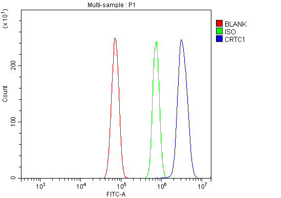 FCM (Flow Cytometry)
(Figure 8. Flow Cytometry analysis of RH35 cells using anti-TORC1/CRTC1 antibody (AAA19251).Overlay histogram showing RH35 cells stained with AAA19251 (Blue line). The cells were blocked with 10% normal goat serum. And then incubated with rabbit anti-TORC1/CRTC1 Antibody (AAA19251, 1μg/1x106 cells) for 30 min at 20 degree C. DyLight®488 conjugated goat anti-rabbit IgG (5-10μg/1x106 cells) was used as secondary antibody for 30 minutes at 20 degree C. Isotype control antibody (Green line) was rabbit IgG (1μg/1x106) used under the same conditions. Unlabelled sample (Red line) was also used as a control.)
FCM (Flow Cytometry)
(Figure 8. Flow Cytometry analysis of RH35 cells using anti-TORC1/CRTC1 antibody (AAA19251).Overlay histogram showing RH35 cells stained with AAA19251 (Blue line). The cells were blocked with 10% normal goat serum. And then incubated with rabbit anti-TORC1/CRTC1 Antibody (AAA19251, 1μg/1x106 cells) for 30 min at 20 degree C. DyLight®488 conjugated goat anti-rabbit IgG (5-10μg/1x106 cells) was used as secondary antibody for 30 minutes at 20 degree C. Isotype control antibody (Green line) was rabbit IgG (1μg/1x106) used under the same conditions. Unlabelled sample (Red line) was also used as a control.)
TORC1/CRTC1, Polyclonal Antibody (Cat# AAA19251)
Full Name
Anti-TORC1/CRTC1 Antibody
Gene Names
CRTC1; MECT1; TORC1; TORC-1; WAMTP1
Reactivity
Human, Mouse, Rat
Applications
Western Blot, Immunohistochemistry, Immunocytochemistry, Immunofluorescence, Flow Cytometry
Purity
Immunogen affinity purified.
Pricing
 Standard Curve (Sample)
Surfactant Associated Protein A (SP-A), ELISA Kit (Cat# AAA13142)
Full Name
Human Surfactant Associated Protein A (SP-A) ELISA Kit
Gene Names
SFTA2; SP-G; SFTPG; UNQ541; GSGL541
Reactivity
Human
Pricing


 Application Data
(Staining of mouse spleen cells with HAMSTER ANTI MOUSE CD29: Alexa Fluor® 647)
Application Data
(Staining of mouse spleen cells with HAMSTER ANTI MOUSE CD29: Alexa Fluor® 647)
Integrin, beta1, Monoclonal Antibody (Cat# AAA14704)
Full Name
Integrin, beta1 (CD29, Very Late Antigen, VLAb1, Platelet gplla, ITGB1, Fibrinogen Receptor beta Subunit, FNRB, Integrin VLA4 Subunit beta, MDF2, MSK12, Very Late Activation Protein beta Polypeptide, VLAB, VLAbeta)
Gene Names
ITGB1; CD29; FNRB; MDF2; VLAB; GPIIA; MSK12; VLA-BETA
Reactivity
Mouse, Rat
Applications
Immunoprecipitation, Flow Cytometry
Purity
Affinity Purified
Purified by Protein G affinity chromatography.
Purified by Protein G affinity chromatography.
Pricing


 FCM (Flow Cytometry)
(Flow cytometry analysis of CD80 overexpressing HEK293 cells using CD80 antibody and control mouse IgG antibody at 10 μg/ml. Blue: Untransfected HEK293 cells. Yellow: CD80 overexpressing HEK293 cells.)
FCM (Flow Cytometry)
(Flow cytometry analysis of CD80 overexpressing HEK293 cells using CD80 antibody and control mouse IgG antibody at 10 μg/ml. Blue: Untransfected HEK293 cells. Yellow: CD80 overexpressing HEK293 cells.)
CD80, Monoclonal Antibody (Cat# AAA10999)
Full Name
CD80 Antibody [11D1]
Gene Names
CD80; B7; BB1; B7-1; B7.1; LAB7; CD28LG; CD28LG1
Reactivity
Human
Applications
Immunohistochemistry, Immunocytochemistry, Immunofluorescence, Flow Cytometry
Purity
Protein A purified
Pricing


 FCM (Flow Cytometry)
(Flow cytometry of Jurkat cells, using anti-STAT5A antibody (Red), compared to a nonspecific negative control antibody (Blue).)
FCM (Flow Cytometry)
(Flow cytometry of Jurkat cells, using anti-STAT5A antibody (Red), compared to a nonspecific negative control antibody (Blue).)
STAT5A, Monoclonal Antibody (Cat# AAA12377)
Full Name
Anti-STAT5A Antibody (clone 9F7) IHC-plus
Gene Names
STAT5A; MGF; STAT5
Reactivity
Dog, Rat, Human, Monkey
Applications
Immunohistochemistry, Immunofluorescence, Western Blot, Flow Cytometry
Purity
Protein A/G purified
Pricing


 Application Data
(Staining of mouse peritoneal macrophages with Rat anti Mouse CD204: Alexa Fluor 488)
Application Data
(Staining of mouse peritoneal macrophages with Rat anti Mouse CD204: Alexa Fluor 488)
CD204, Monoclonal Antibody (Cat# AAA12078)
Full Name
RAT ANTI MOUSE CD204
Gene Names
Msr1; MSR; Scvr; MRS-A; MSR-A; SR-AI; SR-AII; Scara1
Reactivity
Pig, Channel catfish
Applications
Immunohistochemistry, Flow Cytometry, Immunofluorescence, Immunoprecipitation, Western Blot
Pricing

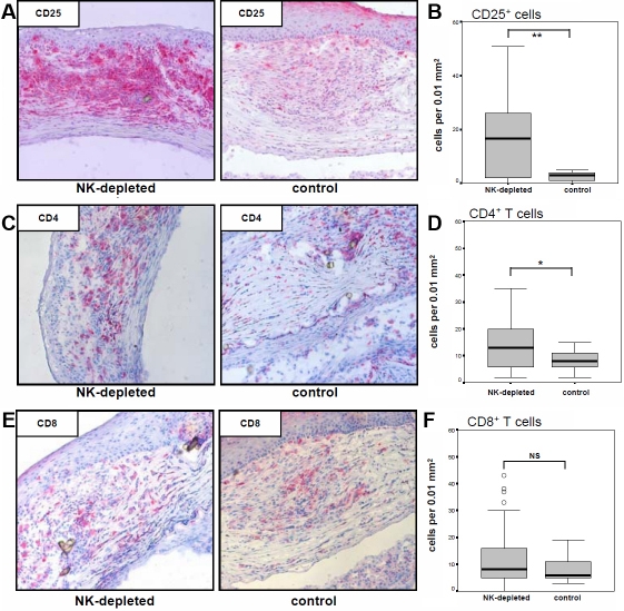


 Application Data
(Immunoperoxidase staining of rat lymph node cryosection with Mouse anti Rat CD25 followed by horseradish peroxidase conjugated Goat anti Mouse IgG1 as a detection reagent. High power)
Application Data
(Immunoperoxidase staining of rat lymph node cryosection with Mouse anti Rat CD25 followed by horseradish peroxidase conjugated Goat anti Mouse IgG1 as a detection reagent. High power)
CD25, Monoclonal Antibody (Cat# AAA12044)
Full Name
MOUSE ANTI RAT CD25:RPE
Gene Names
Il2ra; IL2RAC
Applications
Flow Cytometry
Pricing


 FCM (Flow Cytometry)
(Figure 6. Flow Cytometry analysis of HL-60 cells using anti-MCM6 antibody (AAA19264).Overlay histogram showing HL-60 cells stained with AAA19264 (Blue line). The cells were blocked with 10% normal goat serum. And then incubated with rabbit anti-MCM6 Antibody (AAA19264,1μg/1x106 cells) for 30 min at 20 degree C. DyLight®488 conjugated goat anti-rabbit IgG (5-10μg/1x106 cells) was used as secondary antibody for 30 minutes at 20 degree C. Isotype control antibody (Green line) was rabbit IgG (1μg/1x106) used under the same conditions. Unlabelled sample (Red line) was also used as a control.)
FCM (Flow Cytometry)
(Figure 6. Flow Cytometry analysis of HL-60 cells using anti-MCM6 antibody (AAA19264).Overlay histogram showing HL-60 cells stained with AAA19264 (Blue line). The cells were blocked with 10% normal goat serum. And then incubated with rabbit anti-MCM6 Antibody (AAA19264,1μg/1x106 cells) for 30 min at 20 degree C. DyLight®488 conjugated goat anti-rabbit IgG (5-10μg/1x106 cells) was used as secondary antibody for 30 minutes at 20 degree C. Isotype control antibody (Green line) was rabbit IgG (1μg/1x106) used under the same conditions. Unlabelled sample (Red line) was also used as a control.)
MCM6, Polyclonal Antibody (Cat# AAA19264)
Full Name
Anti-MCM6 Antibody
Gene Names
MCM6; Mis5; P105MCM; MCG40308
Reactivity
Human, Mouse, Rat, Monkey
Applications
Western Blot, Immunohistochemistry, Immunocytochemistry, Immunofluorescence, Flow Cytometry, Direct ELISA
Purity
Immunogen affinity purified.
Pricing


 Application Data
(Published customer image Infiltration of GFP+ BM-cells in infarct and peri-infarct regions. (A-B) Dot plots of viable macrophages/granulocytes (CD11b+CD45high, top right quadrants) and microglia (CD11b+CD45dim, bottom right quadrants) in cortex from BM-chimeric unmanipulated mice and mice exposed to pMCAO. (C) Bar graph showing mean numbers of CD11b+CD45dim microglia and CD11b+CD45high macrophages/granulocytes in BM-chimeric mice 24 hours after pMCAO, subdivided based on expression of GFP (n = 5). Approximately 92% of of the CD45high population were GFP+. (D) Estimation and comparison of mean numbers of CD11b+CD45dim microglia in non-chimeric (n = 10) versus BM-chimeric mice (n = 5) 24 hours after of pMCAO shows significantly fewer CD11b+CD45dim microglial cells in irradiated mice. (E) Overview, showing distribution of infiltrating GFP+ BM-derived cells into infarct (IF) and peri-infarct (P-IF) regions 24 hours after pMCAO. (E-G) By 24 hours, GFP+ single cells (F) and vessel-associated aggregates of GFP+ cells (arrows in G) were observed in infarct and peri-infarct regions. Some of the vessel-associated cells were round, leukocyte-like cells (arrows) while others were elongated cells lining the vasculature (arrow heads in G and in insert). (H) Bar graph showing mean numbers of single GFP+ cells and vessel-associated aggregates of GFP+ cells in ipsi- and contralateral cortex 24 hours after surgery (n = 10). (I-P) Immunohistochemical staining of CD45.1 (I, K), CD45.2 (J, L), IgG2a (M, O) and CD45 (N, P) in ischemic tissue in BM-chimeric (I, J, M, N) and non-chimeric mice (K, L, O, P) 24 hours after pMCAO. N.D, none detected. Scale bars: 200 um (A), 10 um (B, C). 50 um (I-P) *P < 0.05, **P < 0.01, and ***P < 0.001.From: Clausen BH, Lambertsen KL, Babcock AA, Holm TH, Dagnaes-Hansen F, Finsen B. Interleukin-1beta and tumor necrosis factor-alpha are expressed by different subsets of microglia and macrophages after ischemic stroke in mice. J Neuroinflammation. 2008 Oct 23;5:46.)
Application Data
(Published customer image Infiltration of GFP+ BM-cells in infarct and peri-infarct regions. (A-B) Dot plots of viable macrophages/granulocytes (CD11b+CD45high, top right quadrants) and microglia (CD11b+CD45dim, bottom right quadrants) in cortex from BM-chimeric unmanipulated mice and mice exposed to pMCAO. (C) Bar graph showing mean numbers of CD11b+CD45dim microglia and CD11b+CD45high macrophages/granulocytes in BM-chimeric mice 24 hours after pMCAO, subdivided based on expression of GFP (n = 5). Approximately 92% of of the CD45high population were GFP+. (D) Estimation and comparison of mean numbers of CD11b+CD45dim microglia in non-chimeric (n = 10) versus BM-chimeric mice (n = 5) 24 hours after of pMCAO shows significantly fewer CD11b+CD45dim microglial cells in irradiated mice. (E) Overview, showing distribution of infiltrating GFP+ BM-derived cells into infarct (IF) and peri-infarct (P-IF) regions 24 hours after pMCAO. (E-G) By 24 hours, GFP+ single cells (F) and vessel-associated aggregates of GFP+ cells (arrows in G) were observed in infarct and peri-infarct regions. Some of the vessel-associated cells were round, leukocyte-like cells (arrows) while others were elongated cells lining the vasculature (arrow heads in G and in insert). (H) Bar graph showing mean numbers of single GFP+ cells and vessel-associated aggregates of GFP+ cells in ipsi- and contralateral cortex 24 hours after surgery (n = 10). (I-P) Immunohistochemical staining of CD45.1 (I, K), CD45.2 (J, L), IgG2a (M, O) and CD45 (N, P) in ischemic tissue in BM-chimeric (I, J, M, N) and non-chimeric mice (K, L, O, P) 24 hours after pMCAO. N.D, none detected. Scale bars: 200 um (A), 10 um (B, C). 50 um (I-P) *P < 0.05, **P < 0.01, and ***P < 0.001.From: Clausen BH, Lambertsen KL, Babcock AA, Holm TH, Dagnaes-Hansen F, Finsen B. Interleukin-1beta and tumor necrosis factor-alpha are expressed by different subsets of microglia and macrophages after ischemic stroke in mice. J Neuroinflammation. 2008 Oct 23;5:46.)
CD11b, Monoclonal Antibody (Cat# AAA12186)
Full Name
RAT ANTI MOUSE CD11b:RPE
Gene Names
Itgam; CR3; CR3A; MAC1; Cd11b; Ly-40; Mac-1; Mac-1a; CD11b/CD18; F730045J24Rik
Applications
Flow Cytometry
Pricing
 ELISA
(Sandwich ELISA analysis of human PDGF-BB using Mouse anti Human PDGF-BB (AAA12291) as a capture reagent and biotinylated Mouse anti Human PDGF-BB as a detection reagent with purified recombinant PDGF-BB as the antigen. Detection was performed using HRP conjugated streptavidin and substrate.)
ELISA
(Sandwich ELISA analysis of human PDGF-BB using Mouse anti Human PDGF-BB (AAA12291) as a capture reagent and biotinylated Mouse anti Human PDGF-BB as a detection reagent with purified recombinant PDGF-BB as the antigen. Detection was performed using HRP conjugated streptavidin and substrate.)
PDGF-BB, Monoclonal Antibody (Cat# AAA12291)
Full Name
Mouse anti Human PDGF-BB
Gene Names
PDGFB; SIS; SSV; PDGF2; c-sis; PDGF-2
Reactivity
Human
Applications
ELISA
Purity
Purified IgG - liquid
Purified IgG prepared by affinity chromatography on Protein G from tissue culture supernatant
Purified IgG prepared by affinity chromatography on Protein G from tissue culture supernatant
Pricing
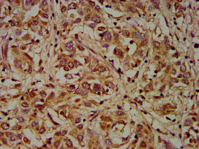
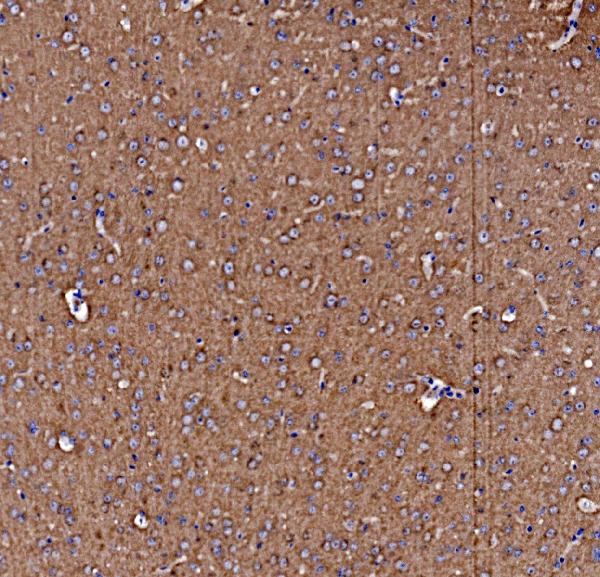
 IHC (Immunohistochemistry)
(IHC image of AAA27003 diluted at 1:200 and staining in paraffin-embedded human skeletal muscle tissue performed on a Leica BondTM system. After dewaxing and hydration, antigen retrieval was mediated by high pressure in a citrate buffer (pH 6.0). Section was blocked with 10% normal goat serum 30min at RT. Then primary antibody (1% BSA) was incubated at 4 degree C overnight. The primary is detected by a biotinylated secondary antibody and visualized using an HRP conjugated SP system.)
IHC (Immunohistochemistry)
(IHC image of AAA27003 diluted at 1:200 and staining in paraffin-embedded human skeletal muscle tissue performed on a Leica BondTM system. After dewaxing and hydration, antigen retrieval was mediated by high pressure in a citrate buffer (pH 6.0). Section was blocked with 10% normal goat serum 30min at RT. Then primary antibody (1% BSA) was incubated at 4 degree C overnight. The primary is detected by a biotinylated secondary antibody and visualized using an HRP conjugated SP system.)
DMD, Polyclonal Antibody (Cat# AAA27003)
Full Name
DMD Antibody
Gene Names
DMD; BMD; CMD3B; MRX85; DXS142; DXS164; DXS206; DXS230; DXS239; DXS268; DXS269; DXS270; DXS272
Reactivity
Human
Applications
Immunohistochemistry
Purity
>95%, Protein G purified
Pricing


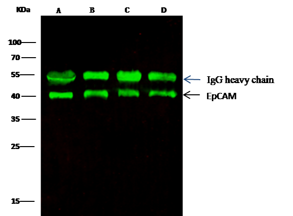 IP (Immunoprecipitation)
(EpCAM was immunoprecipitated using:Lane A:0.5 mg MCF-7 Whole Cell LysateLane B:0.5 mg A431 Whole Cell LysateLane C:0.5 mg HepG2 Whole Cell LysateLane D:0.5 mg Caco-2 Whole Cell Lysate0.5 uL anti-EpCAM rabbit monoclonal antibody and 15 ul of 50 % Protein G agarose.Primary antibody:Anti-EpCAM rabbit monoclonal antibody,at 1:5000 dilution Secondary antibody:Dylight 800-labeled antibody to rabbit IgG (H+L), at 1:5000 dilution Developed using the odssey technique.Performed under reducing conditions.Predicted band size: 40 kDaObserved band size: 40 kDa)
IP (Immunoprecipitation)
(EpCAM was immunoprecipitated using:Lane A:0.5 mg MCF-7 Whole Cell LysateLane B:0.5 mg A431 Whole Cell LysateLane C:0.5 mg HepG2 Whole Cell LysateLane D:0.5 mg Caco-2 Whole Cell Lysate0.5 uL anti-EpCAM rabbit monoclonal antibody and 15 ul of 50 % Protein G agarose.Primary antibody:Anti-EpCAM rabbit monoclonal antibody,at 1:5000 dilution Secondary antibody:Dylight 800-labeled antibody to rabbit IgG (H+L), at 1:5000 dilution Developed using the odssey technique.Performed under reducing conditions.Predicted band size: 40 kDaObserved band size: 40 kDa)
EpCAM, Monoclonal Antibody (Cat# AAA27736)
Full Name
Recombinant Anti-EpCAM Antibody, Rabbit Monoclonal
Gene Names
EPCAM; ESA; KSA; M4S1; MK-1; DIAR5; EGP-2; EGP40; KS1/4; MIC18; TROP1; EGP314; HNPCC8; TACSTD1
Reactivity
Human
Applications
Western Blot, Immunohistochemistry, Flow Cytometry, Immunocytochemistry, Immunofluorescence, Immunoprecipitation
Purity
Protein A
Pricing


 FCM (Flow Cytometry)
(Figure 7. Flow Cytometry analysis of Hela cells using anti-ATG9A antibody (AAA19280).Overlay histogram showing Hela cells stained with AAA19280 (Blue line). The cells were blocked with 10% normal goat serum. And then incubated with rabbit anti-ATG9A Antibody (AAA19280, 1μg/1x106 cells) for 30 min at 20 degree C. DyLight®488 conjugated goat anti-rabbit IgG (5-10μg/1x106 cells) was used as secondary antibody for 30 minutes at 20 degree C. Isotype control antibody (Green line) was rabbit IgG (1μg/1x106) used under the same conditions. Unlabelled sample (Red line) was also used as a control.)
FCM (Flow Cytometry)
(Figure 7. Flow Cytometry analysis of Hela cells using anti-ATG9A antibody (AAA19280).Overlay histogram showing Hela cells stained with AAA19280 (Blue line). The cells were blocked with 10% normal goat serum. And then incubated with rabbit anti-ATG9A Antibody (AAA19280, 1μg/1x106 cells) for 30 min at 20 degree C. DyLight®488 conjugated goat anti-rabbit IgG (5-10μg/1x106 cells) was used as secondary antibody for 30 minutes at 20 degree C. Isotype control antibody (Green line) was rabbit IgG (1μg/1x106) used under the same conditions. Unlabelled sample (Red line) was also used as a control.)
ATG9A, Polyclonal Antibody (Cat# AAA19280)
Full Name
Anti-ATG9A Antibody
Gene Names
ATG9A; mATG9; APG9L1; MGD3208
Reactivity
Human, Mouse, Rat
Applications
WB, IHC-P, FC/FACS/FCM, EIA
Purity
Immunogen affinity purified.
Pricing


 FCM (Flow Cytometry)
(Figure 6. Flow Cytometry analysis of A549 cells using anti-MCM5 antibody (AAA19377).Overlay histogram showing A549 cells stained with AAA19377 (Blue line). The cells were blocked with 10% normal goat serum. And then incubated with mouse anti- MCM5 Antibody (AAA19377, 1μg/1x106 cells) for 30 min at 20 degree C. DyLight®488 conjugated goat anti-mouse IgG (BA1126, 5-10μg/1x106 cells) was used as secondary antibody for 30 minutes at 20 degree C. Isotype control antibody (Green line) was mouse IgG (1μg/1x106) used under the same conditions. Unlabelled sample (Red line) was also used as a control.)
FCM (Flow Cytometry)
(Figure 6. Flow Cytometry analysis of A549 cells using anti-MCM5 antibody (AAA19377).Overlay histogram showing A549 cells stained with AAA19377 (Blue line). The cells were blocked with 10% normal goat serum. And then incubated with mouse anti- MCM5 Antibody (AAA19377, 1μg/1x106 cells) for 30 min at 20 degree C. DyLight®488 conjugated goat anti-mouse IgG (BA1126, 5-10μg/1x106 cells) was used as secondary antibody for 30 minutes at 20 degree C. Isotype control antibody (Green line) was mouse IgG (1μg/1x106) used under the same conditions. Unlabelled sample (Red line) was also used as a control.)
MCM5, Monoclonal Antibody (Cat# AAA19377)
Full Name
Anti-MCM5 Antibody (monoclonal, 4G10)
Gene Names
MCM5; CDC46; P1-CDC46
Reactivity
Human, Mouse, Rat
Applications
WB, IHC-P, ICC, IF, FC/FACS/FCM
Purity
Immunogen affinity purified.
Pricing


 Application Data
(Staining of human peripheral blood lymphocytes with MOUSE ANTI HUMAN CD45RA:FITC (MCA88F))
Application Data
(Staining of human peripheral blood lymphocytes with MOUSE ANTI HUMAN CD45RA:FITC (MCA88F))
CD45RA, Monoclonal Antibody (Cat# AAA26797)
Full Name
CD45RA (CD45 Antigen, B220, GP180, Leukocyte Common Antigen, LCA, L-CA, LY5, LY-5, Protein Tyrosine Phosphatase Receptor Type C Polypeptide, PTPRC, T200, T200 Glycoprotein) (MaxLight 490)
Reactivity
Human, Monkey
Applications
Flow Cytometry, Immunohistochemistry
Purity
Purified by protein G affinity chromatography from tissue culture supernatant.
Pricing


 Application Data
(Staining of CD209 transfected K562 cells with MOUSE ANTI HUMAN CD209: FITC)
Application Data
(Staining of CD209 transfected K562 cells with MOUSE ANTI HUMAN CD209: FITC)
DC-SIGN, Monoclonal Antibody (Cat# AAA26827)
Full Name
DC-SIGN (Dendritic Cell-specific ICAM3 grabbing Non-integrin, C Type Lectin Domain Family 4 Member L, CLEC4L, CD209, CD209 Antigen, CDSIGN, Dendritic Cell-specific ICAM-3-grabbing Non-integrin 1, DC SIGN1, DC-SIGN1, HIV GP120 Binding Protein, MGC129965) (
Reactivity
Human
Applications
FC/FACS, IF
Purity
Purified by protein G affinity chromatography from tissue culture supernatant.
Pricing


 Application Data
(Staining of human peripheral blood granulocytes with Mouse anti Human CD11b:Biotin)
Application Data
(Staining of human peripheral blood granulocytes with Mouse anti Human CD11b:Biotin)
CD11b, Monoclonal Antibody (Cat# AAA11992)
Full Name
MOUSE ANTI HUMAN CD11b
Gene Names
ITGAM; CR3A; MO1A; CD11B; MAC-1; MAC1A; SLEB6
Applications
Immunohistochemistry, Flow Cytometry, Immunoprecipitation
Pricing

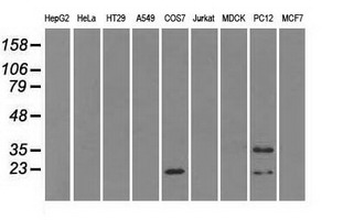


 IP (Immunoprecipitation)
(Immunoprecipitation(IP) of CRYAB by using monoclonal anti-CRYAB antibodies (Negative control: IP without adding anti-CRYAB antibody.). For each experiment, 500ul of DDK tagged CRYAB overexpression lysates (at 1:5 dilution with HEK293T lysate), 2 ug of anti-CRYAB antibody and 20ul (0.1 mg) of goat anti-mouse conjugated magnetic beads were mixed and incubated overnight. After extensive wash to remove any non-specific binding, the immuno-precipitated products were analyzed with rabbit anti-DDK polyclonal antibody.)
IP (Immunoprecipitation)
(Immunoprecipitation(IP) of CRYAB by using monoclonal anti-CRYAB antibodies (Negative control: IP without adding anti-CRYAB antibody.). For each experiment, 500ul of DDK tagged CRYAB overexpression lysates (at 1:5 dilution with HEK293T lysate), 2 ug of anti-CRYAB antibody and 20ul (0.1 mg) of goat anti-mouse conjugated magnetic beads were mixed and incubated overnight. After extensive wash to remove any non-specific binding, the immuno-precipitated products were analyzed with rabbit anti-DDK polyclonal antibody.)
CRYAB / Alpha B Crystallin, Monoclonal Antibody (Cat# AAA12373)
Full Name
Mouse Monoclonal [clone 6D11] (IgG1) to Human CRYAB / Alpha B Crystallin
Gene Names
CRYAB; MFM2; CRYA2; CTPP2; HSPB5; CMD1II; CTRCT16; HEL-S-101
Reactivity
Human, Monkey, Rat
Applications
Immunohistochemistry, Immunofluorescence, Western Blot, Immunoprecipitation, Flow Cytometry
Purity
Protein A/G Purified
Pricing


 Application Data
(Analysis of Protein Array containing more than 19,000 full-length human proteins using HER-2 Mouse Monoclonal Antibody (ERBB2/3080). Z- and S- Score: The Z-score represents the strength of a signal that a monoclonal antibody (MAb) (in combination with a fluorescently-tagged anti-IgG secondary antibody) produces when binding to a particular protein on the HuProtTM array. Z-scores are described in units of standard deviations (SD's) above the mean value of all signals generated on that array. If targets on HuProtTM are arranged in descending order of the Z-score, the S-score is the difference (also in units of SD's) between the Z-score. S-score therefore represents the relative target specificity of a MAb to its intended target. A MAb is considered to specific to its intended target, if the MAb has an S-score of at least 2.5. For example, if a MAb binds to protein X with a Z-score of 43 and to protein Y with a Z-score of 14, then the S-score for the binding of that MAb to protein X is equal to 29.)
Application Data
(Analysis of Protein Array containing more than 19,000 full-length human proteins using HER-2 Mouse Monoclonal Antibody (ERBB2/3080). Z- and S- Score: The Z-score represents the strength of a signal that a monoclonal antibody (MAb) (in combination with a fluorescently-tagged anti-IgG secondary antibody) produces when binding to a particular protein on the HuProtTM array. Z-scores are described in units of standard deviations (SD's) above the mean value of all signals generated on that array. If targets on HuProtTM are arranged in descending order of the Z-score, the S-score is the difference (also in units of SD's) between the Z-score. S-score therefore represents the relative target specificity of a MAb to its intended target. A MAb is considered to specific to its intended target, if the MAb has an S-score of at least 2.5. For example, if a MAb binds to protein X with a Z-score of 43 and to protein Y with a Z-score of 14, then the S-score for the binding of that MAb to protein X is equal to 29.)
HER-2/c-erbB-2/neu/CD340, Monoclonal Antibody (Cat# AAA23912)
Full Name
HER-2/c-erbB-2/neu/CD340
Gene Names
ERBB2; NEU; NGL; HER2; TKR1; CD340; HER-2; MLN 19; HER-2/neu
Reactivity
Human
Applications
Immunohistochemistry
Purity
Purified Ab with BSA and Azide at 200ug/ml OR Purified Ab WITHOUT BSA and Azide at 1.0mg/ml
Pricing


 Application Data
(Published customer image Infiltration of GFP+ BM-cells in infarct and peri-infarct regions. (A-B) Dot plots of viable macrophages/granulocytes (CD11b+CD45high, top right quadrants) and microglia (CD11b+CD45dim, bottom right quadrants) in cortex from BM-chimeric unmanipulated mice and mice exposed to pMCAO. (C) Bar graph showing mean numbers of CD11b+CD45dim microglia and CD11b+CD45high macrophages/granulocytes in BM-chimeric mice 24 hours after pMCAO, subdivided based on expression of GFP (n = 5). Approximately 92% of of the CD45high population were GFP+. (D) Estimation and comparison of mean numbers of CD11b+CD45dim microglia in non-chimeric (n = 10) versus BM-chimeric mice (n = 5) 24 hours after of pMCAO shows significantly fewer CD11b+CD45dim microglial cells in irradiated mice. (E) Overview, showing distribution of infiltrating GFP+ BM-derived cells into infarct (IF) and peri-infarct (P-IF) regions 24 hours after pMCAO. (E-G) By 24 hours, GFP+ single cells (F) and vessel-associated aggregates of GFP+ cells (arrows in G) were observed in infarct and peri-infarct regions. Some of the vessel-associated cells were round, leukocyte-like cells (arrows) while others were elongated cells lining the vasculature (arrow heads in G and in insert). (H) Bar graph showing mean numbers of single GFP+ cells and vessel-associated aggregates of GFP+ cells in ipsi- and contralateral cortex 24 hours after surgery (n = 10). (I-P) Immunohistochemical staining of CD45.1 (I, K), CD45.2 (J, L), IgG2a (M, O) and CD45 (N, P) in ischemic tissue in BM-chimeric (I, J, M, N) and non-chimeric mice (K, L, O, P) 24 hours after pMCAO. N.D, none detected. Scale bars: 200 um (A), 10 um (B, C). 50 um (I-P) *P < 0.05, **P < 0.01, and ***P < 0.001.From: Clausen BH, Lambertsen KL, Babcock AA, Holm TH, Dagnaes-Hansen F, Finsen B. Interleukin-1beta and tumor necrosis factor-alpha are expressed by different subsets of microglia and macrophages after ischemic stroke in mice. J Neuroinflammation. 2008 Oct 23;5:46.)
Application Data
(Published customer image Infiltration of GFP+ BM-cells in infarct and peri-infarct regions. (A-B) Dot plots of viable macrophages/granulocytes (CD11b+CD45high, top right quadrants) and microglia (CD11b+CD45dim, bottom right quadrants) in cortex from BM-chimeric unmanipulated mice and mice exposed to pMCAO. (C) Bar graph showing mean numbers of CD11b+CD45dim microglia and CD11b+CD45high macrophages/granulocytes in BM-chimeric mice 24 hours after pMCAO, subdivided based on expression of GFP (n = 5). Approximately 92% of of the CD45high population were GFP+. (D) Estimation and comparison of mean numbers of CD11b+CD45dim microglia in non-chimeric (n = 10) versus BM-chimeric mice (n = 5) 24 hours after of pMCAO shows significantly fewer CD11b+CD45dim microglial cells in irradiated mice. (E) Overview, showing distribution of infiltrating GFP+ BM-derived cells into infarct (IF) and peri-infarct (P-IF) regions 24 hours after pMCAO. (E-G) By 24 hours, GFP+ single cells (F) and vessel-associated aggregates of GFP+ cells (arrows in G) were observed in infarct and peri-infarct regions. Some of the vessel-associated cells were round, leukocyte-like cells (arrows) while others were elongated cells lining the vasculature (arrow heads in G and in insert). (H) Bar graph showing mean numbers of single GFP+ cells and vessel-associated aggregates of GFP+ cells in ipsi- and contralateral cortex 24 hours after surgery (n = 10). (I-P) Immunohistochemical staining of CD45.1 (I, K), CD45.2 (J, L), IgG2a (M, O) and CD45 (N, P) in ischemic tissue in BM-chimeric (I, J, M, N) and non-chimeric mice (K, L, O, P) 24 hours after pMCAO. N.D, none detected. Scale bars: 200 um (A), 10 um (B, C). 50 um (I-P) *P < 0.05, **P < 0.01, and ***P < 0.001.From: Clausen BH, Lambertsen KL, Babcock AA, Holm TH, Dagnaes-Hansen F, Finsen B. Interleukin-1beta and tumor necrosis factor-alpha are expressed by different subsets of microglia and macrophages after ischemic stroke in mice. J Neuroinflammation. 2008 Oct 23;5:46.)
CD11b, Monoclonal Antibody (Cat# AAA12231)
Full Name
RAT ANTI MOUSE CD11b:Low Endotoxin
Gene Names
Itgam; CR3; CR3A; MAC1; Cd11b; Ly-40; Mac-1; Mac-1a; CD11b/CD18; F730045J24Rik
Applications
Immunohistochemistry, Flow Cytometry, Functional Assay, Immunofluorescence, Immunoprecipitation
Pricing


 Application Data
(Staining of human peripheral blood lymphocytes with MOUSE ANTI HUMAN CD3:ALEXA 647)
Application Data
(Staining of human peripheral blood lymphocytes with MOUSE ANTI HUMAN CD3:ALEXA 647)
CD3, Monoclonal Antibody (Cat# AAA26808)
Full Name
CD3 (MaxLight 550)
Reactivity
Human
Applications
FC/FACS, IHC
Purity
Purified by Protein G Affinity Chromatography
Pricing


 IF (Immunofluorescence)
(Immunofluorescence analysis of NIH/3T3 cells using Acetyl-Histone H3-K9 antibody.)
IF (Immunofluorescence)
(Immunofluorescence analysis of NIH/3T3 cells using Acetyl-Histone H3-K9 antibody.)
Histone H3-K9, Polyclonal Antibody (Cat# AAA29842)
Full Name
Acetyl-Histone H3-K9 pAb
Gene Names
HIST3H3; H3t; H3.4; H3/g; H3FT
Reactivity
Human, Mouse, Rat, Other (Wide Range)
Applications
Western Blot, Immunohistochemistry, Immunofluorescence, Immunoprecipitation, Chromatin Immunoprecipitation, Chromatin Immunoprecipitation
Purity
Affinity purification
Pricing


 Application Data
(Immunoperoxidase staining of mouse lymph node cryosection eith Rat anti Mouse antibody clone R3-63 followed by horseradish peroxidase Goat anti Rat IgG antibody . Medium power)
Application Data
(Immunoperoxidase staining of mouse lymph node cryosection eith Rat anti Mouse antibody clone R3-63 followed by horseradish peroxidase Goat anti Rat IgG antibody . Medium power)
CD13, Monoclonal Antibody (Cat# AAA11954)
Full Name
RAT ANTI MOUSE CD13
Gene Names
Anpep; Apn; AP-M; AP-N; Cd13; P150
Applications
Immunohistochemistry, Flow Cytometry, Immunohistochemistry
Pricing
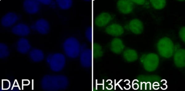


 ChIP (Chromatin Immunoprecipitation)
(Chromatin immunoprecipitation analysis extracts of 293T cells, using TriMethyl-Histone H3-K36 antibody and rabbit IgG. P1 and P2 were located on GAPDH gene. The amount of immunoprecipitated DNA was checked by quantitative PCR. Histogram was constructed by the ratios of the immunoprecipitated DNA to the input.)
ChIP (Chromatin Immunoprecipitation)
(Chromatin immunoprecipitation analysis extracts of 293T cells, using TriMethyl-Histone H3-K36 antibody and rabbit IgG. P1 and P2 were located on GAPDH gene. The amount of immunoprecipitated DNA was checked by quantitative PCR. Histogram was constructed by the ratios of the immunoprecipitated DNA to the input.)
H3K36me3, Antibody (Cat# AAA10657)
Full Name
Histone H3K36me3 Polyclonal Antibody
Gene Names
HIST3H3; H3t; H3.4; H3/g; H3FT
Reactivity
Human, Mouse, Rat, Other (Wide Range)
Applications
Western Blot, Immunohistochemistry, Immunofluorescence, Immunoprecipitation, Chromatin Immunoprecipitation, Chromatin Immunoprecipitation
Purity
Affinity Purification
Pricing


 Application Data
(Staining of human peripheral blood lymphocytes with Mouse anti Human CD4: Pacific Blue)
Application Data
(Staining of human peripheral blood lymphocytes with Mouse anti Human CD4: Pacific Blue)
CD4, Monoclonal Antibody (Cat# AAA11926)
Full Name
MOUSE ANTI HUMAN CD4
Gene Names
CD4; CD4mut
Applications
Immunohistochemistry, Flow Cytometry
Pricing


 Application Data
(Published customer image: Histological staining of control and HBOT wounds at post-wounding days 7 and 29. A -D) H&E staining. E -H) CD34 immunohistochemistry. I+J) CD68 immunohistochemistry.From: uk B, Tong M, Fijneman EMG, van Neck JW (2014) Hyperbaric Oxygen Therapy to Treat Diabetes Impaired Wound Healing in Rats. PLoS ONE 9(10): e108533.)
Application Data
(Published customer image: Histological staining of control and HBOT wounds at post-wounding days 7 and 29. A -D) H&E staining. E -H) CD34 immunohistochemistry. I+J) CD68 immunohistochemistry.From: uk B, Tong M, Fijneman EMG, van Neck JW (2014) Hyperbaric Oxygen Therapy to Treat Diabetes Impaired Wound Healing in Rats. PLoS ONE 9(10): e108533.)
CD68, Monoclonal Antibody (Cat# AAA12148)
Full Name
MOUSE ANTI RAT CD68:FITC
Applications
Flow Cytometry
Pricing


 Application Data
(Published customer image Infiltration of GFP+ BM-cells in infarct and peri-infarct regions. (A-B) Dot plots of viable macrophages/granulocytes (CD11b+CD45high, top right quadrants) and microglia (CD11b+CD45dim, bottom right quadrants) in cortex from BM-chimeric unmanipulated mice and mice exposed to pMCAO. (C) Bar graph showing mean numbers of CD11b+CD45dim microglia and CD11b+CD45high macrophages/granulocytes in BM-chimeric mice 24 hours after pMCAO, subdivided based on expression of GFP (n = 5). Approximately 92% of of the CD45high population were GFP+. (D) Estimation and comparison of mean numbers of CD11b+CD45dim microglia in non-chimeric (n = 10) versus BM-chimeric mice (n = 5) 24 hours after of pMCAO shows significantly fewer CD11b+CD45dim microglial cells in irradiated mice. (E) Overview, showing distribution of infiltrating GFP+ BM-derived cells into infarct (IF) and peri-infarct (P-IF) regions 24 hours after pMCAO. (E-G) By 24 hours, GFP+ single cells (F) and vessel-associated aggregates of GFP+ cells (arrows in G) were observed in infarct and peri-infarct regions. Some of the vessel-associated cells were round, leukocyte-like cells (arrows) while others were elongated cells lining the vasculature (arrow heads in G and in insert). (H) Bar graph showing mean numbers of single GFP+ cells and vessel-associated aggregates of GFP+ cells in ipsi- and contralateral cortex 24 hours after surgery (n = 10). (I-P) Immunohistochemical staining of CD45.1 (I, K), CD45.2 (J, L), IgG2a (M, O) and CD45 (N, P) in ischemic tissue in BM-chimeric (I, J, M, N) and non-chimeric mice (K, L, O, P) 24 hours after pMCAO. N.D, none detected. Scale bars: 200 um (A), 10 um (B, C). 50 um (I-P) *P < 0.05, **P < 0.01, and ***P < 0.001.From: Clausen BH, Lambertsen KL, Babcock AA, Holm TH, Dagnaes-Hansen F, Finsen B. Interleukin-1beta and tumor necrosis factor-alpha are expressed by different subsets of microglia and macrophages after ischemic stroke in mice. J Neuroinflammation. 2008 Oct 23;5:46.)
Application Data
(Published customer image Infiltration of GFP+ BM-cells in infarct and peri-infarct regions. (A-B) Dot plots of viable macrophages/granulocytes (CD11b+CD45high, top right quadrants) and microglia (CD11b+CD45dim, bottom right quadrants) in cortex from BM-chimeric unmanipulated mice and mice exposed to pMCAO. (C) Bar graph showing mean numbers of CD11b+CD45dim microglia and CD11b+CD45high macrophages/granulocytes in BM-chimeric mice 24 hours after pMCAO, subdivided based on expression of GFP (n = 5). Approximately 92% of of the CD45high population were GFP+. (D) Estimation and comparison of mean numbers of CD11b+CD45dim microglia in non-chimeric (n = 10) versus BM-chimeric mice (n = 5) 24 hours after of pMCAO shows significantly fewer CD11b+CD45dim microglial cells in irradiated mice. (E) Overview, showing distribution of infiltrating GFP+ BM-derived cells into infarct (IF) and peri-infarct (P-IF) regions 24 hours after pMCAO. (E-G) By 24 hours, GFP+ single cells (F) and vessel-associated aggregates of GFP+ cells (arrows in G) were observed in infarct and peri-infarct regions. Some of the vessel-associated cells were round, leukocyte-like cells (arrows) while others were elongated cells lining the vasculature (arrow heads in G and in insert). (H) Bar graph showing mean numbers of single GFP+ cells and vessel-associated aggregates of GFP+ cells in ipsi- and contralateral cortex 24 hours after surgery (n = 10). (I-P) Immunohistochemical staining of CD45.1 (I, K), CD45.2 (J, L), IgG2a (M, O) and CD45 (N, P) in ischemic tissue in BM-chimeric (I, J, M, N) and non-chimeric mice (K, L, O, P) 24 hours after pMCAO. N.D, none detected. Scale bars: 200 um (A), 10 um (B, C). 50 um (I-P) *P < 0.05, **P < 0.01, and ***P < 0.001.From: Clausen BH, Lambertsen KL, Babcock AA, Holm TH, Dagnaes-Hansen F, Finsen B. Interleukin-1beta and tumor necrosis factor-alpha are expressed by different subsets of microglia and macrophages after ischemic stroke in mice. J Neuroinflammation. 2008 Oct 23;5:46.)
CD11b, Monoclonal Antibody (Cat# AAA12181)
Full Name
RAT ANTI MOUSE CD11b
Gene Names
Itgam; CR3; CR3A; MAC1; Cd11b; Ly-40; Mac-1; Mac-1a; CD11b/CD18; F730045J24Rik
Reactivity
Human
Applications
Immunohistochemistry, Flow Cytometry, Immunofluorescence, Immunoprecipitation
Pricing


 Application Data
(Staining of mouse thymus with RAT ANTI MOUSE CD150: RPE)
Application Data
(Staining of mouse thymus with RAT ANTI MOUSE CD150: RPE)
CD150, Monoclonal Antibody (Cat# AAA26714)
Full Name
CD150 (CD150 Antigen, Cdw150, 4933415F16, Estm51, Ipo 3, Ipo-3, OTTHUMP00000060252, Signaling Lymphocytic Activation Molecule, SLAM, Signaling Lymphocytic Activation Molecule Family Member 1, SLAMF1 Protein) (AP)
Reactivity
Mouse
Purity
Purified by Protein G Affinity Chromatography
Pricing