Filters
Clonality
Type
Reactivity
Gene Name
Isotype
Host
Application
Clone
1219 results for "Tissue Factor" - showing 300-350


 ICC (Immunocytochemistry)
(ICC staining AQP1 in SW480 cells (red). The nuclear counter stain is DAPI (blue). Cells were fixed in paraformaldehyde, permeabilised with 0.25% Triton X100/PBS.)
ICC (Immunocytochemistry)
(ICC staining AQP1 in SW480 cells (red). The nuclear counter stain is DAPI (blue). Cells were fixed in paraformaldehyde, permeabilised with 0.25% Triton X100/PBS.)
AQP1, Monoclonal Antibody (Cat# AAA30313)
Full Name
AQP1 Antibody
Gene Names
AQP1; CO; CHIP28; AQP-CHIP
Reactivity
Human, Mouse, Rat
Applications
Western Blot, Immunocytochemistry, Immunofluorescence, Immunohistochemistry
Purity
ProA affinity purified
Pricing


 ICC (Immunocytochemistry)
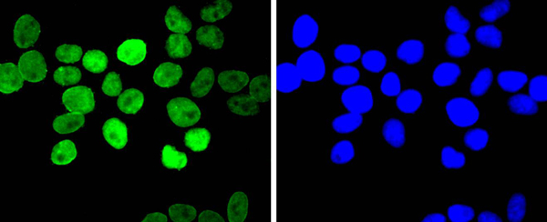
(ICC staining ARID1A in 293 cells (green). The nuclear counter stain is DAPI (blue). Cells were fixed in paraformaldehyde, permeabilised with 0.25% Triton X100/PBS.)
ICC (Immunocytochemistry)
(ICC staining ARID1A in 293 cells (green). The nuclear counter stain is DAPI (blue). Cells were fixed in paraformaldehyde, permeabilised with 0.25% Triton X100/PBS.)
ARID1A, Monoclonal Antibody (Cat# AAA30239)
Full Name
ARID1A Antibody
Gene Names
ARID1A; ELD; B120; OSA1; P270; hELD; BM029; MRD14; hOSA1; BAF250; C1orf4; BAF250a; SMARCF1
Reactivity
Human, Mouse, Rat
Applications
Immunocytochemistry, Immunofluorescence, Immunohistochemistry
Purity
ProA affinity purified
Pricing


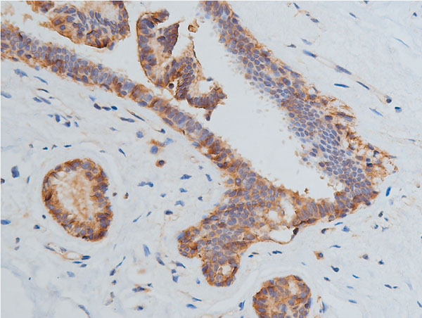 IHC (Immunohistchemistry)
(AAA31091 at 1/200 staining human breast cancer tissue sections by IHC-P. The tissue was formaldehyde fixed and a heat mediated antigen retrieval step in citrate buffer was performed. The tissue was then blocked and incubated with the antibody for 1.5 hours at 22 degree C. An HRP conjugated goat anti-rabbit antibody was used as the secondary.)
IHC (Immunohistchemistry)
(AAA31091 at 1/200 staining human breast cancer tissue sections by IHC-P. The tissue was formaldehyde fixed and a heat mediated antigen retrieval step in citrate buffer was performed. The tissue was then blocked and incubated with the antibody for 1.5 hours at 22 degree C. An HRP conjugated goat anti-rabbit antibody was used as the secondary.)
VEGFR2, Polyclonal Antibody (Cat# AAA31091)
Full Name
VEGFR2 Antibody
Gene Names
KDR; FLK1; CD309; VEGFR; VEGFR2
Reactivity
Human, Mouse, Rat
Applications
Western Blot, Immunohistochemistry, Immunofluorescence, Immunocytochemistry
Purity
The antiserum was purified by peptide affinity chromatography using SulfoLink Coupling Resin.
Pricing


 Application Data
(Analysis of Protein Array containing more than 19, 000 full-length human proteins using PU.1 Mouse Monoclonal Antibody (PU1/2446). Z- and S- Score: The Z-score represents the strength of a signal that a monoclonal antibody (MAb) (in combination with a fluorescently-tagged anti-IgG secondary antibody) produces when binding to a particular protein on the HuProtTM array. Z-scores are described in units of standard deviations (SD's) above the mean value of all signals generated on that array. If targets on HuProtTM are arranged in descending order of the Z-score, the S-score is the difference (also in units of SD's) between the Z-score. S-score therefore represents the relative target specificity of a MAb to its intended target. A MAb is considered to specific to its intended target, if the MAb has an S-score of at least 2.5. For example, if a MAb binds to protein X with a Z-score of 43 and to protein Y with a Z-score of 14, then the S-score for the binding of that MAb to protein X is equal to 29.)
Application Data
(Analysis of Protein Array containing more than 19, 000 full-length human proteins using PU.1 Mouse Monoclonal Antibody (PU1/2446). Z- and S- Score: The Z-score represents the strength of a signal that a monoclonal antibody (MAb) (in combination with a fluorescently-tagged anti-IgG secondary antibody) produces when binding to a particular protein on the HuProtTM array. Z-scores are described in units of standard deviations (SD's) above the mean value of all signals generated on that array. If targets on HuProtTM are arranged in descending order of the Z-score, the S-score is the difference (also in units of SD's) between the Z-score. S-score therefore represents the relative target specificity of a MAb to its intended target. A MAb is considered to specific to its intended target, if the MAb has an S-score of at least 2.5. For example, if a MAb binds to protein X with a Z-score of 43 and to protein Y with a Z-score of 14, then the S-score for the binding of that MAb to protein X is equal to 29.)
PU.1 (SPI-1), Monoclonal Antibody (Cat# AAA23903)
Full Name
PU.1 (SPI-1) (B-Cell Marker)
Gene Names
SPI1; OF; PU.1; SFPI1; SPI-1; SPI-A
Reactivity
Human. Others not known.
Applications
Western Blot, Immunohistochemistry
Pricing


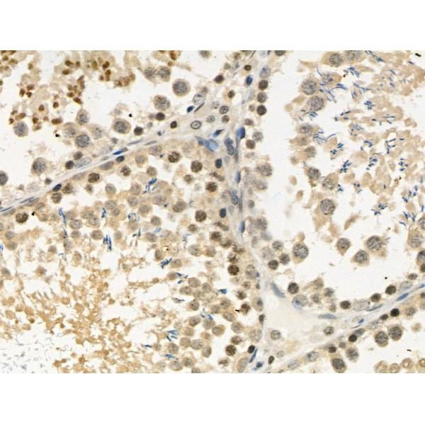

 IHC (Immunohistochemistry)
(At 1/100 staining Mouse kidney tissue by IHC-P. The sample was formaldehyde fixed and a heat mediated antigen retrieval step in citrate buffer was performed. The sample was then blocked and incubated with the primary antibody at 4 degree C overnight. An HRP conjugated anti-Rabbit antibody was used as the secondary antibody.)
IHC (Immunohistochemistry)
(At 1/100 staining Mouse kidney tissue by IHC-P. The sample was formaldehyde fixed and a heat mediated antigen retrieval step in citrate buffer was performed. The sample was then blocked and incubated with the primary antibody at 4 degree C overnight. An HRP conjugated anti-Rabbit antibody was used as the secondary antibody.)
YB1, Polyclonal Antibody (Cat# AAA31294)
Full Name
Phospho-YB1 (Ser102) Antibody
Gene Names
YBX1; YB1; BP-8; CSDB; DBPB; YB-1; CSDA2; NSEP1; NSEP-1; MDR-NF1
Reactivity
Human, Mouse, Rat
Applications
Immunohistochemistry, Peptide ELISA
Purity
The antibody is from purified rabbit serum by affinity purification via sequential chromatography on phospho-peptide and non-phospho-peptide affinity columns.
Pricing
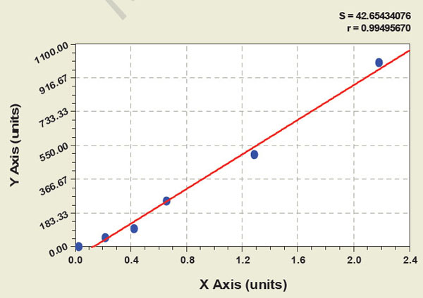
 Standard Curve (Sample)
corticotropin-releasing factor, CRF, ELISA Kit (Cat# AAA11239)
Full Name
Human corticotropin-releasing factor, CRF ELISA Kit
Reactivity
Human
Pricing
 Standard Curve (Sample)
fibroblast growth factor 23, ELISA Kit (Cat# AAA15041)
Full Name
Rat fibroblast growth factor 23, FGF23 ELISA Kit
Reactivity
Rat
Pricing


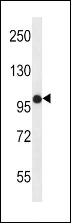
 WB (Western Blot)
(WB Suggested Anti-HCLS1 Antibody Titration: 1.25ug/mlPositive Control: Jurkat cell lysateHCLS1 is supported by BioGPS gene expression data to be expressed in Jurkat)
WB (Western Blot)
(WB Suggested Anti-HCLS1 Antibody Titration: 1.25ug/mlPositive Control: Jurkat cell lysateHCLS1 is supported by BioGPS gene expression data to be expressed in Jurkat)
HCLS1, Polyclonal Antibody (Cat# AAA23398)
Full Name
HCLS1 antibody - N-terminal region
Gene Names
HCLS1; HS1; p75; CTTNL; lckBP1
Reactivity
Cow, Dog, Guinea Pig, Horse, Human, Mouse, Rabbit, Rat
Applications
Immunohistochemistry, Western Blot
Purity
Affinity Purified
Pricing
 Standard Curve (Sample)
Interleukin 5, ELISA Kit (Cat# AAA16941)
Full Name
Monkey Interleukin 5 ELISA Kit
Reactivity
Monkey
Pricing

 IHC (Immunohistochemistry)
(The image on the left is immunohistochemistry of paraffinembedded Human tonsil tissue using POU6F2 Antibody at dilution 1/50, on the right is treated with synthetic peptide. (Original magnification: 200))
IHC (Immunohistochemistry)
(The image on the left is immunohistochemistry of paraffinembedded Human tonsil tissue using POU6F2 Antibody at dilution 1/50, on the right is treated with synthetic peptide. (Original magnification: 200))
POU6F2, Polyclonal Antibody (Cat# AAA29650)
Full Name
POU6F2 Antibody
Gene Names
POU6F2; WT5; WTSL; RPF-1
Reactivity
Human, Mouse
Applications
Western Blot, Immunohistochemistry
Purity
Antigen affinity purification.
Pricing


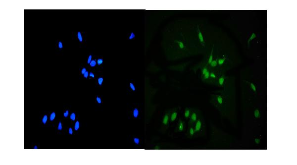
 ICC (Immunocytochemistry)
(ICC staining Apaf-1 in Ags cells (green). The nuclear counter stain is DAPI (blue). Cells were fixed in paraformaldehyde, permeabilised with 0.25% Triton X100/PBS.)
ICC (Immunocytochemistry)
(ICC staining Apaf-1 in Ags cells (green). The nuclear counter stain is DAPI (blue). Cells were fixed in paraformaldehyde, permeabilised with 0.25% Triton X100/PBS.)
Apaf-1, Monoclonal Antibody (Cat# AAA30052)
Full Name
Apaf-1 Antibody
Gene Names
APAF1; CED4; APAF-1
Reactivity
Human, Mouse
Applications
Western Blot, Immunocytochemistry, Immunofluorescence, Immunohistochemistry
Purity
ProA affinity purified
Pricing


 FCM (Flow Cytometry)
(Flow cytometric analysis of Hela cells with Argonaute 2 antibody at 1/50 dilution (red) compared with an unlabelled control (cells without incubation with primary antibody; black). Alexa Fluor 488-conjugated goat anti rabbit IgG was used as the secondary antibody.)
FCM (Flow Cytometry)
(Flow cytometric analysis of Hela cells with Argonaute 2 antibody at 1/50 dilution (red) compared with an unlabelled control (cells without incubation with primary antibody; black). Alexa Fluor 488-conjugated goat anti rabbit IgG was used as the secondary antibody.)
Argonaute 2, Monoclonal Antibody (Cat# AAA30277)
Full Name
Argonaute 2 Antibody
Gene Names
AGO2; Q10; EIF2C2
Reactivity
Human, Mouse, Rat
Applications
Western Blot, Immunocytochemistry, Immunofluorescence, Immunohistochemistry, Immunoprecipitation, Flow Cytometry
Purity
ProA affinity purified
Pricing


 FCM (Flow Cytometry)
(Flow cytometric analysis of THP-1 cells with eEF1A1 antibody at 1/100 dilution (red) compared with an unlabelled control (cells without incubation with primary antibody; black). Alexa Fluor 488-conjugated goat anti-rabbit IgG was used as the secondary antibody.)
FCM (Flow Cytometry)
(Flow cytometric analysis of THP-1 cells with eEF1A1 antibody at 1/100 dilution (red) compared with an unlabelled control (cells without incubation with primary antibody; black). Alexa Fluor 488-conjugated goat anti-rabbit IgG was used as the secondary antibody.)
EEF1A1, Monoclonal Antibody (Cat# AAA30467)
Full Name
EEF1A1 Antibody
Gene Names
EEF1A1; CCS3; EF1A; PTI1; CCS-3; EE1A1; EEF-1; EEF1A; EF-Tu; LENG7; eEF1A-1; GRAF-1EF; HNGC:16303
Reactivity
Human, Mouse, Rat
Applications
Western Blot, Immunocytochemistry, Immunofluorescence, Immunohistochemistry, Flow Cytometry, Immunoprecipitation
Purity
ProA affinity purified
Pricing


 Application Data
(Published customer image: Histological staining of control and HBOT wounds at post-wounding days 7 and 29. A -D) H&E staining. E -H) CD34 immunohistochemistry. I+J) CD68 immunohistochemistry.From: uk B, Tong M, Fijneman EMG, van Neck JW (2014) Hyperbaric Oxygen Therapy to Treat Diabetes Impaired Wound Healing in Rats. PLoS ONE 9(10): e108533.)
Application Data
(Published customer image: Histological staining of control and HBOT wounds at post-wounding days 7 and 29. A -D) H&E staining. E -H) CD34 immunohistochemistry. I+J) CD68 immunohistochemistry.From: uk B, Tong M, Fijneman EMG, van Neck JW (2014) Hyperbaric Oxygen Therapy to Treat Diabetes Impaired Wound Healing in Rats. PLoS ONE 9(10): e108533.)
CD68, Monoclonal Antibody (Cat# AAA12151)
Full Name
MOUSE ANTI RAT CD68
Applications
Immunohistochemistry, Flow Cytometry, Immunofluorescence, Immunoprecipitation, Immunohistochemistry, Radioimmunoassay, Western Blot
Pricing
 Standard Curve (Sample)
Nuclear factor erythroid 2-related factor 2, ELISA Kit (Cat# AAA17229)
Full Name
Chicken Nuclear factor erythroid 2-related factor 2 ELISA Kit
Gene Names
NFE2L2; NRF2
Reactivity
Chicken
Pricing
 Standard Curve (Sample)
nuclear factor-?B p65, NF-kappaB p65, ELISA Kit (Cat# AAA15668)
Full Name
Rat nuclear factor-?B p65, NF-kappaB p65 ELISA Kit
Reactivity
Rat
Pricing


 ICC (Immunocytochemistry)
(ICC staining Phospho-JunD (S255) in MCF-7 cells (green). The nuclear counter stain is DAPI (blue). Cells were fixed in paraformaldehyde, permeabilised with 0.25% Triton X100/PBS.)
ICC (Immunocytochemistry)
(ICC staining Phospho-JunD (S255) in MCF-7 cells (green). The nuclear counter stain is DAPI (blue). Cells were fixed in paraformaldehyde, permeabilised with 0.25% Triton X100/PBS.)
JunD, Monoclonal Antibody (Cat# AAA29784)
Full Name
JunD (Phospho-S255) Antibody
Gene Names
JUND; AP-1
Reactivity
Human, Mouse, Rat
Applications
Western Blot, Immunohistochemistry, Immunocytochemistry, Immunofluorescence, Immunohistochemistry
Purity
ProA affinity purified
Pricing
 WB (Western Blot)
(Western Blot under reducing conditions of human ovarian cancer tissue lysates using AAA14734 2ug/ml. Followed by IgG (HRP) Anti-Mouse. A specific band was detected for Mesothelin at approximately 50kD.)
WB (Western Blot)
(Western Blot under reducing conditions of human ovarian cancer tissue lysates using AAA14734 2ug/ml. Followed by IgG (HRP) Anti-Mouse. A specific band was detected for Mesothelin at approximately 50kD.)
MSLN, Monoclonal Antibody (Cat# AAA14734)
Full Name
MSLN (Mesothelin, CAK1 Antigen, MPF, Pre-pro-megakaryocyte-potentiating Factor, SMR)
Gene Names
MSLN; MPF; SMRP
Reactivity
Human
Applications
EL/EIA, WB, SW
Purity
Affinity Purified
Purified by Protein G affinity chromatography.
Purified by Protein G affinity chromatography.
Pricing


 FCM (Flow Cytometry)
(Flow cytometric analysis of SW480 cells with DR5 antibody at 1/100 dilution (red) compared with an unlabelled control (cells without incubation with primary antibody; black).)
FCM (Flow Cytometry)
(Flow cytometric analysis of SW480 cells with DR5 antibody at 1/100 dilution (red) compared with an unlabelled control (cells without incubation with primary antibody; black).)
DR5, Monoclonal Antibody (Cat# AAA30353)
Full Name
DR5 Antibody
Gene Names
TNFRSF10B; DR5; CD262; KILLER; TRICK2; TRICKB; ZTNFR9; TRAILR2; TRICK2A; TRICK2B; TRAIL-R2; KILLER/DR5
Reactivity
Human
Applications
Western Blot, Immunoprecipitation, Immunohistochemistry, Flow Cytometry
Purity
ProA affinity purified
Pricing


 Application Data
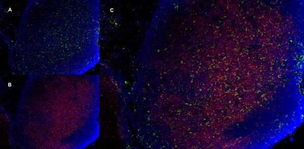
(C:FGFR2/isolectinB4 (C) and FGFR1/isolectinB4 (D) staining of apparent mesenchymal cells and the subpopulation of endothelial cells. Virtually all other dispersed apparent mesenchymal cells express FGFR1 and FGFR2 (merged image in E). F: FGFR2 (F) and FGFR1 (G) staining in clustered cells of epithelial origin (inferred by morphology here) demonstrating that epithelial cells express both FGFR1 and FGFR2 (merged image with DAPI staining in H).)
Application Data
(C:FGFR2/isolectinB4 (C) and FGFR1/isolectinB4 (D) staining of apparent mesenchymal cells and the subpopulation of endothelial cells. Virtually all other dispersed apparent mesenchymal cells express FGFR1 and FGFR2 (merged image in E). F: FGFR2 (F) and FGFR1 (G) staining in clustered cells of epithelial origin (inferred by morphology here) demonstrating that epithelial cells express both FGFR1 and FGFR2 (merged image with DAPI staining in H).)
FGFR2, Polyclonal Antibody (Cat# AAA26855)
Full Name
FGFR2, NT (FGFR2, BEK, KGFR, KSAM, Fibroblast growth factor receptor 2, K-sam, Keratinocyte growth factor receptor, CD332) (Azide free) (HRP)
Gene Names
FGFR2; BEK; JWS; BBDS; CEK3; CFD1; ECT1; KGFR; TK14; TK25; BFR-1; CD332; K-SAM
Reactivity
Human, Monkey, Mouse, Rat
Applications
IHC, EIA, WB
Purity
Purified by Protein G Affinity Chromatography.
Pricing


 FCM (Flow Cytometry)
(Figure 11. Flow Cytometry analysis of U937 cells using anti-NSF antibody (AAA19222).Overlay histogram showing U937 cells stained with AAA19222 (Blue line). The cells were blocked with 10% normal goat serum. And then incubated with rabbit anti-NSF Antibody (AAA19222, 1μg/1x106 cells) for 30 min at 20 degree C. DyLight®488 conjugated goat anti-rabbit IgG (5-10μg/1x106 cells) was used as secondary antibody for 30 minutes at 20 degree C. Isotype control antibody (Green line) was rabbit IgG (1μg/1x106) used under the same conditions. Unlabelled sample (Red line) was also used as a control.)
FCM (Flow Cytometry)
(Figure 11. Flow Cytometry analysis of U937 cells using anti-NSF antibody (AAA19222).Overlay histogram showing U937 cells stained with AAA19222 (Blue line). The cells were blocked with 10% normal goat serum. And then incubated with rabbit anti-NSF Antibody (AAA19222, 1μg/1x106 cells) for 30 min at 20 degree C. DyLight®488 conjugated goat anti-rabbit IgG (5-10μg/1x106 cells) was used as secondary antibody for 30 minutes at 20 degree C. Isotype control antibody (Green line) was rabbit IgG (1μg/1x106) used under the same conditions. Unlabelled sample (Red line) was also used as a control.)
NSF, Polyclonal Antibody (Cat# AAA19222)
Full Name
Anti-NSF Antibody
Gene Names
NSF; SKD2
Reactivity
Human, Mouse, Rat
Applications
Western Blot, Immunohistochemistry, Immunocytochemistry, Immunofluorescence, Flow Cytometry, Direct ELISA
Purity
Immunogen affinity purified.
Pricing
 Standard Curve (Sample)
Lipoprotein associated Phospholipase A2, ELISA Kit (Cat# AAA16475)
Full Name
Human Lipoprotein associated Phospholipase A2 ELISA Kit
Gene Names
PLA2G7; PAFAD; PAFAH; LP-PLA2; LDL-PLA2
Reactivity
Human
Pricing
 Standard Curve (Sample)
Activating Transcription Factor 3 (ATF3), ELISA Kit (Cat# AAA13897)
Full Name
Mouse Activating Transcription Factor 3 (ATF3) ELISA Kit
Gene Names
Atf3; LRG-21
Reactivity
Mouse
Pricing


 FCM (Flow Cytometry)
(Flow cytometric analysis of A549 cells with PRPF8 antibody at 1/100 dilution (red) compared with an unlabelled control (cells without incubation with primary antibody; black).Alexa Fluor 488-conjugated goat anti-rabbit IgG was used as the secondary antibody.)
FCM (Flow Cytometry)
(Flow cytometric analysis of A549 cells with PRPF8 antibody at 1/100 dilution (red) compared with an unlabelled control (cells without incubation with primary antibody; black).Alexa Fluor 488-conjugated goat anti-rabbit IgG was used as the secondary antibody.)
PRPF8, Monoclonal Antibody (Cat# AAA30485)
Full Name
PRPF8 Antibody
Gene Names
PRPF8; PRP8; RP13; HPRP8; PRPC8; SNRNP220
Reactivity
Human, Mouse, Rat
Applications
Western Blot, Immunocytochemistry, Immunohistochemistry, Flow Cytometry
Purity
ProA affinity purified
Pricing


 WB (Western Blot)
(WB Suggested Anti-FOXM1 Antibody Titration: 1 ug/mlPositive Control: Jurkat cell lysate)
WB (Western Blot)
(WB Suggested Anti-FOXM1 Antibody Titration: 1 ug/mlPositive Control: Jurkat cell lysate)
FOXM1, Polyclonal Antibody (Cat# AAA23606)
Full Name
FOXM1 antibody - middle region
Gene Names
FOXM1; MPP2; HFH11; HNF-3; INS-1; MPP-2; PIG29; FKHL16; FOXM1A; FOXM1B; FOXM1C; HFH-11; TRIDENT; MPHOSPH2
Reactivity
Dog, Guinea Pig, Human, Mouse, Rabbit, Rat
Applications
Immunohistochemistry, Western Blot
Purity
Affinity Purified
Pricing


 FCM (Flow Cytometry)
(Figure 6. Flow Cytometry analysis of HL-60 cells using anti-MCM6 antibody (AAA19264).Overlay histogram showing HL-60 cells stained with AAA19264 (Blue line). The cells were blocked with 10% normal goat serum. And then incubated with rabbit anti-MCM6 Antibody (AAA19264,1μg/1x106 cells) for 30 min at 20 degree C. DyLight®488 conjugated goat anti-rabbit IgG (5-10μg/1x106 cells) was used as secondary antibody for 30 minutes at 20 degree C. Isotype control antibody (Green line) was rabbit IgG (1μg/1x106) used under the same conditions. Unlabelled sample (Red line) was also used as a control.)
FCM (Flow Cytometry)
(Figure 6. Flow Cytometry analysis of HL-60 cells using anti-MCM6 antibody (AAA19264).Overlay histogram showing HL-60 cells stained with AAA19264 (Blue line). The cells were blocked with 10% normal goat serum. And then incubated with rabbit anti-MCM6 Antibody (AAA19264,1μg/1x106 cells) for 30 min at 20 degree C. DyLight®488 conjugated goat anti-rabbit IgG (5-10μg/1x106 cells) was used as secondary antibody for 30 minutes at 20 degree C. Isotype control antibody (Green line) was rabbit IgG (1μg/1x106) used under the same conditions. Unlabelled sample (Red line) was also used as a control.)
MCM6, Polyclonal Antibody (Cat# AAA19264)
Full Name
Anti-MCM6 Antibody
Gene Names
MCM6; Mis5; P105MCM; MCG40308
Reactivity
Human, Mouse, Rat, Monkey
Applications
Western Blot, Immunohistochemistry, Immunocytochemistry, Immunofluorescence, Flow Cytometry, Direct ELISA
Purity
Immunogen affinity purified.
Pricing


 Application Data
(Published customer image Infiltration of GFP+ BM-cells in infarct and peri-infarct regions. (A-B) Dot plots of viable macrophages/granulocytes (CD11b+CD45high, top right quadrants) and microglia (CD11b+CD45dim, bottom right quadrants) in cortex from BM-chimeric unmanipulated mice and mice exposed to pMCAO. (C) Bar graph showing mean numbers of CD11b+CD45dim microglia and CD11b+CD45high macrophages/granulocytes in BM-chimeric mice 24 hours after pMCAO, subdivided based on expression of GFP (n = 5). Approximately 92% of of the CD45high population were GFP+. (D) Estimation and comparison of mean numbers of CD11b+CD45dim microglia in non-chimeric (n = 10) versus BM-chimeric mice (n = 5) 24 hours after of pMCAO shows significantly fewer CD11b+CD45dim microglial cells in irradiated mice. (E) Overview, showing distribution of infiltrating GFP+ BM-derived cells into infarct (IF) and peri-infarct (P-IF) regions 24 hours after pMCAO. (E-G) By 24 hours, GFP+ single cells (F) and vessel-associated aggregates of GFP+ cells (arrows in G) were observed in infarct and peri-infarct regions. Some of the vessel-associated cells were round, leukocyte-like cells (arrows) while others were elongated cells lining the vasculature (arrow heads in G and in insert). (H) Bar graph showing mean numbers of single GFP+ cells and vessel-associated aggregates of GFP+ cells in ipsi- and contralateral cortex 24 hours after surgery (n = 10). (I-P) Immunohistochemical staining of CD45.1 (I, K), CD45.2 (J, L), IgG2a (M, O) and CD45 (N, P) in ischemic tissue in BM-chimeric (I, J, M, N) and non-chimeric mice (K, L, O, P) 24 hours after pMCAO. N.D, none detected. Scale bars: 200 um (A), 10 um (B, C). 50 um (I-P) *P < 0.05, **P < 0.01, and ***P < 0.001.From: Clausen BH, Lambertsen KL, Babcock AA, Holm TH, Dagnaes-Hansen F, Finsen B. Interleukin-1beta and tumor necrosis factor-alpha are expressed by different subsets of microglia and macrophages after ischemic stroke in mice. J Neuroinflammation. 2008 Oct 23;5:46.)
Application Data
(Published customer image Infiltration of GFP+ BM-cells in infarct and peri-infarct regions. (A-B) Dot plots of viable macrophages/granulocytes (CD11b+CD45high, top right quadrants) and microglia (CD11b+CD45dim, bottom right quadrants) in cortex from BM-chimeric unmanipulated mice and mice exposed to pMCAO. (C) Bar graph showing mean numbers of CD11b+CD45dim microglia and CD11b+CD45high macrophages/granulocytes in BM-chimeric mice 24 hours after pMCAO, subdivided based on expression of GFP (n = 5). Approximately 92% of of the CD45high population were GFP+. (D) Estimation and comparison of mean numbers of CD11b+CD45dim microglia in non-chimeric (n = 10) versus BM-chimeric mice (n = 5) 24 hours after of pMCAO shows significantly fewer CD11b+CD45dim microglial cells in irradiated mice. (E) Overview, showing distribution of infiltrating GFP+ BM-derived cells into infarct (IF) and peri-infarct (P-IF) regions 24 hours after pMCAO. (E-G) By 24 hours, GFP+ single cells (F) and vessel-associated aggregates of GFP+ cells (arrows in G) were observed in infarct and peri-infarct regions. Some of the vessel-associated cells were round, leukocyte-like cells (arrows) while others were elongated cells lining the vasculature (arrow heads in G and in insert). (H) Bar graph showing mean numbers of single GFP+ cells and vessel-associated aggregates of GFP+ cells in ipsi- and contralateral cortex 24 hours after surgery (n = 10). (I-P) Immunohistochemical staining of CD45.1 (I, K), CD45.2 (J, L), IgG2a (M, O) and CD45 (N, P) in ischemic tissue in BM-chimeric (I, J, M, N) and non-chimeric mice (K, L, O, P) 24 hours after pMCAO. N.D, none detected. Scale bars: 200 um (A), 10 um (B, C). 50 um (I-P) *P < 0.05, **P < 0.01, and ***P < 0.001.From: Clausen BH, Lambertsen KL, Babcock AA, Holm TH, Dagnaes-Hansen F, Finsen B. Interleukin-1beta and tumor necrosis factor-alpha are expressed by different subsets of microglia and macrophages after ischemic stroke in mice. J Neuroinflammation. 2008 Oct 23;5:46.)
CD11b, Monoclonal Antibody (Cat# AAA12186)
Full Name
RAT ANTI MOUSE CD11b:RPE
Gene Names
Itgam; CR3; CR3A; MAC1; Cd11b; Ly-40; Mac-1; Mac-1a; CD11b/CD18; F730045J24Rik
Applications
Flow Cytometry
Pricing
 Standard Curve (Sample)
Macrophage MigRation Inhibitory Factor, ELISA Kit (Cat# AAA16885)
Full Name
Canine Macrophage MigRation Inhibitory Factor ELISA Kit
Gene Names
MIF; GIF; GLIF; MMIF
Reactivity
Canine
Pricing
 Standard Curve (Sample)
Interleukin 18, ELISA Kit (Cat# AAA17037)
Full Name
Porcine Interleukin 18 ELISA Kit
Gene Names
Il18; IL-18
Reactivity
Porcine
Pricing
 ELISA
(Sandwich ELISA analysis of human PDGF-BB using Mouse anti Human PDGF-BB (AAA12291) as a capture reagent and biotinylated Mouse anti Human PDGF-BB as a detection reagent with purified recombinant PDGF-BB as the antigen. Detection was performed using HRP conjugated streptavidin and substrate.)
ELISA
(Sandwich ELISA analysis of human PDGF-BB using Mouse anti Human PDGF-BB (AAA12291) as a capture reagent and biotinylated Mouse anti Human PDGF-BB as a detection reagent with purified recombinant PDGF-BB as the antigen. Detection was performed using HRP conjugated streptavidin and substrate.)
PDGF-BB, Monoclonal Antibody (Cat# AAA12291)
Full Name
Mouse anti Human PDGF-BB
Gene Names
PDGFB; SIS; SSV; PDGF2; c-sis; PDGF-2
Reactivity
Human
Applications
ELISA
Purity
Purified IgG - liquid
Purified IgG prepared by affinity chromatography on Protein G from tissue culture supernatant
Purified IgG prepared by affinity chromatography on Protein G from tissue culture supernatant
Pricing


 WB (Western Blot)
(EIF5 monoclonal antibody, Western Blot analysis of EIF5 expression in Jurkat.)
WB (Western Blot)
(EIF5 monoclonal antibody, Western Blot analysis of EIF5 expression in Jurkat.)
EIF5, Monoclonal Antibody (Cat# AAA25383)
Full Name
EIF5 (Eukaryotic Translation Initiation Factor 5, eIF-5) (HRP)
Gene Names
EIF5; EIF-5; EIF-5A
Reactivity
Human
Applications
Immunohistochemistry, Western Blot
Purity
Purified by Protein A Affinity Chromatography.
Pricing


 FCM (Flow Cytometry)
(Flow cytometric analysis of Jurkat cells with STAT5a antibody at 1/50 dilution (red) compared with an unlabelled control (cells without incubation with primary antibody; black). Alexa Fluor 488-conjugated goat anti rabbit IgG was used as the secondary antibody.)
FCM (Flow Cytometry)
(Flow cytometric analysis of Jurkat cells with STAT5a antibody at 1/50 dilution (red) compared with an unlabelled control (cells without incubation with primary antibody; black). Alexa Fluor 488-conjugated goat anti rabbit IgG was used as the secondary antibody.)
STAT5a, Monoclonal Antibody (Cat# AAA30129)
Full Name
STAT5a Antibody
Gene Names
STAT5A; MGF; STAT5
Reactivity
Human, Mouse, Rat
Applications
Western Blot, Immunocytochemistry, Immunofluorescence, Immunohistochemistry, Immunoprecipitation, Flow Cytometry
Purity
ProA affinity purified
Pricing


 FCM (Flow Cytometry)
(Figure 6. Flow Cytometry analysis of A549 cells using anti-MCM5 antibody (AAA19377).Overlay histogram showing A549 cells stained with AAA19377 (Blue line). The cells were blocked with 10% normal goat serum. And then incubated with mouse anti- MCM5 Antibody (AAA19377, 1μg/1x106 cells) for 30 min at 20 degree C. DyLight®488 conjugated goat anti-mouse IgG (BA1126, 5-10μg/1x106 cells) was used as secondary antibody for 30 minutes at 20 degree C. Isotype control antibody (Green line) was mouse IgG (1μg/1x106) used under the same conditions. Unlabelled sample (Red line) was also used as a control.)
FCM (Flow Cytometry)
(Figure 6. Flow Cytometry analysis of A549 cells using anti-MCM5 antibody (AAA19377).Overlay histogram showing A549 cells stained with AAA19377 (Blue line). The cells were blocked with 10% normal goat serum. And then incubated with mouse anti- MCM5 Antibody (AAA19377, 1μg/1x106 cells) for 30 min at 20 degree C. DyLight®488 conjugated goat anti-mouse IgG (BA1126, 5-10μg/1x106 cells) was used as secondary antibody for 30 minutes at 20 degree C. Isotype control antibody (Green line) was mouse IgG (1μg/1x106) used under the same conditions. Unlabelled sample (Red line) was also used as a control.)
MCM5, Monoclonal Antibody (Cat# AAA19377)
Full Name
Anti-MCM5 Antibody (monoclonal, 4G10)
Gene Names
MCM5; CDC46; P1-CDC46
Reactivity
Human, Mouse, Rat
Applications
WB, IHC-P, ICC, IF, FC/FACS/FCM
Purity
Immunogen affinity purified.
Pricing
 Standard Curve (Sample)
Fibroblast growth factor 15 (FGF15), ELISA Kit (Cat# AAA15900)
Full Name
Mouse Fibroblast growth factor 15, FGF15 ELISA Kit
Reactivity
Mouse
Pricing


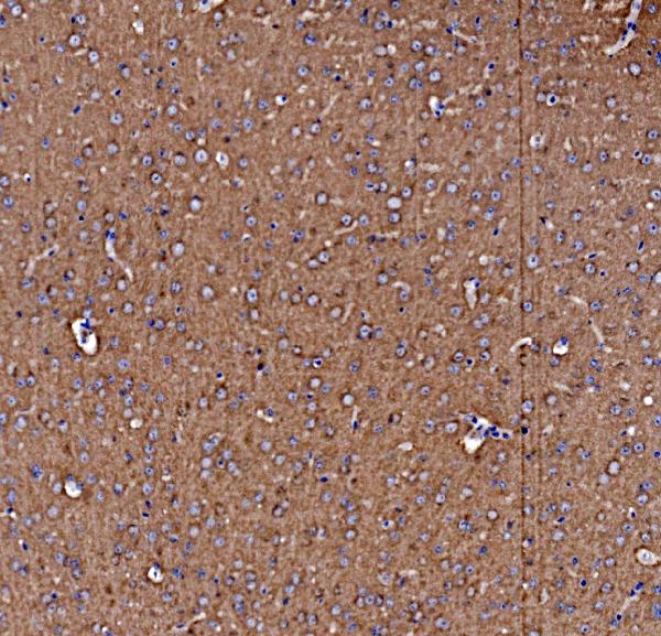
 IHC (Immunohistochemistry)
(Immunohistochemical analysis of paraffin-embedded mouse hippocampus tissue using anti-MEF2A antibody. Counter stained with hematoxylin.)
IHC (Immunohistochemistry)
(Immunohistochemical analysis of paraffin-embedded mouse hippocampus tissue using anti-MEF2A antibody. Counter stained with hematoxylin.)
MEF2A, Monoclonal Antibody (Cat# AAA30354)
Full Name
MEF2A Antibody
Gene Names
MEF2A; mef2; ADCAD1; RSRFC4; RSRFC9
Reactivity
Human, Mouse, Rat
Applications
Western Blot, Immunohistochemistry
Purity
ProA affinity purified
Pricing
 Standard Curve (Sample)
non-metastatic cells 1, protein (NM23A) expressed in, ELISA Kit (Cat# AAA18006)
Full Name
Human Nucleoside diphosphate kinase A, NME1 ELISA Kit
Gene Names
NME1; NB; AWD; NBS; GAAD; NDKA; NM23; NDPKA; NDPK-A; NM23-H1
Reactivity
Human
Pricing


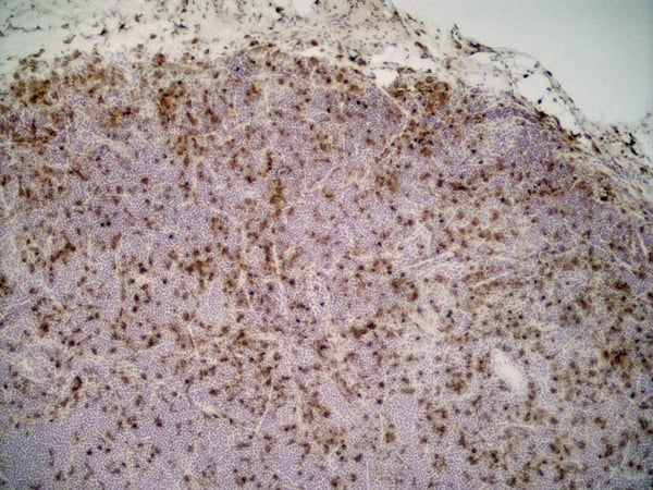
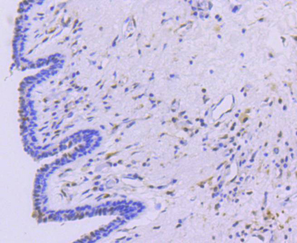
 IHC (Immunohistchemistry)
(At 1/100 staining Human gastric cancer by IHC-P. The sample was formaldehyde fixed and a heat mediated antigen retrieval step in citrate buffer was performed. The sample was then blocked and incubated with the primary antibody at 4 degree C overnight. An HRP conjugated anti-Rabbit antibody was used as the secondary antibody.)
IHC (Immunohistchemistry)
(At 1/100 staining Human gastric cancer by IHC-P. The sample was formaldehyde fixed and a heat mediated antigen retrieval step in citrate buffer was performed. The sample was then blocked and incubated with the primary antibody at 4 degree C overnight. An HRP conjugated anti-Rabbit antibody was used as the secondary antibody.)
SRPK2, Polyclonal Antibody (Cat# AAA31293)
Full Name
Phospho-SRPK2 (Ser494/Ser497) Antibody
Gene Names
SRPK2; SFRSK2
Reactivity
Human, Mouse, Rat
Applications
Immunohistochemistry, Peptide ELISA
Purity
The antibody is from purified rabbit serum by affinity purification via sequential chromatography on phospho-peptide and non-phospho-peptide affinity columns.
Pricing
 Standard Curve (Sample)
Platelet Factor 4, PF-4, ELISA Kit (Cat# AAA11283)
Full Name
Human Platelet Factor 4, PF-4 ELISA Kit
Gene Names
PF4V1; PF4A; CXCL4L1; CXCL4V1; PF4-ALT; SCYB4V1
Reactivity
Human
Pricing
 Standard Curve (Sample)
Coagulation Factor XI, ELISA Kit (Cat# AAA17061)
Full Name
Bovine Coagulation Factor XI ELISA Kit
Gene Names
F11; FXI
Reactivity
Bovine
Pricing
 Standard Curve (Sample)
Proteoglycan 4, ELISA Kit (Cat# AAA17068)
Full Name
Bovine Proteoglycan 4 ELISA Kit
Reactivity
Bovine
Pricing


 IHC (Immunohistochemistry)
(DAB staining on IHC-P; Samples: Human Prostate Gland Cancer Tissue)
IHC (Immunohistochemistry)
(DAB staining on IHC-P; Samples: Human Prostate Gland Cancer Tissue)
Coagulation Factor XIII A1, Polyclonal Antibody (Cat# AAA20937)
Full Name
Polyclonal Antibody to Coagulation Factor XIII A1 Polypeptide (F13A1)
Gene Names
F13A1; F13A
Reactivity
Human, Rat
Applications
WB, ICC, IHC, EIA
Purity
Affinity Chromatography
Pricing


 FCM (Flow Cytometry)
(Flow cytometric analysis of Hela cells with E2F1 antibody at 1/50 dilution (red) compared with an unlabelled control (cells without incubation with primary antibody; black). Alexa Fluor 488-conjugated goat anti rabbit IgG was used as the secondary antibody.)
FCM (Flow Cytometry)
(Flow cytometric analysis of Hela cells with E2F1 antibody at 1/50 dilution (red) compared with an unlabelled control (cells without incubation with primary antibody; black). Alexa Fluor 488-conjugated goat anti rabbit IgG was used as the secondary antibody.)
E2F1, Monoclonal Antibody (Cat# AAA30246)
Full Name
E2F1 Antibody
Gene Names
E2F1; RBP3; E2F-1; RBAP1; RBBP3
Reactivity
Human, Mouse, Rat
Applications
Western Blot, Immunocytochemistry, Immunofluorescence, Immunohistochemistry, Immunoprecipitation, Flow Cytometry
Purity
ProA affinity purified
Pricing


 FCM (Flow Cytometry)
(Figure 8. Flow Cytometry analysis of U20S cells using anti-NFIA antibody (AAA11691).Overlay histogram showing U20S cells stained with AAA11691 (Blue line).The cells were blocked with 10% normal goat serum. And then incubated with rabbit anti-NFIA Antibody (AAA11691,1ug/1x10^6 cells) for 30 min at 20 degree C. DyLight®488 conjugated goat anti-rabbit IgG (5-10ug/1x10^6 cells) was used as secondary antibody for 30 minutes at 20 degree C. Isotype control antibody (Green line) was rabbit IgG (1ug/1x106) used under the same conditions. Unlabelled sample (Red line) was also used as a control.)
FCM (Flow Cytometry)
(Figure 8. Flow Cytometry analysis of U20S cells using anti-NFIA antibody (AAA11691).Overlay histogram showing U20S cells stained with AAA11691 (Blue line).The cells were blocked with 10% normal goat serum. And then incubated with rabbit anti-NFIA Antibody (AAA11691,1ug/1x10^6 cells) for 30 min at 20 degree C. DyLight®488 conjugated goat anti-rabbit IgG (5-10ug/1x10^6 cells) was used as secondary antibody for 30 minutes at 20 degree C. Isotype control antibody (Green line) was rabbit IgG (1ug/1x106) used under the same conditions. Unlabelled sample (Red line) was also used as a control.)
NFIA, Polyclonal Antibody (Cat# AAA11691)
Full Name
Anti-NFIA Antibody
Gene Names
NFIA; CTF; NF1-A; NFI-A; NFI-L; NF-I/A
Reactivity
Human, Mouse, Rat
Applications
Western Blot, Immunohistochemistry
Purity
Immunogen affinity purified.
Pricing


 Application Data
(Published customer image Infiltration of GFP+ BM-cells in infarct and peri-infarct regions. (A-B) Dot plots of viable macrophages/granulocytes (CD11b+CD45high, top right quadrants) and microglia (CD11b+CD45dim, bottom right quadrants) in cortex from BM-chimeric unmanipulated mice and mice exposed to pMCAO. (C) Bar graph showing mean numbers of CD11b+CD45dim microglia and CD11b+CD45high macrophages/granulocytes in BM-chimeric mice 24 hours after pMCAO, subdivided based on expression of GFP (n = 5). Approximately 92% of of the CD45high population were GFP+. (D) Estimation and comparison of mean numbers of CD11b+CD45dim microglia in non-chimeric (n = 10) versus BM-chimeric mice (n = 5) 24 hours after of pMCAO shows significantly fewer CD11b+CD45dim microglial cells in irradiated mice. (E) Overview, showing distribution of infiltrating GFP+ BM-derived cells into infarct (IF) and peri-infarct (P-IF) regions 24 hours after pMCAO. (E-G) By 24 hours, GFP+ single cells (F) and vessel-associated aggregates of GFP+ cells (arrows in G) were observed in infarct and peri-infarct regions. Some of the vessel-associated cells were round, leukocyte-like cells (arrows) while others were elongated cells lining the vasculature (arrow heads in G and in insert). (H) Bar graph showing mean numbers of single GFP+ cells and vessel-associated aggregates of GFP+ cells in ipsi- and contralateral cortex 24 hours after surgery (n = 10). (I-P) Immunohistochemical staining of CD45.1 (I, K), CD45.2 (J, L), IgG2a (M, O) and CD45 (N, P) in ischemic tissue in BM-chimeric (I, J, M, N) and non-chimeric mice (K, L, O, P) 24 hours after pMCAO. N.D, none detected. Scale bars: 200 um (A), 10 um (B, C). 50 um (I-P) *P < 0.05, **P < 0.01, and ***P < 0.001.From: Clausen BH, Lambertsen KL, Babcock AA, Holm TH, Dagnaes-Hansen F, Finsen B. Interleukin-1beta and tumor necrosis factor-alpha are expressed by different subsets of microglia and macrophages after ischemic stroke in mice. J Neuroinflammation. 2008 Oct 23;5:46.)
Application Data
(Published customer image Infiltration of GFP+ BM-cells in infarct and peri-infarct regions. (A-B) Dot plots of viable macrophages/granulocytes (CD11b+CD45high, top right quadrants) and microglia (CD11b+CD45dim, bottom right quadrants) in cortex from BM-chimeric unmanipulated mice and mice exposed to pMCAO. (C) Bar graph showing mean numbers of CD11b+CD45dim microglia and CD11b+CD45high macrophages/granulocytes in BM-chimeric mice 24 hours after pMCAO, subdivided based on expression of GFP (n = 5). Approximately 92% of of the CD45high population were GFP+. (D) Estimation and comparison of mean numbers of CD11b+CD45dim microglia in non-chimeric (n = 10) versus BM-chimeric mice (n = 5) 24 hours after of pMCAO shows significantly fewer CD11b+CD45dim microglial cells in irradiated mice. (E) Overview, showing distribution of infiltrating GFP+ BM-derived cells into infarct (IF) and peri-infarct (P-IF) regions 24 hours after pMCAO. (E-G) By 24 hours, GFP+ single cells (F) and vessel-associated aggregates of GFP+ cells (arrows in G) were observed in infarct and peri-infarct regions. Some of the vessel-associated cells were round, leukocyte-like cells (arrows) while others were elongated cells lining the vasculature (arrow heads in G and in insert). (H) Bar graph showing mean numbers of single GFP+ cells and vessel-associated aggregates of GFP+ cells in ipsi- and contralateral cortex 24 hours after surgery (n = 10). (I-P) Immunohistochemical staining of CD45.1 (I, K), CD45.2 (J, L), IgG2a (M, O) and CD45 (N, P) in ischemic tissue in BM-chimeric (I, J, M, N) and non-chimeric mice (K, L, O, P) 24 hours after pMCAO. N.D, none detected. Scale bars: 200 um (A), 10 um (B, C). 50 um (I-P) *P < 0.05, **P < 0.01, and ***P < 0.001.From: Clausen BH, Lambertsen KL, Babcock AA, Holm TH, Dagnaes-Hansen F, Finsen B. Interleukin-1beta and tumor necrosis factor-alpha are expressed by different subsets of microglia and macrophages after ischemic stroke in mice. J Neuroinflammation. 2008 Oct 23;5:46.)
CD11b, Monoclonal Antibody (Cat# AAA12231)
Full Name
RAT ANTI MOUSE CD11b:Low Endotoxin
Gene Names
Itgam; CR3; CR3A; MAC1; Cd11b; Ly-40; Mac-1; Mac-1a; CD11b/CD18; F730045J24Rik
Applications
Immunohistochemistry, Flow Cytometry, Functional Assay, Immunofluorescence, Immunoprecipitation
Pricing
 Standard Curve (Sample)
TNF-alpha (Tumor Necrosis Factor-alpha), ELISA Kit (Cat# AAA27544)
Full Name
Hamster TNF-alpha (Tumor Necrosis Factor-alpha) ELISA Kit
Reactivity
Hamster
Pricing
 Standard Curve (Sample)
placental growth factor, ELISA Kit (Cat# AAA15437)
Full Name
Rat placenta growth factor, PLGF ELISA Kit
Gene Names
Pgf; Plgf
Reactivity
Rat
Pricing
 Standard Curve (Sample)
S100 calcium binding protein A8, ELISA Kit (Cat# AAA15675)
Full Name
human S100 calcium binding protein A8/calgranulin A, S100A8 ELISA Kit
Gene Names
S100A8; P8; MIF; NIF; CAGA; CFAG; CGLA; L1Ag; MRP8; CP-10; MA387; 60B8AG
Reactivity
Human
Pricing


 WB (Western Blot)
(WB Suggested Anti-NFATC3 Antibody Titration: 0.2-1 ug/mlELISA Titer: 1:62500Positive Control: Hela cell lysate)
WB (Western Blot)
(WB Suggested Anti-NFATC3 Antibody Titration: 0.2-1 ug/mlELISA Titer: 1:62500Positive Control: Hela cell lysate)
NFATC3, Polyclonal Antibody (Cat# AAA23612)
Full Name
NFATC3 antibody - N-terminal region
Gene Names
NFATC3; NFAT4; NFATX; NF-AT4c
Reactivity
Cow, Dog, Horse, Human, Mouse, Rat
Applications
Immunohistochemistry, Western Blot
Purity
Affinity Purified
Pricing


 ICC (Immunocytochemistry)
(ICC staining Lipocalin-2 in SW480 cells (red). The nuclear counter stain is DAPI (blue). Cells were fixed in paraformaldehyde, permeabilised with 0.25% Triton X100/PBS.)
ICC (Immunocytochemistry)
(ICC staining Lipocalin-2 in SW480 cells (red). The nuclear counter stain is DAPI (blue). Cells were fixed in paraformaldehyde, permeabilised with 0.25% Triton X100/PBS.)
Lipocalin-2, Monoclonal Antibody (Cat# AAA30316)
Full Name
Lipocalin-2 Antibody
Reactivity
Human
Applications
Western Blot, Immunocytochemistry, Immunofluorescence, Immunohistochemistry
Purity
ProA affinity purified
Pricing


 IHC (Immunohistochemistry)
(Figure 8. IHC analysis of RbAp48 using anti-RbAp48 antibody (AAA11671).RbAp48 was detected in frozen section of rat small intestine tissue. Heat mediated antigen retrieval was performed in citrate buffer (pH6, epitope retrieval solution) for 20 mins. The tissue section was blocked with 10% goat serum. The tissue section was then incubated with 1ug/ml rabbit anti-RbAp48 Antibody (AAA11671) overnight at 4 degree C. Biotinylated goat anti-rabbit IgG was used as secondary antibody and incubated for 30 minutes at 37 degree C. The tissue section was developed using Strepavidin-Biotin-Complex (SABC) with DAB as the chromogen.)
IHC (Immunohistochemistry)
(Figure 8. IHC analysis of RbAp48 using anti-RbAp48 antibody (AAA11671).RbAp48 was detected in frozen section of rat small intestine tissue. Heat mediated antigen retrieval was performed in citrate buffer (pH6, epitope retrieval solution) for 20 mins. The tissue section was blocked with 10% goat serum. The tissue section was then incubated with 1ug/ml rabbit anti-RbAp48 Antibody (AAA11671) overnight at 4 degree C. Biotinylated goat anti-rabbit IgG was used as secondary antibody and incubated for 30 minutes at 37 degree C. The tissue section was developed using Strepavidin-Biotin-Complex (SABC) with DAB as the chromogen.)
RbAp48, Polyclonal Antibody (Cat# AAA11671)
Full Name
Anti-RbAp48 Antibody
Gene Names
RBBP4; NURF55; RBAP48; lin-53
Reactivity
Human, Mouse, Rat
Applications
Western Blot, Immunohistochemistry
Purity
Immunogen Affinity Purified
Pricing