Filters
Clonality
Type
Reactivity
Gene Name
Isotype
Host
Application
Clone
1219 results for "Tissue Factor" - showing 200-250
 Application Data
CD62L, Monoclonal Antibody (Cat# AAA14858)
Full Name
anti-human CD62L-APC
Reactivity
Human, Horse, Mouse, Guinea Pig, Rat, Dog, Cow
Applications
Flow Cytometry
Pricing


 FCM (Flow Cytometry)
(Flow cytometric analysis of A431 cells with EGFR antibody at 1/50 dilution (red) compared with an unlabelled control (cells without incubation with primary antibody; black). Alexa Fluor 488-conjugated goat anti rabbit IgG was used as the secondary antibody.)
FCM (Flow Cytometry)
(Flow cytometric analysis of A431 cells with EGFR antibody at 1/50 dilution (red) compared with an unlabelled control (cells without incubation with primary antibody; black). Alexa Fluor 488-conjugated goat anti rabbit IgG was used as the secondary antibody.)
EGFR, Monoclonal Antibody (Cat# AAA30021)
Full Name
EGFR Antibody
Gene Names
EGFR; ERBB; HER1; mENA; ERBB1; PIG61
Reactivity
Human, Mouse, Rat
Applications
Western Blot, Immunocytochemistry, Immunofluorescence, Immunohistochemistry, Immunoprecipitation, Flow Cytometry
Purity
ProA affinity purified
Pricing


 FCM (Flow Cytometry)
(Flow cytometric analysis of Hela cells with CREB antibody at 1/50 dilution (blue) compared with an unlabelled control (cells without incubation with primary antibody; red). Alexa Fluor 488-conjugated goat anti rabbit IgG was used as the secondary antibody.)
FCM (Flow Cytometry)
(Flow cytometric analysis of Hela cells with CREB antibody at 1/50 dilution (blue) compared with an unlabelled control (cells without incubation with primary antibody; red). Alexa Fluor 488-conjugated goat anti rabbit IgG was used as the secondary antibody.)
CREB, Monoclonal Antibody (Cat# AAA29973)
Full Name
CREB Antibody
Gene Names
CREB1; CREB
Reactivity
Human, Mouse, Rat, Zebrafish
Applications
Western Blot, Immunocytochemistry, Immunofluorescence, Immunohistochemistry, Immunoprecipitation, Flow Cytometry
Purity
ProA affinity purified
Pricing


 FCM (Flow Cytometry)
(Flow cytometric analysis of Hela cells with Peroxiredoxin 1 antibody at 1/50 dilution (red) compared with an unlabelled control (cells without incubation with primary antibody; black). Alexa Fluor 488-conjugated goat anti rabbit IgG was used as the secondary antibody)
FCM (Flow Cytometry)
(Flow cytometric analysis of Hela cells with Peroxiredoxin 1 antibody at 1/50 dilution (red) compared with an unlabelled control (cells without incubation with primary antibody; black). Alexa Fluor 488-conjugated goat anti rabbit IgG was used as the secondary antibody)
Peroxiredoxin 1, Monoclonal Antibody (Cat# AAA30264)
Full Name
Peroxiredoxin 1 Antibody
Gene Names
PRDX1; PAG; PAGA; PAGB; PRX1; PRXI; MSP23; NKEFA; TDPX2; NKEF-A
Reactivity
Human, Mouse
Applications
Western Blot, Immunocytochemistry, Immunofluorescence, Immunohistochemistry, Flow Cytometry
Purity
ProA affinity purified
Pricing


 WB (Western Blot)
(WB Suggested Anti-HHEX antibody Titration: 1 ug/mLSample Type: Human HepG2)
WB (Western Blot)
(WB Suggested Anti-HHEX antibody Titration: 1 ug/mLSample Type: Human HepG2)
HHEX, Polyclonal Antibody (Cat# AAA23384)
Full Name
HHEX antibody - middle region
Gene Names
HHEX; HEX; PRH; HMPH; PRHX; HOX11L-PEN
Reactivity
Cow, Dog, Guinea Pig, Horse, Human, Mouse, Rat
Applications
Western Blot, Immunohistochemistry
Purity
Affinity Purified
Pricing


 ICC (Immunocytochemistry)
(ICC staining NGF in NIH/3T3 cells (green). The nuclear counter stain is DAPI (blue). Cells were fixed in paraformaldehyde, permeabilised with 0.25% Triton X100/PBS.)
ICC (Immunocytochemistry)
(ICC staining NGF in NIH/3T3 cells (green). The nuclear counter stain is DAPI (blue). Cells were fixed in paraformaldehyde, permeabilised with 0.25% Triton X100/PBS.)
NGF, Monoclonal Antibody (Cat# AAA30040)
Full Name
NGF Antibody
Gene Names
NGF; NGFB; HSAN5; Beta-NGF
Reactivity
Human, Mouse, Rat, Zebrafish
Applications
Western Blot, Immunocytochemistry, Immunofluorescence, Immunohistochemistry
Purity
ProA affinity purified
Pricing


 ICC (Immunocytochemistry)
(ICC staining KLF4 in 293T cells (red). The nuclear counter stain is DAPI (blue). Cells were fixed in paraformaldehyde, permeabilised with 0.25% Triton X100/PBS.)
ICC (Immunocytochemistry)
(ICC staining KLF4 in 293T cells (red). The nuclear counter stain is DAPI (blue). Cells were fixed in paraformaldehyde, permeabilised with 0.25% Triton X100/PBS.)
KLF4, Monoclonal Antibody (Cat# AAA30289)
Full Name
KLF4 Antibody
Gene Names
KLF4; EZF; GKLF
Reactivity
Human, Mouse, Rat
Applications
Western Blot, Immunocytochemistry, Immunofluorescence, Immunohistochemistry
Purity
ProA affinity purified
Pricing


 FCM (Flow Cytometry)
(Flow cytometric analysis of Hela cells with PDGFR alpha antibody at 1/50 dilution (red) compared with an unlabelled control (cells without incubation with primary antibody; black). Alexa Fluor 488-conjugated goat anti rabbit IgG was used as the secondary antibody.)
FCM (Flow Cytometry)
(Flow cytometric analysis of Hela cells with PDGFR alpha antibody at 1/50 dilution (red) compared with an unlabelled control (cells without incubation with primary antibody; black). Alexa Fluor 488-conjugated goat anti rabbit IgG was used as the secondary antibody.)
PDGFR alpha, Monoclonal Antibody (Cat# AAA30282)
Full Name
PDGFR alpha Antibody
Gene Names
PDGFRA; CD140A; PDGFR2; PDGFR-2; RHEPDGFRA
Reactivity
Human, Mouse, Rat
Applications
Western Blot, Immunocytochemistry, Immunohistochemistry, Flow Cytometry
Purity
ProA affinity purified
Pricing


 Application Data
(Published customer image Infiltration of GFP+ BM-cells in infarct and peri-infarct regions. (A-B) Dot plots of viable macrophages/granulocytes (CD11b+CD45high, top right quadrants) and microglia (CD11b+CD45dim, bottom right quadrants) in cortex from BM-chimeric unmanipulated mice and mice exposed to pMCAO. (C) Bar graph showing mean numbers of CD11b+CD45dim microglia and CD11b+CD45high macrophages/granulocytes in BM-chimeric mice 24 hours after pMCAO, subdivided based on expression of GFP (n = 5). Approximately 92% of of the CD45high population were GFP+. (D) Estimation and comparison of mean numbers of CD11b+CD45dim microglia in non-chimeric (n = 10) versus BM-chimeric mice (n = 5) 24 hours after of pMCAO shows significantly fewer CD11b+CD45dim microglial cells in irradiated mice. (E) Overview, showing distribution of infiltrating GFP+ BM-derived cells into infarct (IF) and peri-infarct (P-IF) regions 24 hours after pMCAO. (E-G) By 24 hours, GFP+ single cells (F) and vessel-associated aggregates of GFP+ cells (arrows in G) were observed in infarct and peri-infarct regions. Some of the vessel-associated cells were round, leukocyte-like cells (arrows) while others were elongated cells lining the vasculature (arrow heads in G and in insert). (H) Bar graph showing mean numbers of single GFP+ cells and vessel-associated aggregates of GFP+ cells in ipsi- and contralateral cortex 24 hours after surgery (n = 10). (I-P) Immunohistochemical staining of CD45.1 (I, K), CD45.2 (J, L), IgG2a (M, O) and CD45 (N, P) in ischemic tissue in BM-chimeric (I, J, M, N) and non-chimeric mice (K, L, O, P) 24 hours after pMCAO. N.D, none detected. Scale bars: 200 um (A), 10 um (B, C). 50 um (I-P) *P < 0.05, **P < 0.01, and ***P < 0.001.From: Clausen BH, Lambertsen KL, Babcock AA, Holm TH, Dagnaes-Hansen F, Finsen B. Interleukin-1beta and tumor necrosis factor-alpha are expressed by different subsets of microglia and macrophages after ischemic stroke in mice. J Neuroinflammation. 2008 Oct 23;5:46.)
Application Data
(Published customer image Infiltration of GFP+ BM-cells in infarct and peri-infarct regions. (A-B) Dot plots of viable macrophages/granulocytes (CD11b+CD45high, top right quadrants) and microglia (CD11b+CD45dim, bottom right quadrants) in cortex from BM-chimeric unmanipulated mice and mice exposed to pMCAO. (C) Bar graph showing mean numbers of CD11b+CD45dim microglia and CD11b+CD45high macrophages/granulocytes in BM-chimeric mice 24 hours after pMCAO, subdivided based on expression of GFP (n = 5). Approximately 92% of of the CD45high population were GFP+. (D) Estimation and comparison of mean numbers of CD11b+CD45dim microglia in non-chimeric (n = 10) versus BM-chimeric mice (n = 5) 24 hours after of pMCAO shows significantly fewer CD11b+CD45dim microglial cells in irradiated mice. (E) Overview, showing distribution of infiltrating GFP+ BM-derived cells into infarct (IF) and peri-infarct (P-IF) regions 24 hours after pMCAO. (E-G) By 24 hours, GFP+ single cells (F) and vessel-associated aggregates of GFP+ cells (arrows in G) were observed in infarct and peri-infarct regions. Some of the vessel-associated cells were round, leukocyte-like cells (arrows) while others were elongated cells lining the vasculature (arrow heads in G and in insert). (H) Bar graph showing mean numbers of single GFP+ cells and vessel-associated aggregates of GFP+ cells in ipsi- and contralateral cortex 24 hours after surgery (n = 10). (I-P) Immunohistochemical staining of CD45.1 (I, K), CD45.2 (J, L), IgG2a (M, O) and CD45 (N, P) in ischemic tissue in BM-chimeric (I, J, M, N) and non-chimeric mice (K, L, O, P) 24 hours after pMCAO. N.D, none detected. Scale bars: 200 um (A), 10 um (B, C). 50 um (I-P) *P < 0.05, **P < 0.01, and ***P < 0.001.From: Clausen BH, Lambertsen KL, Babcock AA, Holm TH, Dagnaes-Hansen F, Finsen B. Interleukin-1beta and tumor necrosis factor-alpha are expressed by different subsets of microglia and macrophages after ischemic stroke in mice. J Neuroinflammation. 2008 Oct 23;5:46.)
CD11b, Monoclonal Antibody (Cat# AAA12185)
Full Name
RAT ANTI MOUSE CD11b
Gene Names
Itgam; CR3; CR3A; MAC1; Cd11b; Ly-40; Mac-1; Mac-1a; CD11b/CD18; F730045J24Rik
Applications
Immunohistochemistry, Flow Cytometry, Immunofluorescence, Immunoprecipitation
Pricing


 Application Data
(Published customer image Infiltration of GFP+ BM-cells in infarct and peri-infarct regions. (A-B) Dot plots of viable macrophages/granulocytes (CD11b+CD45high, top right quadrants) and microglia (CD11b+CD45dim, bottom right quadrants) in cortex from BM-chimeric unmanipulated mice and mice exposed to pMCAO. (C) Bar graph showing mean numbers of CD11b+CD45dim microglia and CD11b+CD45high macrophages/granulocytes in BM-chimeric mice 24 hours after pMCAO, subdivided based on expression of GFP (n = 5). Approximately 92% of of the CD45high population were GFP+. (D) Estimation and comparison of mean numbers of CD11b+CD45dim microglia in non-chimeric (n = 10) versus BM-chimeric mice (n = 5) 24 hours after of pMCAO shows significantly fewer CD11b+CD45dim microglial cells in irradiated mice. (E) Overview, showing distribution of infiltrating GFP+ BM-derived cells into infarct (IF) and peri-infarct (P-IF) regions 24 hours after pMCAO. (E-G) By 24 hours, GFP+ single cells (F) and vessel-associated aggregates of GFP+ cells (arrows in G) were observed in infarct and peri-infarct regions. Some of the vessel-associated cells were round, leukocyte-like cells (arrows) while others were elongated cells lining the vasculature (arrow heads in G and in insert). (H) Bar graph showing mean numbers of single GFP+ cells and vessel-associated aggregates of GFP+ cells in ipsi- and contralateral cortex 24 hours after surgery (n = 10). (I-P) Immunohistochemical staining of CD45.1 (I, K), CD45.2 (J, L), IgG2a (M, O) and CD45 (N, P) in ischemic tissue in BM-chimeric (I, J, M, N) and non-chimeric mice (K, L, O, P) 24 hours after pMCAO. N.D, none detected. Scale bars: 200 um (A), 10 um (B, C). 50 um (I-P) *P < 0.05, **P < 0.01, and ***P < 0.001.From: Clausen BH, Lambertsen KL, Babcock AA, Holm TH, Dagnaes-Hansen F, Finsen B. Interleukin-1beta and tumor necrosis factor-alpha are expressed by different subsets of microglia and macrophages after ischemic stroke in mice. J Neuroinflammation. 2008 Oct 23;5:46.)
Application Data
(Published customer image Infiltration of GFP+ BM-cells in infarct and peri-infarct regions. (A-B) Dot plots of viable macrophages/granulocytes (CD11b+CD45high, top right quadrants) and microglia (CD11b+CD45dim, bottom right quadrants) in cortex from BM-chimeric unmanipulated mice and mice exposed to pMCAO. (C) Bar graph showing mean numbers of CD11b+CD45dim microglia and CD11b+CD45high macrophages/granulocytes in BM-chimeric mice 24 hours after pMCAO, subdivided based on expression of GFP (n = 5). Approximately 92% of of the CD45high population were GFP+. (D) Estimation and comparison of mean numbers of CD11b+CD45dim microglia in non-chimeric (n = 10) versus BM-chimeric mice (n = 5) 24 hours after of pMCAO shows significantly fewer CD11b+CD45dim microglial cells in irradiated mice. (E) Overview, showing distribution of infiltrating GFP+ BM-derived cells into infarct (IF) and peri-infarct (P-IF) regions 24 hours after pMCAO. (E-G) By 24 hours, GFP+ single cells (F) and vessel-associated aggregates of GFP+ cells (arrows in G) were observed in infarct and peri-infarct regions. Some of the vessel-associated cells were round, leukocyte-like cells (arrows) while others were elongated cells lining the vasculature (arrow heads in G and in insert). (H) Bar graph showing mean numbers of single GFP+ cells and vessel-associated aggregates of GFP+ cells in ipsi- and contralateral cortex 24 hours after surgery (n = 10). (I-P) Immunohistochemical staining of CD45.1 (I, K), CD45.2 (J, L), IgG2a (M, O) and CD45 (N, P) in ischemic tissue in BM-chimeric (I, J, M, N) and non-chimeric mice (K, L, O, P) 24 hours after pMCAO. N.D, none detected. Scale bars: 200 um (A), 10 um (B, C). 50 um (I-P) *P < 0.05, **P < 0.01, and ***P < 0.001.From: Clausen BH, Lambertsen KL, Babcock AA, Holm TH, Dagnaes-Hansen F, Finsen B. Interleukin-1beta and tumor necrosis factor-alpha are expressed by different subsets of microglia and macrophages after ischemic stroke in mice. J Neuroinflammation. 2008 Oct 23;5:46.)
CD11b, Monoclonal Antibody (Cat# AAA12183)
Full Name
RAT ANTI MOUSE CD11b:FITC
Gene Names
Itgam; CR3; CR3A; MAC1; Cd11b; Ly-40; Mac-1; Mac-1a; CD11b/CD18; F730045J24Rik
Applications
Flow Cytometry
Pricing
 Standard Curve (Sample)
Pregnancy Associated Plasma Protein A (PAPPA), ELISA Kit (Cat# AAA20592)
Full Name
Mouse Pregnancy Associated Plasma Protein A (PAPPA) ELISA Kit
Reactivity
Mouse
Pricing


 FCM (Flow Cytometry)
(Figure 7. Flow Cytometry analysis of U251 cells using anti-Serum Response Factor/SRF antibody (AAA19221).Overlay histogram showing U251 cells stained with AAA19221 (Blue line). The cells were blocked with 10% normal goat serum. And then incubated with rabbit anti-Serum Response Factor/SRF Antibody (AAA19221, 1μg/1x106 cells) for 30 min at 20 degree C. DyLight®488 conjugated goat anti-rabbit IgG (5-10μg/1x106 cells) was used as secondary antibody for 30 minutes at 20 degree C. Isotype control antibody (Green line) was rabbit IgG (1μg/1x106) used under the same conditions. Unlabelled sample (Red line) was also used as a control.)
FCM (Flow Cytometry)
(Figure 7. Flow Cytometry analysis of U251 cells using anti-Serum Response Factor/SRF antibody (AAA19221).Overlay histogram showing U251 cells stained with AAA19221 (Blue line). The cells were blocked with 10% normal goat serum. And then incubated with rabbit anti-Serum Response Factor/SRF Antibody (AAA19221, 1μg/1x106 cells) for 30 min at 20 degree C. DyLight®488 conjugated goat anti-rabbit IgG (5-10μg/1x106 cells) was used as secondary antibody for 30 minutes at 20 degree C. Isotype control antibody (Green line) was rabbit IgG (1μg/1x106) used under the same conditions. Unlabelled sample (Red line) was also used as a control.)
Serum Response Factor/SRF, Polyclonal Antibody (Cat# AAA19221)
Full Name
Anti-Serum Response Factor/SRF Antibody
Gene Names
SRF; MCM1
Reactivity
Human, Mouse, Rat
Applications
Western Blot, Immunohistochemistry, Immunocytochemistry, Immunofluorescence, Flow Cytometry, Direct ELISA
Purity
Immunogen affinity purified.
Pricing


 ICC (Immunocytochemistry)
(ICC staining CD147 in SKOV-3 cells (red). The nuclear counter stain is DAPI (blue). Cells were fixed in paraformaldehyde, permeabilised with 0.25% Triton X100/PBS.)
ICC (Immunocytochemistry)
(ICC staining CD147 in SKOV-3 cells (red). The nuclear counter stain is DAPI (blue). Cells were fixed in paraformaldehyde, permeabilised with 0.25% Triton X100/PBS.)
CD147, Monoclonal Antibody (Cat# AAA30287)
Full Name
CD147 Antibody
Gene Names
BSG; M6; OK; 5F7; TCSF; CD147; EMMPRIN
Reactivity
Human, Mouse, Rat
Applications
Western Blot, Immunocytochemistry, Immunofluorescence, Immunohistochemistry
Purity
ProA affinity purified
Pricing


 Application Data
(Staining of J774 cells with Rat anti Mouse F4/80 antigen Biotin)
Application Data
(Staining of J774 cells with Rat anti Mouse F4/80 antigen Biotin)
F4/80, Monoclonal Antibody (Cat# AAA12169)
Full Name
RAT ANTI MOUSE F4/80
Gene Names
Emr1; Ly71; F4/80; Gpf480; TM7LN3; DD7A5-7; EGF-TM7
Applications
Immunohistochemistry, Fluorescence Microscopy, Flow Cytometry, Immunofluorescence, Immunoprecipitation, Immunohistochemistry, Radioimmunoassay, Immunohistochemistry, Western Blot
Pricing


 Application Data
(Analysis of Protein Array containing more than 19, 000 full-length human proteins using Oct-2 Mouse Monoclonal Antibody (OCT2/2137) Z- and S- Score: The Z-score represents the strength of a signal that a monoclonal antibody (MAb) (in combination with a fluorescently-tagged anti-IgG secondary antibody) produces when binding to a particular protein on the HuProtTM array. Z-scores are described in units of standard deviations (SD's) above the mean value of all signals generated on that array. If targets on HuProtTM are arranged in descending order of the Z-score, the S-score is the difference (also in units of SD's) between the Z-score. S-score therefore represents the relative target specificity of a MAb to its intended target. A MAb is considered to specific to its intended target, if the MAb has an S-score of at least 2.5. For example, if a MAb binds to protein X with a Z-score of 43 and to protein Y with a Z-score of 14, then the S-score for the binding of that MAb to protein X is equal to 29.)
Application Data
(Analysis of Protein Array containing more than 19, 000 full-length human proteins using Oct-2 Mouse Monoclonal Antibody (OCT2/2137) Z- and S- Score: The Z-score represents the strength of a signal that a monoclonal antibody (MAb) (in combination with a fluorescently-tagged anti-IgG secondary antibody) produces when binding to a particular protein on the HuProtTM array. Z-scores are described in units of standard deviations (SD's) above the mean value of all signals generated on that array. If targets on HuProtTM are arranged in descending order of the Z-score, the S-score is the difference (also in units of SD's) between the Z-score. S-score therefore represents the relative target specificity of a MAb to its intended target. A MAb is considered to specific to its intended target, if the MAb has an S-score of at least 2.5. For example, if a MAb binds to protein X with a Z-score of 43 and to protein Y with a Z-score of 14, then the S-score for the binding of that MAb to protein X is equal to 29.)
OCT-2 (POU2F2), Monoclonal Antibody (Cat# AAA23902)
Full Name
OCT-2 (POU2F2) (B-Cell Marker)
Gene Names
POU2F2; OCT2; OTF2; Oct-2
Reactivity
Human. Others not known.
Applications
Western Blot, Immunohistochemistry
Pricing
 Standard Curve (Sample)
IL-18, ELISA Kit (Cat# AAA13491)
Full Name
IL-18 Rat ELISA Kit
Reactivity
Rat
Applications
ELISA
Pricing
 Standard Curve (Sample)
FOXA2 (Forkhead Box Protein A2), ELISA Kit (Cat# AAA27619)
Full Name
Human FOXA2 (Forkhead Box Protein A2) ELISA Kit
Gene Names
FOXA2; HNF3B; TCF3B; HNF-3-beta
Reactivity
Human
Pricing


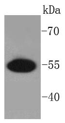
 WB (Western Blot)
(Lanes:Lane1: 400ug rat hippocampalLane2: 300ug rat hippocampalLane3: 200ug rat hippocampalLane4: 100ug rat hippocampalPrimary Antibody Dilution:1:1000Secondary Antibody:Anti-rabbit HRPSecondary Antibody Dilution:1:8000Gene Name:GPM6ASubmitted by:Anonymous)
WB (Western Blot)
(Lanes:Lane1: 400ug rat hippocampalLane2: 300ug rat hippocampalLane3: 200ug rat hippocampalLane4: 100ug rat hippocampalPrimary Antibody Dilution:1:1000Secondary Antibody:Anti-rabbit HRPSecondary Antibody Dilution:1:8000Gene Name:GPM6ASubmitted by:Anonymous)
GPM6A, Polyclonal Antibody (Cat# AAA23595)
Full Name
GPM6A antibody - C-terminal region
Gene Names
GPM6A; M6A; GPM6
Reactivity
Cow, Dog, Guinea Pig, Horse, Human, Mouse, Rabbit, Rat, Zebrafish
Applications
Immunohistochemistry, Western Blot
Purity
Affinity Purified
Pricing


 WB (Western Blot)
(WB Suggested Anti-HDAC1 Antibody Titration: 0.2-1 ug/mlELISA Titer: 1:2500Positive Control: Jurkat cell lysateHDAC1 is supported by BioGPS gene expression data to be expressed in Jurkat)
WB (Western Blot)
(WB Suggested Anti-HDAC1 Antibody Titration: 0.2-1 ug/mlELISA Titer: 1:2500Positive Control: Jurkat cell lysateHDAC1 is supported by BioGPS gene expression data to be expressed in Jurkat)
HDAC1, Polyclonal Antibody (Cat# AAA23405)
Full Name
HDAC1 antibody - middle region
Gene Names
HDAC1; HD1; RPD3; KDAC1; GON-10; RPD3L1
Reactivity
Cow, Dog, Guinea Pig, Horse, Human, Mouse, Rabbit, Rat, Yeast, Zebrafish
Applications
Immunohistochemistry, Western Blot
Purity
Affinity Purified
Pricing


 ICC (Immunocytochemistry)
(ICC staining IRF7 in HepG2 cells (green). The nuclear counter stain is DAPI (blue). Cells were fixed in paraformaldehyde, permeabilised with 0.25% Triton X100/PBS.)
ICC (Immunocytochemistry)
(ICC staining IRF7 in HepG2 cells (green). The nuclear counter stain is DAPI (blue). Cells were fixed in paraformaldehyde, permeabilised with 0.25% Triton X100/PBS.)
IRF7, Monoclonal Antibody (Cat# AAA30150)
Full Name
IRF7 Antibody
Gene Names
IRF7; IRF7A; IRF7B; IRF7C; IRF7H; IRF-7H
Reactivity
Human, Mouse, Rat, Zebrafish
Applications
Western Blot, Immunocytochemistry, Immunofluorescence, Immunohistochemistry, Immunoprecipitation, Flow Cytometry
Purity
ProA affinity purified
Pricing


 Application Data
(Staining of J774 cells with Rat anti Mouse F4/80 antigen Biotin)
Application Data
(Staining of J774 cells with Rat anti Mouse F4/80 antigen Biotin)
F4/80, Monoclonal Antibody (Cat# AAA12163)
Full Name
RAT ANTI MOUSE F4/80:Biotin
Gene Names
Emr1; Ly71; F4/80; Gpf480; TM7LN3; DD7A5-7; EGF-TM7
Applications
Flow Cytometry
Pricing
 Standard Curve (Sample)
interferon regulatory factor 7, ELISA Kit (Cat# AAA18325)
Full Name
Human Interferon regulatory factor 7, IRF7 ELISA Kit
Gene Names
IRF7; IRF7A; IRF7B; IRF7C; IRF7H; IRF-7H
Reactivity
Human
Pricing


 ICC (Immunocytochemistry)
(ICC staining Phospho-GSK3 (alpha+beta) (Y216+Y279) in MCF-7 cells (green). The nuclear counter stain is DAPI (blue). Cells were fixed in paraformaldehyde, permeabilised with 0.25% Triton X100/PBS.)
ICC (Immunocytochemistry)
(ICC staining Phospho-GSK3 (alpha+beta) (Y216+Y279) in MCF-7 cells (green). The nuclear counter stain is DAPI (blue). Cells were fixed in paraformaldehyde, permeabilised with 0.25% Triton X100/PBS.)
GSK3 (alpha+beta), Monoclonal Antibody (Cat# AAA29772)
Full Name
GSK3 (alpha+beta) (Phospho-Y216+Y279) Antibody
Reactivity
Human, Mouse, Rat
Applications
Western Blot, Immunocytochemistry, Immunohistochemistry, Immunoprecipitation
Purity
ProA affinity purified
Pricing
 Standard Curve (Sample)
NGF, ELISA Kit (Cat# AAA13485)
Full Name
NGF Human ELISA Kit
Gene Names
NGF; NGFB; HSAN5; Beta-NGF
Reactivity
Human
Applications
ELISA
Pricing
 Standard Curve (Sample)
platelet-derived growth factor receptor, beta polypeptide, ELISA Kit (Cat# AAA18160)
Full Name
Mouse Beta-type platelet-derived growth factor receptor, PDGFRB ELISA Kit
Gene Names
Pdgfrb; Pdgfr; CD140b; PDGFR-1; AI528809
Reactivity
Mouse
Pricing
 Standard Curve (Sample)
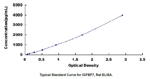
Insulin Like Growth Factor Binding Protein 7 (IGFBP7), ELISA Kit (Cat# AAA23011)
Full Name
Rat Insulin Like Growth Factor Binding Protein 7 (IGFBP7) ELISA Kit
Gene Names
IGFBP7; AGM; PSF; TAF; FSTL2; IBP-7; MAC25; IGFBP-7; RAMSVPS; IGFBP-7v; IGFBPRP1
Reactivity
Rat
Pricing


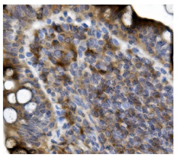
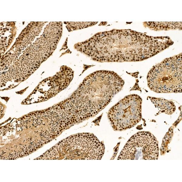
 IHC (Immunohistchemistry)
(Immunohistochemistry of TFF3 in human small intestine tissue with TFF3 antibody at 2 μg/ml.)
IHC (Immunohistchemistry)
(Immunohistochemistry of TFF3 in human small intestine tissue with TFF3 antibody at 2 μg/ml.)
TFF3, Polyclonal Antibody (Cat# AAA10911)
Full Name
TFF3 Antibody
Gene Names
TFF3; ITF; P1B; TFI
Reactivity
Human, Mouse, Rat
Applications
Western Blot, Immunohistochemistry, Immunofluorescence
Purity
TFF3 Antibody is affinity chromatography purified via peptide column.
Pricing


 FCM (Flow Cytometry)
(Flow cytometric analysis of Hela cells with M6PR antibody at 1/50 dilution (blue) compared with an unlabelled control (cells without incubation with primary antibody; red). Alexa Fluor 488-conjugated Goat anti mouse IgG was used as the secondary antibody.)
FCM (Flow Cytometry)
(Flow cytometric analysis of Hela cells with M6PR antibody at 1/50 dilution (blue) compared with an unlabelled control (cells without incubation with primary antibody; red). Alexa Fluor 488-conjugated Goat anti mouse IgG was used as the secondary antibody.)
IGF2R/M6PR, Polyclonal Antibody (Cat# AAA29869)
Full Name
IGF2R/M6PR Antibody
Gene Names
IGF2R; MPR1; MPRI; CD222; CIMPR; M6P-R
Reactivity
Human
Applications
Immunocytochemistry, Immunohistochemistry, Flow Cytometry
Purity
Peptide affinity purified
Pricing


 FCM (Flow Cytometry)
(Flow cytometric analysis of Hela cells with Phospho-eIF-2a (S51) antibody at 1/50 dilution (blue) compared with an unlabelled control (cells without incubation with primary antibody; red). Alexa Fluor 488-conjugated goat anti rabbit IgG was used as the secondary antibody.)
FCM (Flow Cytometry)
(Flow cytometric analysis of Hela cells with Phospho-eIF-2a (S51) antibody at 1/50 dilution (blue) compared with an unlabelled control (cells without incubation with primary antibody; red). Alexa Fluor 488-conjugated goat anti rabbit IgG was used as the secondary antibody.)
eIF-2a, Monoclonal Antibody (Cat# AAA29766)
Full Name
eIF-2a (Phospho-S51) Antibody
Gene Names
EIF2A; CDA02; EIF-2A; MST089; MSTP004; MSTP089
Reactivity
Human, Mouse, Rat
Applications
Western Blot, Immunohistochemistry, Flow Cytometry, Immunocytochemistry, Immunofluorescence, Immunohistochemistry, Immunoprecipitation, Flow Cytometry
Purity
ProA affinity purified
Pricing


 Application Data
(Staining of J774 cells with Rat anti Mouse F4/80 antigen Biotin)
Application Data
(Staining of J774 cells with Rat anti Mouse F4/80 antigen Biotin)
F4/80, Monoclonal Antibody (Cat# AAA12174)
Full Name
RAT ANTI MOUSE F4/80
Gene Names
Emr1; Ly71; F4/80; Gpf480; TM7LN3; DD7A5-7; EGF-TM7
Applications
Immunohistochemistry, Fluorescence Microscopy, Flow Cytometry, Immunofluorescence, Immunoprecipitation, Immunohistochemistry, Radioimmunoassay, Immunohistochemistry, Western Blot
Pricing


 WB (Western Blot)
(WB Suggested Anti-PTX3 Antibody Titration: 0.2-1 ug/mlELISA Titer: 1:312500Positive Control: MCF7 cell lysate)
WB (Western Blot)
(WB Suggested Anti-PTX3 Antibody Titration: 0.2-1 ug/mlELISA Titer: 1:312500Positive Control: MCF7 cell lysate)
PTX3, Polyclonal Antibody (Cat# AAA23569)
Full Name
PTX3 antibody - N-terminal region
Gene Names
PTX3; TSG-14; TNFAIP5
Reactivity
Cow, Guinea Pig, Human, Mouse, Rabbit, Rat
Applications
Immunohistochemistry, Western Blot
Purity
Affinity Purified
Pricing


 ICC (Immunocytochemistry)
(ICC staining STAT6 in NIH/3T3 cells (green). The nuclear counter stain is DAPI (blue). Cells were fixed in paraformaldehyde, permeabilised with 0.25% Triton X100/PBS.)
ICC (Immunocytochemistry)
(ICC staining STAT6 in NIH/3T3 cells (green). The nuclear counter stain is DAPI (blue). Cells were fixed in paraformaldehyde, permeabilised with 0.25% Triton X100/PBS.)
STAT6, Monoclonal Antibody (Cat# AAA30030)
Full Name
STAT6 Antibody
Gene Names
STAT6; STAT6B; STAT6C; D12S1644; IL-4-STAT
Reactivity
Human, Mouse
Applications
Western Blot, Immunocytochemistry, Immunohistochemistry, Immunoprecipitation
Purity
ProA affinity purified
Pricing


 IF (Immunofluorescence)
(Figure 6. IF analysis of EEF2 using anti- EEF2 antibody (AAA19228).EEF2 was detected in immunocytochemical section of MCF-7 cells. Enzyme antigen retrieval was performed using IHC enzyme antigen retrieval reagent for 15 mins. The cells were blocked with 10% goat serum. And then incubated with 5μg/mL rabbit anti-EEF2 Antibody (AAA19228) overnight at 4 degree C. DyLight®488 Conjugated Goat Anti-Rabbit IgG was used as secondary antibody at 1:100 dilution and incubated for 30 minutes at 37 degree C. The section was counterstained with DAPI. Visualize using a fluorescence microscope and filter sets appropriate for the label used.)
IF (Immunofluorescence)
(Figure 6. IF analysis of EEF2 using anti- EEF2 antibody (AAA19228).EEF2 was detected in immunocytochemical section of MCF-7 cells. Enzyme antigen retrieval was performed using IHC enzyme antigen retrieval reagent for 15 mins. The cells were blocked with 10% goat serum. And then incubated with 5μg/mL rabbit anti-EEF2 Antibody (AAA19228) overnight at 4 degree C. DyLight®488 Conjugated Goat Anti-Rabbit IgG was used as secondary antibody at 1:100 dilution and incubated for 30 minutes at 37 degree C. The section was counterstained with DAPI. Visualize using a fluorescence microscope and filter sets appropriate for the label used.)
EEF2, Polyclonal Antibody (Cat# AAA19228)
Full Name
Anti-EEF2 Antibody
Gene Names
EEF2; EF2; EF-2; EEF-2
Reactivity
Human
Applications
Western Blot, Immunohistochemistry, Immunocytochemistry, Immunofluorescence, Direct ELISA
Purity
Immunogen affinity purified.
Pricing


 FCM (Flow Cytometry)
(Figure 6. Flow Cytometry analysis of U87 cells using anti-GRB10 antibody (AAA19244).Overlay histogram showing U87 cells stained with AAA19244 (Blue line). The cells were blocked with 10% normal goat serum. And then incubated with rabbit anti-GRB10 Antibody (AAA19244,1μg/1x106 cells) for 30 min at 20 degree C. DyLight®488 conjugated goat anti-rabbit IgG (5-10μg/1x106 cells) was used as secondary antibody for 30 minutes at 20 degree C. Isotype control antibody (Green line) was rabbit IgG (1μg/1x106) used under the same conditions. Unlabelled sample (Red line) was also used as a control.)
FCM (Flow Cytometry)
(Figure 6. Flow Cytometry analysis of U87 cells using anti-GRB10 antibody (AAA19244).Overlay histogram showing U87 cells stained with AAA19244 (Blue line). The cells were blocked with 10% normal goat serum. And then incubated with rabbit anti-GRB10 Antibody (AAA19244,1μg/1x106 cells) for 30 min at 20 degree C. DyLight®488 conjugated goat anti-rabbit IgG (5-10μg/1x106 cells) was used as secondary antibody for 30 minutes at 20 degree C. Isotype control antibody (Green line) was rabbit IgG (1μg/1x106) used under the same conditions. Unlabelled sample (Red line) was also used as a control.)
GRB10, Polyclonal Antibody (Cat# AAA19244)
Full Name
Anti-GRB10 Antibody
Gene Names
GRB10; RSS; IRBP; MEG1; GRB-IR; Grb-10
Reactivity
Human, Mouse, Rat
Applications
Western Blot, Immunohistochemistry, Flow Cytometry, Direct ELISA
Purity
Immunogen affinity purified.
Pricing
 Standard Curve (Sample)
interleukin 10, ELISA Kit (Cat# AAA15218)
Full Name
Chicken Interleukin 10, IL-10 ELISA Kit
Gene Names
IL10; IL-10
Reactivity
Chicken
Pricing


 WB (Western Blot)
(WB Suggested Anti-PAX4 Antibody Titration: 0.2-1 ug/mlPositive Control: Jurkat cell lysate)
WB (Western Blot)
(WB Suggested Anti-PAX4 Antibody Titration: 0.2-1 ug/mlPositive Control: Jurkat cell lysate)
PAX4, Polyclonal Antibody (Cat# AAA23402)
Full Name
PAX4 antibody - middle region
Gene Names
PAX4; KPD; MODY9
Reactivity
Cow, Dog, Guinea Pig, Horse, Human, Mouse, Rabbit, Rat
Applications
Western Blot
Purity
Affinity Purified
Pricing


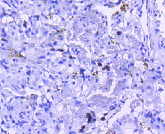
 IHC (Immunohistchemistry)
(DAB staining on IHC-P; Samples: Mouse Small intestine Tissue; Primary Ab: 30ug/ml Rabbit Anti-Mouse LTBP1 Antibody Second Ab: 2ug/mL HRP-Linked Caprine Anti-Rabbit IgG Polyclonal Antibody (Catalog:))
IHC (Immunohistchemistry)
(DAB staining on IHC-P; Samples: Mouse Small intestine Tissue; Primary Ab: 30ug/ml Rabbit Anti-Mouse LTBP1 Antibody Second Ab: 2ug/mL HRP-Linked Caprine Anti-Rabbit IgG Polyclonal Antibody (Catalog:))
Latent Transforming Growth Factor Beta Binding Protein 1, Polyclonal Antibody (Cat# AAA20909)
Full Name
Polyclonal Antibody to Latent Transforming Growth Factor Beta Binding Protein 1 (LTBP1)
Gene Names
Ltbp1; Tgfb; Ltbp-1; Ltbp1L; 9830146M04; b2b1000Clo; 9430031G15Rik; TGF-beta1-BP-1
Reactivity
Mouse
Applications
WB, IHC, ICC, IP
Purity
Antigen-specific affinity chromatography followed by Protein A affinity chromatography
Pricing


 FCM (Flow Cytometry)
(Flow cytometric analysis of Hela cells with PRP19 antibody at 1/50 dilution (red) compared with an unlabelled control (cells without incubation with primary antibody; black). Alexa Fluor 488-conjugated goat anti rabbit IgG was used as the secondary antibody.)
FCM (Flow Cytometry)
(Flow cytometric analysis of Hela cells with PRP19 antibody at 1/50 dilution (red) compared with an unlabelled control (cells without incubation with primary antibody; black). Alexa Fluor 488-conjugated goat anti rabbit IgG was used as the secondary antibody.)
PRP19, Monoclonal Antibody (Cat# AAA30128)
Full Name
PRP19 Antibody
Gene Names
PRPF19; PSO4; SNEV; PRP19; UBOX4; hPSO4; NMP200
Reactivity
Human, Mouse, Rat
Applications
Western Blot, Immunocytochemistry, Immunohistochemistry, Flow Cytometry
Purity
ProA affinity purified
Pricing


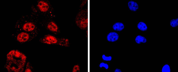
 ICC (Immunocytochemistry)
(ICC staining ATF5 in PC-12 cells (green). The nuclear counter stain is DAPI (blue). Cells were fixed in paraformaldehyde, permeabilised with 0.25% Triton X100/PBS.)
ICC (Immunocytochemistry)
(ICC staining ATF5 in PC-12 cells (green). The nuclear counter stain is DAPI (blue). Cells were fixed in paraformaldehyde, permeabilised with 0.25% Triton X100/PBS.)
ATF5, Monoclonal Antibody (Cat# AAA30200)
Full Name
ATF5 Antibody
Gene Names
ATF5; ATFX; HMFN0395
Reactivity
Human, Mouse, Rat
Applications
Western Blot, Immunocytochemistry, Immunofluorescence, Immunohistochemistry, Immunoprecipitation
Purity
ProA affinity purified
Pricing


 ICC (Immunocytochemistry)
(Anti- MCM7 Picoband antibody, AAA11649,ICCICC: A549 Cell)
ICC (Immunocytochemistry)
(Anti- MCM7 Picoband antibody, AAA11649,ICCICC: A549 Cell)
MCM7, Polyclonal Antibody (Cat# AAA11649)
Full Name
Anti-MCM7 Antibody
Gene Names
MCM7; MCM2; CDC47; P85MCM; P1CDC47; PNAS146; PPP1R104; P1.1-MCM3
Reactivity
Human, Mouse, Rat
Applications
Western Blot, Immunohistochemistry, Immunocytochemistry
Purity
Immunogen Affinity Purified
Pricing


 Application Data
(Staining of J774 cells with Rat anti Mouse F4/80 antigen Biotin)
Application Data
(Staining of J774 cells with Rat anti Mouse F4/80 antigen Biotin)
F4/80, Monoclonal Antibody (Cat# AAA12164)
Full Name
RAT ANTI MOUSE F4/80:Biotin
Gene Names
Emr1; Ly71; F4/80; Gpf480; TM7LN3; DD7A5-7; EGF-TM7
Applications
Flow Cytometry
Pricing


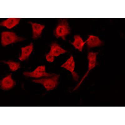 Application Data
(At 25 degree C. The primary antibody was diluted at 1/200 and incubated with the sample for 1 hour at 37 degree C. An Alexa Fluor 594 conjugated goat anti-rabbit IgG (H+L) Ab, diluted at 1/600, was used as the secondary antibody.)
Application Data
(At 25 degree C. The primary antibody was diluted at 1/200 and incubated with the sample for 1 hour at 37 degree C. An Alexa Fluor 594 conjugated goat anti-rabbit IgG (H+L) Ab, diluted at 1/600, was used as the secondary antibody.)
Retinoblastoma, Polyclonal Antibody (Cat# AAA31401)
Full Name
Phospho-Retinoblastoma (Ser788) Antibody
Gene Names
RB1; RB; pRb; OSRC; pp110; p105-Rb
Reactivity
Human, Mouse, Rat
Predicted Reactivity: Pig (100%), Bovine (100%), Horse (100%), Sheep (100%), Rabbit (100%), Dog (100%), Chicken (92%), Xenopus (92%)
Predicted Reactivity: Pig (100%), Bovine (100%), Horse (100%), Sheep (100%), Rabbit (100%), Dog (100%), Chicken (92%), Xenopus (92%)
Applications
Western Blot, Immunohistochemistry, Immunofluorescence, Immunocytochemistry, Peptide ELISA
Purity
The antibody is from purified rabbit serum by affinity purification via sequential chromatography on phospho-peptide and non-phospho-peptide affinity columns.
Pricing


 WB (Western Blot)
(Host: RabbitTarget: GATA2Positive control (+): Hela (HL)Negative control (-): 293T (2T)Antibody concentration: 1ug/ml)
WB (Western Blot)
(Host: RabbitTarget: GATA2Positive control (+): Hela (HL)Negative control (-): 293T (2T)Antibody concentration: 1ug/ml)
GATA2, Polyclonal Antibody (Cat# AAA23392)
Full Name
GATA2 antibody - N-terminal region
Gene Names
GATA2; DCML; IMD21; NFE1B; MONOMAC
Reactivity
Tested Reactivity: Human, Mouse
Predicted Reactivity: Cow, Dog, Guinea Pig, Horse, Human, Mouse, Rabbit, Rat
Predicted Reactivity: Cow, Dog, Guinea Pig, Horse, Human, Mouse, Rabbit, Rat
Applications
Immunohistochemistry, Western Blot
Purity
Protein A purified
Pricing


 Application Data
(At 25 degree C. Samples were then incubated with primary Ab(At 37 degree C. An AlexaFluor594 conjugated goat anti-rabbit IgG(H+L) Ab(Red) and an AlexaFluor488 conjugated goat anti-mouse IgG(H+L) Ab(Green) were used as the secondary antibody.The nuclear counter stain is DAPI(blue).)
Application Data
(At 25 degree C. Samples were then incubated with primary Ab(At 37 degree C. An AlexaFluor594 conjugated goat anti-rabbit IgG(H+L) Ab(Red) and an AlexaFluor488 conjugated goat anti-mouse IgG(H+L) Ab(Green) were used as the secondary antibody.The nuclear counter stain is DAPI(blue).)
PDX1, Polyclonal Antibody (Cat# AAA31430)
Full Name
Phospho-PDX1 (Ser66) Antibody
Gene Names
PDX1; GSF; IPF1; IUF1; IDX-1; MODY4; PDX-1; STF-1
Reactivity
Human, Mouse, Rat
Predicted Reactivity: Pig (88%), Bovine (100%), Horse (100%), Sheep (100%), Rabbit (100%), Dog (100%), Chicken (88%), Xenopus (100%)
Predicted Reactivity: Pig (88%), Bovine (100%), Horse (100%), Sheep (100%), Rabbit (100%), Dog (100%), Chicken (88%), Xenopus (100%)
Applications
Western Blot, Immunohistochemistry, Immunofluorescence, Immunocytochemistry, Peptide ELISA
Purity
The antibody is from purified rabbit serum by affinity purification via sequential chromatography on phospho-peptide and non-phospho-peptide affinity columns.
Pricing


 Application Data
(At 25 degree C. Samples were then incubated with primary Ab(At 37 degree C. An AlexaFluor594 conjugated goat anti-rabbit IgG(H+L) Ab(Red) and an AlexaFluor488 conjugated goat anti-mouse IgG(H+L) Ab(Green) were used as the secondary antibody.The nuclear counter stain is DAPI (blue).)
Application Data
(At 25 degree C. Samples were then incubated with primary Ab(At 37 degree C. An AlexaFluor594 conjugated goat anti-rabbit IgG(H+L) Ab(Red) and an AlexaFluor488 conjugated goat anti-mouse IgG(H+L) Ab(Green) were used as the secondary antibody.The nuclear counter stain is DAPI (blue).)
RPA32/RPA2, Polyclonal Antibody (Cat# AAA31304)
Full Name
Phospho-RPA32/RPA2 (Ser4/Ser8) Antibody
Gene Names
RPA2; REPA2; RPA32
Reactivity
Human, Mouse, Rat
Applications
Immunohistochemistry, Immunofluorescence, Immunocytochemistry, Peptide ELISA
Purity
The antibody is from purified rabbit serum by affinity purification via sequential chromatography on phospho-peptide and non-phospho-peptide affinity columns.
Pricing


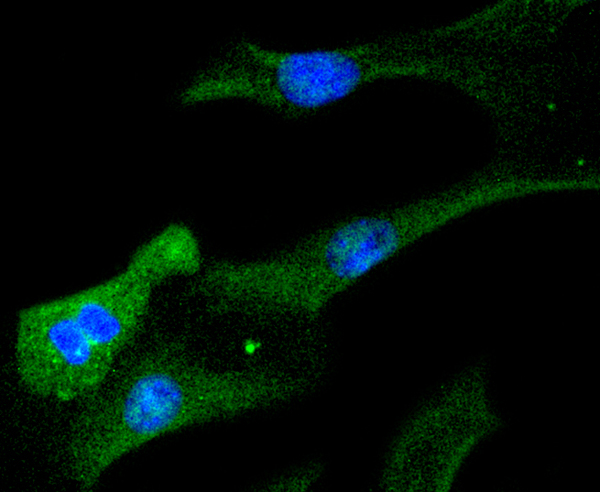
 ICC (Immunocytochemistry)
(ICC staining MCSF in Hela cells (green). The nuclear counter stain is DAPI (blue). Cells were fixed in paraformaldehyde, permeabilised with 0.25% Triton X100/PBS.)
ICC (Immunocytochemistry)
(ICC staining MCSF in Hela cells (green). The nuclear counter stain is DAPI (blue). Cells were fixed in paraformaldehyde, permeabilised with 0.25% Triton X100/PBS.)
MCSF, Monoclonal Antibody (Cat# AAA30082)
Full Name
MCSF Antibody
Gene Names
CSF1; MCSF; CSF-1
Reactivity
Human
Applications
Western Blot, Immunohistochemistry, Immunocytochemistry, Immunoprecipitation
Purity
ProA affinity purified
Pricing


 FCM (Flow Cytometry)
(Flow cytometric analysis of Hela cells with HDAC2 antibody at 1/50 dilution (red) compared with an unlabelled control (cells without incubation with primary antibody; black). Alexa Fluor 488-conjugated goat anti rabbit IgG was used as the secondary antibody.)
FCM (Flow Cytometry)
(Flow cytometric analysis of Hela cells with HDAC2 antibody at 1/50 dilution (red) compared with an unlabelled control (cells without incubation with primary antibody; black). Alexa Fluor 488-conjugated goat anti rabbit IgG was used as the secondary antibody.)
HDAC2, Monoclonal Antibody (Cat# AAA30065)
Full Name
HDAC2 Antibody
Gene Names
HDAC2; HD2; RPD3; YAF1
Reactivity
Human, Mouse, Rat
Applications
Western Blot, Immunocytochemistry, Immunofluorescence, Immunohistochemistry, Immunoprecipitation, Flow Cytometry
Purity
ProA affinity purified
Pricing


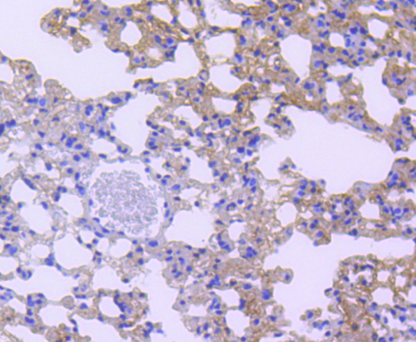
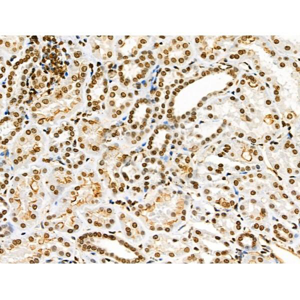
 IHC (Immunohistochemistry)
(Immunochemical staining RBBP4 in mouse lung with rabbit polyclonal antibody at 1:2000 dilution, formalin-fixed paraffin embedded sections.)
IHC (Immunohistochemistry)
(Immunochemical staining RBBP4 in mouse lung with rabbit polyclonal antibody at 1:2000 dilution, formalin-fixed paraffin embedded sections.)
RbAp48, Polyclonal Antibody (Cat# AAA27724)
Full Name
Anti-RbAp48 Antibody, Rabbit Polyclonal
Gene Names
RBBP4; NURF55; RBAP48; lin-53
Reactivity
Human, Mouse
Applications
Western Blot, Immunohistochemistry, Immunocytochemistry, Immunofluorescence
Purity
Protein A & Antigen Affinity
Pricing
 Standard Curve (Sample)
Interleukin 12, ELISA Kit (Cat# AAA17599)
Full Name
Human Interleukin 12 ELISA Kit
Gene Names
IL12A; P35; CLMF; NFSK; NKSF1; IL-12A
Reactivity
Human
Pricing
 Standard Curve (Sample)
coagulation factor VII (serum prothrombin conversion accelerator), ELISA Kit (Cat# AAA18375)
Full Name
Mouse coagulation factor VII, FVII ELISA Kit
Gene Names
F7; Cf7; FVII; mfVII; AI132620
Reactivity
Mouse
Pricing