Filters
Clonality
Type
Reactivity
Gene Name
Isotype
Host
Application
Clone
437 results for " Signal Proteins" - showing 200-250


 FCM (Flow Cytometry)
(Figure 8. Flow Cytometry analysis of U20S cells using anti-Dynamin 1 antibody (AAA19160).Overlay histogram showing U20S cells stained with AAA19160 (Blue line).The cells were blocked with 10% normal goat serum. And then incubated with rabbit anti-DNM1 Antibody (AAA19160,1ug/1x10^6 cells) for 30 min at 20 degree C. DyLight®488 conjugated goat anti-rabbit IgG (5-10ug/1x10^6 cells) was used as secondary antibody for 30 minutes at 20 degree C. Isotype control antibody (Green line) was rabbit IgG (1ug/1x106) used under the same conditions. Unlabelled sample (Red line) was also used as a control.)
FCM (Flow Cytometry)
(Figure 8. Flow Cytometry analysis of U20S cells using anti-Dynamin 1 antibody (AAA19160).Overlay histogram showing U20S cells stained with AAA19160 (Blue line).The cells were blocked with 10% normal goat serum. And then incubated with rabbit anti-DNM1 Antibody (AAA19160,1ug/1x10^6 cells) for 30 min at 20 degree C. DyLight®488 conjugated goat anti-rabbit IgG (5-10ug/1x10^6 cells) was used as secondary antibody for 30 minutes at 20 degree C. Isotype control antibody (Green line) was rabbit IgG (1ug/1x106) used under the same conditions. Unlabelled sample (Red line) was also used as a control.)
Dynamin 1, Polyclonal Antibody (Cat# AAA19160)
Full Name
Anti-Dynamin 1 Picoband antibody
Gene Names
DNM1; DNM; EIEE31
Reactivity
Human, Mouse, Rat
No cross reactivity with other proteins.
No cross reactivity with other proteins.
Applications
EIA, FC/FACS, IHC, ICC, WB
Pricing


 FCM (Flow Cytometry)
(Figure 7. Flow Cytometry analysis of Hela cells using anti- ETF/TEAD2 antibody (AAA19325).Overlay histogram showing Hela cells stained withAAA19325 (Blue line). The cells were blocked with 10% normal goat serum. And then incubated with rabbit anti- ETF/TEAD2 Antibody (AAA19325, 1μg/1x106 cells) for 30 min at 20 degree C. DyLight®488 conjugated goat anti-rabbit IgG (5-10μg/1x106 cells) was used as secondary antibody for 30 minutes at 20 degree C. Isotype control antibody (Green line) was rabbit IgG (1μg/1x106) used under the same conditions. Unlabelled sample (Red line) was also used as a control.)
FCM (Flow Cytometry)
(Figure 7. Flow Cytometry analysis of Hela cells using anti- ETF/TEAD2 antibody (AAA19325).Overlay histogram showing Hela cells stained withAAA19325 (Blue line). The cells were blocked with 10% normal goat serum. And then incubated with rabbit anti- ETF/TEAD2 Antibody (AAA19325, 1μg/1x106 cells) for 30 min at 20 degree C. DyLight®488 conjugated goat anti-rabbit IgG (5-10μg/1x106 cells) was used as secondary antibody for 30 minutes at 20 degree C. Isotype control antibody (Green line) was rabbit IgG (1μg/1x106) used under the same conditions. Unlabelled sample (Red line) was also used as a control.)
ETF/TEAD2, Polyclonal Antibody (Cat# AAA19325)
Full Name
Anti-ETF/TEAD2 Antibody
Gene Names
TEAD2; ETF; TEF4; TEF-4; TEAD-2
Reactivity
Human, Mouse, Rat
Applications
WB, IHC-P, FC/FACS/FCM, EIA
Purity
Immunogen affinity purified.
Pricing


 FCM (Flow Cytometry)
(Figure 6. Flow Cytometry analysis of A431 cells using anti-NDUFB5 antibody (AAA19333).Overlay histogram showing A431 cells stained with AAA19333 (Blue line). The cells were blocked with 10% normal goat serum. And then incubated with rabbit anti-NDUFB5 Antibody (AAA19333, 1μg/1x106 cells) for 30 min at 20 degree C. DyLight®488 conjugated goat anti-rabbit IgG (5-10μg/1x106 cells) was used as secondary antibody for 30 minutes at 20 degree C. Isotype control antibody (Green line) was rabbit IgG (1μg/1x106) used under the same conditions. Unlabelled sample (Red line) was also used as a control.)
FCM (Flow Cytometry)
(Figure 6. Flow Cytometry analysis of A431 cells using anti-NDUFB5 antibody (AAA19333).Overlay histogram showing A431 cells stained with AAA19333 (Blue line). The cells were blocked with 10% normal goat serum. And then incubated with rabbit anti-NDUFB5 Antibody (AAA19333, 1μg/1x106 cells) for 30 min at 20 degree C. DyLight®488 conjugated goat anti-rabbit IgG (5-10μg/1x106 cells) was used as secondary antibody for 30 minutes at 20 degree C. Isotype control antibody (Green line) was rabbit IgG (1μg/1x106) used under the same conditions. Unlabelled sample (Red line) was also used as a control.)
NDUFB5, Polyclonal Antibody (Cat# AAA19333)
Full Name
Anti-NDUFB5 Antibody
Gene Names
NDUFB5; SGDH; CISGDH
Reactivity
Human, Mouse, Rat
Applications
WB, IHC-P, ICC, IF, FC/FACS/FCM, EIA
Purity
Immunogen affinity purified.
Pricing


 IHC (Immunohistochemistry)
(Figure 7. IHC analysis of VEGF Receptor 3 using anti-VEGF Receptor 3 antibody (AAA19147).VEGF Receptor 3 was detected in paraffin-embedded section of mouse liver tissue. Heat mediated antigen retrieval was performed in citrate buffer (pH6, epitope retrieval solution) for 20 mins. The tissue section was blocked with 10% goat serum. The tissue section was then incubated with 1ug/ml rabbit anti-VEGF Receptor 3 Antibody (AAA19147) overnight at 4 degree C. Biotinylated goat anti-rabbit IgG was used as secondary antibody and incubated for 30 minutes at 37 degree C. The tissue section was developed using Strepavidin-Biotin-Complex (SABC) with DAB as the chromogen.)
IHC (Immunohistochemistry)
(Figure 7. IHC analysis of VEGF Receptor 3 using anti-VEGF Receptor 3 antibody (AAA19147).VEGF Receptor 3 was detected in paraffin-embedded section of mouse liver tissue. Heat mediated antigen retrieval was performed in citrate buffer (pH6, epitope retrieval solution) for 20 mins. The tissue section was blocked with 10% goat serum. The tissue section was then incubated with 1ug/ml rabbit anti-VEGF Receptor 3 Antibody (AAA19147) overnight at 4 degree C. Biotinylated goat anti-rabbit IgG was used as secondary antibody and incubated for 30 minutes at 37 degree C. The tissue section was developed using Strepavidin-Biotin-Complex (SABC) with DAB as the chromogen.)
VEGF Receptor 3, Polyclonal Antibody (Cat# AAA19147)
Full Name
Anti-VEGF Receptor 3 Picoband antibody
Gene Names
FLT4; PCL; FLT-4; FLT41; LMPH1A; VEGFR3; VEGFR-3
Reactivity
Human, Mouse, Rat
Applications
EIA, IHC, WB, FC
Purity
Immunoggen affinity purified
Pricing


 FCM (Flow Cytometry)
(Figure 11. Flow Cytometry analysis of THP-1 cells using anti-CDC123 antibody (AAA19323).Overlay histogram showing THP-1 cells stained with AAA19323 (Blue line). The cells were blocked with 10% normal goat serum. And then incubated with rabbit anti-CDC123 Antibody (AAA19323, 1μg/1x106 cells) for 30 min at 20 degree C. DyLight®488 conjugated goat anti-rabbit IgG (5-10μg/1x106 cells) was used as secondary antibody for 30 minutes at 20 degree C. Isotype control antibody (Green line) was rabbit IgG (1μg/1x106) used under the same conditions. Unlabelled sample (Red line) was also used as a control.)
FCM (Flow Cytometry)
(Figure 11. Flow Cytometry analysis of THP-1 cells using anti-CDC123 antibody (AAA19323).Overlay histogram showing THP-1 cells stained with AAA19323 (Blue line). The cells were blocked with 10% normal goat serum. And then incubated with rabbit anti-CDC123 Antibody (AAA19323, 1μg/1x106 cells) for 30 min at 20 degree C. DyLight®488 conjugated goat anti-rabbit IgG (5-10μg/1x106 cells) was used as secondary antibody for 30 minutes at 20 degree C. Isotype control antibody (Green line) was rabbit IgG (1μg/1x106) used under the same conditions. Unlabelled sample (Red line) was also used as a control.)
CDC123, Polyclonal Antibody (Cat# AAA19323)
Full Name
Anti-CDC123 Antibody
Gene Names
CDC123; D123; C10orf7
Reactivity
Human
Applications
WB, IHC-P, ICC, IF, FC/FACS/FCM, EIA
Purity
Immunogen affinity purified.
Pricing


 FCM (Flow Cytometry)
(Figure 7. Flow Cytometry analysis of U87 cells using anti-MPI antibody (AAA19349).Overlay histogram showing U87 cells stained with AAA19349 (Blue line). The cells were blocked with 10% normal goat serum. And then incubated with mouse anti- MPI Antibody (AAA19349, 1μg/1x106 cells) for 30 min at 20 degree C. DyLight®488 conjugated goat anti-mouse IgG (BA1126, 5-10μg/1x106 cells) was used as secondary antibody for 30 minutes at 20 degree C. Isotype control antibody (Green line) was mouse IgG (1μg/1x106) used under the same conditions. Unlabelled sample (Red line) was also used as a control.)
FCM (Flow Cytometry)
(Figure 7. Flow Cytometry analysis of U87 cells using anti-MPI antibody (AAA19349).Overlay histogram showing U87 cells stained with AAA19349 (Blue line). The cells were blocked with 10% normal goat serum. And then incubated with mouse anti- MPI Antibody (AAA19349, 1μg/1x106 cells) for 30 min at 20 degree C. DyLight®488 conjugated goat anti-mouse IgG (BA1126, 5-10μg/1x106 cells) was used as secondary antibody for 30 minutes at 20 degree C. Isotype control antibody (Green line) was mouse IgG (1μg/1x106) used under the same conditions. Unlabelled sample (Red line) was also used as a control.)
MPI, Monoclonal Antibody (Cat# AAA19349)
Full Name
Anti-MPI Antibody (monoclonal, 5G5)
Gene Names
MPI; PMI; PMI1; CDG1B
Reactivity
Human, Rat
Applications
WB, IHC-P, ICC, IF, FC/FACS/FCM
Purity
Immunogen affinity purified.
Pricing


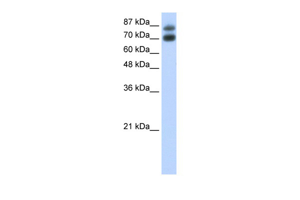
 WB (Western Blot)
(WB Suggested Anti-A1BG AntibodyTitration: 1.25 ug/mlPositive Control: HepG2 Whole CellA1BG is supported by BioGPS gene expression data to be expressed in HepG2)
WB (Western Blot)
(WB Suggested Anti-A1BG AntibodyTitration: 1.25 ug/mlPositive Control: HepG2 Whole CellA1BG is supported by BioGPS gene expression data to be expressed in HepG2)
A1BG, Polyclonal Antibody (Cat# AAA23428)
Full Name
A1BG antibody - N-terminal region
Gene Names
A1BG; A1B; ABG; GAB; HYST2477
Reactivity
Human
Applications
WB
Purity
Affinity Purified
Pricing


 WB (Western Blot)
(WB Suggested Anti-SOX4 Antibody Titration: 1 ug/mlPositive Control: Hela cell lysate)
WB (Western Blot)
(WB Suggested Anti-SOX4 Antibody Titration: 1 ug/mlPositive Control: Hela cell lysate)
SOX4, Polyclonal Antibody (Cat# AAA23462)
Full Name
SOX4 antibody - N-terminal region
Gene Names
SOX4; EVI16
Reactivity
Cow, Dog, Goat, Guinea Pig, Horse, Human, Mouse, Rabbit, Rat, Zebrafish
Applications
IHC, WB
Purity
Affinity Purified
Pricing


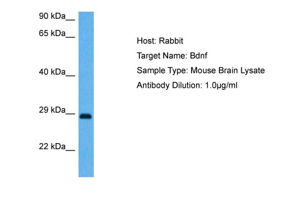


 Application Data
(Surface Plasmon Resonance Kinetic Characterization of Polyclonal Antibody Affinity. Purified polyclonal antibodies were immobilized on a Protein A/G coated Carterra LSA sensor chip at concentrations of 5, and 50 ug/mL in duplicate. Antibodies on the surface were exposed to interaction with peptides sequentially via microfluidic controlled flow at 333nM peptide concentration for kinetic characterization of the binders for affinity and specificity, followed by curve fitting using the Kinetics software. Kd determinations for both concentrations were averaged and results and standard deviation are shown.)
Application Data
(Surface Plasmon Resonance Kinetic Characterization of Polyclonal Antibody Affinity. Purified polyclonal antibodies were immobilized on a Protein A/G coated Carterra LSA sensor chip at concentrations of 5, and 50 ug/mL in duplicate. Antibodies on the surface were exposed to interaction with peptides sequentially via microfluidic controlled flow at 333nM peptide concentration for kinetic characterization of the binders for affinity and specificity, followed by curve fitting using the Kinetics software. Kd determinations for both concentrations were averaged and results and standard deviation are shown.)
BDNF, Polyclonal Antibody (Cat# AAA23497)
Full Name
BDNF antibody - middle region
Gene Names
BDNF; ANON2; BULN2
Reactivity
Tested Species Reactivity: Human, Mouse, Monkey
Predicted Species Reactivity: Human, Mouse, Rat, Dog, Horse, Pig, Rabbit
Predicted Species Reactivity: Human, Mouse, Rat, Dog, Horse, Pig, Rabbit
Applications
IHC, WB
Purity
Affinity Purified
Pricing


 IHC (Immunohistochemistry)
(Figure 7. IHC analysis of PARK7 / DJ1 using anti-PARK7 / DJ1 antibody (AAA19139).PARK7 / DJ1 was detected in paraffin-embedded section of rat testis tissue. Heat mediated antigen retrieval was performed in citrate buffer (pH6, epitope retrieval solution) for 20 mins. The tissue section was blocked with 10% goat serum. The tissue section was then incubated with 1ug/ml rabbit anti-PARK7 / DJ1 Antibody (AAA19139) overnight at 4 degree C. Biotinylated goat anti-rabbit IgG was used as secondary antibody and incubated for 30 minutes at 37 degree C. The tissue section was developed using Strepavidin-Biotin-Complex (SABC) with DAB as the chromogen.)
IHC (Immunohistochemistry)
(Figure 7. IHC analysis of PARK7 / DJ1 using anti-PARK7 / DJ1 antibody (AAA19139).PARK7 / DJ1 was detected in paraffin-embedded section of rat testis tissue. Heat mediated antigen retrieval was performed in citrate buffer (pH6, epitope retrieval solution) for 20 mins. The tissue section was blocked with 10% goat serum. The tissue section was then incubated with 1ug/ml rabbit anti-PARK7 / DJ1 Antibody (AAA19139) overnight at 4 degree C. Biotinylated goat anti-rabbit IgG was used as secondary antibody and incubated for 30 minutes at 37 degree C. The tissue section was developed using Strepavidin-Biotin-Complex (SABC) with DAB as the chromogen.)
PARK7/DJ1, Polyclonal Antibody (Cat# AAA19139)
Full Name
Anti-PARK7/DJ1 Picoband Antibody
Gene Names
Park7; Dj1; CAP1; DJ-1; SP22
Reactivity
Mouse, Rat
No cross reactivity with other proteins.
No cross reactivity with other proteins.
Applications
EIA, IHC, WB
Purity
Immunogen affinity purified
Pricing


 FCM (Flow Cytometry)
(Figure 6. Flow Cytometry analysis of HEPA1-6 cells using anti-Rad9/Rad9a antibody (AAA19288).Overlay histogram showing HEPA1-6 cells stained with AAA19288 (Blue line). The cells were blocked with 10% normal goat serum. And then incubated with rabbit anti-Rad9/Rad9a Antibody (AAA19288,1μg/1x106 cells) for 30 min at 20 degree C. DyLight®488 conjugated goat anti-rabbit IgG (5-10μg/1x106 cells) was used as secondary antibody for 30 minutes at 20 degree C. Isotype control antibody (Green line) was rabbit IgG (1μg/1x106) used under the same conditions. Unlabelled sample (Red line) was also used as a control.)
FCM (Flow Cytometry)
(Figure 6. Flow Cytometry analysis of HEPA1-6 cells using anti-Rad9/Rad9a antibody (AAA19288).Overlay histogram showing HEPA1-6 cells stained with AAA19288 (Blue line). The cells were blocked with 10% normal goat serum. And then incubated with rabbit anti-Rad9/Rad9a Antibody (AAA19288,1μg/1x106 cells) for 30 min at 20 degree C. DyLight®488 conjugated goat anti-rabbit IgG (5-10μg/1x106 cells) was used as secondary antibody for 30 minutes at 20 degree C. Isotype control antibody (Green line) was rabbit IgG (1μg/1x106) used under the same conditions. Unlabelled sample (Red line) was also used as a control.)
Rad9/Rad9a, Polyclonal Antibody (Cat# AAA19288)
Full Name
Anti-Rad9/Rad9a Antibody
Gene Names
Rad9a; Rad9
Reactivity
Mouse, Rat
Applications
WB, IHC-P, FC/FACS/FCM, EIA
Purity
Immunogen affinity purified.
Pricing


 IF (Immunofluorescence)
(Figure 6. IF analysis of SCG10/STMN2 using anti- SCG10/STMN2 antibody (AAA19299).SCG10/STMN2 was detected in immunocytochemical section of U20S cells. Enzyme antigen retrieval was performed using IHC enzyme antigen retrieval reagent for 15 mins. The cells were blocked with 10% goat serum. And then incubated with 5μg/mL rabbit anti- SCG10/STMN2 Antibody (AAA19299) overnight at 4 degree C. DyLight®488 Conjugated Goat Anti-Rabbit IgG was used as secondary antibody at 1:100 dilution and incubated for 30 minutes at 37 degree C. The section was counterstained with DAPI. Visualize using a fluorescence microscope and filter sets appropriate for the label used.)
IF (Immunofluorescence)
(Figure 6. IF analysis of SCG10/STMN2 using anti- SCG10/STMN2 antibody (AAA19299).SCG10/STMN2 was detected in immunocytochemical section of U20S cells. Enzyme antigen retrieval was performed using IHC enzyme antigen retrieval reagent for 15 mins. The cells were blocked with 10% goat serum. And then incubated with 5μg/mL rabbit anti- SCG10/STMN2 Antibody (AAA19299) overnight at 4 degree C. DyLight®488 Conjugated Goat Anti-Rabbit IgG was used as secondary antibody at 1:100 dilution and incubated for 30 minutes at 37 degree C. The section was counterstained with DAPI. Visualize using a fluorescence microscope and filter sets appropriate for the label used.)
SCG10/STMN2, Polyclonal Antibody (Cat# AAA19299)
Full Name
Anti-SCG10/STMN2 Antibody
Gene Names
STMN2; SCG10; SCGN10
Reactivity
Human, Mouse, Rat
Applications
WB, IHC-P, ICC, IF, FC/FACS/FCM, EIA
Purity
Immunogen affinity purified.
Pricing
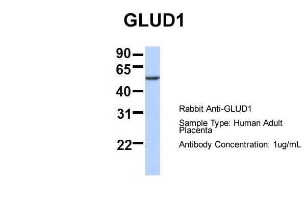


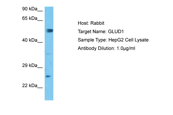


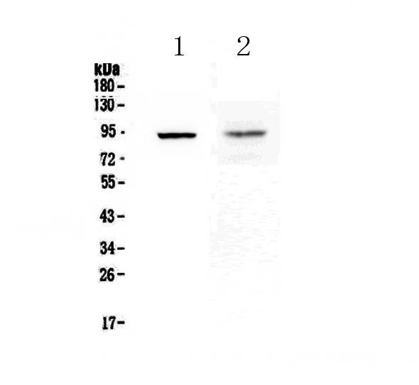
 WB (Western Blot)
(lanes 5: rat kidney cordexlanes 6: rat kidney proximal tubules prepped from cortexlanes 7: LLCPK-F+ pig kidney proximal tubule tissue culture lysatelanes 8: rat brain supernatant)
WB (Western Blot)
(lanes 5: rat kidney cordexlanes 6: rat kidney proximal tubules prepped from cortexlanes 7: LLCPK-F+ pig kidney proximal tubule tissue culture lysatelanes 8: rat brain supernatant)
GLUD1, Polyclonal Antibody (Cat# AAA23521)
Full Name
GLUD1 antibody - N-terminal region
Gene Names
GLUD1; GDH; GDH1; GLUD
Reactivity
Cow, Dog, Guinea Pig, Horse, Human, Mouse, Pig, Rabbit, Rat, Sheep, Zebrafish
Applications
IHC, WB
Purity
Affinity Purified
Pricing


 WB (Western Blot)
(ZEB2 antibody - middle region validated by WB using H9 human embryonic stem cells at 1:1000.)
WB (Western Blot)
(ZEB2 antibody - middle region validated by WB using H9 human embryonic stem cells at 1:1000.)
ZEB2, Polyclonal Antibody (Cat# AAA23465)
Full Name
ZEB2 antibody - middle region
Gene Names
ZEB2; SIP1; SIP-1; ZFHX1B; HSPC082; SMADIP1
Reactivity
Cow, Guinea Pig, Horse, Human, Mouse, Pig, Rat, Zebrafish
Applications
IHC, WB
Purity
Affinity Purified
Pricing


 IHC (Immunohistchemistry)
(Figure 6. IHC analysis of Annexin VI using anti-Annexin VI antibody (AAA19170).Annexin VI was detected in paraffin-embedded section of rat spleen tissue. Heat mediated antigen retrieval was performed in citrate buffer (pH6, epitope retrieval solution) for 20 mins. The tissue section was blocked with 10% goat serum. The tissue section was then incubated with 2ug/ml rabbit anti-Annexin VI Antibody (AAA19170) overnight at 4 degree C. Biotinylated goat anti-rabbit IgG was used as secondary antibody and incubated for 30 minutes at 37 degree C. The tissue section was developed using Strepavidin-Biotin-Complex (SABC) with DAB as the chromogen.)
IHC (Immunohistchemistry)
(Figure 6. IHC analysis of Annexin VI using anti-Annexin VI antibody (AAA19170).Annexin VI was detected in paraffin-embedded section of rat spleen tissue. Heat mediated antigen retrieval was performed in citrate buffer (pH6, epitope retrieval solution) for 20 mins. The tissue section was blocked with 10% goat serum. The tissue section was then incubated with 2ug/ml rabbit anti-Annexin VI Antibody (AAA19170) overnight at 4 degree C. Biotinylated goat anti-rabbit IgG was used as secondary antibody and incubated for 30 minutes at 37 degree C. The tissue section was developed using Strepavidin-Biotin-Complex (SABC) with DAB as the chromogen.)
Annexin VI, Polyclonal Antibody (Cat# AAA19170)
Full Name
Anti-Annexin VI Picoband Antibody
Gene Names
ANXA6; ANX6; CBP68
Reactivity
Human, Mouse, Rat
No cross reactivity with other proteins.
No cross reactivity with other proteins.
Applications
EIA, IHC, WB
Pricing


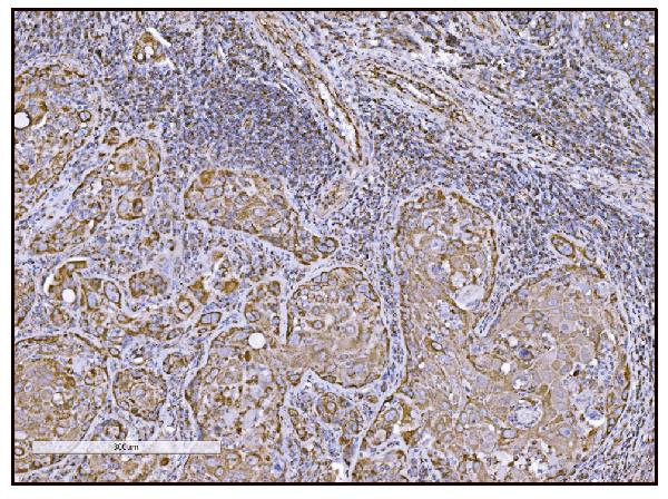


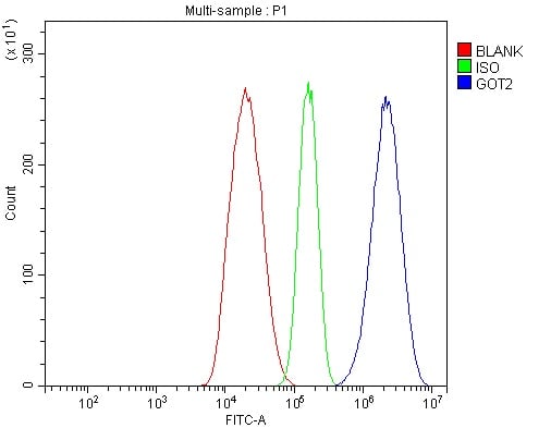


 IHC (Immunohistochemistry)
(Figure 14. IHC analysis of FABP-1/GOT2 using anti-FABP-1/GOT2 antibody (AAA19309).FABP-1/GOT2 was detected in paraffin-embedded section of rat lymph node tissue. Heat mediated antigen retrieval was performed in EDTA buffer (pH8. 0, epitope retrieval solution). The tissue section was blocked with 10% goat serum. The tissue section was then incubated with 2μg/ml rabbit anti-FABP-1/GOT2 Antibody (AAA19309) overnight at 4 degree C. Biotinylated goat anti-rabbit IgG was used as secondary antibody and incubated for 30 minutes at 37 degree C. The tissue section was developed using Strepavidin-Biotin-Complex (SABC) (Catalog # with DAB as the chromogen.)
IHC (Immunohistochemistry)
(Figure 14. IHC analysis of FABP-1/GOT2 using anti-FABP-1/GOT2 antibody (AAA19309).FABP-1/GOT2 was detected in paraffin-embedded section of rat lymph node tissue. Heat mediated antigen retrieval was performed in EDTA buffer (pH8. 0, epitope retrieval solution). The tissue section was blocked with 10% goat serum. The tissue section was then incubated with 2μg/ml rabbit anti-FABP-1/GOT2 Antibody (AAA19309) overnight at 4 degree C. Biotinylated goat anti-rabbit IgG was used as secondary antibody and incubated for 30 minutes at 37 degree C. The tissue section was developed using Strepavidin-Biotin-Complex (SABC) (Catalog # with DAB as the chromogen.)
FABP-1/GOT2, Polyclonal Antibody (Cat# AAA19309)
Full Name
Anti-FABP-1/GOT2 Antibody
Gene Names
GOT2; KAT4; KATIV; mitAAT
Reactivity
Human, Mouse, Rat
Applications
WB, IHC-P, ICC, IF, FC/FACS/FCM, EIA
Purity
Immunogen affinity purified.
Pricing


 WB (Western Blot)
(WB Suggested Anti-NTRK3 Antibody Titration: 0.2-1 ug/mlELISA Titer: 1:312500Positive Control: Human Liver)
WB (Western Blot)
(WB Suggested Anti-NTRK3 Antibody Titration: 0.2-1 ug/mlELISA Titer: 1:312500Positive Control: Human Liver)
NTRK3, Polyclonal Antibody (Cat# AAA23541)
Full Name
NTRK3 antibody - C-terminal region
Gene Names
NTRK3; TRKC; GP145-TrkC; gp145(trkC)
Reactivity
Cow, Guinea Pig, Horse, Human, Mouse, Pig, Rabbit, Rat, Zebrafish
Applications
WB
Purity
Affinity Purified
Pricing


 WB (Western Blot)
(Western blot analysis of lysate from HepG2 cell line using PPT1 Antibody. AAA28640 was diluted at 1:1000 at each lane. A goat anti-mouse IgG H&L(HRP) at 1:3000 dilution was used as the secondary antibody. Lysate at 35ug per lane.)
WB (Western Blot)
(Western blot analysis of lysate from HepG2 cell line using PPT1 Antibody. AAA28640 was diluted at 1:1000 at each lane. A goat anti-mouse IgG H&L(HRP) at 1:3000 dilution was used as the secondary antibody. Lysate at 35ug per lane.)
PPT1, Monoclonal Antibody (Cat# AAA28640)
Full Name
PPT1 Antibody
Gene Names
PPT1; PPT; CLN1; INCL
Reactivity
Human
Applications
WB, EIA, FC/FACS, IHC
Purity
Purified Mouse Monoclonal Antibody (Mab)
Pricing


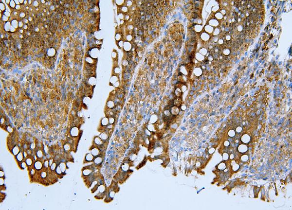
 IHC (Immunohistochemistry)
(Figure 8. IHC analysis of PRDM1/Blimp1 using anti-PRDM1/Blimp1 antibody (AAA19133).PRDM1/Blimp1 was detected in paraffin-embedded section of rat small intestine tissue. Heat mediated antigen retrieval was performed in citrate buffer (pH6, epitope retrieval solution) for 20 mins. The tissue section was blocked with 10% goat serum. The tissue section was then incubated with 1ug/ml rabbit anti-PRDM1/Blimp1 Antibody (AAA19133) overnight at 4 degree C. Biotinylated goat anti-rabbit IgG was used as secondary antibody and incubated for 30 minutes at 37 degree C. The tissue section was developed using Strepavidin-Biotin-Complex (SABC) with DAB as the chromogen.)
IHC (Immunohistochemistry)
(Figure 8. IHC analysis of PRDM1/Blimp1 using anti-PRDM1/Blimp1 antibody (AAA19133).PRDM1/Blimp1 was detected in paraffin-embedded section of rat small intestine tissue. Heat mediated antigen retrieval was performed in citrate buffer (pH6, epitope retrieval solution) for 20 mins. The tissue section was blocked with 10% goat serum. The tissue section was then incubated with 1ug/ml rabbit anti-PRDM1/Blimp1 Antibody (AAA19133) overnight at 4 degree C. Biotinylated goat anti-rabbit IgG was used as secondary antibody and incubated for 30 minutes at 37 degree C. The tissue section was developed using Strepavidin-Biotin-Complex (SABC) with DAB as the chromogen.)
PRDM1/Blimp1, Polyclonal Antibody (Cat# AAA19133)
Full Name
Anti-PRDM1/Blimp1 Picoband antibody
Gene Names
PRDM1; BLIMP1; PRDI-BF1
Reactivity
Human, Mouse, Rat
No cross reactivity with other proteins.
No cross reactivity with other proteins.
Applications
EIA, IHC, WB
Pricing


 IHC (Immunohistchemistry)
(Figure 6. IHC analysis of CPI17 alpha using anti- CPI17 alpha antibody (AAA19176).CPI17 alpha was detected in paraffin-embedded section of human placenta tissues. Heat mediated antigen retrieval was performed in citrate buffer (pH6, epitope retrieval solution) for 20 mins. The tissue section was blocked with 10% goat serum. The tissue section was then incubated with 1ug/ml rabbit anti- CPI17 alpha Antibody (AAA19176) overnight at 4 degree C. Biotinylated goat anti-rabbit IgG was used as secondary antibody and incubated for 30 minutes at 37 degree C. The tissue section was developed using Strepavidin-Biotin-Complex (SABC) with DAB as the chromogen.)
IHC (Immunohistchemistry)
(Figure 6. IHC analysis of CPI17 alpha using anti- CPI17 alpha antibody (AAA19176).CPI17 alpha was detected in paraffin-embedded section of human placenta tissues. Heat mediated antigen retrieval was performed in citrate buffer (pH6, epitope retrieval solution) for 20 mins. The tissue section was blocked with 10% goat serum. The tissue section was then incubated with 1ug/ml rabbit anti- CPI17 alpha Antibody (AAA19176) overnight at 4 degree C. Biotinylated goat anti-rabbit IgG was used as secondary antibody and incubated for 30 minutes at 37 degree C. The tissue section was developed using Strepavidin-Biotin-Complex (SABC) with DAB as the chromogen.)
CPI17 alpha, Polyclonal Antibody (Cat# AAA19176)
Full Name
Anti-CPI17 alpha Picoband Antibody
Gene Names
PPP1R14A; CPI17; CPI-17; PPP1INL
Reactivity
Human, Mouse, Rat
No cross reactivity with other proteins
No cross reactivity with other proteins
Applications
IHC, WB
Purity
Immunogen affinity purified
Pricing


 FCM (Flow Cytometry)
(Figure 7. Flow Cytometry analysis of 293T cells using anti-OTULIN antibody (AAA19319).Overlay histogram showing 293T cells stained with AAA19319 (Blue line). The cells were blocked with 10% normal goat serum. And then incubated with rabbit anti-OTULIN Antibody (AAA19319, 1μg/1x106 cells) for 30 min at 20 degree C. DyLight®488 conjugated goat anti-rabbit IgG (5-10μg/1x106 cells) was used as secondary antibody for 30 minutes at 20 degree C. Isotype control antibody (Green line) was rabbit IgG (1μg/1x106) used under the same conditions. Unlabelled sample (Red line) was also used as a control.)
FCM (Flow Cytometry)
(Figure 7. Flow Cytometry analysis of 293T cells using anti-OTULIN antibody (AAA19319).Overlay histogram showing 293T cells stained with AAA19319 (Blue line). The cells were blocked with 10% normal goat serum. And then incubated with rabbit anti-OTULIN Antibody (AAA19319, 1μg/1x106 cells) for 30 min at 20 degree C. DyLight®488 conjugated goat anti-rabbit IgG (5-10μg/1x106 cells) was used as secondary antibody for 30 minutes at 20 degree C. Isotype control antibody (Green line) was rabbit IgG (1μg/1x106) used under the same conditions. Unlabelled sample (Red line) was also used as a control.)
OTULIN, Polyclonal Antibody (Cat# AAA19319)
Full Name
Anti-OTULIN Antibody
Gene Names
OTULIN; GUM; FAM105B
Reactivity
Human, Mouse, Rat
Applications
WB, IHC-P, ICC, IF, FC/FACS/FCM, EIA
Purity
Immunogen affinity purified.
Pricing
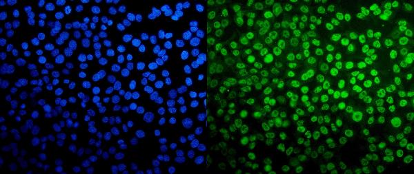


 FCM (Flow Cytometry)
(Figure 13. Flow Cytometry analysis of Hela cells using anti-TAF4 antibody (AAA19307).Overlay histogram showing Hela cells stained with AAA19307 (Blue line). The cells were blocked with 10% normal goat serum. And then incubated with rabbit anti-TAF4 Antibody (AAA19307, 1μg/1x106 cells) for 30 min at 20 degree C. DyLight®488 conjugated goat anti-rabbit IgG (5-10μg/1x106 cells) was used as secondary antibody for 30 minutes at 20 degree C. Isotype control antibody (Green line) was rabbit IgG (1μg/1x106) used under the same conditions. Unlabelled sample (Red line) was also used as a control.)
FCM (Flow Cytometry)
(Figure 13. Flow Cytometry analysis of Hela cells using anti-TAF4 antibody (AAA19307).Overlay histogram showing Hela cells stained with AAA19307 (Blue line). The cells were blocked with 10% normal goat serum. And then incubated with rabbit anti-TAF4 Antibody (AAA19307, 1μg/1x106 cells) for 30 min at 20 degree C. DyLight®488 conjugated goat anti-rabbit IgG (5-10μg/1x106 cells) was used as secondary antibody for 30 minutes at 20 degree C. Isotype control antibody (Green line) was rabbit IgG (1μg/1x106) used under the same conditions. Unlabelled sample (Red line) was also used as a control.)
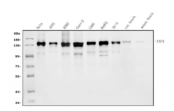
TAF4, Polyclonal Antibody (Cat# AAA19307)
Full Name
Anti-TAF4 Antibody
Gene Names
TAF4; TAF2C; TAF4A; TAF2C1; TAFII130; TAFII135
Reactivity
Human, Mouse, Rat
Applications
WB, IHC-P, ICC, IF, EIA
Purity
Immunogen affinity purified.
Pricing


 WB (Western Blot)
(WB Suggested Anti-HNRPL Antibody Titration: 0.2-1 ug/mlELISA Titer: 1:62500Positive Control: Jurkat cell lysateHNRNPL is strongly supported by BioGPS gene expression data to be expressed in Human Jurkat cells)
WB (Western Blot)
(WB Suggested Anti-HNRPL Antibody Titration: 0.2-1 ug/mlELISA Titer: 1:62500Positive Control: Jurkat cell lysateHNRNPL is strongly supported by BioGPS gene expression data to be expressed in Human Jurkat cells)
HNRPL, Polyclonal Antibody (Cat# AAA23474)
Full Name
HNRPL antibody - N-terminal region
Gene Names
HNRNPL; HNRPL; hnRNP-L; P/OKcl.14
Reactivity
Cow, Dog, Guinea Pig, Horse, Human, Mouse, Rabbit, Rat
Applications
IF, IP, IHC, WB
Purity
Affinity Purified
Pricing


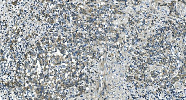
 IHC (Immunohistchemistry)
(Figure 6. IHC analysis of Cdc20 using anti-Cdc20 antibody (AAA19132).Cdc20 was detected in paraffin-embedded section of human mammary cancer tissue. Heat mediated antigen retrieval was performed in citrate buffer (pH6, epitope retrieval solution) for 20 mins. The tissue section was blocked with 10% goat serum. The tissue section was then incubated with 1ug/ml rabbit anti-Cdc20 Antibody (AAA19132) overnight at 4 degree C. Biotinylated goat anti-rabbit IgG was used as secondary antibody and incubated for 30 minutes at 37 degree C. The tissue section was developed using Strepavidin-Biotin-Complex (SABC) with DAB as the chromogen.)
IHC (Immunohistchemistry)
(Figure 6. IHC analysis of Cdc20 using anti-Cdc20 antibody (AAA19132).Cdc20 was detected in paraffin-embedded section of human mammary cancer tissue. Heat mediated antigen retrieval was performed in citrate buffer (pH6, epitope retrieval solution) for 20 mins. The tissue section was blocked with 10% goat serum. The tissue section was then incubated with 1ug/ml rabbit anti-Cdc20 Antibody (AAA19132) overnight at 4 degree C. Biotinylated goat anti-rabbit IgG was used as secondary antibody and incubated for 30 minutes at 37 degree C. The tissue section was developed using Strepavidin-Biotin-Complex (SABC) with DAB as the chromogen.)
Cdc20/P55 Cdc, Polyclonal Antibody (Cat# AAA19132)
Full Name
Anti-Cdc20/P55 Cdc Picoband Antibody
Gene Names
CDC20; CDC20A; p55CDC; bA276H19.3
Reactivity
Human, Mouse, Rat
No cross reactivity with other proteins.
No cross reactivity with other proteins.
Applications
IHC, WB
Purity
Immunogen affinity purified
Pricing


 FCM (Flow Cytometry)
(Figure 11. Flow Cytometry analysis of A431 cells using anti-SAMHD1 antibody (AAA19355).Overlay histogram showing A431 cells stained with AAA19355 (Blue line). The cells were blocked with 10% normal goat serum. And then incubated with mouse anti- SAMHD1 Antibody (AAA19355, 1μg/1x106 cells) for 30 min at 20 degree C. DyLight®488 conjugated goat anti-mouse IgG (BA1126, 5-10μg/1x106 cells) was used as secondary antibody for 30 minutes at 20 degree C. Isotype control antibody (Green line) was mouse IgG (1μg/1x106) used under the same conditions. Unlabelled sample (Red line) was also used as a control.)
FCM (Flow Cytometry)
(Figure 11. Flow Cytometry analysis of A431 cells using anti-SAMHD1 antibody (AAA19355).Overlay histogram showing A431 cells stained with AAA19355 (Blue line). The cells were blocked with 10% normal goat serum. And then incubated with mouse anti- SAMHD1 Antibody (AAA19355, 1μg/1x106 cells) for 30 min at 20 degree C. DyLight®488 conjugated goat anti-mouse IgG (BA1126, 5-10μg/1x106 cells) was used as secondary antibody for 30 minutes at 20 degree C. Isotype control antibody (Green line) was mouse IgG (1μg/1x106) used under the same conditions. Unlabelled sample (Red line) was also used as a control.)
SAMHD1, Monoclonal Antibody (Cat# AAA19355)
Full Name
Anti-SAMHD1 Antibody (monoclonal, 3B9)
Gene Names
SAMHD1; DCIP; CHBL2; HDDC1; MOP-5; SBBI88
Reactivity
Human
Applications
WB, IHC-P, ICC, IF, FC/FACS/FCM
Purity
Immunogen affinity purified.
Pricing


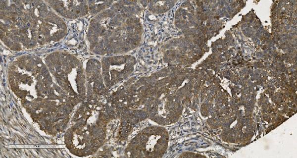
 IHC (Immunohistchemistry)
(Figure 6. IHC analysis of Musashi 1/Msi1 using anti- Musashi 1/Msi1 antibody (AAA19172).Musashi 1/Msi1 was detected in paraffin-embedded section of human mammary cancer tissues. Heat mediated antigen retrieval was performed in citrate buffer (pH6, epitope retrieval solution) for 20 mins. The tissue section was blocked with 10% goat serum. The tissue section was then incubated with 1ug/ml rabbit anti- Musashi 1/Msi1 Antibody (AAA19172) overnight at 4 degree C. Biotinylated goat anti-rabbit IgG was used as secondary antibody and incubated for 30 minutes at 37 degree C. The tissue section was developed using Strepavidin-Biotin-Complex (SABC) with DAB as the chromogen.)
IHC (Immunohistchemistry)
(Figure 6. IHC analysis of Musashi 1/Msi1 using anti- Musashi 1/Msi1 antibody (AAA19172).Musashi 1/Msi1 was detected in paraffin-embedded section of human mammary cancer tissues. Heat mediated antigen retrieval was performed in citrate buffer (pH6, epitope retrieval solution) for 20 mins. The tissue section was blocked with 10% goat serum. The tissue section was then incubated with 1ug/ml rabbit anti- Musashi 1/Msi1 Antibody (AAA19172) overnight at 4 degree C. Biotinylated goat anti-rabbit IgG was used as secondary antibody and incubated for 30 minutes at 37 degree C. The tissue section was developed using Strepavidin-Biotin-Complex (SABC) with DAB as the chromogen.)
Musashi 1/Msi1, Polyclonal Antibody (Cat# AAA19172)
Full Name
Anti-Musashi 1/Msi1 Picoband Antibody
Reactivity
Human, Mouse, Rat
No cross reactivity with other proteins
No cross reactivity with other proteins
Applications
IHC, WB
Purity
Immunogen affinity purified
Pricing


 FCM (Flow Cytometry)
(Figure 6. Flow Cytometry analysis of HEPA1-6 cells using anti-PAR4/Pawr antibody (AAA19278).Overlay histogram showing HEPA1-6 cells stained with AAA19278 (Blue line). The cells were blocked with 10% normal goat serum. And then incubated with rabbit anti-PAR4/Pawr Antibody (AAA19278, 1μg/1x106 cells) for 30 min at 20 degree C. DyLight®488 conjugated goat anti-rabbit IgG (5-10μg/1x106 cells) was used as secondary antibody for 30 minutes at 20 degree C. Isotype control antibody (Green line) was rabbit IgG (1μg/1x106) used under the same conditions. Unlabelled sample (Red line) was also used as a control.)
FCM (Flow Cytometry)
(Figure 6. Flow Cytometry analysis of HEPA1-6 cells using anti-PAR4/Pawr antibody (AAA19278).Overlay histogram showing HEPA1-6 cells stained with AAA19278 (Blue line). The cells were blocked with 10% normal goat serum. And then incubated with rabbit anti-PAR4/Pawr Antibody (AAA19278, 1μg/1x106 cells) for 30 min at 20 degree C. DyLight®488 conjugated goat anti-rabbit IgG (5-10μg/1x106 cells) was used as secondary antibody for 30 minutes at 20 degree C. Isotype control antibody (Green line) was rabbit IgG (1μg/1x106) used under the same conditions. Unlabelled sample (Red line) was also used as a control.)
PAR4/Pawr, Polyclonal Antibody (Cat# AAA19278)
Full Name
Anti-PAR4/Pawr Antibody
Gene Names
Pawr; PAR4; Par-4; 2310001G03Rik
Reactivity
Mouse, Rat
Applications
WB, IHC-P, ICC, IF, FC/FACS/FCM, EIA
Purity
Immunogen affinity purified.
Pricing


 FCM (Flow Cytometry)
(Figure 10. Flow Cytometry analysis of A431 cells using anti-CDC27 antibody (AAA19285).Overlay histogram showing A431 cells stained with AAA19285 (Blue line). The cells were blocked with 10% normal goat serum. And then incubated with rabbit anti-CDC27 Antibody (AAA19285, 1μg/1x106 cells) for 30 min at 20 degree C. DyLight®488 conjugated goat anti-rabbit IgG (5-10μg/1x106 cells) was used as secondary antibody for 30 minutes at 20 degree C. Isotype control antibody (Green line) was rabbit IgG (1μg/1x106) used under the same conditions. Unlabelled sample (Red line) was also used as a control.)
FCM (Flow Cytometry)
(Figure 10. Flow Cytometry analysis of A431 cells using anti-CDC27 antibody (AAA19285).Overlay histogram showing A431 cells stained with AAA19285 (Blue line). The cells were blocked with 10% normal goat serum. And then incubated with rabbit anti-CDC27 Antibody (AAA19285, 1μg/1x106 cells) for 30 min at 20 degree C. DyLight®488 conjugated goat anti-rabbit IgG (5-10μg/1x106 cells) was used as secondary antibody for 30 minutes at 20 degree C. Isotype control antibody (Green line) was rabbit IgG (1μg/1x106) used under the same conditions. Unlabelled sample (Red line) was also used as a control.)
CDC27, Polyclonal Antibody (Cat# AAA19285)
Full Name
Anti-CDC27 Antibody
Gene Names
CDC27; APC3; HNUC; NUC2; ANAPC3; CDC27Hs; D0S1430E; D17S978E
Reactivity
Human, Mouse, Rat
Applications
WB, IHC-P, ICC, IF, FC/FACS/FCM, EIA
Purity
Immunogen affinity purified.
Pricing


 IHC (Immunohistochemistry)
(Figure 8. IHC analysis of TAGLN/Transgelin using anti-TAGLN/Transgelin antibody (AAA19286).TAGLN/Transgelin was detected in paraffin-embedded section of rat testis tissue. Heat mediated antigen retrieval was performed in EDTA buffer (pH8. 0, epitope retrieval solution). The tissue section was blocked with 10% goat serum. The tissue section was then incubated with 2μg/ml rabbit anti-TAGLN/Transgelin Antibody (AAA19286) overnight at 4 degree C. Biotinylated goat anti-rabbit IgG was used as secondary antibody and incubated for 30 minutes at 37 degree C. The tissue section was developed using Strepavidin-Biotin-Complex (SABC) (Catalog # with DAB as the chromogen.)
IHC (Immunohistochemistry)
(Figure 8. IHC analysis of TAGLN/Transgelin using anti-TAGLN/Transgelin antibody (AAA19286).TAGLN/Transgelin was detected in paraffin-embedded section of rat testis tissue. Heat mediated antigen retrieval was performed in EDTA buffer (pH8. 0, epitope retrieval solution). The tissue section was blocked with 10% goat serum. The tissue section was then incubated with 2μg/ml rabbit anti-TAGLN/Transgelin Antibody (AAA19286) overnight at 4 degree C. Biotinylated goat anti-rabbit IgG was used as secondary antibody and incubated for 30 minutes at 37 degree C. The tissue section was developed using Strepavidin-Biotin-Complex (SABC) (Catalog # with DAB as the chromogen.)
TAGLN/Transgelin, Polyclonal Antibody (Cat# AAA19286)
Full Name
Anti-TAGLN/Transgelin Antibody
Gene Names
TAGLN; SM22; SMCC; TAGLN1; WS3-10
Reactivity
Human, Mouse, Rat
Applications
WB, IHC-P, ICC, IF, FC/FACS/FCM, EIA
Purity
Immunogen affinity purified.
Pricing
 WB (Western Blot)
(WB Suggested Anti-MOSC1 Antibody Titration: 0.2-1 ug/mlELISA Titer: 1:1562500Positive Control: Human Placenta)
WB (Western Blot)
(WB Suggested Anti-MOSC1 Antibody Titration: 0.2-1 ug/mlELISA Titer: 1:1562500Positive Control: Human Placenta)
MARC1, Polyclonal Antibody (Cat# AAA23536)
Full Name
MARC1 Antibody - C-terminal region
Gene Names
MARC1; MOSC1
Reactivity
Tested Reactivity: Human
Predicted Reactivity: Cow, Dog, Guinea Pig, Horse, Human, Mouse, Rabbit, Rat, Zebrafish
Predicted Reactivity: Cow, Dog, Guinea Pig, Horse, Human, Mouse, Rabbit, Rat, Zebrafish
Pricing


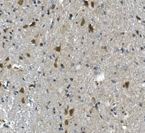
 IHC (Immunohistchemistry)
(Figure 6. IHC analysis of PP2A-alpha/PPP2CA using anti-PP2A-alpha/PPP2CA antibody (AAA19366).PP2A-alpha/PPP2CA was detected in paraffin-embedded section of rat brain tissue. Heat mediated antigen retrieval was performed in EDTA buffer (pH8. 0, epitope retrieval solution). The tissue section was blocked with 10% goat serum. The tissue section was then incubated with 1μg/ml mouse anti-PP2A-alpha/PPP2CA Antibody (AAA19366) overnight at 4 degree C. Biotinylated goat anti-mouse IgG was used as secondary antibody and incubated for 30 minutes at 37 degree C. The tissue section was developed using Strepavidin-Biotin-Complex (SABC) (Catalog # with DAB as the chromogen.)
IHC (Immunohistchemistry)
(Figure 6. IHC analysis of PP2A-alpha/PPP2CA using anti-PP2A-alpha/PPP2CA antibody (AAA19366).PP2A-alpha/PPP2CA was detected in paraffin-embedded section of rat brain tissue. Heat mediated antigen retrieval was performed in EDTA buffer (pH8. 0, epitope retrieval solution). The tissue section was blocked with 10% goat serum. The tissue section was then incubated with 1μg/ml mouse anti-PP2A-alpha/PPP2CA Antibody (AAA19366) overnight at 4 degree C. Biotinylated goat anti-mouse IgG was used as secondary antibody and incubated for 30 minutes at 37 degree C. The tissue section was developed using Strepavidin-Biotin-Complex (SABC) (Catalog # with DAB as the chromogen.)
PP2A-alpha/PPP2CA, Monoclonal Antibody (Cat# AAA19366)
Full Name
Anti-PP2A-alpha/PPP2CA Antibody (monoclonal, 3B6)
Gene Names
PPP2CA; RP-C; PP2Ac; PP2CA; PP2Calpha
Reactivity
Human, Monkey, Mouse, Rat
Applications
WB, IHC-P, ICC, IF, FC/FACS/FCM
Purity
Immunogen affinity purified.
Pricing


 IF (Immunofluorescence)
(Figure 9. IF analysis of ApoER2/LRP8 using anti- ApoER2/LRP8 antibody (AAA19275).ApoER2/LRP8 was detected in immunocytochemical section of HepG2 cells. Enzyme antigen retrieval was performed using IHC enzyme antigen retrieval reagent for 15 mins. The cells were blocked with 10% goat serum. And then incubated with 4μg/mL rabbit anti- ApoER2/LRP8 Antibody (AAA19275) overnight at 4 degree C. DyLight®488 Conjugated Goat Anti-Rabbit IgG was used as secondary antibody at 1:100 dilution and incubated for 30 minutes at 37 degree C. The section was counterstained with DAPI. Visualize using a fluorescence microscope and filter sets appropriate for the label used.)
IF (Immunofluorescence)
(Figure 9. IF analysis of ApoER2/LRP8 using anti- ApoER2/LRP8 antibody (AAA19275).ApoER2/LRP8 was detected in immunocytochemical section of HepG2 cells. Enzyme antigen retrieval was performed using IHC enzyme antigen retrieval reagent for 15 mins. The cells were blocked with 10% goat serum. And then incubated with 4μg/mL rabbit anti- ApoER2/LRP8 Antibody (AAA19275) overnight at 4 degree C. DyLight®488 Conjugated Goat Anti-Rabbit IgG was used as secondary antibody at 1:100 dilution and incubated for 30 minutes at 37 degree C. The section was counterstained with DAPI. Visualize using a fluorescence microscope and filter sets appropriate for the label used.)
ApoER2/LRP8, Polyclonal Antibody (Cat# AAA19275)
Full Name
Anti-ApoER2/LRP8 Antibody
Gene Names
LRP8; MCI1; LRP-8; APOER2; HSZ75190
Reactivity
Human, Mouse, Rat
Applications
WB, IHC-P, ICC, IF, FC/FACS/FCM, EIA
Purity
Immunogen affinity purified.
Pricing


 IF (Immunofluorescence)
(Figure 15. IF analysis of LSM2 using anti- LSM2 antibody (AAA19316).LSM2 was detected in immunocytochemical section of MCF-7 cells. Enzyme antigen retrieval was performed using IHC enzyme antigen retrieval reagent for 15 mins. The cells were blocked with 10% goat serum. And then incubated with 5μg/mL rabbit anti- LSM2 Antibody (AAA19316) overnight at 4 degree C. DyLight®488 Conjugated Goat Anti-Rabbit IgG was used as secondary antibody at 1:100 dilution and incubated for 30 minutes at 37 degree C. The section was counterstained with DAPI. Visualize using a fluorescence microscope and filter sets appropriate for the label used.)
IF (Immunofluorescence)
(Figure 15. IF analysis of LSM2 using anti- LSM2 antibody (AAA19316).LSM2 was detected in immunocytochemical section of MCF-7 cells. Enzyme antigen retrieval was performed using IHC enzyme antigen retrieval reagent for 15 mins. The cells were blocked with 10% goat serum. And then incubated with 5μg/mL rabbit anti- LSM2 Antibody (AAA19316) overnight at 4 degree C. DyLight®488 Conjugated Goat Anti-Rabbit IgG was used as secondary antibody at 1:100 dilution and incubated for 30 minutes at 37 degree C. The section was counterstained with DAPI. Visualize using a fluorescence microscope and filter sets appropriate for the label used.)
LSM2, Polyclonal Antibody (Cat# AAA19316)
Full Name
Anti-LSM2 Antibody
Gene Names
LSM2; G7B; snRNP; C6orf28; YBL026W
Reactivity
Human, Mouse, Rat, Monkey
Applications
WB, IHC-P, ICC, IF, FC/FACS/FCM
Purity
Immunogen affinity purified.
Pricing
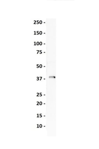
 WB (Western Blot)
(Western Blot detection against Immunogen (37kD).)
WB (Western Blot)
(Western Blot detection against Immunogen (37kD).)
KHDRBS1, Monoclonal Antibody (Cat# AAA24252)
Full Name
KHDRBS1 (KH Domain-containing, RNA-binding, Signal Transduction-associated Protein 1, FLJ34027, GAP-associated Tyrosine Phosphoprotein p62, p21 Ras GTPase-activating Protein-associated p62, p62, p68, Src-associated in Mitosis 68kD Protein, Sam68) (AP)
Gene Names
KHDRBS1; p62; p68; Sam68
Reactivity
Human
Applications
EIA, IHC, WB
Purity
Purified by Protein A Affinity Chromatography.
Pricing


 FCM (Flow Cytometry)
(Figure 6. Flow Cytometry analysis of THP-1 cells using anti-TLR1 antibody (AAA19134).Overlay histogram showing THP-1 cells stained with AAA19134 (Blue line).The cells were blocked with 10% normal goat serum. And then incubated with rabbit anti-TLR1 Antibody (AAA19134,1ug/1x10^6 cells) for 30 min at 20 degree C. DyLight®488 conjugated goat anti-rabbit IgG (5-10ug/1x10^6 cells) was used as secondary antibody for 30 minutes at 20 degree C. Isotype control antibody (Green line) was rabbit IgG (1ug/1x106) used under the same conditions. Unlabelled sample (Red line) was also used as a control.)
FCM (Flow Cytometry)
(Figure 6. Flow Cytometry analysis of THP-1 cells using anti-TLR1 antibody (AAA19134).Overlay histogram showing THP-1 cells stained with AAA19134 (Blue line).The cells were blocked with 10% normal goat serum. And then incubated with rabbit anti-TLR1 Antibody (AAA19134,1ug/1x10^6 cells) for 30 min at 20 degree C. DyLight®488 conjugated goat anti-rabbit IgG (5-10ug/1x10^6 cells) was used as secondary antibody for 30 minutes at 20 degree C. Isotype control antibody (Green line) was rabbit IgG (1ug/1x106) used under the same conditions. Unlabelled sample (Red line) was also used as a control.)
TLR1, Polyclonal Antibody (Cat# AAA19134)
Full Name
Anti-TLR1 Picoband antibody
Gene Names
TLR1; TIL; CD281; rsc786; TIL. LPRS5
Reactivity
Human, Mouse, Rat
No cross reactivity with other proteins.
No cross reactivity with other proteins.
Applications
EIA, FC/FACS, IHC, ICC, WB
Pricing


 Application Data
(Analysis of Protein Array containing more than 19,000 full-length human proteins using AMACR/p504S Mouse Monoclonal Antibody (AMACR/1864). Z- and S- Score: The Z-score represents the strength of a signal that a monoclonal antibody (MAb) (in combination with a fluorescently-tagged anti-IgG secondary antibody) produces when binding to a particular protein on the HuProtTM array. Z-scores are described in units of standard deviations (SD's) above the mean value of all signals generated on that array. If targets on HuProtTM are arranged in descending order of the Z-score, the S-score is the difference (also in units of SD's) between the Z-score. S-score therefore represents the relative target specificity of a MAb to its intended target. A MAb is considered to specific to its intended target, if the MAb has an S-score of at least 2.5. For example, if a MAb binds to protein X with a Z-score of 43 and to protein Y with a Z-score of 14, then the S-score for the binding of that MAb to protein X is equal to 29.)
Application Data
(Analysis of Protein Array containing more than 19,000 full-length human proteins using AMACR/p504S Mouse Monoclonal Antibody (AMACR/1864). Z- and S- Score: The Z-score represents the strength of a signal that a monoclonal antibody (MAb) (in combination with a fluorescently-tagged anti-IgG secondary antibody) produces when binding to a particular protein on the HuProtTM array. Z-scores are described in units of standard deviations (SD's) above the mean value of all signals generated on that array. If targets on HuProtTM are arranged in descending order of the Z-score, the S-score is the difference (also in units of SD's) between the Z-score. S-score therefore represents the relative target specificity of a MAb to its intended target. A MAb is considered to specific to its intended target, if the MAb has an S-score of at least 2.5. For example, if a MAb binds to protein X with a Z-score of 43 and to protein Y with a Z-score of 14, then the S-score for the binding of that MAb to protein X is equal to 29.)
AMACR/p504S, Monoclonal Antibody (Cat# AAA23906)
Full Name
AMACR/p504S (Prostate Cancer Marker)
Gene Names
AMACR; RM; RACE; CBAS4; P504S; AMACRD
Reactivity
Human. Others not known.
Applications
Western Blot, Immunohistochemistry
Purity
Purified Ab with BSA and Azide at 200ug/ml OR Purified Ab WITHOUT BSA and Azide at 1.0mg/ml
Pricing


 IHC (Immunohistochemistry)
(Figure 11. IHC analysis of U2AF65/U2AF2 using anti U2AF65/U2AF2 antibody (AAA19376).U2AF65/U2AF2 was detected in paraffin-embedded section of human gallbladder adenocarcinoma tissue. Heat mediated antigen retrieval was performed in EDTA buffer (pH8. 0, epitope retrieval solution). The tissue section was blocked with 10% goat serum. The tissue section was then incubated with 2μg/ml mouse anti-U2AF65/U2AF2 Antibody (AAA19376) overnight at 4 degree C. Biotinylated goat anti-mouse IgG was used as secondary antibody and incubated for 30 minutes at 37 degree C. The tissue section was developed using Strepavidin-Biotin-Complex (SABC) (Catalog # with DAB as the chromogen.)
IHC (Immunohistochemistry)
(Figure 11. IHC analysis of U2AF65/U2AF2 using anti U2AF65/U2AF2 antibody (AAA19376).U2AF65/U2AF2 was detected in paraffin-embedded section of human gallbladder adenocarcinoma tissue. Heat mediated antigen retrieval was performed in EDTA buffer (pH8. 0, epitope retrieval solution). The tissue section was blocked with 10% goat serum. The tissue section was then incubated with 2μg/ml mouse anti-U2AF65/U2AF2 Antibody (AAA19376) overnight at 4 degree C. Biotinylated goat anti-mouse IgG was used as secondary antibody and incubated for 30 minutes at 37 degree C. The tissue section was developed using Strepavidin-Biotin-Complex (SABC) (Catalog # with DAB as the chromogen.)
U2AF65/U2AF2, Monoclonal Antibody (Cat# AAA19376)
Full Name
Anti-U2AF65/U2AF2 Antibody (monoclonal, 3G9)
Gene Names
U2AF2; U2AF65
Reactivity
Human, Mouse, Rat
Applications
WB, IHC-P, ICC, IF, FC/FACS/FCM
Purity
Immunogen affinity purified.
Pricing


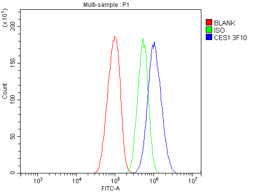


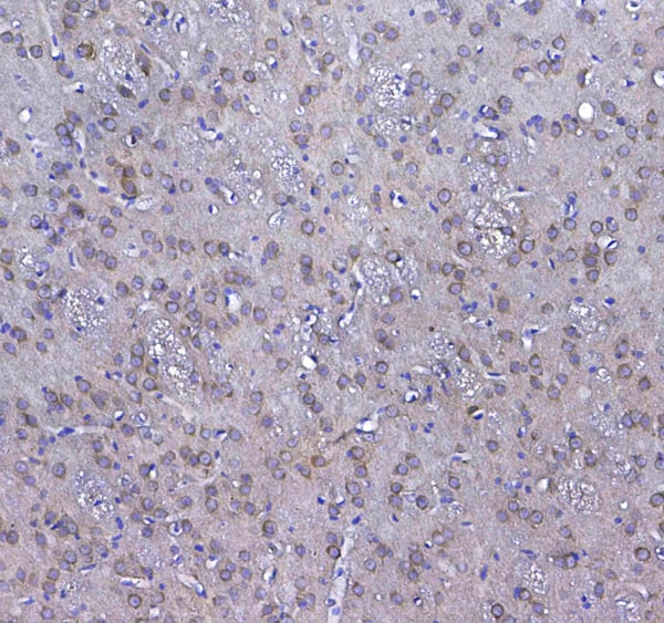
 IHC (Immunohistochemistry)
(Figure 8. IHC analysis of Liver Carboxylesterase 1/CES1 using anti-Liver Carboxylesterase 1/CES1 antibody (AAA19363).Liver Carboxylesterase 1/CES1 was detected in paraffin-embedded section of rat liver tissue. Heat mediated antigen retrieval was performed in EDTA buffer (pH8. 0, epitope retrieval solution). The tissue section was blocked with 10% goat serum. The tissue section was then incubated with 2μg/ml mouse anti-Liver Carboxylesterase 1/CES1 Antibody (AAA19363) overnight at 4 degree C. Biotinylated goat anti-mouse IgG was used as secondary antibody and incubated for 30 minutes at 37 degree C. The tissue section was developed using Strepavidin-Biotin-Complex (SABC) (Catalog # with DAB as the chromogen.)
IHC (Immunohistochemistry)
(Figure 8. IHC analysis of Liver Carboxylesterase 1/CES1 using anti-Liver Carboxylesterase 1/CES1 antibody (AAA19363).Liver Carboxylesterase 1/CES1 was detected in paraffin-embedded section of rat liver tissue. Heat mediated antigen retrieval was performed in EDTA buffer (pH8. 0, epitope retrieval solution). The tissue section was blocked with 10% goat serum. The tissue section was then incubated with 2μg/ml mouse anti-Liver Carboxylesterase 1/CES1 Antibody (AAA19363) overnight at 4 degree C. Biotinylated goat anti-mouse IgG was used as secondary antibody and incubated for 30 minutes at 37 degree C. The tissue section was developed using Strepavidin-Biotin-Complex (SABC) (Catalog # with DAB as the chromogen.)
Liver Carboxylesterase 1/CES1, Monoclonal Antibody (Cat# AAA19363)
Full Name
Anti-Liver Carboxylesterase 1/CES1 Antibody (monoclonal, 3F10)
Gene Names
CES1; CEH; REH; TGH; ACAT; CE-1; CES2; HMSE; SES1; HMSE1; PCE-1; hCE-1
Reactivity
Human, Mouse, Rat
Applications
WB, IHC-P, ICC, IF, FC/FACS/FCM
Purity
Immunogen affinity purified.
Pricing


 FCM (Flow Cytometry)
(Figure 8. Flow Cytometry analysis of SiHa cells using anti-Astrin/Deepest/SPAG5 antibody (AAA19313).Overlay histogram showing SiHa cells stained with AAA19313 (Blue line). The cells were blocked with 10% normal goat serum. And then incubated with rabbit anti-Astrin/Deepest/SPAG5 Antibody (AAA19313, 1μg/1x106 cells) for 30 min at 20 degree C. DyLight®488 conjugated goat anti-rabbit IgG (5-10μg/1x106 cells) was used as secondary antibody for 30 minutes at 20 degree C. Isotype control antibody (Green line) was rabbit IgG (1μg/1x106) used under the same conditions. Unlabelled sample (Red line) was also used as a control.)
FCM (Flow Cytometry)
(Figure 8. Flow Cytometry analysis of SiHa cells using anti-Astrin/Deepest/SPAG5 antibody (AAA19313).Overlay histogram showing SiHa cells stained with AAA19313 (Blue line). The cells were blocked with 10% normal goat serum. And then incubated with rabbit anti-Astrin/Deepest/SPAG5 Antibody (AAA19313, 1μg/1x106 cells) for 30 min at 20 degree C. DyLight®488 conjugated goat anti-rabbit IgG (5-10μg/1x106 cells) was used as secondary antibody for 30 minutes at 20 degree C. Isotype control antibody (Green line) was rabbit IgG (1μg/1x106) used under the same conditions. Unlabelled sample (Red line) was also used as a control.)
Astrin/Deepest/SPAG5, Polyclonal Antibody (Cat# AAA19313)
Full Name
Anti-Astrin/Deepest/SPAG5 Antibody
Gene Names
SPAG5; MAP126; DEEPEST; hMAP126
Reactivity
Human
Applications
WB, IHC-P, ICC, IF, FC/FACS/FCM, EIA
Purity
Immunogen affinity purified.
Pricing


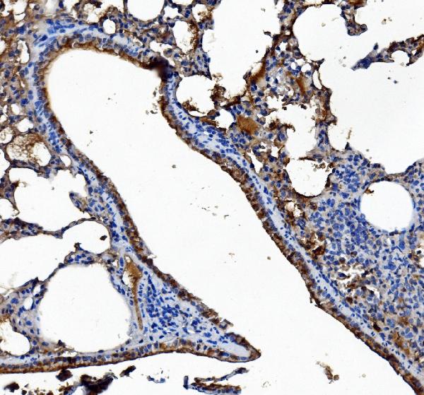
 IHC (Immunohistochemistry)
(Immunohistochemistry of paraffin-embedded mouse brain using SRP19 antibody at dilution of 1:100 (40x lens).)
IHC (Immunohistochemistry)
(Immunohistochemistry of paraffin-embedded mouse brain using SRP19 antibody at dilution of 1:100 (40x lens).)
SRP19, Polyclonal Antibody (Cat# AAA28153)
Full Name
SRP19 Polyclonal Antibody
Reactivity
Human, Mouse, Rat
Applications
WB, IHC, IF
Purity
Affinity Purification
Pricing


 IHC (Immunohistchemistry)
(Figure 6. IHC analysis of GLO1 using anti-GLO1 antibody (AAA19155).GLO1 was detected in paraffin-embedded section of rat spleen tissue. Heat mediated antigen retrieval was performed in citrate buffer (pH6, epitope retrieval solution) for 20 mins. The tissue section was blocked with 10% goat serum. The tissue section was then incubated with 1ug/ml rabbit anti-GLO1 Antibody (AAA19155) overnight at 4 degree C. Biotinylated goat anti-rabbit IgG was used as secondary antibody and incubated for 30 minutes at 37 degree C. The tissue section was developed using Strepavidin-Biotin-Complex (SABC) with DAB as the chromogen.)
IHC (Immunohistchemistry)
(Figure 6. IHC analysis of GLO1 using anti-GLO1 antibody (AAA19155).GLO1 was detected in paraffin-embedded section of rat spleen tissue. Heat mediated antigen retrieval was performed in citrate buffer (pH6, epitope retrieval solution) for 20 mins. The tissue section was blocked with 10% goat serum. The tissue section was then incubated with 1ug/ml rabbit anti-GLO1 Antibody (AAA19155) overnight at 4 degree C. Biotinylated goat anti-rabbit IgG was used as secondary antibody and incubated for 30 minutes at 37 degree C. The tissue section was developed using Strepavidin-Biotin-Complex (SABC) with DAB as the chromogen.)
GLO1/Glyoxalase I, Polyclonal Antibody (Cat# AAA19155)
Full Name
Anti-GLO1/Glyoxalase I Picoband antibody
Gene Names
GLO1; GLYI; GLOD1; HEL-S-74
Reactivity
Human, Mouse, Rat
No cross reactivity with other proteins.
No cross reactivity with other proteins.
Applications
EIA, IHC, WB
Pricing


 IHC (Immunohistchemistry)
(Figure 6. IHC analysis of Synaptopodin/SYNPO using anti-Synaptopodin/SYNPO antibody (AAA19271).Synaptopodin/SYNPO was detected in paraffin-embedded section of mouse brain tissue. Heat mediated antigen retrieval was performed in EDTA buffer (pH8. 0, epitope retrieval solution). The tissue section was blocked with 10% goat serum. The tissue section was then incubated with 2μg/ml rabbit anti-Synaptopodin/SYNPO Antibody (AAA19271) overnight at 4 degree C. Biotinylated goat anti-rabbit IgG was used as secondary antibody and incubated for 30 minutes at 37 degree C. The tissue section was developed using Strepavidin-Biotin-Complex (SABC) (Catalog # with DAB as the chromogen.)
IHC (Immunohistchemistry)
(Figure 6. IHC analysis of Synaptopodin/SYNPO using anti-Synaptopodin/SYNPO antibody (AAA19271).Synaptopodin/SYNPO was detected in paraffin-embedded section of mouse brain tissue. Heat mediated antigen retrieval was performed in EDTA buffer (pH8. 0, epitope retrieval solution). The tissue section was blocked with 10% goat serum. The tissue section was then incubated with 2μg/ml rabbit anti-Synaptopodin/SYNPO Antibody (AAA19271) overnight at 4 degree C. Biotinylated goat anti-rabbit IgG was used as secondary antibody and incubated for 30 minutes at 37 degree C. The tissue section was developed using Strepavidin-Biotin-Complex (SABC) (Catalog # with DAB as the chromogen.)
Synaptopodin/SYNPO, Polyclonal Antibody (Cat# AAA19271)
Full Name
Anti-Synaptopodin/SYNPO Antibody
Reactivity
Human, Mouse, Rat
Applications
WB, IHC-P, ICC, IF, FC/FACS/FCM
Purity
Immunogen affinity purified.
Pricing


 FCM (Flow Cytometry)
(Figure 7. Flow Cytometry analysis of THP-1 cells using anti-PNPT1 antibody (AAA19306).Overlay histogram showing THP-1 cells stained with AAA19306 (Blue line). The cells were blocked with 10% normal goat serum. And then incubated with rabbit anti-PNPT1 Antibody (AAA19306, 1μg/1x106 cells) for 30 min at 20 degree C. DyLight®488 conjugated goat anti-rabbit IgG (5-10μg/1x106 cells) was used as secondary antibody for 30 minutes at 20 degree C. Isotype control antibody (Green line) was rabbit IgG (1μg/1x106) used under the same conditions. Unlabelled sample (Red line) was also used as a control.)
FCM (Flow Cytometry)
(Figure 7. Flow Cytometry analysis of THP-1 cells using anti-PNPT1 antibody (AAA19306).Overlay histogram showing THP-1 cells stained with AAA19306 (Blue line). The cells were blocked with 10% normal goat serum. And then incubated with rabbit anti-PNPT1 Antibody (AAA19306, 1μg/1x106 cells) for 30 min at 20 degree C. DyLight®488 conjugated goat anti-rabbit IgG (5-10μg/1x106 cells) was used as secondary antibody for 30 minutes at 20 degree C. Isotype control antibody (Green line) was rabbit IgG (1μg/1x106) used under the same conditions. Unlabelled sample (Red line) was also used as a control.)
PNPT1, Polyclonal Antibody (Cat# AAA19306)
Full Name
Anti-PNPT1 Antibody
Gene Names
PNPT1; OLD35; DFNB70; PNPASE; old-35; COXPD13
Reactivity
Human, Mouse, Rat
Applications
WB, IHC-P, ICC, IF, FC/FACS/FCM, EIA
Purity
Immunogen affinity purified.
Pricing


 WB (Western Blot)
(WB Suggested Anti-KCNK9 Antibody Titration: 1 ug/mlPositive Control: HepG2 cell lysate)
WB (Western Blot)
(WB Suggested Anti-KCNK9 Antibody Titration: 1 ug/mlPositive Control: HepG2 cell lysate)
KCNK9, Polyclonal Antibody (Cat# AAA23441)
Full Name
KCNK9 antibody - N-terminal region
Gene Names
KCNK9; KT3.2; TASK3; K2p9.1; TASK-3
Reactivity
Cow, Guinea Pig, Horse, Human, Mouse, Rabbit, Rat, Dog
Applications
IHC, WB
Purity
Affinity Purified
Pricing


 IP (Immunoprecipitation)
(Immunoprecipitation analysis of 200ug extracts of HeLa cells using 3ug NF-kB p65/RelA antibody . Western blot was performed from the immunoprecipitate using NF-kB p65/RelA antibody at a dilition of 1:1000.)
IP (Immunoprecipitation)
(Immunoprecipitation analysis of 200ug extracts of HeLa cells using 3ug NF-kB p65/RelA antibody . Western blot was performed from the immunoprecipitate using NF-kB p65/RelA antibody at a dilition of 1:1000.)
NF-kB p65/RelA, Monoclonal Antibody (Cat# AAA28381)
Full Name
[KO Validated] NF-kB p65/RelA Rabbit mAb
Gene Names
RELA; p65; NFKB3
Reactivity
Human, Mouse, Rat
Applications
WB, IHC, IP
Purity
Affinity purification
Pricing


 IHC (Immunohistochemistry)
(Figure 8. IHC analysis of GSTM3 using anti-GSTM3 antibody (AAA19168).GSTM3 was detected in paraffin-embedded section of rat testis tissue. Heat mediated antigen retrieval was performed in citrate buffer (pH6, epitope retrieval solution) for 20 mins. The tissue section was blocked with 10% goat serum. The tissue section was then incubated with 1ug/ml rabbit anti-GSTM3 Antibody (AAA19168) overnight at 4 degree C. Biotinylated goat anti-rabbit IgG was used as secondary antibody and incubated for 30 minutes at 37 degree C. The tissue section was developed using Strepavidin-Biotin-Complex (SABC) with DAB as the chromogen.)
IHC (Immunohistochemistry)
(Figure 8. IHC analysis of GSTM3 using anti-GSTM3 antibody (AAA19168).GSTM3 was detected in paraffin-embedded section of rat testis tissue. Heat mediated antigen retrieval was performed in citrate buffer (pH6, epitope retrieval solution) for 20 mins. The tissue section was blocked with 10% goat serum. The tissue section was then incubated with 1ug/ml rabbit anti-GSTM3 Antibody (AAA19168) overnight at 4 degree C. Biotinylated goat anti-rabbit IgG was used as secondary antibody and incubated for 30 minutes at 37 degree C. The tissue section was developed using Strepavidin-Biotin-Complex (SABC) with DAB as the chromogen.)
GSTM3, Polyclonal Antibody (Cat# AAA19168)
Full Name
Anti-GSTM3 Picoband antibody
Gene Names
GSTM3; GST5; GSTB; GTM3; GSTM3-3
Reactivity
Human, Mouse, Rat
No cross reactivity with other proteins.
No cross reactivity with other proteins.
Applications
EIA, IHC, WB
Pricing


 WB (Western Blot)
(WB Suggested Anti-POSTN AntibodyTitration: 1 ug/mlPositive Control: 293T cells lysate)
WB (Western Blot)
(WB Suggested Anti-POSTN AntibodyTitration: 1 ug/mlPositive Control: 293T cells lysate)
POSTN, Polyclonal Antibody (Cat# AAA23493)
Full Name
POSTN antibody - middle region
Gene Names
POSTN; PN; OSF2; OSF-2; PDLPOSTN
Reactivity
Cow, Dog, Guinea Pig, Horse, Human, Mouse, Pig, Rabbit, Rat
Applications
IHC, WB
Purity
Affinity Purified
Pricing


 WB (Western Blot)
(WB Suggested Anti-SLC39A5 Antibody Titration: 0.5ug/mlPositive Control: HepG2 cell lysate)
WB (Western Blot)
(WB Suggested Anti-SLC39A5 Antibody Titration: 0.5ug/mlPositive Control: HepG2 cell lysate)
SLC39A5, Polyclonal Antibody (Cat# AAA23515)
Full Name
SLC39A5 antibody - N-terminal region
Gene Names
SLC39A5; ZIP5; MYP24; LZT-Hs7
Reactivity
Cow, Dog, Guinea Pig, Horse, Human, Mouse, Rabbit, Rat
Applications
IHC, WB
Purity
Affinity Purified
Pricing


 WB (Western Blot)
(WB Suggested Anti-HNRPUL1 Antibody Titration: 0.25ug/mlELISA Titer: 1:312500Positive Control: Raji cell lysateHNRNPUL1 is supported by BioGPS gene expression data to be expressed in Raji)
WB (Western Blot)
(WB Suggested Anti-HNRPUL1 Antibody Titration: 0.25ug/mlELISA Titer: 1:312500Positive Control: Raji cell lysateHNRNPUL1 is supported by BioGPS gene expression data to be expressed in Raji)
HNRPUL1, Polyclonal Antibody (Cat# AAA23483)
Full Name
HNRPUL1 antibody - C-terminal region
Gene Names
HNRNPUL1; E1BAP5; E1B-AP5; HNRPUL1
Reactivity
Cow, Dog, Guinea Pig, Horse, Human, Mouse, Pig, Rabbit, Rat
Applications
IF, IP, IHC, WB
Purity
Protein A purified
Pricing


 FCM (Flow Cytometry)
(Figure 7. Flow Cytometry analysis of CACO-2 cells using anti-Carbonic Anhydrase 13/CA13 antibody (AAA19326).Overlay histogram showing CACO-2 cells stained with AAA19326 (Blue line). The cells were blocked with 10% normal goat serum. And then incubated with rabbit anti-Carbonic Anhydrase 13/CA13 Antibody (AAA19326, 1μg/1x106 cells) for 30 min at 20 degree C. DyLight®488 conjugated goat anti-rabbit IgG (5-10μg/1x106 cells) was used as secondary antibody for 30 minutes at 20 degree C. Isotype control antibody (Green line) was rabbit IgG (1μg/1x106) used under the same conditions. Unlabelled sample (Red line) was also used as a control.)
FCM (Flow Cytometry)
(Figure 7. Flow Cytometry analysis of CACO-2 cells using anti-Carbonic Anhydrase 13/CA13 antibody (AAA19326).Overlay histogram showing CACO-2 cells stained with AAA19326 (Blue line). The cells were blocked with 10% normal goat serum. And then incubated with rabbit anti-Carbonic Anhydrase 13/CA13 Antibody (AAA19326, 1μg/1x106 cells) for 30 min at 20 degree C. DyLight®488 conjugated goat anti-rabbit IgG (5-10μg/1x106 cells) was used as secondary antibody for 30 minutes at 20 degree C. Isotype control antibody (Green line) was rabbit IgG (1μg/1x106) used under the same conditions. Unlabelled sample (Red line) was also used as a control.)
Carbonic Anhydrase 13/CA13, Polyclonal Antibody (Cat# AAA19326)
Full Name
Anti-Carbonic Anhydrase 13/CA13 Antibody
Gene Names
CA13; CAXIII
Reactivity
Human, Mouse, Rat
Applications
WB, IHC-P, ICC, IF, FC/FACS/FCM
Purity
Immunogen affinity purified.
Pricing