Filters
Clonality
Type
Reactivity
Gene Name
Isotype
Host
Application
Clone
437 results for " Signal Proteins" - showing 50-100


 IF (Immunofluorescence)
(Immunofluorescence analysis of U-2 OS cells using WNT10B antibody at dilution of 1:100. Blue: DAPI for nuclear staining.)
IF (Immunofluorescence)
(Immunofluorescence analysis of U-2 OS cells using WNT10B antibody at dilution of 1:100. Blue: DAPI for nuclear staining.)
WNT10B, Polyclonal Antibody (Cat# AAA28334)
Full Name
WNT10B Rabbit pAb
Gene Names
WNT10B; SHFM6; WNT-12
Reactivity
Human, Mouse, Rat
Applications
Western Blot, Immunohistochemistry, Immunofluorescence
Purity
Affinity purification
Pricing


 FCM (Flow Cytometry)
(Figure 6. Flow Cytometry analysis of Hela cells using anti-Caspase-9/CASP9 antibody (AAA19215).Overlay histogram showing Hela cells stained with AAA19215 (Blue line). The cells were blocked with 10% normal goat serum. And then incubated with rabbit anti-Caspase-9/CASP9 Antibody (AAA19215, 1 μg/1x106 cells) for 30 min at 20 degree C. DyLight®488 conjugated goat anti-rabbit IgG (5-10 μg/1x106 cells) was used as secondary antibody for 30 minutes at 20 degree C. Isotype control antibody (Green line) was rabbit IgG (1 μg/1x106) used under the same conditions. Unlabelled sample (Red line) was also used as a control.)
FCM (Flow Cytometry)
(Figure 6. Flow Cytometry analysis of Hela cells using anti-Caspase-9/CASP9 antibody (AAA19215).Overlay histogram showing Hela cells stained with AAA19215 (Blue line). The cells were blocked with 10% normal goat serum. And then incubated with rabbit anti-Caspase-9/CASP9 Antibody (AAA19215, 1 μg/1x106 cells) for 30 min at 20 degree C. DyLight®488 conjugated goat anti-rabbit IgG (5-10 μg/1x106 cells) was used as secondary antibody for 30 minutes at 20 degree C. Isotype control antibody (Green line) was rabbit IgG (1 μg/1x106) used under the same conditions. Unlabelled sample (Red line) was also used as a control.)
Caspase-9/CASP9, Polyclonal Antibody (Cat# AAA19215)
Full Name
Anti-Caspase-9/CASP9 Antibody
Gene Names
CASP9; MCH6; APAF3; APAF-3; PPP1R56; ICE-LAP6
Reactivity
Human
Applications
Western Blot, Immunohistochemistry, Flow Cytometry, Direct ELISA
Purity
Immunogen affinity purified.
Pricing


 Application Data
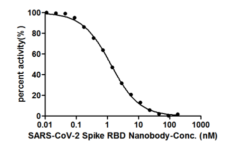
(ELISA: Immobilize various types of SARS proteins at concentration of 2mg/ml on solid substrate, then react with SARS-CoV-2 Spike RBD Nanobody at concentration of 100mg/ml, 10mg/ml and 1mg/ml. It shows the SARS-CoV-2 Spike RBD Nanobody (AAA27051) is specific for SARS-CoV-2-S1-RBD protein, without any cross-reactivity with MERS-CoV, SARS-CoV, HCoV-OC43 or HCoV-229E.)
Application Data
(ELISA: Immobilize various types of SARS proteins at concentration of 2mg/ml on solid substrate, then react with SARS-CoV-2 Spike RBD Nanobody at concentration of 100mg/ml, 10mg/ml and 1mg/ml. It shows the SARS-CoV-2 Spike RBD Nanobody (AAA27051) is specific for SARS-CoV-2-S1-RBD protein, without any cross-reactivity with MERS-CoV, SARS-CoV, HCoV-OC43 or HCoV-229E.)
COVID 19 Spike RBD Coronavirus, Monoclonal Recombinant Antibody (Cat# AAA27051)
Full Name
SARS-CoV-2 Spike RBD Nanobody
Reactivity
Human Novel Coronavirus (SARS-CoV-2/ 2019-nCoV)
Applications
Colloidal Gold Immunochromatographic Assay, Neutralization Assay
Purity
Affinity-chromatography
Pricing


 FCM (Flow Cytometry)
(Figure 6. Flow Cytometry analysis of Hela cells using anti-CD147/Emmprin antibody (AAA11678).Overlay histogram showing Hela cells stained with AAA11678 (Blue line).The cells were blocked with 10% normal goat serum. And then incubated with rabbit anti-CD147/Emmprin Antibody (AAA11678,1ug/1x10^6 cells) for 30 min at 20 degree C. DyLight 488 conjugated goat anti-rabbit IgG (5-10ug/1x10^6 cells) was used as secondary antibody for 30 minutes at 20 degree C. Isotype control antibody (Green line) was rabbit IgG (1ug/1x106) used under the same conditions. Unlabelled sample (Red line) was also used as a control.)
FCM (Flow Cytometry)
(Figure 6. Flow Cytometry analysis of Hela cells using anti-CD147/Emmprin antibody (AAA11678).Overlay histogram showing Hela cells stained with AAA11678 (Blue line).The cells were blocked with 10% normal goat serum. And then incubated with rabbit anti-CD147/Emmprin Antibody (AAA11678,1ug/1x10^6 cells) for 30 min at 20 degree C. DyLight 488 conjugated goat anti-rabbit IgG (5-10ug/1x10^6 cells) was used as secondary antibody for 30 minutes at 20 degree C. Isotype control antibody (Green line) was rabbit IgG (1ug/1x106) used under the same conditions. Unlabelled sample (Red line) was also used as a control.)
CD147/Emmprin, Polyclonal Antibody (Cat# AAA11678)
Full Name
Anti-CD147/Emmprin Antibody
Gene Names
BSG; OK; 5F7; TCSF; CD147; EMMPRIN
Reactivity
Human, Mouse, Rat
Applications
Western Blot, Immunohistochemistry
Purity
Immunogen affinity purified.
Pricing
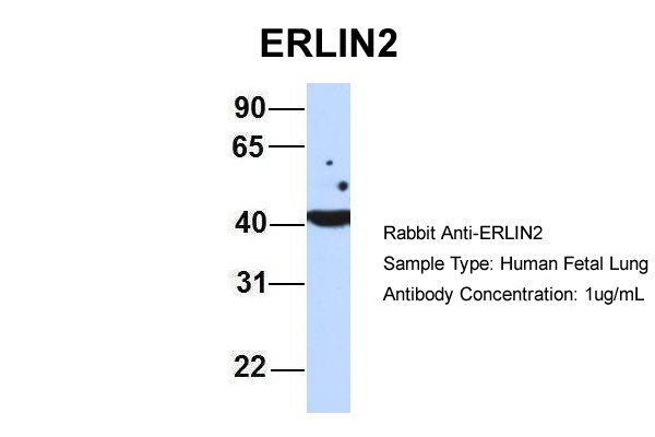


 WB (Western Blot)
(WB Suggested Anti-ERLIN2 Antibody Titration: 0.2-1 ug/mlELISA Titer: 1:62500Positive Control: 293T cell lysate.ERLIN2 is strongly supported by BioGPS gene expression data to be expressed in HEK293T)
WB (Western Blot)
(WB Suggested Anti-ERLIN2 Antibody Titration: 0.2-1 ug/mlELISA Titer: 1:62500Positive Control: 293T cell lysate.ERLIN2 is strongly supported by BioGPS gene expression data to be expressed in HEK293T)
ERLIN2, Polyclonal Antibody (Cat# AAA23581)
Full Name
ERLIN2 antibody - middle region
Gene Names
ERLIN2; NET32; SPFH2; SPG18; C8orf2; Erlin-2
Reactivity
Human, Mouse, Pig, Rabbit, Rat
Applications
Western Blot
Purity
Affinity Purified
Pricing


 FCM (Flow Cytometry)
(Figure 7. Flow Cytometry analysis of U20S cells using anti-SAMHD1 antibody (AAA19223).Overlay histogram showing U20S cells stained with AAA19223 (Blue line). The cells were blocked with 10% normal goat serum. And then incubated with rabbit anti-SAMHD1 Antibody (AAA19223, 1μg/1x106 cells) for 30 min at 20 degree C. DyLight®488 conjugated goat anti-rabbit IgG (5-10μg/1x106 cells) was used as secondary antibody for 30 minutes at 20 degree C. Isotype control antibody (Green line) was rabbit IgG (1μg/1x106) used under the same conditions. Unlabelled sample (Red line) was also used as a control.)
FCM (Flow Cytometry)
(Figure 7. Flow Cytometry analysis of U20S cells using anti-SAMHD1 antibody (AAA19223).Overlay histogram showing U20S cells stained with AAA19223 (Blue line). The cells were blocked with 10% normal goat serum. And then incubated with rabbit anti-SAMHD1 Antibody (AAA19223, 1μg/1x106 cells) for 30 min at 20 degree C. DyLight®488 conjugated goat anti-rabbit IgG (5-10μg/1x106 cells) was used as secondary antibody for 30 minutes at 20 degree C. Isotype control antibody (Green line) was rabbit IgG (1μg/1x106) used under the same conditions. Unlabelled sample (Red line) was also used as a control.)
SAMHD1, Polyclonal Antibody (Cat# AAA19223)
Full Name
Anti-SAMHD1 Antibody
Gene Names
SAMHD1; DCIP; CHBL2; HDDC1; MOP-5; SBBI88
Reactivity
Human, Mouse, Rat
Applications
Western Blot, Immunohistochemistry, Immunocytochemistry, Immunofluorescence, Flow Cytometry, Direct ELISA
Purity
Immunogen affinity purified.
Pricing


 IF (Immunofluorescence)
(Immunofluorescence analysis of L929 cells using Bax Rabbit pAb at dilution of 1:100. Blue: DAPI for nuclear staining.)
IF (Immunofluorescence)
(Immunofluorescence analysis of L929 cells using Bax Rabbit pAb at dilution of 1:100. Blue: DAPI for nuclear staining.)
Bax, Polyclonal Antibody (Cat# AAA28318)
Full Name
Bax Rabbit pAb
Gene Names
BAX; BCL2L4
Reactivity
Human, Mouse, Rat
Applications
Western Blot, Immunohistochemistry, Immunofluorescence
Purity
Affinity purification
Pricing


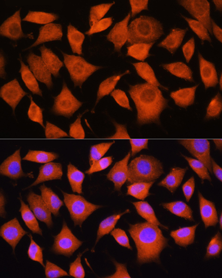
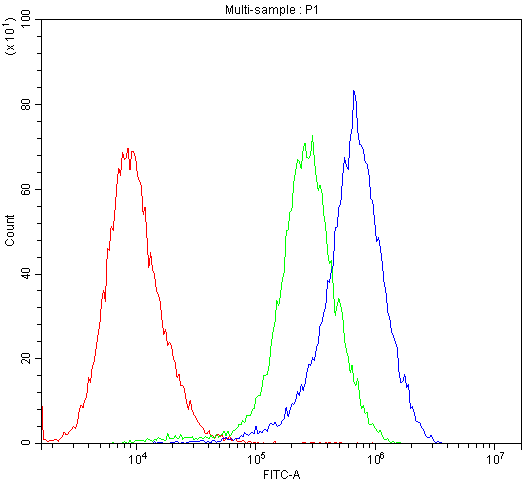
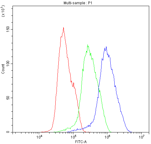
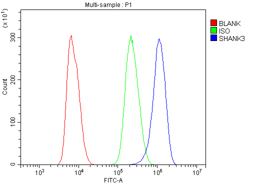 FCM (Flow Cytometry)
(Figure 6. Flow Cytometry analysis of NRK cells using anti-SHANK3 antibody (AAA19237).Overlay histogram showing NRK cells stained with AAA19237 (Blue line). The cells were blocked with 10% normal goat serum. And then incubated with rabbit anti-SHANK3 Antibody (AAA19237,1μg/1x106 cells) for 30 min at 20 degree C. DyLight®488 conjugated goat anti-rabbit IgG (5-10μg/1x106 cells) was used as secondary antibody for 30 minutes at 20 degree C. Isotype control antibody (Green line) was rabbit IgG (1μg/1x106) used under the same conditions. Unlabelled sample (Red line) was also used as a control.)
FCM (Flow Cytometry)
(Figure 6. Flow Cytometry analysis of NRK cells using anti-SHANK3 antibody (AAA19237).Overlay histogram showing NRK cells stained with AAA19237 (Blue line). The cells were blocked with 10% normal goat serum. And then incubated with rabbit anti-SHANK3 Antibody (AAA19237,1μg/1x106 cells) for 30 min at 20 degree C. DyLight®488 conjugated goat anti-rabbit IgG (5-10μg/1x106 cells) was used as secondary antibody for 30 minutes at 20 degree C. Isotype control antibody (Green line) was rabbit IgG (1μg/1x106) used under the same conditions. Unlabelled sample (Red line) was also used as a control.)
SHANK3, Polyclonal Antibody (Cat# AAA19237)
Full Name
Anti-SHANK3 Antibody
Gene Names
SHANK3; PSAP2; SCZD15; PROSAP2; SPANK-2; DEL22q13.3
Reactivity
Human, Mouse, Rat
Applications
Western Blot, Immunohistochemistry, Flow Cytometry, Direct ELISA
Purity
Immunogen affinity purified.
Pricing


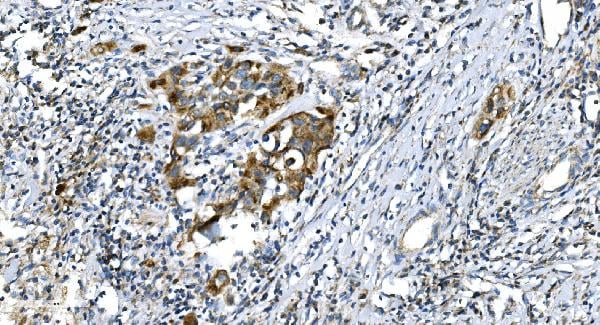
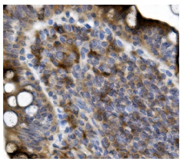
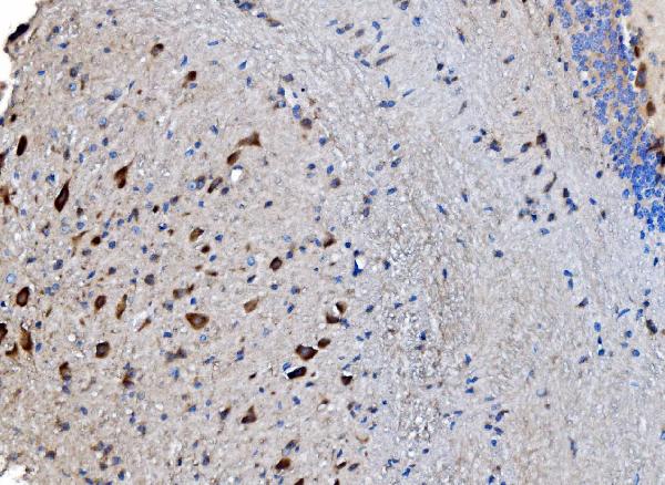
 IHC (Immunohistochemistry)
(Figure 8. IHC analysis of FOXP1 using anti-FOXP1 antibody (AAA19227).FOXP1 was detected in paraffin-embedded section of rat brain tissue. Heat mediated antigen retrieval was performed in EDTA buffer (pH8. 0, epitope retrieval solution). The tissue section was blocked with 10% goat serum. The tissue section was then incubated with 2μg/ml rabbit anti-FOXP1 Antibody (AAA19227) overnight at 4 degree C. Biotinylated goat anti-rabbit IgG was used as secondary antibody and incubated for 30 minutes at 37 degree C. The tissue section was developed using Strepavidin-Biotin-Complex (SABC) (Catalog # with DAB as the chromogen.)
IHC (Immunohistochemistry)
(Figure 8. IHC analysis of FOXP1 using anti-FOXP1 antibody (AAA19227).FOXP1 was detected in paraffin-embedded section of rat brain tissue. Heat mediated antigen retrieval was performed in EDTA buffer (pH8. 0, epitope retrieval solution). The tissue section was blocked with 10% goat serum. The tissue section was then incubated with 2μg/ml rabbit anti-FOXP1 Antibody (AAA19227) overnight at 4 degree C. Biotinylated goat anti-rabbit IgG was used as secondary antibody and incubated for 30 minutes at 37 degree C. The tissue section was developed using Strepavidin-Biotin-Complex (SABC) (Catalog # with DAB as the chromogen.)
FOXP1, Polyclonal Antibody (Cat# AAA19227)
Full Name
Anti-FOXP1 Antibody
Gene Names
FOXP1; QRF1; 12CC4; hFKH1B; HSPC215
Reactivity
Human, Mouse, Rat, Monkey
Applications
Western Blot, Immunohistochemistry
Purity
Immunogen affinity purified.
Pricing


 FCM (Flow Cytometry)
(Figure 7. Flow Cytometry analysis of SiHa cells using anti-POR antibody (AAA11661).Overlay histogram showing SiHa cells stained with AAA11661 (Blue line).The cells were blocked with 10% normal goat serum. And then incubated with rabbit anti-POR Antibody (AAA11661,1ug/1x10^6 cells) for 30 min at 20 degree C. DyLight®488 conjugated goat anti-rabbit IgG (5-10ug/1x10^6 cells) was used as secondary antibody for 30 minutes at 20 degree C. Isotype control antibody (Green line) was rabbit IgG (1ug/1x106) used under the same conditions. Unlabelled sample (Red line) was also used as a control.)
FCM (Flow Cytometry)
(Figure 7. Flow Cytometry analysis of SiHa cells using anti-POR antibody (AAA11661).Overlay histogram showing SiHa cells stained with AAA11661 (Blue line).The cells were blocked with 10% normal goat serum. And then incubated with rabbit anti-POR Antibody (AAA11661,1ug/1x10^6 cells) for 30 min at 20 degree C. DyLight®488 conjugated goat anti-rabbit IgG (5-10ug/1x10^6 cells) was used as secondary antibody for 30 minutes at 20 degree C. Isotype control antibody (Green line) was rabbit IgG (1ug/1x106) used under the same conditions. Unlabelled sample (Red line) was also used as a control.)
POR, Polyclonal Antibody (Cat# AAA11661)
Full Name
Anti-POR Antibody
Gene Names
POR; CPR; CYPOR; P450R
Reactivity
Human, Mouse, Rat
Applications
Western Blot, Immunohistochemistry
Purity
Immunogen Affinity Purified
Pricing


 Strength of Signal
(Analysis of Protein Array containing more than 19,000 full-length human proteins using Vimentin (VIM) Mouse Monoclonal Antibody (VM452)Z- and S- Score: The Z-score represents the strength of a signal that a monoclonal antibody (MAb) (in combination with a fluorescently-tagged anti-IgG secondary antibody) produces when binding to a particular protein on the HuProtTM array. Z-scores are described in units of standard deviations (SD's) above the mean value of all signals generated on that array. If targets on HuProtTM are arranged in descending order of the Z-score, the S-score is the difference (also in units of SD's) between the Z-score. S-score therefore represents the relative target specificity of a MAb to its intended target. A MAb is considered to specific to its intended target, if the MAb has an S-score of at least 2.5. For example, if a MAb binds to protein X with a Z-score of 43 and to protein Y with a Z-score of 14, then the S-score for the binding of that MAb to protein X is equal to 29.)
Strength of Signal
(Analysis of Protein Array containing more than 19,000 full-length human proteins using Vimentin (VIM) Mouse Monoclonal Antibody (VM452)Z- and S- Score: The Z-score represents the strength of a signal that a monoclonal antibody (MAb) (in combination with a fluorescently-tagged anti-IgG secondary antibody) produces when binding to a particular protein on the HuProtTM array. Z-scores are described in units of standard deviations (SD's) above the mean value of all signals generated on that array. If targets on HuProtTM are arranged in descending order of the Z-score, the S-score is the difference (also in units of SD's) between the Z-score. S-score therefore represents the relative target specificity of a MAb to its intended target. A MAb is considered to specific to its intended target, if the MAb has an S-score of at least 2.5. For example, if a MAb binds to protein X with a Z-score of 43 and to protein Y with a Z-score of 14, then the S-score for the binding of that MAb to protein X is equal to 29.)
Vimentin, Monoclonal Antibody (Cat# AAA13810)
Full Name
Vimentin (Mesenchymal Cell Marker) Mouse Monoclonal Antibody
Gene Names
VIM; HEL113; CTRCT30
Reactivity
Human, Cow, Dog, Cat, Pig, Goat, Chicken.
Does not react with Mouse and Rat
Does not react with Mouse and Rat
Applications
Flow Cytometry, Immunofluorescence, Western Blot, Immunohistochemistry
Pricing


 WB (Western Blot)
(WB Suggested Anti-SSBP2 Antibody Titration: 0.2-1 ug/mlELISA Titer: 1:7812500Positive Control: Raji cell lysate)
WB (Western Blot)
(WB Suggested Anti-SSBP2 Antibody Titration: 0.2-1 ug/mlELISA Titer: 1:7812500Positive Control: Raji cell lysate)
SSBP2, Polyclonal Antibody (Cat# AAA23390)
Full Name
SSBP2 antibody - N-terminal region
Gene Names
SSBP2; HSPC116; SOSS-B2
Reactivity
Cow, Dog, Guinea Pig, Horse, Human, Mouse, Rabbit, Rat, Zebrafish
Applications
Immunohistochemistry, Western Blot
Purity
Affinity Purified
Pricing


 FCM (Flow Cytometry)
(Figure 7. Flow Cytometry analysis of A431 cells using anti-BIK antibody (AAA11666).Overlay histogram showing A431 cells stained with AAA11666 (Blue line).The cells were blocked with 10% normal goat serum. And then incubated with rabbit anti-BIK Antibody (AAA11666,1ug/1x10^6 cells) for 30 min at 20 degree C. DyLight®488 conjugated goat anti-rabbit IgG (5-10ug/1x10^6 cells) was used as secondary antibody for 30 minutes at 20 degree C. Isotype control antibody (Green line) was rabbit IgG (1ug/1x106) used under the same conditions. Unlabelled sample (Red line) was also used as a control.)
FCM (Flow Cytometry)
(Figure 7. Flow Cytometry analysis of A431 cells using anti-BIK antibody (AAA11666).Overlay histogram showing A431 cells stained with AAA11666 (Blue line).The cells were blocked with 10% normal goat serum. And then incubated with rabbit anti-BIK Antibody (AAA11666,1ug/1x10^6 cells) for 30 min at 20 degree C. DyLight®488 conjugated goat anti-rabbit IgG (5-10ug/1x10^6 cells) was used as secondary antibody for 30 minutes at 20 degree C. Isotype control antibody (Green line) was rabbit IgG (1ug/1x106) used under the same conditions. Unlabelled sample (Red line) was also used as a control.)
Bik, Polyclonal Antibody (Cat# AAA11666)
Full Name
Anti-Bik Antibody
Gene Names
BIK; BP4; NBK; BIP1
Reactivity
Human, Mouse, Rat
Applications
Western Blot, Immunohistochemistry
Purity
Immunogen Affinity Purified
Pricing


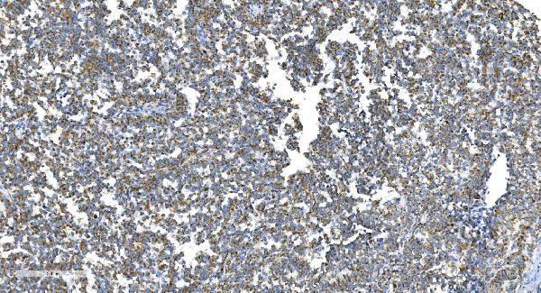
 Application Data
(Analysis of Protein Array containing more than 19, 000 full-length human proteins using CD10 Mouse Monoclonal Antibody (MME/1870)Z- and S- Score: The Z-score represents the strength of a signal that a monoclonal antibody (MAb) (in combination with a fluorescently-tagged anti-IgG secondary antibody) produces when binding to a particular protein on the HuProtTM array. Z-scores are described in units of standard deviations (SD's) above the mean value of all signals generated on that array. If targets on HuProtTM are arranged in descending order of the Z-score, the S-score is the difference (also in units of SD's) between the Z-score. S-score therefore represents the relative target specificity of a MAb to its intended target. A MAb is considered to specific to its intended target, if the MAb has an S-score of at least 2.5. For example, if a MAb binds to protein X with a Z-score of 43 and to protein Y with a Z-score of 14, then the S-score for the binding of that MAb to protein X is equal to 29.)
Application Data
(Analysis of Protein Array containing more than 19, 000 full-length human proteins using CD10 Mouse Monoclonal Antibody (MME/1870)Z- and S- Score: The Z-score represents the strength of a signal that a monoclonal antibody (MAb) (in combination with a fluorescently-tagged anti-IgG secondary antibody) produces when binding to a particular protein on the HuProtTM array. Z-scores are described in units of standard deviations (SD's) above the mean value of all signals generated on that array. If targets on HuProtTM are arranged in descending order of the Z-score, the S-score is the difference (also in units of SD's) between the Z-score. S-score therefore represents the relative target specificity of a MAb to its intended target. A MAb is considered to specific to its intended target, if the MAb has an S-score of at least 2.5. For example, if a MAb binds to protein X with a Z-score of 43 and to protein Y with a Z-score of 14, then the S-score for the binding of that MAb to protein X is equal to 29.)
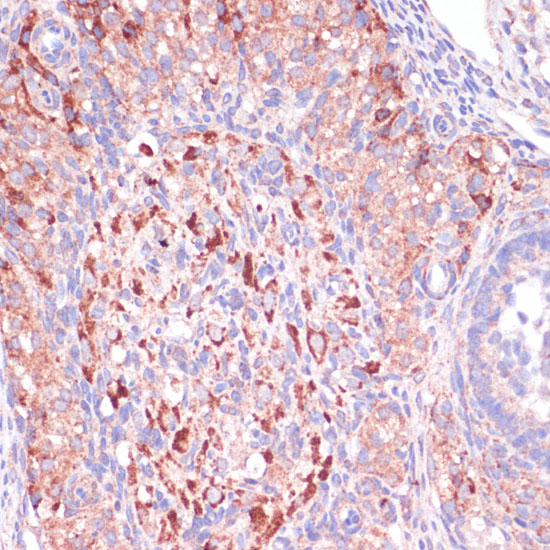
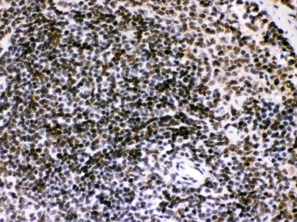
CD10, Monoclonal Antibody (Cat# AAA23901)
Full Name
CD10 (Membrane Metalloendopeptidase)
Gene Names
MME; NEP; SFE; CD10; CALLA; CMT2T; SCA43
Reactivity
Human. Others not tested.
Applications
Immunohistochemistry
Pricing


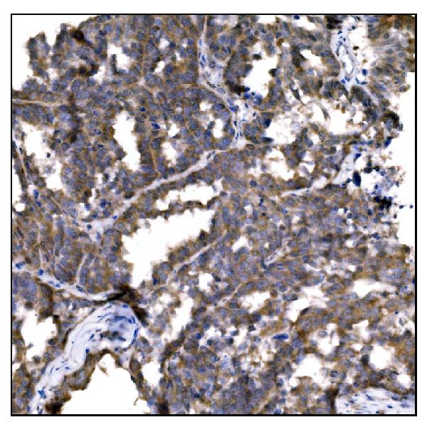
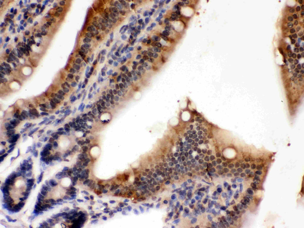
 IHC (Immunohistochemistry)
(Figure 13. IHC analysis of EEF2/Elongation factor 2 using anti-EEF2/Elongation factor 2 antibody (AAA19229).EEF2/Elongation factor 2 was detected in paraffin-embedded section of rat brain tissue. Heat mediated antigen retrieval was performed in EDTA buffer (pH8. 0, epitope retrieval solution). The tissue section was blocked with 10% goat serum. The tissue section was then incubated with 2μg/ml rabbit anti-EEF2/Elongation factor 2 Antibody (AAA19229) overnight at 4 degree C. Biotinylated goat anti-rabbit IgG was used as secondary antibody and incubated for 30 minutes at 37 degree C. The tissue section was developed using Strepavidin-Biotin-Complex (SABC) (Catalog # with DAB as the chromogen.)
IHC (Immunohistochemistry)
(Figure 13. IHC analysis of EEF2/Elongation factor 2 using anti-EEF2/Elongation factor 2 antibody (AAA19229).EEF2/Elongation factor 2 was detected in paraffin-embedded section of rat brain tissue. Heat mediated antigen retrieval was performed in EDTA buffer (pH8. 0, epitope retrieval solution). The tissue section was blocked with 10% goat serum. The tissue section was then incubated with 2μg/ml rabbit anti-EEF2/Elongation factor 2 Antibody (AAA19229) overnight at 4 degree C. Biotinylated goat anti-rabbit IgG was used as secondary antibody and incubated for 30 minutes at 37 degree C. The tissue section was developed using Strepavidin-Biotin-Complex (SABC) (Catalog # with DAB as the chromogen.)
EEF2/Elongation factor 2, Polyclonal Antibody (Cat# AAA19229)
Full Name
Anti-EEF2/Elongation factor 2 Antibody
Gene Names
EEF2; EF2; EF-2; EEF-2
Reactivity
Human, Mouse, Rat, Monkey
Applications
Western Blot, Immunohistochemistry, Immunocytochemistry, Immunofluorescence, Flow Cytometry
Purity
Immunogen affinity purified.
Pricing


 FCM (Flow Cytometry)
(Figure 12. Flow Cytometry analysis of A549 cells using anti-Transketolase/TKT antibody (AAA19367).Overlay histogram showing A549 cells stained with AAA19367 (Blue line). The cells were blocked with 10% normal goat serum. And then incubated with mouse anti- Transketolase/TKT Antibody (AAA19367, 1μg/1x106 cells) for 30 min at 20 degree C. DyLight®488 conjugated goat anti-mouse IgG (BA1126, 5-10μg/1x106 cells) was used as secondary antibody for 30 minutes at 20 degree C. Isotype control antibody (Green line) was mouse IgG (1μg/1x106) used under the same conditions. Unlabelled sample (Red line) was also used as a control.)
FCM (Flow Cytometry)
(Figure 12. Flow Cytometry analysis of A549 cells using anti-Transketolase/TKT antibody (AAA19367).Overlay histogram showing A549 cells stained with AAA19367 (Blue line). The cells were blocked with 10% normal goat serum. And then incubated with mouse anti- Transketolase/TKT Antibody (AAA19367, 1μg/1x106 cells) for 30 min at 20 degree C. DyLight®488 conjugated goat anti-mouse IgG (BA1126, 5-10μg/1x106 cells) was used as secondary antibody for 30 minutes at 20 degree C. Isotype control antibody (Green line) was mouse IgG (1μg/1x106) used under the same conditions. Unlabelled sample (Red line) was also used as a control.)
Transketolase/TKT, Monoclonal Antibody (Cat# AAA19367)
Full Name
Anti-Transketolase/TKT Antibody (monoclonal, 3E5)
Gene Names
TKT; TK; TKT1; HEL107
Reactivity
Human, Mouse, Rat
Applications
WB, IHC-P, ICC, IF, FC/FACS/FCM
Purity
Immunogen affinity purified.
Pricing


 Application Data
(Analysis of Protein Array containing more than 19,000 full-length human proteins using ERCC1 Mouse Monoclonal Antibody (ERCC1/2318). Z- and S- Score: The Z-score represents the strength of a signal that a monoclonal antibody (MAb) (in combination with a fluorescently-tagged anti-IgG secondary antibody) produces when binding to a particular protein on the HuProtTM array. Z-scores are described in units of standard deviations (SD's) above the mean value of all signals generated on that array. If targets on HuProtTM are arranged in descending order of the Z-score, the S-score is the difference (also in units of SD's) between the Z-score. S-score therefore represents the relative target specificity of a MAb to its intended target. A MAb is considered to specific to its intended target, if the MAb has an S-score of at least 2.5. For example, if a MAb binds to protein X with a Z-score of 43 and to protein Y with a Z-score of 14, then the S-score for the binding of that MAb to protein X is equal to 29.)
Application Data
(Analysis of Protein Array containing more than 19,000 full-length human proteins using ERCC1 Mouse Monoclonal Antibody (ERCC1/2318). Z- and S- Score: The Z-score represents the strength of a signal that a monoclonal antibody (MAb) (in combination with a fluorescently-tagged anti-IgG secondary antibody) produces when binding to a particular protein on the HuProtTM array. Z-scores are described in units of standard deviations (SD's) above the mean value of all signals generated on that array. If targets on HuProtTM are arranged in descending order of the Z-score, the S-score is the difference (also in units of SD's) between the Z-score. S-score therefore represents the relative target specificity of a MAb to its intended target. A MAb is considered to specific to its intended target, if the MAb has an S-score of at least 2.5. For example, if a MAb binds to protein X with a Z-score of 43 and to protein Y with a Z-score of 14, then the S-score for the binding of that MAb to protein X is equal to 29.)
ERCC1/RAD10, Monoclonal Antibody (Cat# AAA23913)
Full Name
ERCC1/RAD10 (Tumor Progression Marker)
Gene Names
ERCC1; UV20; COFS4; RAD10
Reactivity
Human
Applications
Immunohistochemistry
Purity
Purified Ab with BSA and Azide at 200ug/ml OR Purified Ab WITHOUT BSA and Azide at 1.0mg/ml
Pricing


 FCM (Flow Cytometry)
(Figure 6. Flow Cytometry analysis of MCF-7 cells using anti-ALK-1/ACVRL1 antibody (AAA19242).Overlay histogram showing MCF-7 cells stained with AAA19242 (Blue line). The cells were blocked with 10% normal goat serum. And then incubated with rabbit anti-ALK-1/ACVRL1 Antibody (AAA19242, 1μg/1x106 cells) for 30 min at 20 degree C. DyLight®488 conjugated goat anti-rabbit IgG (5-10μg/1x106 cells) was used as secondary antibody for 30 minutes at 20 degree C. Isotype control antibody (Green line) was rabbit IgG (1μg/1x106) used under the same conditions. Unlabelled sample (Red line) was also used as a control.)
FCM (Flow Cytometry)
(Figure 6. Flow Cytometry analysis of MCF-7 cells using anti-ALK-1/ACVRL1 antibody (AAA19242).Overlay histogram showing MCF-7 cells stained with AAA19242 (Blue line). The cells were blocked with 10% normal goat serum. And then incubated with rabbit anti-ALK-1/ACVRL1 Antibody (AAA19242, 1μg/1x106 cells) for 30 min at 20 degree C. DyLight®488 conjugated goat anti-rabbit IgG (5-10μg/1x106 cells) was used as secondary antibody for 30 minutes at 20 degree C. Isotype control antibody (Green line) was rabbit IgG (1μg/1x106) used under the same conditions. Unlabelled sample (Red line) was also used as a control.)
ALK-1/ACVRL1, Polyclonal Antibody (Cat# AAA19242)
Full Name
Anti-ALK-1/ACVRL1 Antibody
Gene Names
ACVRL1; HHT; ALK1; HHT2; ORW2; SKR3; ALK-1; TSR-I; ACVRLK1
Reactivity
Human, Mouse, Rat
Applications
Western Blot, Immunohistochemistry, Flow Cytometry, Direct ELISA
Purity
Immunogen affinity purified.
Pricing


 FCM (Flow Cytometry)
(Figure 13. Flow Cytometry analysis of SiHa cells using anti-DRP1/DNM1L antibody (AAA19220).Overlay histogram showing SiHa cells stained with AAA19220 (Blue line). The cells were blocked with 10% normal goat serum. And then incubated with rabbit anti-DRP1/DNM1L Antibody (AAA19220, 1μg/1x106 cells) for 30 min at 20 degree C. DyLight®488 conjugated goat anti-rabbit IgG (5-10μg/1x106 cells) was used as secondary antibody for 30 minutes at 20 degree C. Isotype control antibody (Green line) was rabbit IgG (1μg/1x106) used under the same conditions. Unlabelled sample (Red line) was also used as a control.)
FCM (Flow Cytometry)
(Figure 13. Flow Cytometry analysis of SiHa cells using anti-DRP1/DNM1L antibody (AAA19220).Overlay histogram showing SiHa cells stained with AAA19220 (Blue line). The cells were blocked with 10% normal goat serum. And then incubated with rabbit anti-DRP1/DNM1L Antibody (AAA19220, 1μg/1x106 cells) for 30 min at 20 degree C. DyLight®488 conjugated goat anti-rabbit IgG (5-10μg/1x106 cells) was used as secondary antibody for 30 minutes at 20 degree C. Isotype control antibody (Green line) was rabbit IgG (1μg/1x106) used under the same conditions. Unlabelled sample (Red line) was also used as a control.)
DRP1/DNM1L, Polyclonal Antibody (Cat# AAA19220)
Full Name
Anti-DRP1/DNM1L Antibody
Gene Names
DNM1L; DLP1; DRP1; DVLP; EMPF; DYMPLE; HDYNIV
Reactivity
Human, Mouse, Rat
Applications
Western Blot, Immunohistochemistry, Immunocytochemistry, Immunofluorescence, Flow Cytometry, Direct ELISA
Purity
Immunogen affinity purified.
Pricing


 FCM (Flow Cytometry)
(Figure 6. Flow Cytometry analysis of MCF-7 cells using anti-Caspase-7/CASP7 antibody (AAA19232).Overlay histogram showing MCF-7 cells stained with AAA19232 (Blue line). The cells were blocked with 10% normal goat serum. And then incubated with rabbit anti-Caspase-7/CASP7 Antibody (AAA19232, 1μg/1x106 cells) for 30 min at 20 degree C. DyLight®488 conjugated goat anti-rabbit IgG (5-10μg/1x106 cells) was used as secondary antibody for 30 minutes at 20 degree C. Isotype control antibody (Green line) was rabbit IgG (1μg/1x106) used under the same conditions. Unlabelled sample (Red line) was also used as a control.)
FCM (Flow Cytometry)
(Figure 6. Flow Cytometry analysis of MCF-7 cells using anti-Caspase-7/CASP7 antibody (AAA19232).Overlay histogram showing MCF-7 cells stained with AAA19232 (Blue line). The cells were blocked with 10% normal goat serum. And then incubated with rabbit anti-Caspase-7/CASP7 Antibody (AAA19232, 1μg/1x106 cells) for 30 min at 20 degree C. DyLight®488 conjugated goat anti-rabbit IgG (5-10μg/1x106 cells) was used as secondary antibody for 30 minutes at 20 degree C. Isotype control antibody (Green line) was rabbit IgG (1μg/1x106) used under the same conditions. Unlabelled sample (Red line) was also used as a control.)
Caspase-7/CASP7, Polyclonal Antibody (Cat# AAA19232)
Full Name
Anti-Caspase-7/CASP7 Antibody
Gene Names
CASP7; MCH3; CMH-1; LICE2; CASP-7; ICE-LAP3
Reactivity
Human, Rat
Applications
Western Blot, Immunohistochemistry, Immunocytochemistry, Immunofluorescence, Flow Cytometry, Direct ELISA
Purity
Immunogen affinity purified.
Pricing


 FCM (Flow Cytometry)
(Figure 6. Flow Cytometry analysis of PC-3 cells using anti-APP antibody (AAA11597).Overlay histogram showing PC-3 cells stained with AAA11597 (Blue line).The cells were blocked with 10% normal goat serum. And then incubated with rabbit anti-APP Antibody (AAA11597,1ug/1x10^6 cells) for 30 min at 20 degree C. DyLight®488 conjugated goat anti-rabbit IgG (5-10ug/1x10^6 cells) was used as secondary antibody for 30 minutes at 20 degree C. Isotype control antibody (Green line) was rabbit IgG (1ug/1x106) used under the same conditions. Unlabelled sample (Red line) was also used as a control.)
FCM (Flow Cytometry)
(Figure 6. Flow Cytometry analysis of PC-3 cells using anti-APP antibody (AAA11597).Overlay histogram showing PC-3 cells stained with AAA11597 (Blue line).The cells were blocked with 10% normal goat serum. And then incubated with rabbit anti-APP Antibody (AAA11597,1ug/1x10^6 cells) for 30 min at 20 degree C. DyLight®488 conjugated goat anti-rabbit IgG (5-10ug/1x10^6 cells) was used as secondary antibody for 30 minutes at 20 degree C. Isotype control antibody (Green line) was rabbit IgG (1ug/1x106) used under the same conditions. Unlabelled sample (Red line) was also used as a control.)
beta Amyloid, Polyclonal Antibody (Cat# AAA11597)
Full Name
Anti-beta Amyloid Antibody
Gene Names
APP; AAA; AD1; PN2; ABPP; APPI; CVAP; ABETA; PN-II; CTFgamma
Reactivity
Human, Mouse, Rat
Applications
Western Blot, Immunohistochemistry
Purity
Immunogen affinity purified.
Pricing
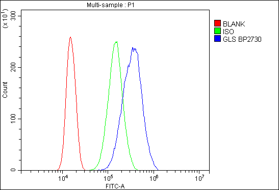


 FCM (Flow Cytometry)
(Figure 6. Flow Cytometry analysis of SiHa cells using anti-Glutaminase/GLS antibody (AAA19239).Overlay histogram showing SiHa cells stained with AAA19239 (Blue line). The cells were blocked with 10% normal goat serum. And then incubated with rabbit anti-Glutaminase/GLS Antibody (AAA19239,1μg/1x106 cells) for 30 min at 20 degree C. DyLight®488 conjugated goat anti-rabbit IgG (5-10μg/1x106 cells) was used as secondary antibody for 30 minutes at 20 degree C. Isotype control antibody (Green line) was rabbit IgG (1μg/1x106) used under the same conditions. Unlabelled sample (Red line) was also used as a control.)
FCM (Flow Cytometry)
(Figure 6. Flow Cytometry analysis of SiHa cells using anti-Glutaminase/GLS antibody (AAA19239).Overlay histogram showing SiHa cells stained with AAA19239 (Blue line). The cells were blocked with 10% normal goat serum. And then incubated with rabbit anti-Glutaminase/GLS Antibody (AAA19239,1μg/1x106 cells) for 30 min at 20 degree C. DyLight®488 conjugated goat anti-rabbit IgG (5-10μg/1x106 cells) was used as secondary antibody for 30 minutes at 20 degree C. Isotype control antibody (Green line) was rabbit IgG (1μg/1x106) used under the same conditions. Unlabelled sample (Red line) was also used as a control.)
Glutaminase/GLS, Polyclonal Antibody (Cat# AAA19239)
Full Name
Anti-Glutaminase/GLS Antibody
Gene Names
GLS; GAC; GAM; KGA; GLS1; AAD20
Reactivity
Human, Mouse, Rat, Monkey
Applications
Western Blot, Immunohistochemistry, Immunocytochemistry, Immunofluorescence, Flow Cytometry, Direct ELISA
Purity
Immunogen affinity purified.
Pricing


 FCM (Flow Cytometry)
(Figure 7. Flow Cytometry analysis of U251 cells using anti-Serum Response Factor/SRF antibody (AAA19221).Overlay histogram showing U251 cells stained with AAA19221 (Blue line). The cells were blocked with 10% normal goat serum. And then incubated with rabbit anti-Serum Response Factor/SRF Antibody (AAA19221, 1μg/1x106 cells) for 30 min at 20 degree C. DyLight®488 conjugated goat anti-rabbit IgG (5-10μg/1x106 cells) was used as secondary antibody for 30 minutes at 20 degree C. Isotype control antibody (Green line) was rabbit IgG (1μg/1x106) used under the same conditions. Unlabelled sample (Red line) was also used as a control.)
FCM (Flow Cytometry)
(Figure 7. Flow Cytometry analysis of U251 cells using anti-Serum Response Factor/SRF antibody (AAA19221).Overlay histogram showing U251 cells stained with AAA19221 (Blue line). The cells were blocked with 10% normal goat serum. And then incubated with rabbit anti-Serum Response Factor/SRF Antibody (AAA19221, 1μg/1x106 cells) for 30 min at 20 degree C. DyLight®488 conjugated goat anti-rabbit IgG (5-10μg/1x106 cells) was used as secondary antibody for 30 minutes at 20 degree C. Isotype control antibody (Green line) was rabbit IgG (1μg/1x106) used under the same conditions. Unlabelled sample (Red line) was also used as a control.)
Serum Response Factor/SRF, Polyclonal Antibody (Cat# AAA19221)
Full Name
Anti-Serum Response Factor/SRF Antibody
Gene Names
SRF; MCM1
Reactivity
Human, Mouse, Rat
Applications
Western Blot, Immunohistochemistry, Immunocytochemistry, Immunofluorescence, Flow Cytometry, Direct ELISA
Purity
Immunogen affinity purified.
Pricing


 Application Data
(Analysis of Protein Array containing more than 19, 000 full-length human proteins using Oct-2 Mouse Monoclonal Antibody (OCT2/2137) Z- and S- Score: The Z-score represents the strength of a signal that a monoclonal antibody (MAb) (in combination with a fluorescently-tagged anti-IgG secondary antibody) produces when binding to a particular protein on the HuProtTM array. Z-scores are described in units of standard deviations (SD's) above the mean value of all signals generated on that array. If targets on HuProtTM are arranged in descending order of the Z-score, the S-score is the difference (also in units of SD's) between the Z-score. S-score therefore represents the relative target specificity of a MAb to its intended target. A MAb is considered to specific to its intended target, if the MAb has an S-score of at least 2.5. For example, if a MAb binds to protein X with a Z-score of 43 and to protein Y with a Z-score of 14, then the S-score for the binding of that MAb to protein X is equal to 29.)
Application Data
(Analysis of Protein Array containing more than 19, 000 full-length human proteins using Oct-2 Mouse Monoclonal Antibody (OCT2/2137) Z- and S- Score: The Z-score represents the strength of a signal that a monoclonal antibody (MAb) (in combination with a fluorescently-tagged anti-IgG secondary antibody) produces when binding to a particular protein on the HuProtTM array. Z-scores are described in units of standard deviations (SD's) above the mean value of all signals generated on that array. If targets on HuProtTM are arranged in descending order of the Z-score, the S-score is the difference (also in units of SD's) between the Z-score. S-score therefore represents the relative target specificity of a MAb to its intended target. A MAb is considered to specific to its intended target, if the MAb has an S-score of at least 2.5. For example, if a MAb binds to protein X with a Z-score of 43 and to protein Y with a Z-score of 14, then the S-score for the binding of that MAb to protein X is equal to 29.)
OCT-2 (POU2F2), Monoclonal Antibody (Cat# AAA23902)
Full Name
OCT-2 (POU2F2) (B-Cell Marker)
Gene Names
POU2F2; OCT2; OTF2; Oct-2
Reactivity
Human. Others not known.
Applications
Western Blot, Immunohistochemistry
Pricing


 FCM (Flow Cytometry)
(Figure 7. Flow Cytometry analysis of U87 cells using anti-PDE6 beta/PDE6B antibody (AAA19261).Overlay histogram showing U87 cells stained with AAA19261 (Blue line). The cells were blocked with 10% normal goat serum. And then incubated with rabbit anti-PDE6 beta/PDE6B Antibody (AAA19261,1μg/1x106 cells) for 30 min at 20 degree C. DyLight®488 conjugated goat anti-rabbit IgG (5-10μg/1x106 cells) was used as secondary antibody for 30 minutes at 20 degree C. Isotype control antibody (Green line) was rabbit IgG (1μg/1x106) used under the same conditions. Unlabelled sample (Red line) was also used as a control.)
FCM (Flow Cytometry)
(Figure 7. Flow Cytometry analysis of U87 cells using anti-PDE6 beta/PDE6B antibody (AAA19261).Overlay histogram showing U87 cells stained with AAA19261 (Blue line). The cells were blocked with 10% normal goat serum. And then incubated with rabbit anti-PDE6 beta/PDE6B Antibody (AAA19261,1μg/1x106 cells) for 30 min at 20 degree C. DyLight®488 conjugated goat anti-rabbit IgG (5-10μg/1x106 cells) was used as secondary antibody for 30 minutes at 20 degree C. Isotype control antibody (Green line) was rabbit IgG (1μg/1x106) used under the same conditions. Unlabelled sample (Red line) was also used as a control.)
PDE6 beta/PDE6B, Polyclonal Antibody (Cat# AAA19261)
Full Name
Anti-PDE6 beta/PDE6B Antibody
Reactivity
Human, Mouse, Rat
Applications
Western Blot, Immunohistochemistry, Immunocytochemistry, Immunofluorescence, Flow Cytometry, Direct ELISA
Purity
Immunogen affinity purified.
Pricing


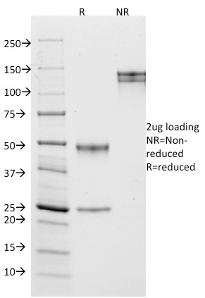
 SDS-PAGE
(SDS-PAGE Analysis Purified HSP60 Mouse Monoclonal Antibody (CPTC-HSPD1-1). Confirmation of Purity and Integrity of Antibody.)
SDS-PAGE
(SDS-PAGE Analysis Purified HSP60 Mouse Monoclonal Antibody (CPTC-HSPD1-1). Confirmation of Purity and Integrity of Antibody.)
HSP60 (Heat Shock Protein 60), Monoclonal Antibody (Cat# AAA23919)
Full Name
HSP60 (Heat Shock Protein 60) (Mitochondrial Marker)
Gene Names
HSPD1; HLD4; CPN60; GROEL; HSP60; HSP65; SPG13; HSP-60; HuCHA60
Reactivity
Human
Applications
Flow Cytometry, Immunofluorescence, Western Blot, Immunohistochemistry
Purity
Purified Ab with BSA and Azide at 200ug/ml OR Purified Ab WITHOUT BSA and Azide at 1.0mg/ml
Pricing


 FCM (Flow Cytometry)
(Figure 6. Flow Cytometry analysis of PC-3 cells using anti-ADK antibody (AAA11690).Overlay histogram showing PC-3 cells stained with AAA11690 (Blue line).The cells were blocked with 10% normal goat serum. And then incubated with rabbit anti-ADK Antibody (AAA11690,1ug/1x10^6 cells) for 30 min at 20 degree C. DyLight®488 conjugated goat anti-rabbit IgG (5-10ug/1x10^6 cells) was used as secondary antibody for 30 minutes at 20 degree C. Isotype control antibody (Green line) was rabbit IgG (1ug/1x106) used under the same conditions. Unlabelled sample (Red line) was also used as a control.)
FCM (Flow Cytometry)
(Figure 6. Flow Cytometry analysis of PC-3 cells using anti-ADK antibody (AAA11690).Overlay histogram showing PC-3 cells stained with AAA11690 (Blue line).The cells were blocked with 10% normal goat serum. And then incubated with rabbit anti-ADK Antibody (AAA11690,1ug/1x10^6 cells) for 30 min at 20 degree C. DyLight®488 conjugated goat anti-rabbit IgG (5-10ug/1x10^6 cells) was used as secondary antibody for 30 minutes at 20 degree C. Isotype control antibody (Green line) was rabbit IgG (1ug/1x106) used under the same conditions. Unlabelled sample (Red line) was also used as a control.)
ADK, Polyclonal Antibody (Cat# AAA11690)
Full Name
Anti-ADK Antibody
Gene Names
ADK; AK
Reactivity
Human, Mouse, Rat
Applications
Western Blot, Immunohistochemistry
Purity
Immunogen affinity purified.
Pricing


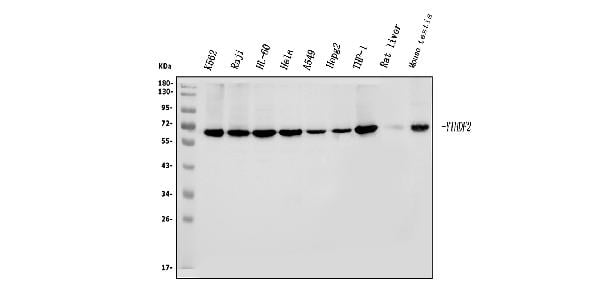
 FCM (Flow Cytometry)
(Figure 8. Flow Cytometry analysis of THP-1 cells using anti-YTHDF2 antibody (AAA19260).Overlay histogram showing THP-1 cells stained with AAA19260 (Blue line). The cells were blocked with 10% normal goat serum. And then incubated with rabbit anti-YTHDF2 Antibody (AAA19260, 1μg/1x106 cells) for 30 min at 20 degree C. DyLight®488 conjugated goat anti-rabbit IgG (5-10μg/1x106 cells) was used as secondary antibody for 30 minutes at 20 degree C. Isotype control antibody (Green line) was rabbit IgG (1μg/1x106) used under the same conditions. Unlabelled sample (Red line) was also used as a control.)
FCM (Flow Cytometry)
(Figure 8. Flow Cytometry analysis of THP-1 cells using anti-YTHDF2 antibody (AAA19260).Overlay histogram showing THP-1 cells stained with AAA19260 (Blue line). The cells were blocked with 10% normal goat serum. And then incubated with rabbit anti-YTHDF2 Antibody (AAA19260, 1μg/1x106 cells) for 30 min at 20 degree C. DyLight®488 conjugated goat anti-rabbit IgG (5-10μg/1x106 cells) was used as secondary antibody for 30 minutes at 20 degree C. Isotype control antibody (Green line) was rabbit IgG (1μg/1x106) used under the same conditions. Unlabelled sample (Red line) was also used as a control.)
YTHDF2, Polyclonal Antibody (Cat# AAA19260)
Full Name
Anti-YTHDF2 Antibody
Gene Names
YTHDF2; HGRG8; NY-REN-2
Reactivity
Human, Mouse, Rat
Applications
Western Blot, Immunohistochemistry, Immunocytochemistry, Immunofluorescence, Flow Cytometry
Purity
Immunogen affinity purified.
Pricing


 WB (Western Blot)
(WB Suggested Anti-PTX3 Antibody Titration: 0.2-1 ug/mlELISA Titer: 1:312500Positive Control: MCF7 cell lysate)
WB (Western Blot)
(WB Suggested Anti-PTX3 Antibody Titration: 0.2-1 ug/mlELISA Titer: 1:312500Positive Control: MCF7 cell lysate)
PTX3, Polyclonal Antibody (Cat# AAA23569)
Full Name
PTX3 antibody - N-terminal region
Gene Names
PTX3; TSG-14; TNFAIP5
Reactivity
Cow, Guinea Pig, Human, Mouse, Rabbit, Rat
Applications
Immunohistochemistry, Western Blot
Purity
Affinity Purified
Pricing


 IF (Immunofluorescence)
(Figure 6. IF analysis of EEF2 using anti- EEF2 antibody (AAA19228).EEF2 was detected in immunocytochemical section of MCF-7 cells. Enzyme antigen retrieval was performed using IHC enzyme antigen retrieval reagent for 15 mins. The cells were blocked with 10% goat serum. And then incubated with 5μg/mL rabbit anti-EEF2 Antibody (AAA19228) overnight at 4 degree C. DyLight®488 Conjugated Goat Anti-Rabbit IgG was used as secondary antibody at 1:100 dilution and incubated for 30 minutes at 37 degree C. The section was counterstained with DAPI. Visualize using a fluorescence microscope and filter sets appropriate for the label used.)
IF (Immunofluorescence)
(Figure 6. IF analysis of EEF2 using anti- EEF2 antibody (AAA19228).EEF2 was detected in immunocytochemical section of MCF-7 cells. Enzyme antigen retrieval was performed using IHC enzyme antigen retrieval reagent for 15 mins. The cells were blocked with 10% goat serum. And then incubated with 5μg/mL rabbit anti-EEF2 Antibody (AAA19228) overnight at 4 degree C. DyLight®488 Conjugated Goat Anti-Rabbit IgG was used as secondary antibody at 1:100 dilution and incubated for 30 minutes at 37 degree C. The section was counterstained with DAPI. Visualize using a fluorescence microscope and filter sets appropriate for the label used.)
EEF2, Polyclonal Antibody (Cat# AAA19228)
Full Name
Anti-EEF2 Antibody
Gene Names
EEF2; EF2; EF-2; EEF-2
Reactivity
Human
Applications
Western Blot, Immunohistochemistry, Immunocytochemistry, Immunofluorescence, Direct ELISA
Purity
Immunogen affinity purified.
Pricing


 FCM (Flow Cytometry)
(Figure 6. Flow Cytometry analysis of U87 cells using anti-GRB10 antibody (AAA19244).Overlay histogram showing U87 cells stained with AAA19244 (Blue line). The cells were blocked with 10% normal goat serum. And then incubated with rabbit anti-GRB10 Antibody (AAA19244,1μg/1x106 cells) for 30 min at 20 degree C. DyLight®488 conjugated goat anti-rabbit IgG (5-10μg/1x106 cells) was used as secondary antibody for 30 minutes at 20 degree C. Isotype control antibody (Green line) was rabbit IgG (1μg/1x106) used under the same conditions. Unlabelled sample (Red line) was also used as a control.)
FCM (Flow Cytometry)
(Figure 6. Flow Cytometry analysis of U87 cells using anti-GRB10 antibody (AAA19244).Overlay histogram showing U87 cells stained with AAA19244 (Blue line). The cells were blocked with 10% normal goat serum. And then incubated with rabbit anti-GRB10 Antibody (AAA19244,1μg/1x106 cells) for 30 min at 20 degree C. DyLight®488 conjugated goat anti-rabbit IgG (5-10μg/1x106 cells) was used as secondary antibody for 30 minutes at 20 degree C. Isotype control antibody (Green line) was rabbit IgG (1μg/1x106) used under the same conditions. Unlabelled sample (Red line) was also used as a control.)
GRB10, Polyclonal Antibody (Cat# AAA19244)
Full Name
Anti-GRB10 Antibody
Gene Names
GRB10; RSS; IRBP; MEG1; GRB-IR; Grb-10
Reactivity
Human, Mouse, Rat
Applications
Western Blot, Immunohistochemistry, Flow Cytometry, Direct ELISA
Purity
Immunogen affinity purified.
Pricing


 WB (Western Blot)
(Anti-SLC10A1 Antibody (C-term)at 1:2000 dilution + K562 whole cell lysatesLysates/proteins at 20 ug per lane.SecondaryGoat Anti-Rabbit IgG, (H+L), Peroxidase conjugated at 1/10000 dilutionPredicted band size : 38 kDaBlocking/Dilution buffer: 5% NFDM/TBST.)
WB (Western Blot)
(Anti-SLC10A1 Antibody (C-term)at 1:2000 dilution + K562 whole cell lysatesLysates/proteins at 20 ug per lane.SecondaryGoat Anti-Rabbit IgG, (H+L), Peroxidase conjugated at 1/10000 dilutionPredicted band size : 38 kDaBlocking/Dilution buffer: 5% NFDM/TBST.)
SLC10A1, Polyclonal Antibody (Cat# AAA28775)
Full Name
SLC10A1 Antibody (C-term)
Gene Names
SLC10A1; NTCP
Reactivity
Human
Applications
Western Blot, Flow Cytometry
Purity
Peptide Affinity Purified Rabbit Polyclonal Antibody (Pab)
Pricing


 FCM (Flow Cytometry)
(Figure 7. Flow Cytometry analysis of THP-1 cells using anti-PCBP2/hnRNP E2 antibody (AAA19259).Overlay histogram showing THP-1 cells stained with AAA19259 (Blue line). The cells were blocked with 10% normal goat serum. And then incubated with rabbit anti-PCBP2/hnRNP E2 Antibody (AAA19259, 1μg/1x106 cells) for 30 min at 20 degree C. DyLight®488 conjugated goat anti-rabbit IgG (5-10μg/1x106 cells) was used as secondary antibody for 30 minutes at 20 degree C. Isotype control antibody (Green line) was rabbit IgG (1μg/1x106) used under the same conditions. Unlabelled sample (Red line) was also used as a control.)
FCM (Flow Cytometry)
(Figure 7. Flow Cytometry analysis of THP-1 cells using anti-PCBP2/hnRNP E2 antibody (AAA19259).Overlay histogram showing THP-1 cells stained with AAA19259 (Blue line). The cells were blocked with 10% normal goat serum. And then incubated with rabbit anti-PCBP2/hnRNP E2 Antibody (AAA19259, 1μg/1x106 cells) for 30 min at 20 degree C. DyLight®488 conjugated goat anti-rabbit IgG (5-10μg/1x106 cells) was used as secondary antibody for 30 minutes at 20 degree C. Isotype control antibody (Green line) was rabbit IgG (1μg/1x106) used under the same conditions. Unlabelled sample (Red line) was also used as a control.)
PCBP2/hnRNP E2, Polyclonal Antibody (Cat# AAA19259)
Full Name
Anti-PCBP2/hnRNP E2 Antibody
Gene Names
PCBP2; HNRPE2; HNRNPE2; hnRNP-E2
Reactivity
Human, Mouse, Rat
Applications
Western Blot, Immunohistochemistry, Flow Cytometry, Direct ELISA
Purity
Immunogen affinity purified.
Pricing


 IHC (Immunohistchemistry)
(Figure 6. IHC analysis of PDPK1 using anti-PDPK1 antibody (AAA11643).PDPK1 was detected in frozen section of mouse small intestine tissue. Heat mediated antigen retrieval was performed in citrate buffer (pH6, epitope retrieval solution) for 20 mins. The tissue section was blocked with 10% goat serum. The tissue section was then incubated with 1ug/ml rabbit anti-PDPK1 Antibody (AAA11643) overnight at 4 degree C. Biotinylated goat anti-rabbit IgG was used as secondary antibody and incubated for 30 minutes at 37 degree C. The tissue section was developed using Strepavidin-Biotin-Complex (SABC) with DAB as the chromogen.)
IHC (Immunohistchemistry)
(Figure 6. IHC analysis of PDPK1 using anti-PDPK1 antibody (AAA11643).PDPK1 was detected in frozen section of mouse small intestine tissue. Heat mediated antigen retrieval was performed in citrate buffer (pH6, epitope retrieval solution) for 20 mins. The tissue section was blocked with 10% goat serum. The tissue section was then incubated with 1ug/ml rabbit anti-PDPK1 Antibody (AAA11643) overnight at 4 degree C. Biotinylated goat anti-rabbit IgG was used as secondary antibody and incubated for 30 minutes at 37 degree C. The tissue section was developed using Strepavidin-Biotin-Complex (SABC) with DAB as the chromogen.)
PDPK1, Polyclonal Antibody (Cat# AAA11643)
Full Name
Anti-PDPK1 Antibody
Gene Names
PDPK1; PDK1; PDPK2; PDPK2P; PRO0461
Reactivity
Human, Mouse, Rat
Applications
Western Blot, Immunohistochemistry
Purity
Immunogen Affinity Purified
Pricing


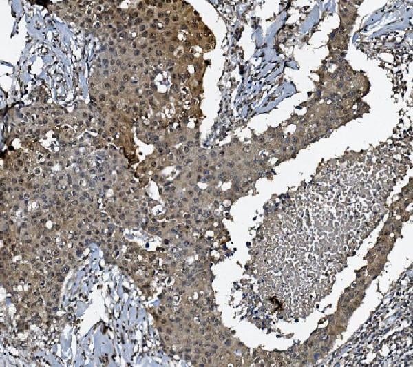
 IHC (Immunohistochemistry)
(Figure 8. IHC analysis of COMT using anti-COMT antibody (AAA11647).COMT was detected in frozen section of rat lung tissue. Heat mediated antigen retrieval was performed in citrate buffer (pH6, epitope retrieval solution) for 20 mins. The tissue section was blocked with 10% goat serum. The tissue section was then incubated with 1ug/ml rabbit anti-COMT Antibody (AAA11647) overnight at 4 degree C. Biotinylated goat anti-rabbit IgG was used as secondary antibody and incubated for 30 minutes at 37 degree C. The tissue section was developed using Strepavidin-Biotin-Complex (SABC) with DAB as the chromogen.)
IHC (Immunohistochemistry)
(Figure 8. IHC analysis of COMT using anti-COMT antibody (AAA11647).COMT was detected in frozen section of rat lung tissue. Heat mediated antigen retrieval was performed in citrate buffer (pH6, epitope retrieval solution) for 20 mins. The tissue section was blocked with 10% goat serum. The tissue section was then incubated with 1ug/ml rabbit anti-COMT Antibody (AAA11647) overnight at 4 degree C. Biotinylated goat anti-rabbit IgG was used as secondary antibody and incubated for 30 minutes at 37 degree C. The tissue section was developed using Strepavidin-Biotin-Complex (SABC) with DAB as the chromogen.)
COMT, Polyclonal Antibody (Cat# AAA11647)
Full Name
Anti-COMT Antibody
Gene Names
COMT; HEL-S-98n
Reactivity
Human, Mouse, Rat
Applications
Western Blot, Immunohistochemistry, Immunocytochemistry
Purity
Immunogen Affinity Purified
Pricing

 SDS-PAGE
Thrombin/Antithrombin Complex (TAT), Recombinant Protein (Cat# AAA21174)
Full Name
Recombinant Thrombin/Antithrombin Complex (TAT)
Reactivity
Homo sapiens (Human)
Applications
WB
Purity
> 80%
Pricing


 FCM (Flow Cytometry)
(Figure 6. Flow Cytometry analysis of U-87 cells using anti-SSH3BP1 antibody (AAA11656).Overlay histogram showing U-87 cells stained with AAA11656 (Blue line).The cells were blocked with 10% normal goat serum. And then incubated with rabbit anti-SSH3BP1 Antibody (AAA11656,1ug/1x10^6 cells) for 30 min at 20 degree C. DyLight®488 conjugated goat anti-rabbit IgG (5-10ug/1x10^6 cells) was used as secondary antibody for 30 minutes at 20 degree C. Isotype control antibody (Green line) was rabbit IgG (1ug/1x106) used under the same conditions. Unlabelled sample (Red line) was also used as a control.)
FCM (Flow Cytometry)
(Figure 6. Flow Cytometry analysis of U-87 cells using anti-SSH3BP1 antibody (AAA11656).Overlay histogram showing U-87 cells stained with AAA11656 (Blue line).The cells were blocked with 10% normal goat serum. And then incubated with rabbit anti-SSH3BP1 Antibody (AAA11656,1ug/1x10^6 cells) for 30 min at 20 degree C. DyLight®488 conjugated goat anti-rabbit IgG (5-10ug/1x10^6 cells) was used as secondary antibody for 30 minutes at 20 degree C. Isotype control antibody (Green line) was rabbit IgG (1ug/1x106) used under the same conditions. Unlabelled sample (Red line) was also used as a control.)
SSH3BP1, Polyclonal Antibody (Cat# AAA11656)
Full Name
Anti-SSH3BP1 Antibody
Gene Names
ABI1; E3B1; ABI-1; ABLBP4; NAP1BP; SSH3BP; SSH3BP1
Reactivity
Human, Mouse, Rat
Applications
Western Blot, Immunohistochemistry
Purity
Immunogen Affinity Purified
Pricing


 FCM (Flow Cytometry)
(Figure 7. Flow Cytometry analysis of HELA cells using anti-MCU antibody (AAA19226).Overlay histogram showing HELA cells stained with AAA19226 (Blue line). The cells were blocked with 10% normal goat serum. And then incubated with rabbit anti-MCU Antibody (AAA19226, 1μg/1x106 cells) for 30 min at 20 degree C. DyLight®488 conjugated goat anti-rabbit IgG (5-10μg/1x106 cells) was used as secondary antibody for 30 minutes at 20 degree C. Isotype control antibody (Green line) was rabbit IgG (1μg/1x106) used under the same conditions. Unlabelled sample (Red line) was also used as a control.)
FCM (Flow Cytometry)
(Figure 7. Flow Cytometry analysis of HELA cells using anti-MCU antibody (AAA19226).Overlay histogram showing HELA cells stained with AAA19226 (Blue line). The cells were blocked with 10% normal goat serum. And then incubated with rabbit anti-MCU Antibody (AAA19226, 1μg/1x106 cells) for 30 min at 20 degree C. DyLight®488 conjugated goat anti-rabbit IgG (5-10μg/1x106 cells) was used as secondary antibody for 30 minutes at 20 degree C. Isotype control antibody (Green line) was rabbit IgG (1μg/1x106) used under the same conditions. Unlabelled sample (Red line) was also used as a control.)
MCU, Polyclonal Antibody (Cat# AAA19226)
Full Name
Anti-MCU Antibody
Gene Names
MCU; C10orf42; CCDC109A
Reactivity
Human, Mouse, Rat, Monkey
Applications
Western Blot, Immunohistochemistry, Flow Cytometry, Direct ELISA
Purity
Immunogen affinity purified.
Pricing


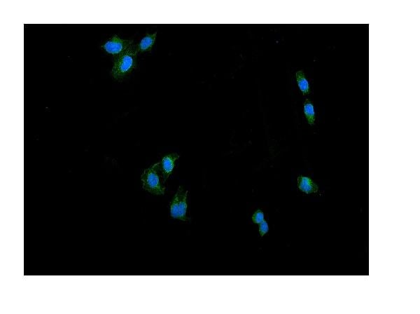
 IF (Immunofluorescence)
(Immunofluorescence analysis of U-2 OS cells using YAP1 Rabbit pAb at dilution of 1:150 (40x lens). Blue: DAPI for nuclear staining.)
IF (Immunofluorescence)
(Immunofluorescence analysis of U-2 OS cells using YAP1 Rabbit pAb at dilution of 1:150 (40x lens). Blue: DAPI for nuclear staining.)
YAP1, Polyclonal Antibody (Cat# AAA28345)
Full Name
YAP1 Rabbit pAb
Gene Names
YAP1; YAP; YKI; YAP2; YAP65
Reactivity
Human, Mouse, Rat
Applications
Western Blot, Immunohistochemistry, Immunofluorescence
Purity
Affinity purification
Pricing


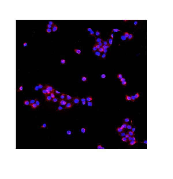
 IF (Immunofluorescence)
(Figure 9. IF analysis of P4HB using anti- P4HB antibody (AAA19258).P4HB was detected in immunocytochemical section of T-47D cells. Enzyme antigen retrieval was performed using IHC enzyme antigen retrieval reagent for 15 mins. The cells were blocked with 10% goat serum. And then incubated with 5μg/mL rabbit anti- P4HB Antibody (AAA19258) overnight at 4 degree C. DyLight®488 Conjugated Goat Anti-Rabbit IgG was used as secondary antibody at 1:100 dilution and incubated for 30 minutes at 37 degree C. The section was counterstained with DAPI. Visualize using a fluorescence microscope and filter sets appropriate for the label used.)
IF (Immunofluorescence)
(Figure 9. IF analysis of P4HB using anti- P4HB antibody (AAA19258).P4HB was detected in immunocytochemical section of T-47D cells. Enzyme antigen retrieval was performed using IHC enzyme antigen retrieval reagent for 15 mins. The cells were blocked with 10% goat serum. And then incubated with 5μg/mL rabbit anti- P4HB Antibody (AAA19258) overnight at 4 degree C. DyLight®488 Conjugated Goat Anti-Rabbit IgG was used as secondary antibody at 1:100 dilution and incubated for 30 minutes at 37 degree C. The section was counterstained with DAPI. Visualize using a fluorescence microscope and filter sets appropriate for the label used.)
P4HB, Polyclonal Antibody (Cat# AAA19258)
Full Name
Anti-P4HB Antibody
Gene Names
P4HB; DSI; GIT; PDI; PHDB; PDIA1; PO4DB; PO4HB; PROHB; ERBA2L; P4Hbeta
Reactivity
Human, Monkey, Mouse, Rat
Applications
Western Blot, Immunohistochemistry, Immunocytochemistry, Immunofluorescence, Flow Cytometry, Direct ELISA
Purity
Immunogen affinity purified.
Pricing


 WB (Western Blot)
(Western blot analysis of lysate from mouse lung tissue lysate, using Mlkl Antibody (C-term). AAA28711 was diluted at 1:1000. A goat anti-rabbit IgG H&L(HRP) at 1:10000 dilution was used as the secondary antibody. Lysate at 20ug.)
WB (Western Blot)
(Western blot analysis of lysate from mouse lung tissue lysate, using Mlkl Antibody (C-term). AAA28711 was diluted at 1:1000. A goat anti-rabbit IgG H&L(HRP) at 1:10000 dilution was used as the secondary antibody. Lysate at 20ug.)
M Mlkl, Polyclonal Antibody (Cat# AAA28711)
Full Name
M Mlkl Antibody (C-term)
Gene Names
Mlkl; 9130019I15Rik
Reactivity
Mouse
Applications
Western Blot
Purity
Peptide Affinity Purified Rabbit Polyclonal Antibody (Pab)
Pricing


 FCM (Flow Cytometry)
(Figure 9. Flow Cytometry analysis of SiHa cells using anti-DCTN1/p150-glued antibody (AAA19253).Overlay histogram showing SiHa cells stained with AAA19253 (Blue line). The cells were blocked with 10% normal goat serum. And then incubated with rabbit anti-DCTN1/p150-glued Antibody (AAA19253, 1μg/1x106 cells) for 30 min at 20 degree C. DyLight®488 conjugated goat anti-rabbit IgG (5-10μg/1x106 cells) was used as secondary antibody for 30 minutes at 20 degree C. Isotype control antibody (Green line) was rabbit IgG (1μg/1x106) used under the same conditions. Unlabelled sample (Red line) was also used as a control.)
FCM (Flow Cytometry)
(Figure 9. Flow Cytometry analysis of SiHa cells using anti-DCTN1/p150-glued antibody (AAA19253).Overlay histogram showing SiHa cells stained with AAA19253 (Blue line). The cells were blocked with 10% normal goat serum. And then incubated with rabbit anti-DCTN1/p150-glued Antibody (AAA19253, 1μg/1x106 cells) for 30 min at 20 degree C. DyLight®488 conjugated goat anti-rabbit IgG (5-10μg/1x106 cells) was used as secondary antibody for 30 minutes at 20 degree C. Isotype control antibody (Green line) was rabbit IgG (1μg/1x106) used under the same conditions. Unlabelled sample (Red line) was also used as a control.)
DCTN1/p150-glued, Polyclonal Antibody (Cat# AAA19253)
Full Name
Anti-DCTN1/p150-glued Antibody
Gene Names
DCTN1; P135; DP-150; DAP-150
Reactivity
Human, Mouse, Rat
Applications
Western Blot, Immunohistochemistry, Immunocytochemistry, Immunofluorescence, Flow Cytometry, Direct ELISA
Purity
Immunogen affinity purified.
Pricing


 WB (Western Blot)
(KHDRBS1 monoclonal antibody Western Blot analysis of KHDRBS1 expression in A-431)
WB (Western Blot)
(KHDRBS1 monoclonal antibody Western Blot analysis of KHDRBS1 expression in A-431)
KHDRBS1, Monoclonal Antibody (Cat# AAA25730)
Full Name
KHDRBS1 (KH Domain-containing, RNA-binding, Signal Transduction-associated Protein 1, FLJ34027, GAP-associated Tyrosine Phosphoprotein p62, p21 Ras GTPase-activating Protein-associated p62, p62, p68, Src-associated in Mitosis 68kD Protein, Sam68) (PE)
Gene Names
KHDRBS1; p62; p68; Sam68
Reactivity
Human
Applications
Immunofluorescence, Immunohistochemistry, Western Blot
Purity
Purified by Protein A Affinity Chromatography.
Pricing


 FCM (Flow Cytometry)
(Figure 8. Flow Cytometry analysis of U251 cells using anti-GABARAP antibody (AAA19250).Overlay histogram showing U251 cells stained with AAA19250 (Blue line). The cells were blocked with 10% normal goat serum. And then incubated with rabbit anti-GABARAP Antibody (AAA19250, 1μg/1x106 cells) for 30 min at 20 degree C. DyLight®488 conjugated goat anti-rabbit IgG (5-10μg/1x106 cells) was used as secondary antibody for 30 minutes at 20 degree C. Isotype control antibody (Green line) was rabbit IgG (1μg/1x106) used under the same conditions. Unlabelled sample (Red line) was also used as a control.)
FCM (Flow Cytometry)
(Figure 8. Flow Cytometry analysis of U251 cells using anti-GABARAP antibody (AAA19250).Overlay histogram showing U251 cells stained with AAA19250 (Blue line). The cells were blocked with 10% normal goat serum. And then incubated with rabbit anti-GABARAP Antibody (AAA19250, 1μg/1x106 cells) for 30 min at 20 degree C. DyLight®488 conjugated goat anti-rabbit IgG (5-10μg/1x106 cells) was used as secondary antibody for 30 minutes at 20 degree C. Isotype control antibody (Green line) was rabbit IgG (1μg/1x106) used under the same conditions. Unlabelled sample (Red line) was also used as a control.)
GABARAP, Polyclonal Antibody (Cat# AAA19250)
Full Name
Anti-GABARAP Antibody
Gene Names
GABARAP; MM46; ATG8A; GABARAP-a
Reactivity
Human, Mouse, Rat
Applications
Western Blot, Immunohistochemistry, Flow Cytometry
Purity
Immunogen affinity purified.
Pricing


 ICC (Immunocytochemistry)
(Figure 7. IHC analysis of ERAB using anti-ERAB antibody (AAA11668).ERAB was detected in immunocytochemical section of SMMC-7721 cell. Heat mediated antigen retrieval was performed in citrate buffer (pH6, epitope retrieval solution) for 20 mins. The tissue section was blocked with 10% goat serum. The tissue section was then incubated with 1ug/ml rabbit anti-ERAB Antibody (AAA11668) overnight at 4 degree C. Biotinylated goat anti-rabbit IgG was used as secondary antibody and incubated for 30 minutes at 37 degree C. The tissue section was developed using Strepavidin-Biotin-Complex (SABC) with DAB as the chromogen.)
ICC (Immunocytochemistry)
(Figure 7. IHC analysis of ERAB using anti-ERAB antibody (AAA11668).ERAB was detected in immunocytochemical section of SMMC-7721 cell. Heat mediated antigen retrieval was performed in citrate buffer (pH6, epitope retrieval solution) for 20 mins. The tissue section was blocked with 10% goat serum. The tissue section was then incubated with 1ug/ml rabbit anti-ERAB Antibody (AAA11668) overnight at 4 degree C. Biotinylated goat anti-rabbit IgG was used as secondary antibody and incubated for 30 minutes at 37 degree C. The tissue section was developed using Strepavidin-Biotin-Complex (SABC) with DAB as the chromogen.)
ERAB, Polyclonal Antibody (Cat# AAA11668)
Full Name
Anti-ERAB Antibody
Gene Names
HSD17B10; ABAD; CAMR; ERAB; HCD2; MHBD; HADH2; MRPP2; MRX17; MRX31; SCHAD; MRXS10; SDR5C1; 17b-HSD10; DUPXp11.22
Reactivity
Human, Mouse
Applications
Western Blot, Immunohistochemistry
Purity
Immunogen Affinity Purified
Pricing


 IP (Immunoprecipitation)
(Immunoprecipitation analysis of 300ug extracts of HeLa cells using 3ug CDK4 antibody . Western blot was performed from the immunoprecipitate using CDK4 antibody at a dilition of 1:1000.)
IP (Immunoprecipitation)
(Immunoprecipitation analysis of 300ug extracts of HeLa cells using 3ug CDK4 antibody . Western blot was performed from the immunoprecipitate using CDK4 antibody at a dilition of 1:1000.)
CDK4, Polyclonal Antibody (Cat# AAA28337)
Full Name
CDK4 Rabbit pAb
Gene Names
CDK4; CMM3; PSK-J3
Reactivity
Human, Mouse, Rat
Applications
Western Blot, Immunohistochemistry, Immunofluorescence, Immunoprecipitation
Purity
Affinity purification
Pricing


 WB (Western Blot)
(KHDRBS1 monoclonal antibody Western Blot analysis of KHDRBS1 expression in A-431)
WB (Western Blot)
(KHDRBS1 monoclonal antibody Western Blot analysis of KHDRBS1 expression in A-431)
KHDRBS1, Monoclonal Antibody (Cat# AAA25140)
Full Name
KHDRBS1 (KH Domain-containing, RNA-binding, Signal Transduction-associated Protein 1, FLJ34027, GAP-associated Tyrosine Phosphoprotein p62, p21 Ras GTPase-activating Protein-associated p62, p62, p68, Src-associated in Mitosis 68kD Protein, Sam68) (FITC)
Gene Names
KHDRBS1; p62; p68; Sam68
Reactivity
Human
Applications
Immunofluorescence, Immunohistochemistry, Western Blot
Purity
Purified by Protein A Affinity Chromatography.
Pricing


 FCM (Flow Cytometry)
(Figure 6. Flow Cytometry analysis of PC-3 cells using anti-ATG14L antibody (AAA11658).Overlay histogram showing PC-3 cells stained with AAA11658 (Blue line).The cells were blocked with 10% normal goat serum. And then incubated with rabbit anti-ATG14L Antibody (AAA11658,1ug/1x10^6 cells) for 30 min at 20 degree C. DyLight®488 conjugated goat anti-rabbit IgG (5-10ug/1x10^6 cells) was used as secondary antibody for 30 minutes at 20 degree C. Isotype control antibody (Green line) was rabbit IgG (1ug/1x106) used under the same conditions. Unlabelled sample (Red line) was also used as a control.)
FCM (Flow Cytometry)
(Figure 6. Flow Cytometry analysis of PC-3 cells using anti-ATG14L antibody (AAA11658).Overlay histogram showing PC-3 cells stained with AAA11658 (Blue line).The cells were blocked with 10% normal goat serum. And then incubated with rabbit anti-ATG14L Antibody (AAA11658,1ug/1x10^6 cells) for 30 min at 20 degree C. DyLight®488 conjugated goat anti-rabbit IgG (5-10ug/1x10^6 cells) was used as secondary antibody for 30 minutes at 20 degree C. Isotype control antibody (Green line) was rabbit IgG (1ug/1x106) used under the same conditions. Unlabelled sample (Red line) was also used as a control.)
ATG14L, Polyclonal Antibody (Cat# AAA11658)
Full Name
Anti-ATG14L Antibody
Gene Names
ATG14; ATG14L; BARKOR; KIAA0831
Reactivity
Human, Rat
Applications
Western Blot, Immunohistochemistry
Purity
Immunogen Affinity Purified
Pricing
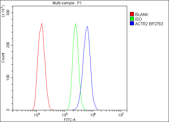


 FCM (Flow Cytometry)
(Figure 6. Flow Cytometry analysis of C6 cells using anti-Arp2/ACTR2 antibody (AAA19284).Overlay histogram showing C6 cells stained with AAA19284 (Blue line). The cells were blocked with 10% normal goat serum. And then incubated with rabbit anti-Arp2/ACTR2 Antibody (AAA19284,1μg/1x106 cells) for 30 min at 20 degree C. DyLight®488 conjugated goat anti-rabbit IgG (5-10μg/1x106 cells) was used as secondary antibody for 30 minutes at 20 degree C. Isotype control antibody (Green line) was rabbit IgG (1μg/1x106) used under the same conditions. Unlabelled sample (Red line) was also used as a control.)
FCM (Flow Cytometry)
(Figure 6. Flow Cytometry analysis of C6 cells using anti-Arp2/ACTR2 antibody (AAA19284).Overlay histogram showing C6 cells stained with AAA19284 (Blue line). The cells were blocked with 10% normal goat serum. And then incubated with rabbit anti-Arp2/ACTR2 Antibody (AAA19284,1μg/1x106 cells) for 30 min at 20 degree C. DyLight®488 conjugated goat anti-rabbit IgG (5-10μg/1x106 cells) was used as secondary antibody for 30 minutes at 20 degree C. Isotype control antibody (Green line) was rabbit IgG (1μg/1x106) used under the same conditions. Unlabelled sample (Red line) was also used as a control.)
Arp2/ACTR2, Polyclonal Antibody (Cat# AAA19284)
Full Name
Anti-Arp2/ACTR2 Antibody
Gene Names
ACTR2; ARP2
Reactivity
Human, Mouse, Monkey, Rat
Applications
WB, IHC-P, ICC, IF, FC/FACS/FCM, EIA
Purity
Immunogen affinity purified.
Pricing


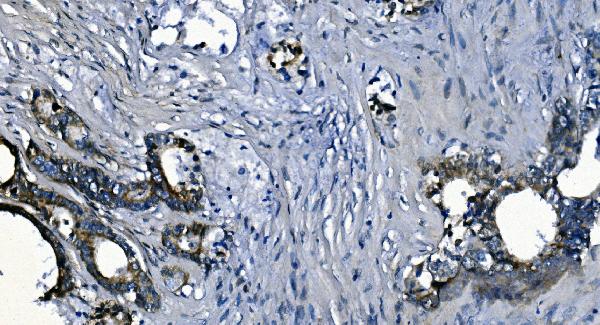
 IHC (Immunohistchemistry)
(Immunohistochemistry of paraffin-embedded rat heart using IRS1 Rabbit pAb at dilution of 1:100 (40x lens).Perform high pressure antigen retrieval with 10 mM citrate buffer pH 6.0 before commencing with IHC staining protocol.)
IHC (Immunohistchemistry)
(Immunohistochemistry of paraffin-embedded rat heart using IRS1 Rabbit pAb at dilution of 1:100 (40x lens).Perform high pressure antigen retrieval with 10 mM citrate buffer pH 6.0 before commencing with IHC staining protocol.)
IRS1, Polyclonal Antibody (Cat# AAA28342)
Full Name
IRS1 Rabbit pAb
Gene Names
IRS1; HIRS-1
Reactivity
Human, Mouse, Rat
Applications
Western Blot, Immunohistochemistry, Immunofluorescence
Purity
Affinity purification
Pricing