Filters
Clonality
Type
Reactivity
Gene Name
Isotype
Host
Application
Clone
437 results for " Signal Proteins" - showing 400-437
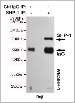 IP (Immunoprecipitation)
(Immunoprecipitation analysis of Raji cell lysates using SHP-1 mouse mAb.)
IP (Immunoprecipitation)
(Immunoprecipitation analysis of Raji cell lysates using SHP-1 mouse mAb.)
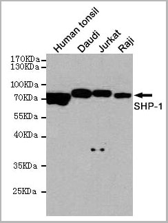
SHP-1, Monoclonal Antibody (Cat# AAA14102)
Full Name
Anti-SHP-1 Mouse mAb
Gene Names
PTPN6; HCP; HCPH; SHP1; SHP-1; HPTP1C; PTP-1C; SHP-1L; SH-PTP1
Reactivity
Human
Applications
Western Blot, Immunoprecipitation
Purity
Affinity purified
Pricing
 WB (Western Blot)
(Western blot analysis of C3 expression in HepG2 cell lysate (AAA11693). Electrophoresis was performed on a 5-20% SDS-PAGE gel at 70V (stacking gel) / 90V (resolving gel) for 2-3 hours. The sample well of each lane was loaded with 50ug of sample under reducing conditions. After Electrophoresis, proteins were transferred to a Nitrocellulose membrane at 150mA for 50-90 minutes. Blocked the membrane with 5% Non-fat Milk/TBS for 1.5 hour at RT. The membrane was incubated with rabbit anti-C3 monoclonal antibody (AAA11693) overnight at 4°C, then washed with TBS-0.1%Tween 3 times with 5 minutes each and probed with goat anti-rabbit IgG-HRP secondary antibody at a dilution of 1:10000 for 1.5 hour at RT. The signal is developed using an Enhanced Chemiluminescent detection (ECL) kit (please inquire) with Tanon 5200 system. A specific band was detected for C3.)
WB (Western Blot)
(Western blot analysis of C3 expression in HepG2 cell lysate (AAA11693). Electrophoresis was performed on a 5-20% SDS-PAGE gel at 70V (stacking gel) / 90V (resolving gel) for 2-3 hours. The sample well of each lane was loaded with 50ug of sample under reducing conditions. After Electrophoresis, proteins were transferred to a Nitrocellulose membrane at 150mA for 50-90 minutes. Blocked the membrane with 5% Non-fat Milk/TBS for 1.5 hour at RT. The membrane was incubated with rabbit anti-C3 monoclonal antibody (AAA11693) overnight at 4°C, then washed with TBS-0.1%Tween 3 times with 5 minutes each and probed with goat anti-rabbit IgG-HRP secondary antibody at a dilution of 1:10000 for 1.5 hour at RT. The signal is developed using an Enhanced Chemiluminescent detection (ECL) kit (please inquire) with Tanon 5200 system. A specific band was detected for C3.)
C3/Complement C3, Monoclonal Antibody (Cat# AAA11693)
Full Name
Anti-C3/Complement C3 Rabbit Monoclonal Antibody
Gene Names
C3; ASP; C3a; C3b; AHUS5; ARMD9; CPAMD1; HEL-S-62p
Reactivity
Human
Applications
Flow Cytometry, Immunofluorescence, Immunocytochemistry, Western Blot
Purity
Affinity-chromatography
Pricing
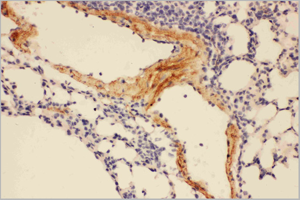
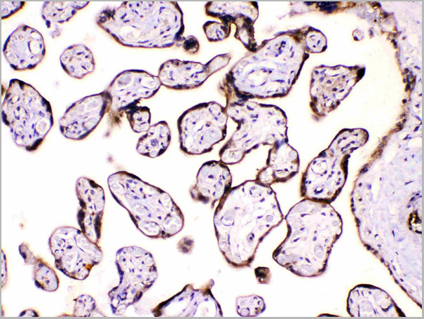
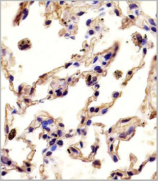
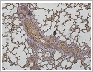
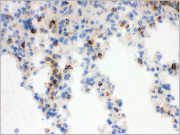
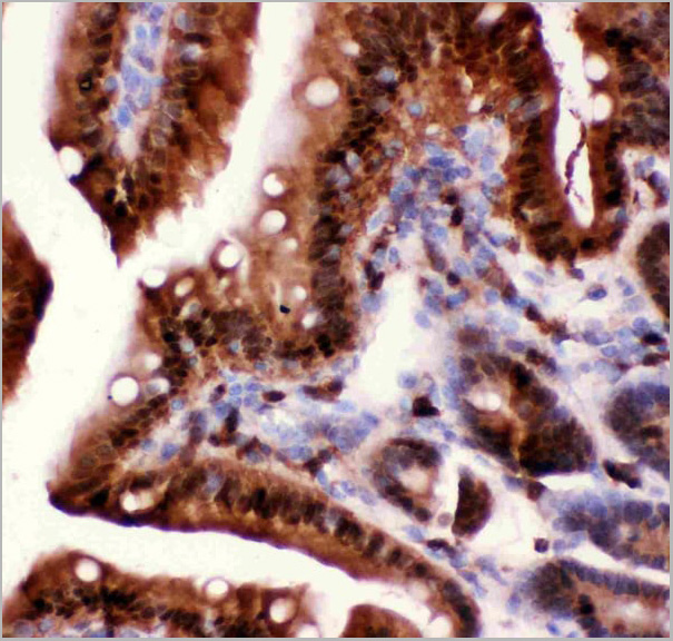
 IHC (Immunohistochemistry)
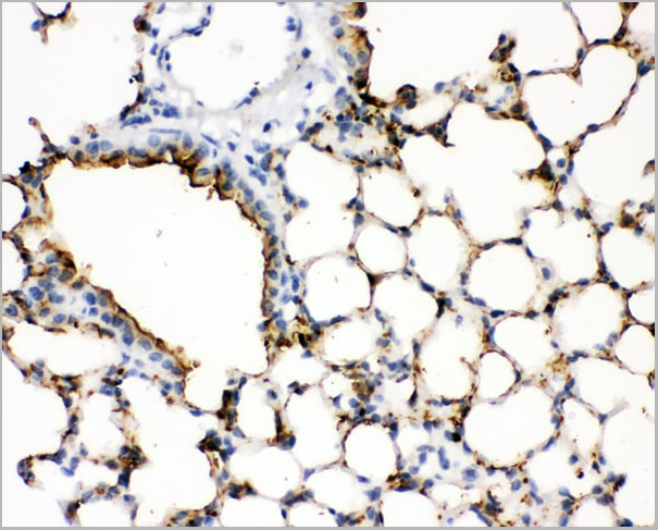
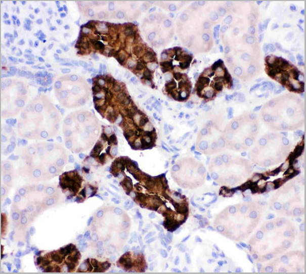
(Figure 3. IHC analysis of SFTPA1 using anti- SFTPA1 antibody (AAA11521).SFTPA1 was detected in paraffin-embedded section of mouse lung tissues. Heat mediated antigen retrieval was performed in citrate buffer (pH6, epitope retrieval solution) for 20 mins. The tissue section was blocked with 10% goat serum. The tissue section was then incubated with 1ug/ml rabbit anti- SFTPA1 Antibody (AAA11521) overnight at 4 degree C. Biotinylated goat anti-rabbit IgG was used as secondary antibody and incubated for 30 minutes at 37 degree C. The tissue section was developed using Strepavidin-Biotin-Complex (SABC) with DAB as the chromogen.)
IHC (Immunohistochemistry)
(Figure 3. IHC analysis of SFTPA1 using anti- SFTPA1 antibody (AAA11521).SFTPA1 was detected in paraffin-embedded section of mouse lung tissues. Heat mediated antigen retrieval was performed in citrate buffer (pH6, epitope retrieval solution) for 20 mins. The tissue section was blocked with 10% goat serum. The tissue section was then incubated with 1ug/ml rabbit anti- SFTPA1 Antibody (AAA11521) overnight at 4 degree C. Biotinylated goat anti-rabbit IgG was used as secondary antibody and incubated for 30 minutes at 37 degree C. The tissue section was developed using Strepavidin-Biotin-Complex (SABC) with DAB as the chromogen.)
Surfactant Protein A, Polyclonal Antibody (Cat# AAA11521)
Full Name
Polyclonal Anti-SFTPA1 Antibody
Gene Names
Sftpa1; SP-A; Sftp1; Sftp-1
Reactivity
Mouse, Rat
Applications
Western Blot, Immunohistochemistry
Purity
Immunogen affinity purified.
Pricing


 Application Data
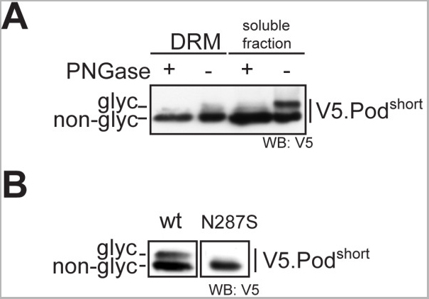
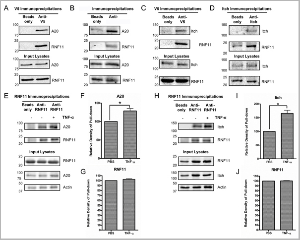
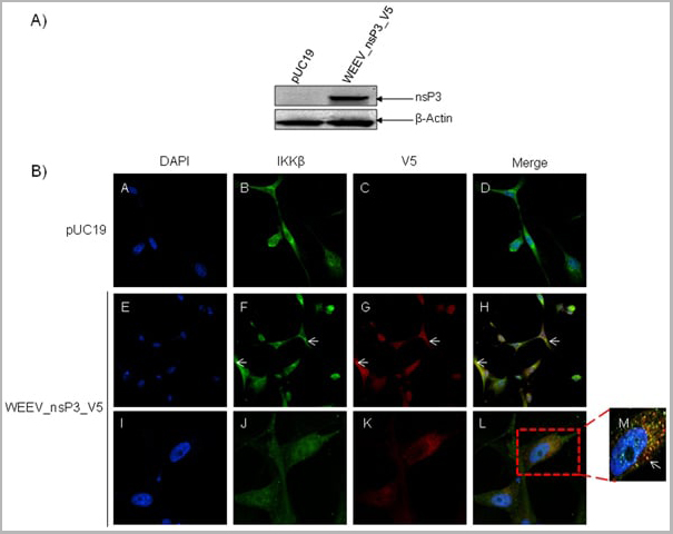
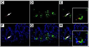
(Published customer image: Mouse anti V5 tag antibody, clone SV5-Pk1 used for the detection of V5 tagged WEEV_nsP3 protein by western blotting and immunofluorescenceImage caption: WEEV nsP3 interaction with host IKKbeta. A) U87MGs were transfected in a 6-well plate with 5 ug of pUC19 and WEEV_nsP3_HA for 24 hours. Cell lysates were resolved using SDS-PAGE and subsequently immunoblotted with V5 antibody and beta-actin served as a loading control. B) U87MGs were transfected with WEEV_nsP3_V5; cells were fixed after 24 hours and stained with antibodies against the endogenous IKKbeta and the V5 tag. Cells were incubated with appropriate secondary Alexa Fluor antibodies and the nuclei stained with DAPI. Co-localization of IKKbeta with WEEV_nsP3_V5 (yellow) was observed as shown by the arrows. B) Panels E -H serve as an example of transfected cells in a given field of view that show co-localization of IKKbeta and WEEV_nsP3_V5 24 hours post transfection. Panels I-L represent magnified images of other cells showing co-localization of IKKbeta and WEEV_nsP3_V5. Panel M is a magnified image of panel L. The co-localization was confirmed by Z-stack analysis. Co-localization was calculated to be approximately in 61% of cells (163 cells were counted of which 44% demonstrated expression of nsP3. Of those cells that expressed nsP3, 61% showed co-localization of both proteins). Images were taken using Nikon Eclipse TE2000-U at 60x magnification and are representative of 2 independent experiments.From: Amaya M, Voss K, Sampey G, Senina S, de la Fuente C, et al. (2014) The Role of IKKbeta in Venezuelan Equine Encephalitis Virus Infection. PLoS ONE 9(2): e86745.)
Application Data
(Published customer image: Mouse anti V5 tag antibody, clone SV5-Pk1 used for the detection of V5 tagged WEEV_nsP3 protein by western blotting and immunofluorescenceImage caption: WEEV nsP3 interaction with host IKKbeta. A) U87MGs were transfected in a 6-well plate with 5 ug of pUC19 and WEEV_nsP3_HA for 24 hours. Cell lysates were resolved using SDS-PAGE and subsequently immunoblotted with V5 antibody and beta-actin served as a loading control. B) U87MGs were transfected with WEEV_nsP3_V5; cells were fixed after 24 hours and stained with antibodies against the endogenous IKKbeta and the V5 tag. Cells were incubated with appropriate secondary Alexa Fluor antibodies and the nuclei stained with DAPI. Co-localization of IKKbeta with WEEV_nsP3_V5 (yellow) was observed as shown by the arrows. B) Panels E -H serve as an example of transfected cells in a given field of view that show co-localization of IKKbeta and WEEV_nsP3_V5 24 hours post transfection. Panels I-L represent magnified images of other cells showing co-localization of IKKbeta and WEEV_nsP3_V5. Panel M is a magnified image of panel L. The co-localization was confirmed by Z-stack analysis. Co-localization was calculated to be approximately in 61% of cells (163 cells were counted of which 44% demonstrated expression of nsP3. Of those cells that expressed nsP3, 61% showed co-localization of both proteins). Images were taken using Nikon Eclipse TE2000-U at 60x magnification and are representative of 2 independent experiments.From: Amaya M, Voss K, Sampey G, Senina S, de la Fuente C, et al. (2014) The Role of IKKbeta in Venezuelan Equine Encephalitis Virus Infection. PLoS ONE 9(2): e86745.)
V5-TAG, Monoclonal Antibody (Cat# AAA12211)
Full Name
MOUSE ANTI V5-TAG
Applications
Immunohistochemistry, Flow Cytometry, Immunofluorescence, Immunoprecipitation, Western Blot, Radioimmunoassay
Pricing
 WB (Western Blot)
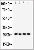
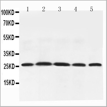
(Figure 6. Western blot analysis of Adiponectin using anti-Adiponectin antibody (AAA11602).Electrophoresis was performed on a 5-20% SDS-PAGE gel at 70V (Stacking gel) / 90V (Resolving gel) for 2-3 hours. The sample well of each lane was loaded with 50ug of sample under reducing conditions.Lane 1: Rat Testis Tissue Lysate,Lane 2: Rat Spleen Tissue Lysate,Lane 3: Rat Skeletal Muscle Tissue Lysate,Lane 4: Rat Cardiac Muscle Tissue Lysate,Lane 5: Rat Liver Tissue Lysate,After Electrophoresis, proteins were transferred to a Nitrocellulose membrane at 150mA for 50-90 minutes. Blocked the membrane with 5% Non-fat Milk/ TBS for 1.5 hour at RT. The membrane was incubated with rabbit anti-Adiponectin antigen affinity purified polyclonal antibody at 0.5ug/mL overnight at 4 degree C, then washed with TBS-0.1%Tween 3 times with 5 minutes each and probed with a goat anti-rabbit IgG-HRP secondary antibody at a dilution of 1:10000 for 1.5 hour at RT. The signal is developed using an Enhanced Chemiluminescent detection (ECL) kit with Tanon 5200 system. A specific band was detected for Adiponectin at approximately 26KD. The expected band size for Adiponectin is at 26KD.)
WB (Western Blot)
(Figure 6. Western blot analysis of Adiponectin using anti-Adiponectin antibody (AAA11602).Electrophoresis was performed on a 5-20% SDS-PAGE gel at 70V (Stacking gel) / 90V (Resolving gel) for 2-3 hours. The sample well of each lane was loaded with 50ug of sample under reducing conditions.Lane 1: Rat Testis Tissue Lysate,Lane 2: Rat Spleen Tissue Lysate,Lane 3: Rat Skeletal Muscle Tissue Lysate,Lane 4: Rat Cardiac Muscle Tissue Lysate,Lane 5: Rat Liver Tissue Lysate,After Electrophoresis, proteins were transferred to a Nitrocellulose membrane at 150mA for 50-90 minutes. Blocked the membrane with 5% Non-fat Milk/ TBS for 1.5 hour at RT. The membrane was incubated with rabbit anti-Adiponectin antigen affinity purified polyclonal antibody at 0.5ug/mL overnight at 4 degree C, then washed with TBS-0.1%Tween 3 times with 5 minutes each and probed with a goat anti-rabbit IgG-HRP secondary antibody at a dilution of 1:10000 for 1.5 hour at RT. The signal is developed using an Enhanced Chemiluminescent detection (ECL) kit with Tanon 5200 system. A specific band was detected for Adiponectin at approximately 26KD. The expected band size for Adiponectin is at 26KD.)
Adiponectin, Polyclonal Antibody (Cat# AAA11602)
Full Name
Anti-Adiponectin antibody
Gene Names
Adipoq; Acdc; Acrp30
Reactivity
Mouse, Rat
Applications
Western Blot, Immunohistochemistry
Purity
Immunogen affinity purified.
Pricing
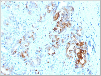
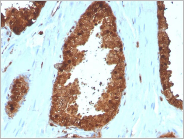
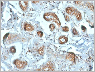
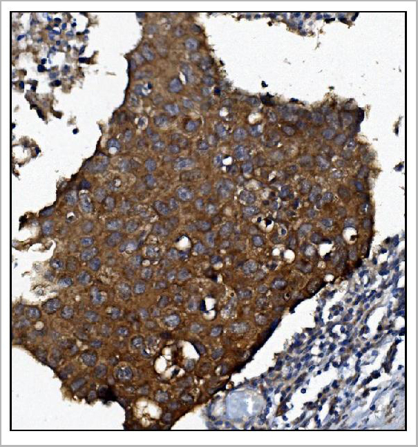
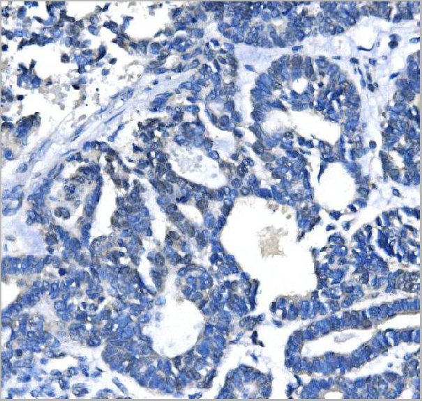
 IHC (Immunohistchemistry)
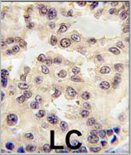
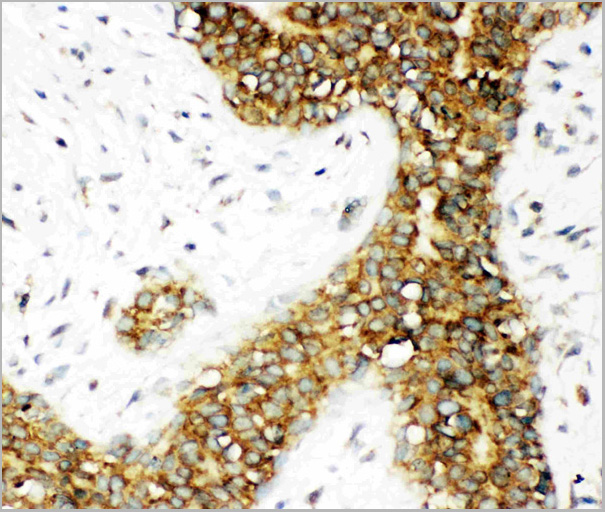
(Formalin-fixed and paraffin-embedded human lung carcinoma tissue reacted with EN1 antibody (N-term) (Cat.#AAA28756), which was peroxidase-conjugated to the secondary antibody, followed by DAB staining. This data demonstrates the use of this antibody for immunohistochemistry; clinical relevance has not been evaluated.)
IHC (Immunohistchemistry)
(Formalin-fixed and paraffin-embedded human lung carcinoma tissue reacted with EN1 antibody (N-term) (Cat.#AAA28756), which was peroxidase-conjugated to the secondary antibody, followed by DAB staining. This data demonstrates the use of this antibody for immunohistochemistry; clinical relevance has not been evaluated.)
EN1 (Engrailed 1), Polyclonal Antibody (Cat# AAA28756)
Full Name
EN1 (Engrailed 1) Antibody (N-term)
Reactivity
Human, Mouse
Applications
Western Blot, Immunohistochemistry, Immunofluorescence
Purity
This antibody is purified through a protein A column, followed by peptide affinity purification.
Pricing
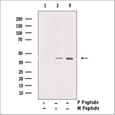
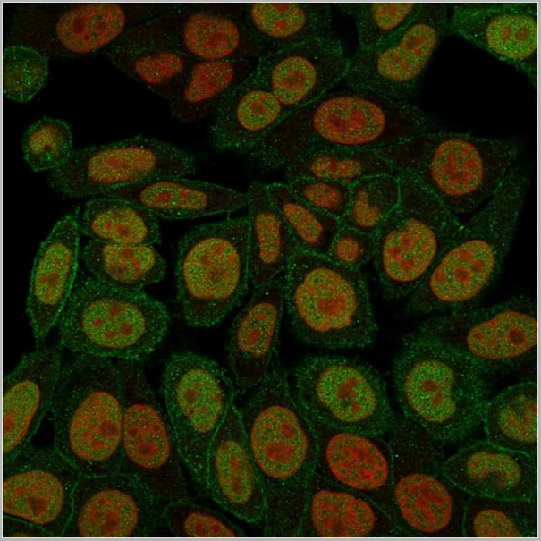
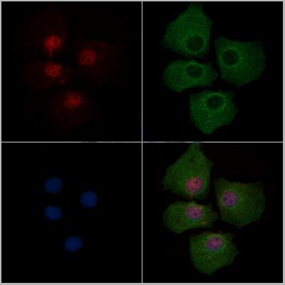 Application Data
(At 25 degree C. Samples were then incubated with primary Ab(At 37 degree C. An AlexaFluor594 conjugated goat anti-rabbit IgG(H+L) Ab(Red) and an AlexaFluor488 conjugated goat anti-mouse IgG(H+L) Ab(Green) were used as the secondary antibody.The nuclear counter stain is DAPI(blue).)
Application Data
(At 25 degree C. Samples were then incubated with primary Ab(At 37 degree C. An AlexaFluor594 conjugated goat anti-rabbit IgG(H+L) Ab(Red) and an AlexaFluor488 conjugated goat anti-mouse IgG(H+L) Ab(Green) were used as the secondary antibody.The nuclear counter stain is DAPI(blue).)
PAR4, Polyclonal Antibody (Cat# AAA31375)
Full Name
Phospho-PAR4 (Thr163) Antibody
Gene Names
PAWR; PAR4; Par-4
Reactivity
Human, Mouse, Rat
Predicted Reactivity: Pig (100%), Zebrafish (92%), Rabbit (100%), Chicken (100%), Xenopus (92%)
Predicted Reactivity: Pig (100%), Zebrafish (92%), Rabbit (100%), Chicken (100%), Xenopus (92%)
Applications
Western Blot, Immunohistochemistry, Immunofluorescence, Immunocytochemistry, Peptide ELISA
Purity
The antibody is from purified rabbit serum by affinity purification via sequential chromatography on phospho-peptide and non-phospho-peptide affinity columns.
Pricing
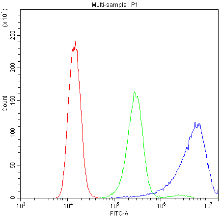
 FCM (Flow Cytometry)
(Figure 6. Flow Cytometry analysis of U20S cells using anti-Calpastatin antibody (AAA11679).Overlay histogram showing U20S cells stained with AAA11679 (Blue line).The cells were blocked with 10% normal goat serum. And then incubated with rabbit anti-Calpastatin Antibody (AAA11679,1ug/1x10^6 cells) for 30 min at 20 degree C. DyLight ®488 conjugated goat anti-rabbit IgG (5-10ug/1x10^6 cells) was used as secondary antibody for 30 minutes at 20 degree C. Isotype control antibody (Green line) was rabbit IgG (1ug/1x106) used under the same conditions. Unlabelled sample (Red line) was also used as a control.)
FCM (Flow Cytometry)
(Figure 6. Flow Cytometry analysis of U20S cells using anti-Calpastatin antibody (AAA11679).Overlay histogram showing U20S cells stained with AAA11679 (Blue line).The cells were blocked with 10% normal goat serum. And then incubated with rabbit anti-Calpastatin Antibody (AAA11679,1ug/1x10^6 cells) for 30 min at 20 degree C. DyLight ®488 conjugated goat anti-rabbit IgG (5-10ug/1x10^6 cells) was used as secondary antibody for 30 minutes at 20 degree C. Isotype control antibody (Green line) was rabbit IgG (1ug/1x106) used under the same conditions. Unlabelled sample (Red line) was also used as a control.)
Calpastatin, Polyclonal Antibody (Cat# AAA11679)
Full Name
Anti-Calpastatin Antibody
Gene Names
CAST; BS-17; PLACK
Reactivity
Human
Applications
Western Blot, Immunohistochemistry
Purity
Immunogen affinity purified.
Pricing

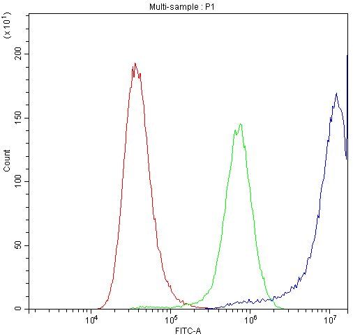
 FCM (Flow Cytometry)
(Figure 7. Flow Cytometry analysis of U251 cells using anti-PNP antibody (AAA11680).Overlay histogram showing U251 cells stained with AAA11680 (Blue line).The cells were blocked with 10% normal goat serum. And then incubated with rabbit anti-PNP Antibody (AAA11680,1ug/1x10^6 cells) for 30 min at 20 degree C. DyLight ®488 conjugated goat anti-rabbit IgG (5-10ug/1x10^6 cells) was used as secondary antibody for 30 minutes at 20 degree C. Isotype control antibody (Green line) was rabbit IgG (1ug/1x106) used under the same conditions. Unlabelled sample (Red line) was also used as a control.)
FCM (Flow Cytometry)
(Figure 7. Flow Cytometry analysis of U251 cells using anti-PNP antibody (AAA11680).Overlay histogram showing U251 cells stained with AAA11680 (Blue line).The cells were blocked with 10% normal goat serum. And then incubated with rabbit anti-PNP Antibody (AAA11680,1ug/1x10^6 cells) for 30 min at 20 degree C. DyLight ®488 conjugated goat anti-rabbit IgG (5-10ug/1x10^6 cells) was used as secondary antibody for 30 minutes at 20 degree C. Isotype control antibody (Green line) was rabbit IgG (1ug/1x106) used under the same conditions. Unlabelled sample (Red line) was also used as a control.)
PNP, Polyclonal Antibody (Cat# AAA11680)
Full Name
Anti-PNP Antibody
Gene Names
PNP; NP; PUNP; PRO1837
Reactivity
Human, Mouse, Rat
Applications
Western Blot, Immunohistochemistry
Purity
Immunogen affinity purified.
Pricing
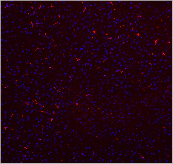
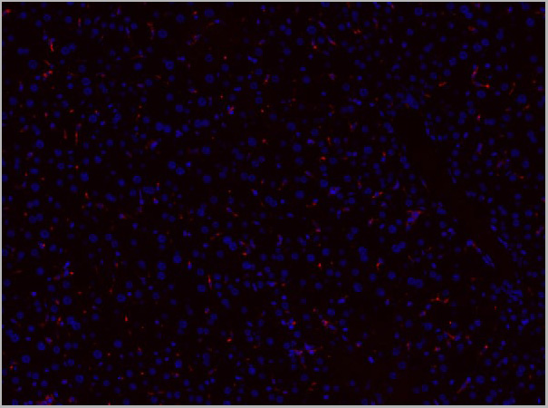

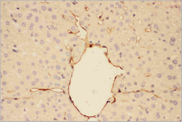
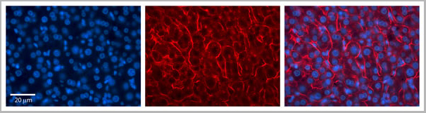
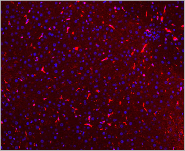 IF (Immunofluorescence)
(Figure 7. IF analysis of CD68 using anti- CD68 antibody CD68 was detected in paraffin-embedded section of mouse liver tissues. Heat mediated antigen retrieval was performed in citrate buffer (pH6, epitope retrieval solution) for 20 mins. The tissue section was blocked with 10% goat serum. The tissue section was then incubated with 1ug/mL rabbit anti- CD68 Antibody overnight at 4 degree C. Cy3 Conjugated Goat Anti-Rabbit IgG was used as secondary antibody at 1:100 dilution and incubated for 30 minutes at 37 degree C. The section was counterstained with DAPI. Visualize using a fluorescence microscope and filter sets appropriate for the label used.)
IF (Immunofluorescence)
(Figure 7. IF analysis of CD68 using anti- CD68 antibody CD68 was detected in paraffin-embedded section of mouse liver tissues. Heat mediated antigen retrieval was performed in citrate buffer (pH6, epitope retrieval solution) for 20 mins. The tissue section was blocked with 10% goat serum. The tissue section was then incubated with 1ug/mL rabbit anti- CD68 Antibody overnight at 4 degree C. Cy3 Conjugated Goat Anti-Rabbit IgG was used as secondary antibody at 1:100 dilution and incubated for 30 minutes at 37 degree C. The section was counterstained with DAPI. Visualize using a fluorescence microscope and filter sets appropriate for the label used.)
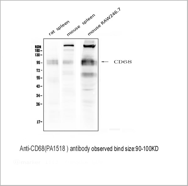
CD68, Polyclonal Antibody (Cat# AAA11512)
Full Name
Anti-CD68 antibody
Gene Names
Cd68; Lamp4; gp110; Scard1
Reactivity
Mouse, Rat
Applications
Western Blot, Immunohistochemistry, Immunohistochemistry
Purity
Immunogen affinity purified.
Pricing


 Application Data
(Published customer image: Mouse anti V5 tag antibody, clone SV5-Pk1 used for the detection of V5 tagged WEEV_nsP3 protein by western blotting and immunofluorescenceImage caption: WEEV nsP3 interaction with host IKKbeta. A) U87MGs were transfected in a 6-well plate with 5 ug of pUC19 and WEEV_nsP3_HA for 24 hours. Cell lysates were resolved using SDS-PAGE and subsequently immunoblotted with V5 antibody and beta-actin served as a loading control. B) U87MGs were transfected with WEEV_nsP3_V5; cells were fixed after 24 hours and stained with antibodies against the endogenous IKKbeta and the V5 tag. Cells were incubated with appropriate secondary Alexa Fluor antibodies and the nuclei stained with DAPI. Co-localization of IKKbeta with WEEV_nsP3_V5 (yellow) was observed as shown by the arrows. B) Panels E -H serve as an example of transfected cells in a given field of view that show co-localization of IKKbeta and WEEV_nsP3_V5 24 hours post transfection. Panels I-L represent magnified images of other cells showing co-localization of IKKbeta and WEEV_nsP3_V5. Panel M is a magnified image of panel L. The co-localization was confirmed by Z-stack analysis. Co-localization was calculated to be approximately in 61% of cells (163 cells were counted of which 44% demonstrated expression of nsP3. Of those cells that expressed nsP3, 61% showed co-localization of both proteins). Images were taken using Nikon Eclipse TE2000-U at 60x magnification and are representative of 2 independent experiments.From: Amaya M, Voss K, Sampey G, Senina S, de la Fuente C, et al. (2014) The Role of IKKbeta in Venezuelan Equine Encephalitis Virus Infection. PLoS ONE 9(2): e86745.)
Application Data
(Published customer image: Mouse anti V5 tag antibody, clone SV5-Pk1 used for the detection of V5 tagged WEEV_nsP3 protein by western blotting and immunofluorescenceImage caption: WEEV nsP3 interaction with host IKKbeta. A) U87MGs were transfected in a 6-well plate with 5 ug of pUC19 and WEEV_nsP3_HA for 24 hours. Cell lysates were resolved using SDS-PAGE and subsequently immunoblotted with V5 antibody and beta-actin served as a loading control. B) U87MGs were transfected with WEEV_nsP3_V5; cells were fixed after 24 hours and stained with antibodies against the endogenous IKKbeta and the V5 tag. Cells were incubated with appropriate secondary Alexa Fluor antibodies and the nuclei stained with DAPI. Co-localization of IKKbeta with WEEV_nsP3_V5 (yellow) was observed as shown by the arrows. B) Panels E -H serve as an example of transfected cells in a given field of view that show co-localization of IKKbeta and WEEV_nsP3_V5 24 hours post transfection. Panels I-L represent magnified images of other cells showing co-localization of IKKbeta and WEEV_nsP3_V5. Panel M is a magnified image of panel L. The co-localization was confirmed by Z-stack analysis. Co-localization was calculated to be approximately in 61% of cells (163 cells were counted of which 44% demonstrated expression of nsP3. Of those cells that expressed nsP3, 61% showed co-localization of both proteins). Images were taken using Nikon Eclipse TE2000-U at 60x magnification and are representative of 2 independent experiments.From: Amaya M, Voss K, Sampey G, Senina S, de la Fuente C, et al. (2014) The Role of IKKbeta in Venezuelan Equine Encephalitis Virus Infection. PLoS ONE 9(2): e86745.)
V5-TAG, Monoclonal Antibody (Cat# AAA11850)
Full Name
MOUSE ANTI V5-TAG:Biotin
Applications
Immunohistochemistry, Western Blot
Pricing
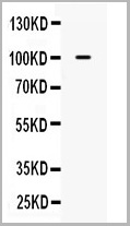
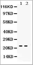
 WB (Western Blot)
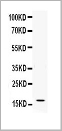
(Figure. Western blot analysis of IFN gamma using anti- IFN gamma antibody (AAA11517).Electrophoresis was performed on a 5-20% SDS-PAGE gel at 70V (Stacking gel) / 90V (Resolving gel) for 2-3 hours. The sample well of each lane was loaded with 50ug of sample under reducing conditions.Lane: Recombinant Human IFN gamma Protein 0.5ngAfter Electrophoresis, proteins were transferred to a Nitrocellulose membrane at 150mA for 50-90 minutes. Blocked the membrane with 5% Non-fat Milk/ TBS for 1.5 hour at RT. The membrane was incubated with rabbit anti- IFN gamma antigen affinity purified polyclonal antibody at 0.5ug/mL overnight at 4 degree C, then washed with TBS-0.1%Tween 3 times with 5 minutes each and probed with a goat anti-rabbit IgG-HRP secondary antibody at a dilution of 1:10000 for 1.5 hour at RT. The signal is developed using an Enhanced Chemiluminescent detection (ECL) kit with Tanon 5200 system. A specific band was detected for IFN gamma at approximately 17KD. The expected band size for IFN gamma is at 17KD.)
WB (Western Blot)
(Figure. Western blot analysis of IFN gamma using anti- IFN gamma antibody (AAA11517).Electrophoresis was performed on a 5-20% SDS-PAGE gel at 70V (Stacking gel) / 90V (Resolving gel) for 2-3 hours. The sample well of each lane was loaded with 50ug of sample under reducing conditions.Lane: Recombinant Human IFN gamma Protein 0.5ngAfter Electrophoresis, proteins were transferred to a Nitrocellulose membrane at 150mA for 50-90 minutes. Blocked the membrane with 5% Non-fat Milk/ TBS for 1.5 hour at RT. The membrane was incubated with rabbit anti- IFN gamma antigen affinity purified polyclonal antibody at 0.5ug/mL overnight at 4 degree C, then washed with TBS-0.1%Tween 3 times with 5 minutes each and probed with a goat anti-rabbit IgG-HRP secondary antibody at a dilution of 1:10000 for 1.5 hour at RT. The signal is developed using an Enhanced Chemiluminescent detection (ECL) kit with Tanon 5200 system. A specific band was detected for IFN gamma at approximately 17KD. The expected band size for IFN gamma is at 17KD.)
Interferon gamma, Polyclonal Antibody (Cat# AAA11517)
Full Name
Anti-human Interferon gamma antibody
Gene Names
IFNG; IFG; IFI
Reactivity
Human
Applications
Western Blot, Immunohistochemistry, Neutralization Assay, Immunoprecipitation
Purity
Immunogen affinity purified.
Pricing
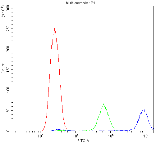
 FCM (Flow Cytometry)
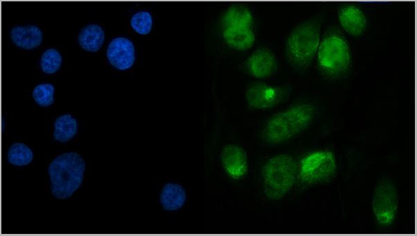
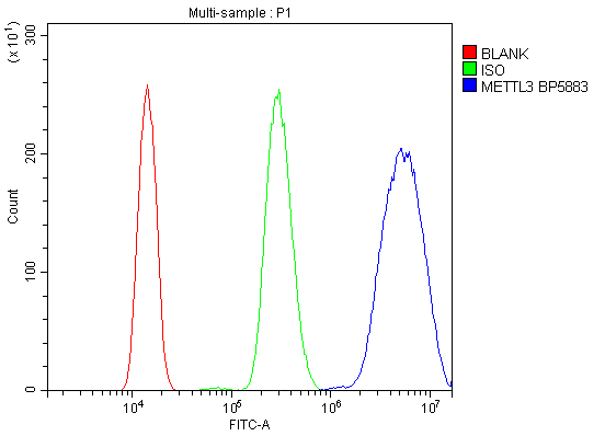
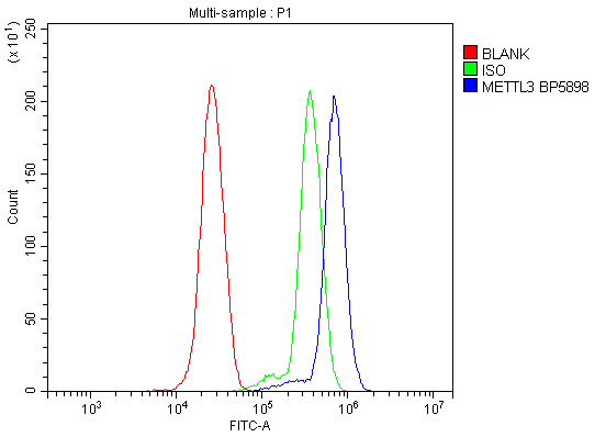
(Figure 6. Flow Cytometry analysis of HL-60 cells using anti-METTL3 antibody (AAA19247).Overlay histogram showing HL-60 cells stained with AAA19247 (Blue line). The cells were blocked with 10% normal goat serum. And then incubated with rabbit anti-METTL3 Antibody (AAA19247, 1μg/1x106 cells) for 30 min at 20 degree C. DyLight®488 conjugated goat anti-rabbit IgG (5-10μg/1x106 cells) was used as secondary antibody for 30 minutes at 20 degree C. Isotype control antibody (Green line) was rabbit IgG (1μg/1x106) used under the same conditions. Unlabelled sample (Red line) was also used as a control.)
FCM (Flow Cytometry)
(Figure 6. Flow Cytometry analysis of HL-60 cells using anti-METTL3 antibody (AAA19247).Overlay histogram showing HL-60 cells stained with AAA19247 (Blue line). The cells were blocked with 10% normal goat serum. And then incubated with rabbit anti-METTL3 Antibody (AAA19247, 1μg/1x106 cells) for 30 min at 20 degree C. DyLight®488 conjugated goat anti-rabbit IgG (5-10μg/1x106 cells) was used as secondary antibody for 30 minutes at 20 degree C. Isotype control antibody (Green line) was rabbit IgG (1μg/1x106) used under the same conditions. Unlabelled sample (Red line) was also used as a control.)
METTL3, Polyclonal Antibody (Cat# AAA19247)
Full Name
Anti-METTL3 Antibody
Gene Names
METTL3; M6A; IME4; Spo8; MT-A70
Reactivity
Human, Mouse, Rat
Applications
Western Blot, Immunohistochemistry, Immunocytochemistry, Immunofluorescence, Flow Cytometry, Direct ELISA
Purity
Immunogen affinity purified.
Pricing
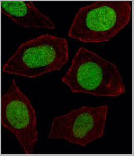
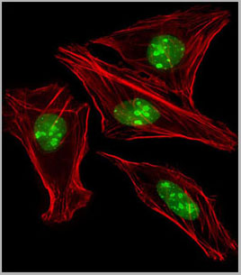
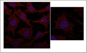
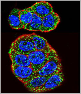
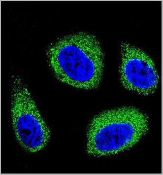
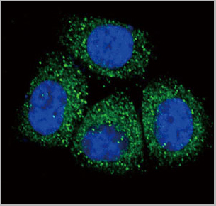
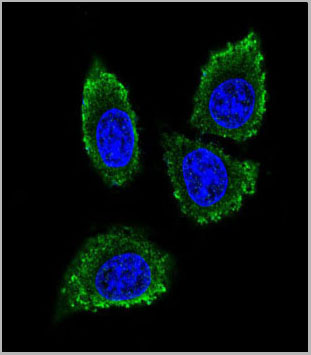 IF (Immunofluorescence)
(Confocal immunofluorescent analysis of MMP12 Antibody (C-term) with 293 cell followed by Alexa Fluor® 488-conjugated goat anti-rabbit lgG (green). DAPI was used to stain the cell nuclear (blue).)
IF (Immunofluorescence)
(Confocal immunofluorescent analysis of MMP12 Antibody (C-term) with 293 cell followed by Alexa Fluor® 488-conjugated goat anti-rabbit lgG (green). DAPI was used to stain the cell nuclear (blue).)
MMP12, Polyclonal Antibody (Cat# AAA28713)
Full Name
MMP12 Antibody (C-term)
Gene Names
MMP12; ME; HME; MME; MMP-12
Reactivity
Human, mouse
Applications
Western Blot, Immunohistochemistry, Immunofluorescence
Purity
Purified Rabbit Polyclonal Antibody (Pab)
Pricing
 FCM (Flow Cytometry)
(Figure 7. Flow Cytometry analysis of Hela cells using anti-Calbindin antibody (AAA11603).Overlay histogram showing Hela cells stained with AAA11603 (Blue line).The cells were blocked with 10% normal goat serum. And then incubated with rabbit anti-Calbindin Antibody (AAA11603,1ug/1x10^6 cells) for 30 min at 20 degree C. DyLight®488 conjugated goat anti-rabbit IgG (5-10ug/1x10^6 cells) was used as secondary antibody for 30 minutes at 20 degree C. Isotype control antibody (Green line) was rabbit IgG (1ug/1x106) used under the same conditions. Unlabelled sample (Red line) was also used as a control.)
FCM (Flow Cytometry)
(Figure 7. Flow Cytometry analysis of Hela cells using anti-Calbindin antibody (AAA11603).Overlay histogram showing Hela cells stained with AAA11603 (Blue line).The cells were blocked with 10% normal goat serum. And then incubated with rabbit anti-Calbindin Antibody (AAA11603,1ug/1x10^6 cells) for 30 min at 20 degree C. DyLight®488 conjugated goat anti-rabbit IgG (5-10ug/1x10^6 cells) was used as secondary antibody for 30 minutes at 20 degree C. Isotype control antibody (Green line) was rabbit IgG (1ug/1x106) used under the same conditions. Unlabelled sample (Red line) was also used as a control.)
Calbindin, Polyclonal Antibody (Cat# AAA11603)
Full Name
Anti-Calbindin Antibody
Gene Names
CALB1; CALB
Reactivity
Human, Mouse, Rat
Applications
Western Blot, Immunohistochemistry
Purity
Immunogen affinity purified.
Pricing
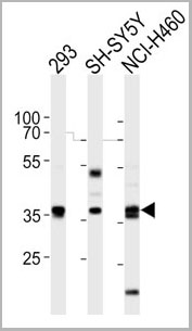
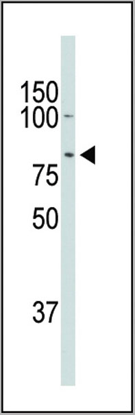
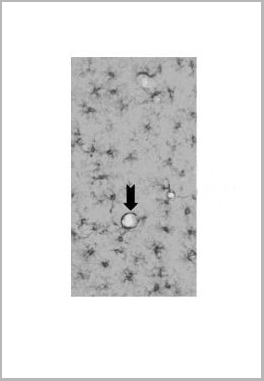
 WB (Western Blot)
(DRD2 Antibody (C-term) western blot analysis in mouse NIH-3T3 cell line lysates (35ug/lane).This demonstrates the DRD2 antibody detected the DRD2 protein (arrow).)
WB (Western Blot)
(DRD2 Antibody (C-term) western blot analysis in mouse NIH-3T3 cell line lysates (35ug/lane).This demonstrates the DRD2 antibody detected the DRD2 protein (arrow).)
DRD2, Polyclonal Antibody (Cat# AAA28691)
Full Name
DRD2 Antibody (C-term)
Gene Names
DRD2; D2R; D2DR
Reactivity
Human, mouse
Applications
Western Blot
Purity
Peptide Affinity Purified Rabbit Polyclonal Antibody (Pab)
Pricing
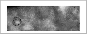
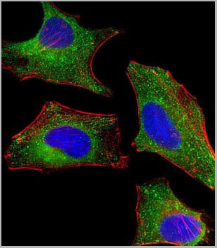
 IF (Immunofluorescence)
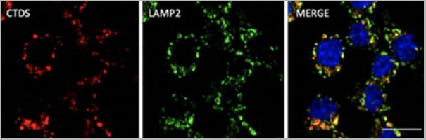
(Immunofluorescence - anti-CTSD Ab at 1/100 dilution in RAW264.7 cells; cells were fixed with PFA and permeabilized with 0.05% saponin)
IF (Immunofluorescence)
(Immunofluorescence - anti-CTSD Ab at 1/100 dilution in RAW264.7 cells; cells were fixed with PFA and permeabilized with 0.05% saponin)
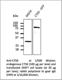
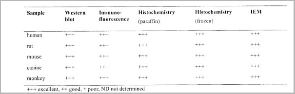
Cathepsin D, Polyclonal Antibody (Cat# AAA13859)
Full Name
Cathepsin D Polyclonal Antibody
Gene Names
CTSD; CPSD; CLN10; HEL-S-130P
Reactivity
Reacts against human, rat, mouse, canine and monkey proteins.
Applications
Western Blot, Immunofluorescence, Immunohistochemistry, Immunohistochemistry
Pricing
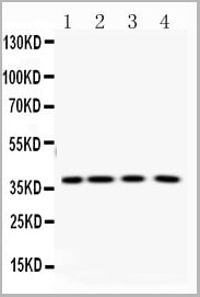
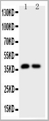
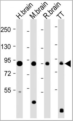 WB (Western Blot)
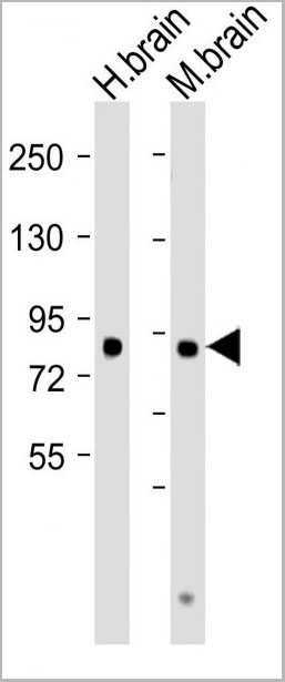
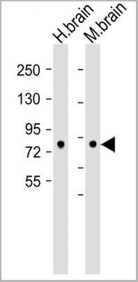
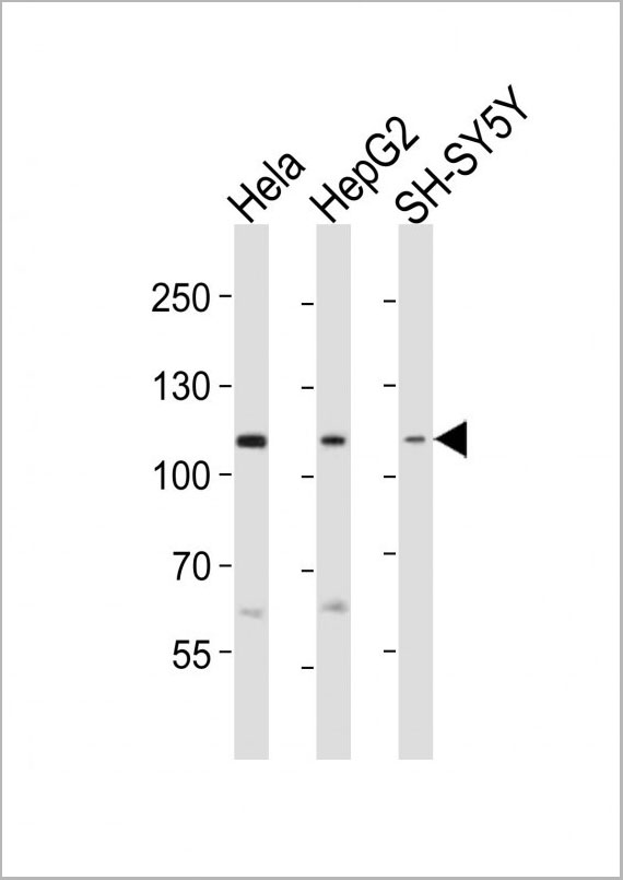
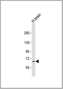
(All lanes : Anti-hDCAMKL1-V705 at 1:8000 dilution Lane 1: Human brain lysate Lane 2: Mouse brain lysate Lane 3: Rat brain lysate Lane 4: TT whole cell lysate Lysates/proteins at 20 ug per lane. Secondary Goat Anti-Rabbit IgG, (H+L), Peroxidase conjugated at 1/10000 dilution. Predicted band size : 82 kDa Blocking/Dilution buffer: 5% NFDM/TBST)
WB (Western Blot)
(All lanes : Anti-hDCAMKL1-V705 at 1:8000 dilution Lane 1: Human brain lysate Lane 2: Mouse brain lysate Lane 3: Rat brain lysate Lane 4: TT whole cell lysate Lysates/proteins at 20 ug per lane. Secondary Goat Anti-Rabbit IgG, (H+L), Peroxidase conjugated at 1/10000 dilution. Predicted band size : 82 kDa Blocking/Dilution buffer: 5% NFDM/TBST)
DCAMKL1, Polyclonal Antibody (Cat# AAA28779)
Full Name
DCAMKL1 Antibody (C-term)
Gene Names
DCLK1; CL1; DCLK; CLICK1; DCDC3A; DCAMKL1
Reactivity
Human, Mouse
Applications
Western Blot
Purity
Purified Rabbit Polyclonal Antibody (Pab)
Pricing
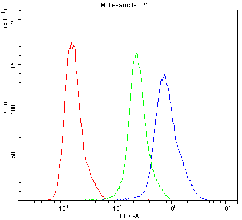
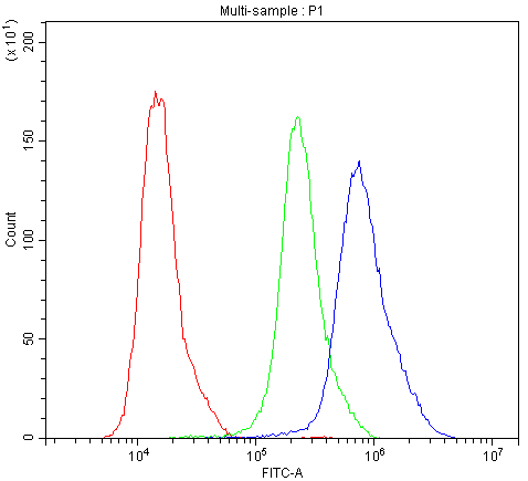
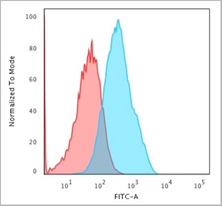
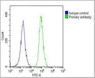
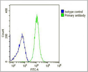
 FCM (Flow Cytometry)
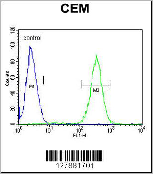
(SLC5A12 Antibody (C-term) flow cytometric analysis of CEM cells (right histogram) compared to a negative control cell (left histogram).FITC-conjugated goat-anti-rabbit secondary antibodies were used for the analysis.)
FCM (Flow Cytometry)
(SLC5A12 Antibody (C-term) flow cytometric analysis of CEM cells (right histogram) compared to a negative control cell (left histogram).FITC-conjugated goat-anti-rabbit secondary antibodies were used for the analysis.)
SLC5A12, Polyclonal Antibody (Cat# AAA28799)
Full Name
SLC5A12 Antibody (C-term)
Gene Names
SLC5A12; SMCT2
Reactivity
Human
Applications
Western Blot, Immunohistochemistry, Flow Cytometry
Pricing
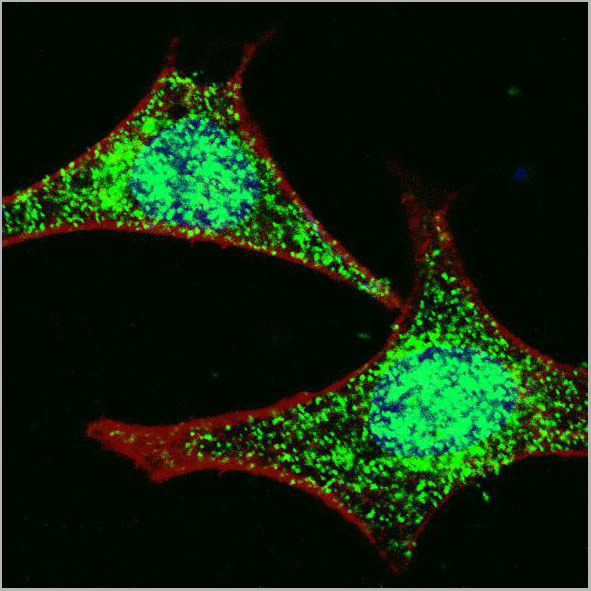
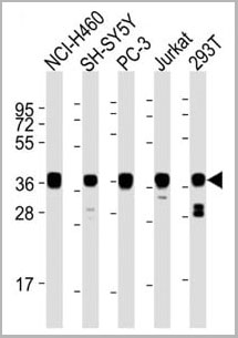
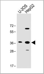
 IF (Immunofluorescence)
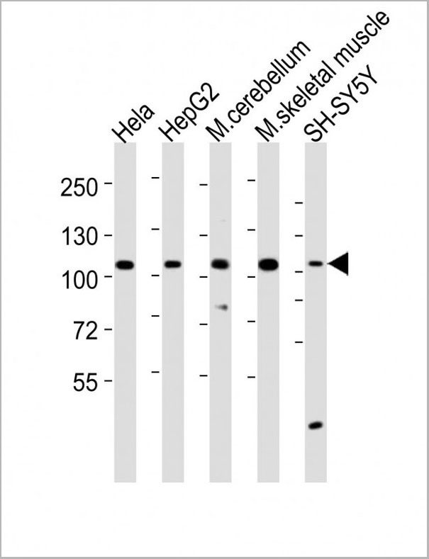
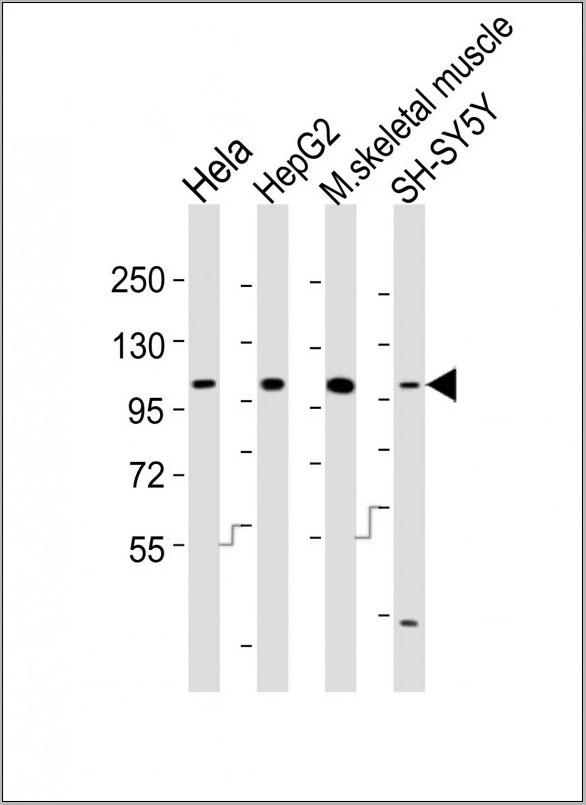
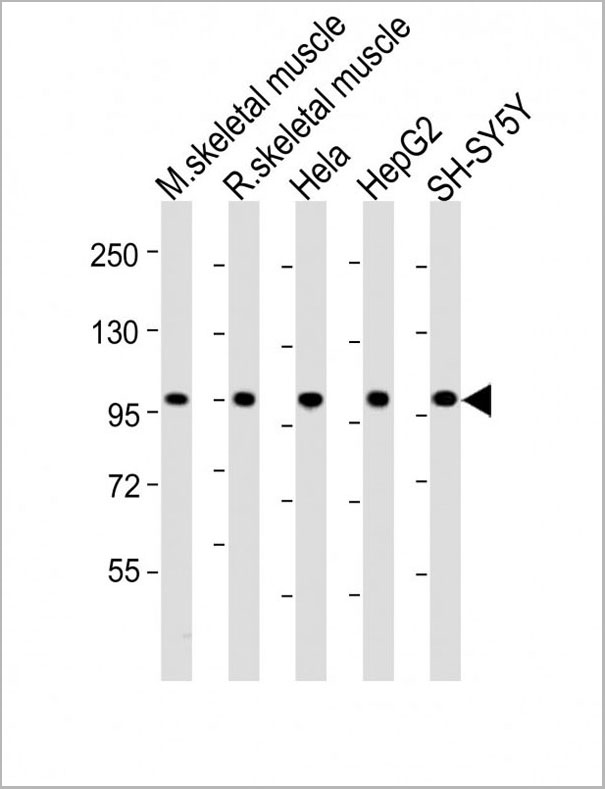
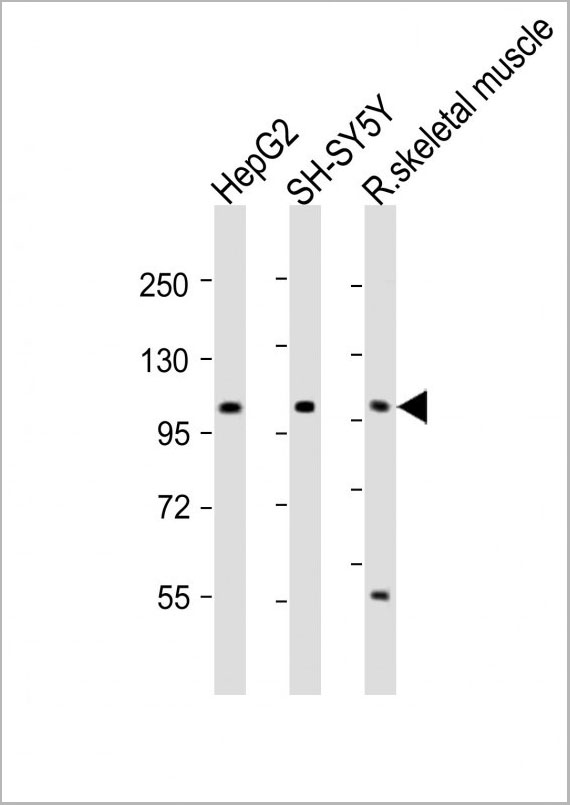
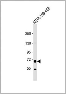
(LGR5/GPR49 Antibody (loop2) western blot analysis in HepG2 cell line lysates (35ug/lane).This demonstrates the LGR5/GPR49 antibody detected the LGR5/GPR49 protein (arrow).)
IF (Immunofluorescence)
(LGR5/GPR49 Antibody (loop2) western blot analysis in HepG2 cell line lysates (35ug/lane).This demonstrates the LGR5/GPR49 antibody detected the LGR5/GPR49 protein (arrow).)
LGR5/GPR49, Polyclonal Antibody (Cat# AAA28712)
Full Name
LGR5/GPR49 Antibody (loop2)
Gene Names
LGR5; FEX; HG38; GPR49; GPR67; GRP49
Reactivity
Human, mouse, rat
Applications
Western Blot, Immunofluorescence, Flow Cytometry
Purity
Purified Rabbit Polyclonal Antibody (Pab)
Pricing
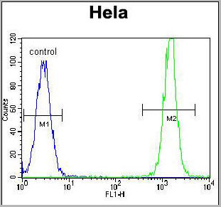 FCM (Flow Cytometry)
(KRAS Antibody (C-term)(AAA28748) flow cytometric analysis of Hela cells (right histogram) compared to a negative control cell (left histogram).FITC-conjugated goat-anti-rabbit secondary antibodies were used for the analysis.)
FCM (Flow Cytometry)
(KRAS Antibody (C-term)(AAA28748) flow cytometric analysis of Hela cells (right histogram) compared to a negative control cell (left histogram).FITC-conjugated goat-anti-rabbit secondary antibodies were used for the analysis.)
KRAS, Polyclonal Antibody (Cat# AAA28748)
Full Name
KRAS Antibody (C-term)
Gene Names
KRAS; NS; NS3; CFC2; KRAS1; KRAS2; RASK2; KI-RAS; C-K-RAS; K-RAS2A; K-RAS2B; K-RAS4A; K-RAS4B
Reactivity
Human, Mouse, Rat (Predicted: Rat)
Applications
Western Blot, Immunofluorescence, Flow Cytometry
Purity
This antibody is purified through a protein A column, followed by peptide affinity purification.
Pricing

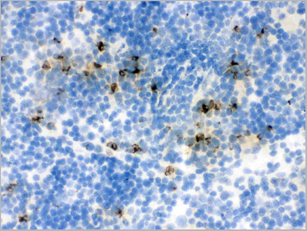
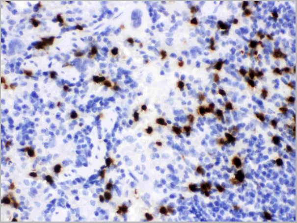
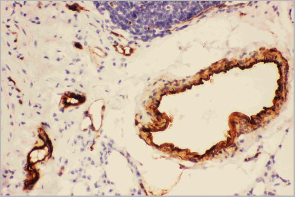
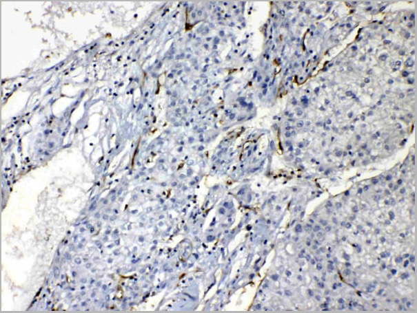
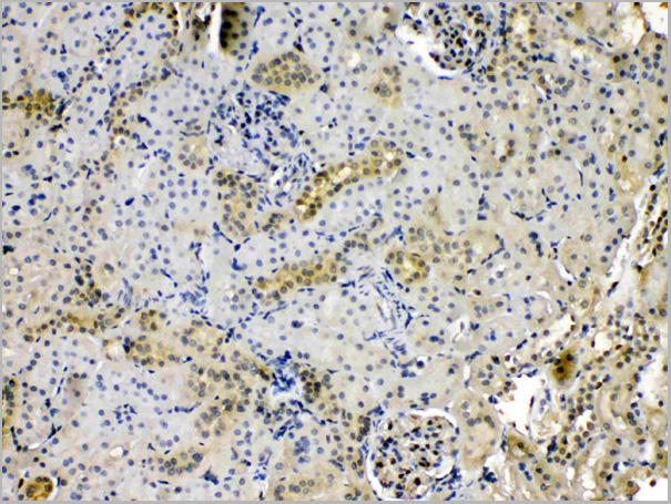
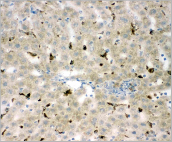
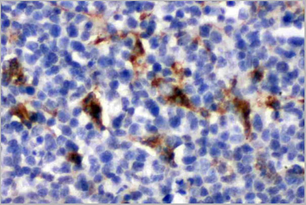
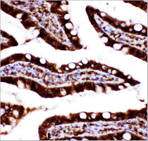
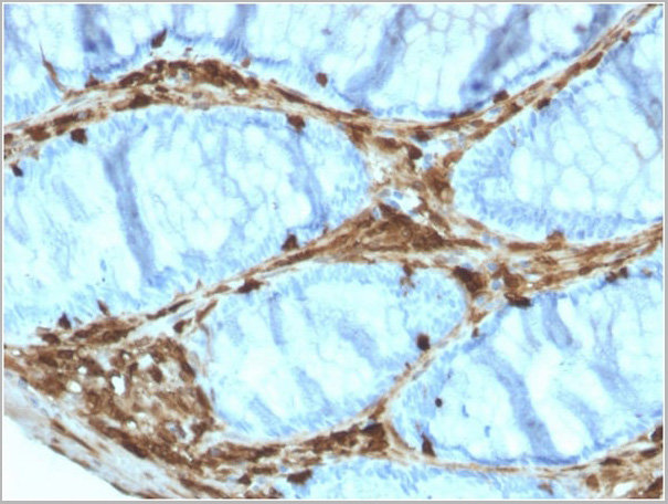
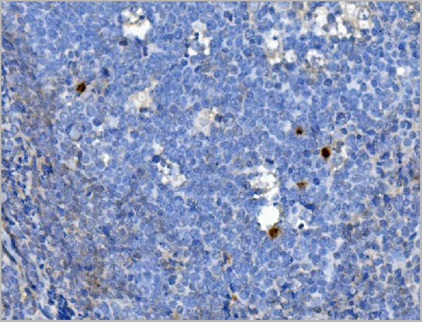
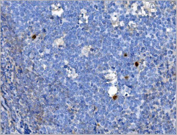
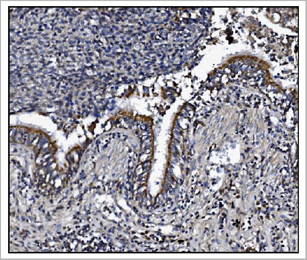
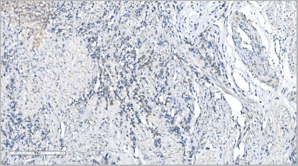
 IHC (Immunohistochemistry)
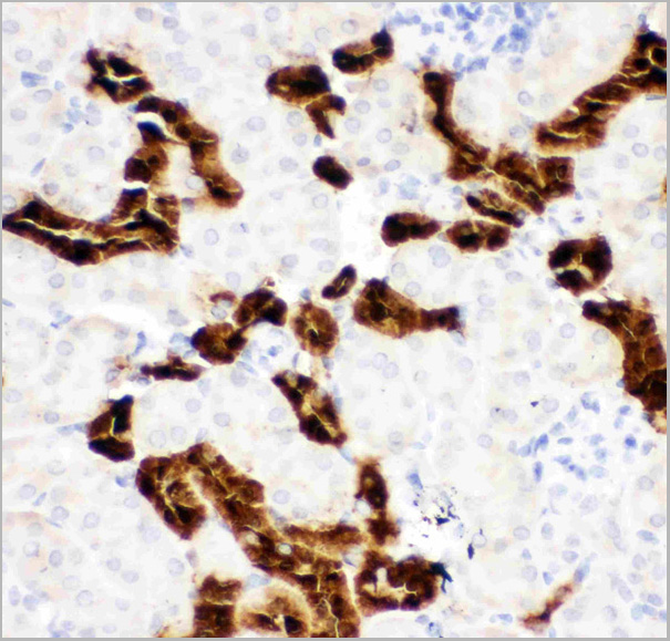
(Figure 7. IHC analysis of Lipocalin 2 using anti-Lipocalin 2 antibody (AAA11670).Lipocalin 2 was detected in frozen section of rat spleen tissue. Heat mediated antigen retrieval was performed in citrate buffer (pH6, epitope retrieval solution) for 20 mins. The tissue section was blocked with 10% goat serum. The tissue section was then incubated with 1ug/ml rabbit anti-Lipocalin 2 Antibody (AAA11670) overnight at 4 degree C. Biotinylated goat anti-rabbit IgG was used as secondary antibody and incubated for 30 minutes at 37 degree C. The tissue section was developed using Strepavidin-Biotin-Complex (SABC) with DAB as the chromogen.)
IHC (Immunohistochemistry)
(Figure 7. IHC analysis of Lipocalin 2 using anti-Lipocalin 2 antibody (AAA11670).Lipocalin 2 was detected in frozen section of rat spleen tissue. Heat mediated antigen retrieval was performed in citrate buffer (pH6, epitope retrieval solution) for 20 mins. The tissue section was blocked with 10% goat serum. The tissue section was then incubated with 1ug/ml rabbit anti-Lipocalin 2 Antibody (AAA11670) overnight at 4 degree C. Biotinylated goat anti-rabbit IgG was used as secondary antibody and incubated for 30 minutes at 37 degree C. The tissue section was developed using Strepavidin-Biotin-Complex (SABC) with DAB as the chromogen.)
Lipocalin 2, Polyclonal Antibody (Cat# AAA11670)
Full Name
Anti-Lipocalin 2 Antibody
Gene Names
Lcn2; 24p3; Sip24; AW212229
Reactivity
Mouse, Rat
Applications
Western Blot, Immunohistochemistry
Purity
Immunogen Affinity Purified
Pricing
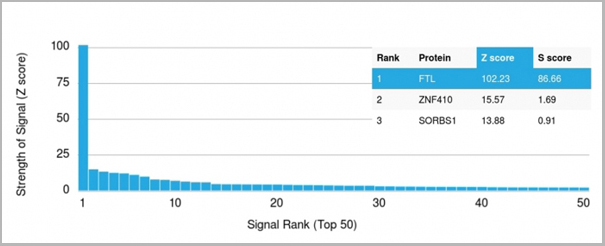
 Application Data
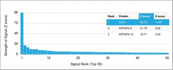
(Analysis of Protein Array containing more than 19,000 full-length human proteins using Estrogen Receptor alpha Mouse Monoclonal Antibody (ESR1/1904) Z- and S- Score: The Z-score represents the strength of a signal that a monoclonal antibody (MAb) (in combination with a fluorescently-tagged anti-IgG secondary antibody) produces when binding to a particular protein on the HuProtTM array. Z-scores are described in units of standard deviations (SD’s) above the mean value of all signals generated on that array. If targets on HuProtTM are arranged in descending order of the Z-score, the S-score is the difference (also in units of SD’s) between the Z-score. S-score therefore represents the relative target specificity of a MAb to its intended target. A MAb is considered to specific to its intended target, if the MAb has an S-score of at least 2.5. For example, if a MAb binds to protein X with a Z-score of 43 and to protein Y with a Z-score of 14, then the S-score for the binding of that MAb to protein X is equal to 29.)
Application Data
(Analysis of Protein Array containing more than 19,000 full-length human proteins using Estrogen Receptor alpha Mouse Monoclonal Antibody (ESR1/1904) Z- and S- Score: The Z-score represents the strength of a signal that a monoclonal antibody (MAb) (in combination with a fluorescently-tagged anti-IgG secondary antibody) produces when binding to a particular protein on the HuProtTM array. Z-scores are described in units of standard deviations (SD’s) above the mean value of all signals generated on that array. If targets on HuProtTM are arranged in descending order of the Z-score, the S-score is the difference (also in units of SD’s) between the Z-score. S-score therefore represents the relative target specificity of a MAb to its intended target. A MAb is considered to specific to its intended target, if the MAb has an S-score of at least 2.5. For example, if a MAb binds to protein X with a Z-score of 43 and to protein Y with a Z-score of 14, then the S-score for the binding of that MAb to protein X is equal to 29.)
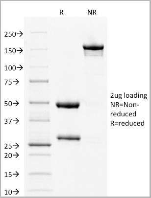
Estrogen Receptor, alpha, Monoclonal Antibody (Cat# AAA23914)
Full Name
Estrogen Receptor, alpha (Marker of Estrogen Dependence)
Gene Names
ESR1; ER; ESR; Era; ESRA; ESTRR; NR3A1
Reactivity
Human
Applications
Western Blot, Immunohistochemistry
Purity
Purified Ab with BSA and Azide at 200ug/ml OR Purified Ab WITHOUT BSA and Azide at 1.0mg/ml
Pricing
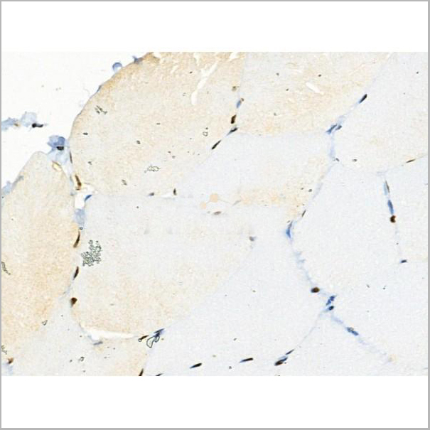
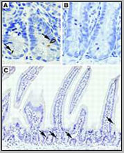
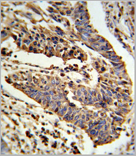
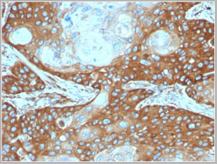
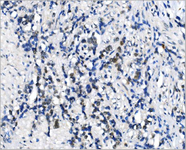
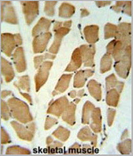 IHC (Immunohistchemistry)
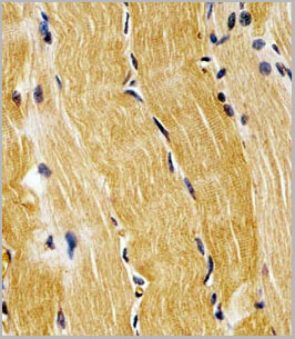
(GLG1 Antibody (C-term) (AAA28703) IHCanalysis in formalin fixed and paraffinembedded skeletal muscle followed byperoxidase conjugation of the secondaryantibody and DAB staining. This data demonstrates the use of the GLG1 Antibody (C-term) for immunohistochemistry. Clinicalrelevance has not been evaluated)
IHC (Immunohistchemistry)
(GLG1 Antibody (C-term) (AAA28703) IHCanalysis in formalin fixed and paraffinembedded skeletal muscle followed byperoxidase conjugation of the secondaryantibody and DAB staining. This data demonstrates the use of the GLG1 Antibody (C-term) for immunohistochemistry. Clinicalrelevance has not been evaluated)
GLG1, Polyclonal Antibody (Cat# AAA28703)
Full Name
GLG1 Antibody (C-term)
Gene Names
GLG1; CFR-1; ESL-1; MG160; MG-160
Reactivity
Human, mouse (Predicted Reactivity: Chicken, Hamster, Rat)
Applications
Western Blot, Immunohistochemistry
Purity
Peptide Affinity Purified Rabbit Polyclonal Antibody (Pab)
Pricing
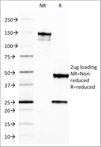
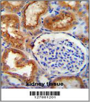
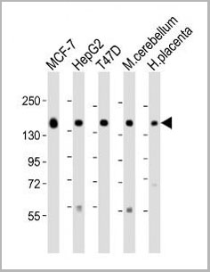
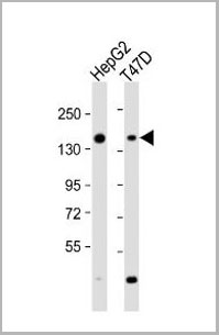
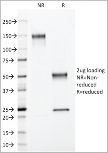
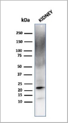 WB (Western Blot)
(Western Blot Analysis of human kidney cell lysate using Ferritin, Light ChainMouse Monoclonal Antibody (FTL/1387).)
WB (Western Blot)
(Western Blot Analysis of human kidney cell lysate using Ferritin, Light ChainMouse Monoclonal Antibody (FTL/1387).)
Ferritin, Light Chain (FTL), Monoclonal Antibody (Cat# AAA23907)
Full Name
Ferritin, Light Chain (FTL) (Microglia Marker)
Gene Names
FTL; LFTD; NBIA3
Reactivity
Human
Applications
Western Blot, Immunohistochemistry
Pricing
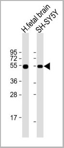
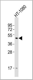
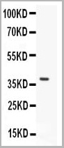
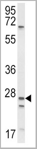
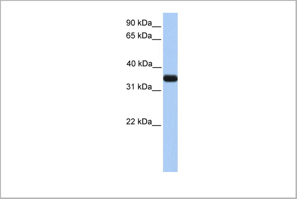
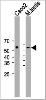 WB (Western Blot)
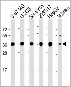
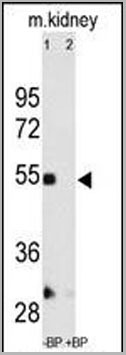
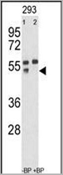
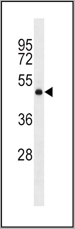
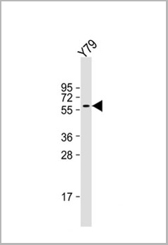
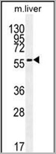
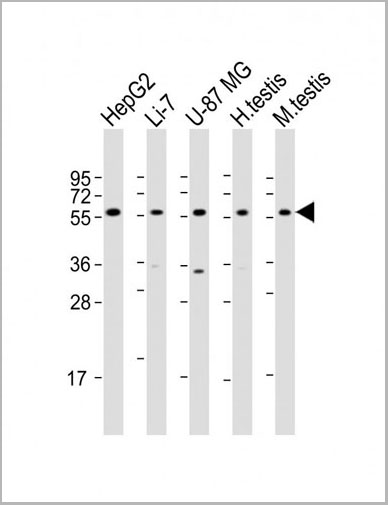
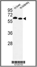
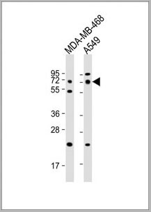
(All lanes : Anti-DHCR7 Antibody (C-term) at 1:2000 dilution Lane 1: Caco2 whole cell lysateLane 2: mouse testis lysate Lysates/proteins at 20 ug per lane. Secondary Goat Anti-Rabbit IgG, (H+L), Peroxidase conjugated (please inquire) at 1/15000 dilution.Observed band size : 55kDa Blocking/Dilution buffer: 5% NFDM/TBST.)
WB (Western Blot)
(All lanes : Anti-DHCR7 Antibody (C-term) at 1:2000 dilution Lane 1: Caco2 whole cell lysateLane 2: mouse testis lysate Lysates/proteins at 20 ug per lane. Secondary Goat Anti-Rabbit IgG, (H+L), Peroxidase conjugated (please inquire) at 1/15000 dilution.Observed band size : 55kDa Blocking/Dilution buffer: 5% NFDM/TBST.)
DHCR7, Polyclonal Antibody (Cat# AAA28753)
Full Name
DHCR7 Antibody (C-term)
Gene Names
DHCR7; SLOS
Reactivity
Human, Mouse
Applications
Western Blot, Immunohistochemistry, Immunofluorescence
Purity
This antibody is purified through a protein A column, followed by peptide affinity purification.
Pricing

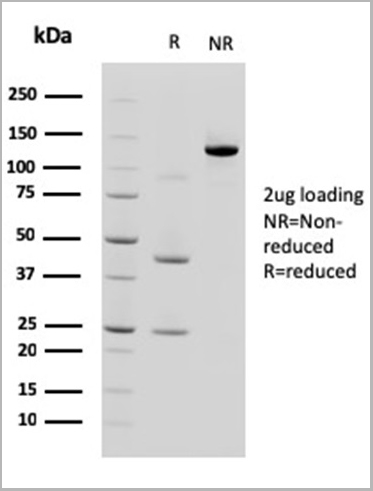
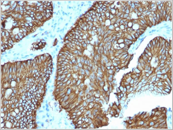
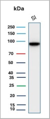 WB (Western Blot)
(Western Blot Analysis of human Small Intestine tissue lysate using CDH17 Mouse Monoclonal Antibody (CDH17/2618).)
WB (Western Blot)
(Western Blot Analysis of human Small Intestine tissue lysate using CDH17 Mouse Monoclonal Antibody (CDH17/2618).)
Cadherin 17/LI Cadherin, Monoclonal Antibody (Cat# AAA23911)
Full Name
Cadherin 17/LI Cadherin (Liver-Intestine Marker)
Gene Names
CDH17; HPT1; CDH16; HPT-1
Reactivity
Human
Applications
Flow Cytometry, Immunohistochemistry, Western Blot
Pricing
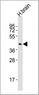
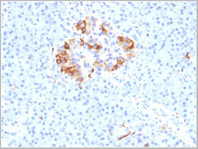
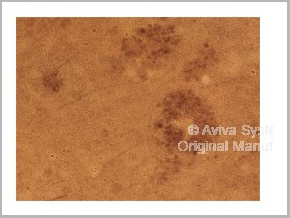
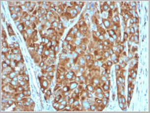
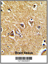 IHC (Immunohistochemistry)
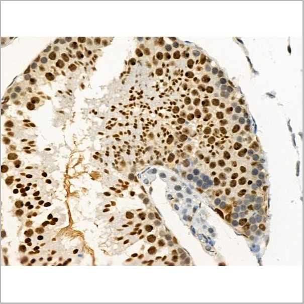
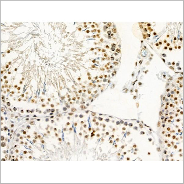
(Formalin-fixed and paraffin-embedded human brain tissue reacted with mouse Prodh Antibody (Center), which was peroxidase-conjugated to the secondary antibody, followed by DAB staining. This data demonstrates the use of this antibody for immunohistochemistry; clinical relevance has not been evaluated.)
IHC (Immunohistochemistry)
(Formalin-fixed and paraffin-embedded human brain tissue reacted with mouse Prodh Antibody (Center), which was peroxidase-conjugated to the secondary antibody, followed by DAB staining. This data demonstrates the use of this antibody for immunohistochemistry; clinical relevance has not been evaluated.)
PRODH, Polyclonal Antibody (Cat# AAA28717)
Full Name
PRODH Antibody (Center)
Gene Names
Prodh; Pro1; Pro-1; Ym24d07
Reactivity
Human, mouse
Applications
Flow Cytometry, Immunohistochemistry, Western Blot
Purity
Peptide Affinity Purified Rabbit Polyclonal Antibody (Pab)
Pricing
 FCM (Flow Cytometry)
(Figure 7. Flow Cytometry analysis of K562 cells using anti-CASP3 antibody (AAA11633).Overlay histogram showing K562 cells stained with AAA11633 (Blue line).The cells were blocked with 10% normal goat serum. And then incubated with rabbit anti-CASP3 Antibody (AAA11633,1ug/1x10^6 cells) for 30 min at 20 degree C. DyLight ®488 conjugated goat anti-rabbit IgG (5-10ug/1x10^6 cells) was used as secondary antibody for 30 minutes at 20 degree C. Isotype control antibody (Green line) was rabbit IgG (1ug/1x106) used under the same conditions. Unlabelled sample (Red line) was also used as a control.)
FCM (Flow Cytometry)
(Figure 7. Flow Cytometry analysis of K562 cells using anti-CASP3 antibody (AAA11633).Overlay histogram showing K562 cells stained with AAA11633 (Blue line).The cells were blocked with 10% normal goat serum. And then incubated with rabbit anti-CASP3 Antibody (AAA11633,1ug/1x10^6 cells) for 30 min at 20 degree C. DyLight ®488 conjugated goat anti-rabbit IgG (5-10ug/1x10^6 cells) was used as secondary antibody for 30 minutes at 20 degree C. Isotype control antibody (Green line) was rabbit IgG (1ug/1x106) used under the same conditions. Unlabelled sample (Red line) was also used as a control.)
Caspase-3, Polyclonal Antibody (Cat# AAA11633)
Full Name
Anti-Caspase-3 Antibody
Gene Names
CASP3; CPP32; SCA-1; CPP32B
Reactivity
Human, Mouse, Rat. No cross reactivity with other proteins.
Applications
Western Blot, Immunohistochemistry
Purity
Immunogen affinity purified.
Pricing
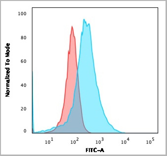
 FCM (Flow Cytometry)
(SCNN1A Antibody (Center) flow cytometric analysis of WiDr cells (right histogram) compared to a negative control cell (left histogram).FITC-conjugated goat-anti-rabbit secondary antibodies were used for the analysis.)
FCM (Flow Cytometry)
(SCNN1A Antibody (Center) flow cytometric analysis of WiDr cells (right histogram) compared to a negative control cell (left histogram).FITC-conjugated goat-anti-rabbit secondary antibodies were used for the analysis.)
SCNN1A, Polyclonal Antibody (Cat# AAA28784)
Full Name
SCNN1A Antibody (Center)
Gene Names
SCNN1A; BESC2; ENaCa; SCNEA; SCNN1; ENaCalpha
Reactivity
Human
Applications
Western Blot, Immunohistochemistry, Immunofluorescence, Flow Cytometry
Purity
Peptide Affinity Purified Rabbit Polyclonal Antibody (Pab)
Pricing

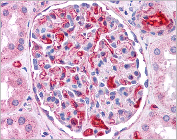
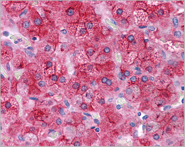
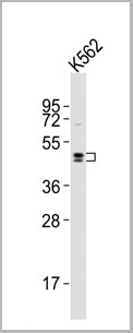
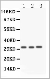
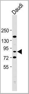
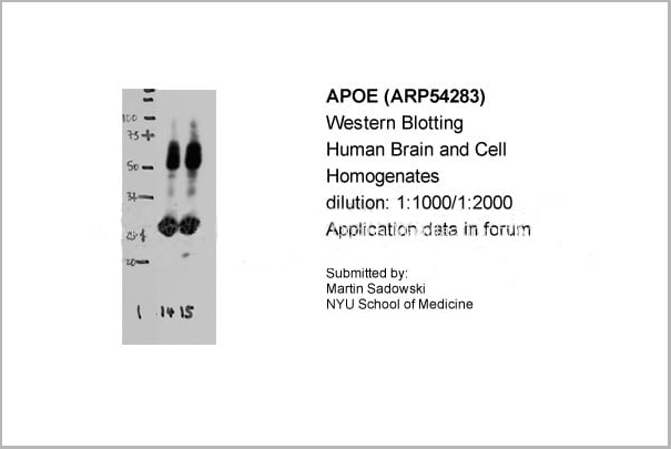 WB (Western Blot)
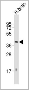
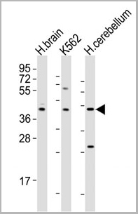
(Human Brain and cell HomogenatesDilution : 1:1000/ 1:2000)
WB (Western Blot)
(Human Brain and cell HomogenatesDilution : 1:1000/ 1:2000)
APOE, Polyclonal Antibody (Cat# AAA23552)
Full Name
APOE antibody - N-terminal region
Gene Names
APOE; AD2; LPG; APO-E; ApoE4; LDLCQ5
Reactivity
Human
Applications
Western Blot, Immunohistochemistry
Purity
Affinity Purified
Pricing
 Application Data
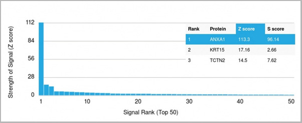
(Analysis of Protein Array containing more than 19,000 full-length human proteins using Annexin A1 Mouse Monoclonal Antibody (ANXA1/3566). Z- and S- Score: The Z-score represents the strength of a signal that a monoclonal antibody (MAb) (in combination with a fluorescently-tagged anti-IgG secondary antibody) produces when binding to a particular protein on the HuProtTM array. Z-scores are described in units of standard deviations (SD’s) above the mean value of all signals generated on that array. If targets on HuProtTM are arranged in descending order of the Z-score, the S-score is the difference (also in units of SD’s) between the Z-score. S-score therefore represents the relative target specificity of a MAb to its intended target. A MAb is considered to specific to its intended target, if the MAb has an S-score of at least 2.5. For example, if a MAb binds to protein X with a Z-score of 43 and to protein Y with a Z-score of 14, then the S-score for the binding of that MAb to protein X is equal to 29.)
Application Data
(Analysis of Protein Array containing more than 19,000 full-length human proteins using Annexin A1 Mouse Monoclonal Antibody (ANXA1/3566). Z- and S- Score: The Z-score represents the strength of a signal that a monoclonal antibody (MAb) (in combination with a fluorescently-tagged anti-IgG secondary antibody) produces when binding to a particular protein on the HuProtTM array. Z-scores are described in units of standard deviations (SD’s) above the mean value of all signals generated on that array. If targets on HuProtTM are arranged in descending order of the Z-score, the S-score is the difference (also in units of SD’s) between the Z-score. S-score therefore represents the relative target specificity of a MAb to its intended target. A MAb is considered to specific to its intended target, if the MAb has an S-score of at least 2.5. For example, if a MAb binds to protein X with a Z-score of 43 and to protein Y with a Z-score of 14, then the S-score for the binding of that MAb to protein X is equal to 29.)
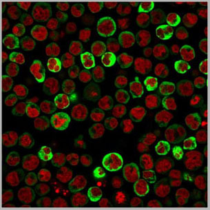
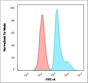
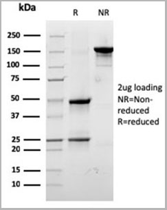
Annexin A1, Monoclonal Antibody (Cat# AAA23918)
Full Name
Annexin A1/(Hairy Cell Leukemia Marker)
Gene Names
ANXA1; ANX1; LPC1
Reactivity
Human
Applications
Flow Cytometry, Immunofluorescence, Immunohistochemistry
Purity
Purified Ab with BSA and Azide at 200ug/ml OR Purified Ab WITHOUT BSA and Azide at 1.0mg/ml
Pricing
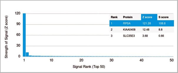 Application Data
(Analysis of Protein Array containing more than 19,000 full-length human proteins using Laminin Receptor Monospecific Mouse Monoclonal Antibody (RPSA/2699) Z- and S- Score: The Z-score represents the strength of a signal that a monoclonal antibody (MAb) (in combination with a fluorescently-tagged anti-IgG secondary antibody) produces when binding to a particular protein on the HuProtTM array. Z-scores are described in units of standard deviations (SD's) above the mean value of all signals generated on that array. If targets on HuProtTM are arranged in descending order of the Z-score, the S-score is the difference (also in units of SD's) between the Z-score. S-score therefore represents the relative target specificity of a MAb to its intended target. A MAb is considered to specific to its intended target, if the MAb has an S-score of at least 2.5. For example, if a MAb binds to protein X with a Z-score of 43 and to protein Y with a Z-score of 14, then the S-score for the binding of that MAb to protein X is equal to 29.)
Application Data
(Analysis of Protein Array containing more than 19,000 full-length human proteins using Laminin Receptor Monospecific Mouse Monoclonal Antibody (RPSA/2699) Z- and S- Score: The Z-score represents the strength of a signal that a monoclonal antibody (MAb) (in combination with a fluorescently-tagged anti-IgG secondary antibody) produces when binding to a particular protein on the HuProtTM array. Z-scores are described in units of standard deviations (SD's) above the mean value of all signals generated on that array. If targets on HuProtTM are arranged in descending order of the Z-score, the S-score is the difference (also in units of SD's) between the Z-score. S-score therefore represents the relative target specificity of a MAb to its intended target. A MAb is considered to specific to its intended target, if the MAb has an S-score of at least 2.5. For example, if a MAb binds to protein X with a Z-score of 43 and to protein Y with a Z-score of 14, then the S-score for the binding of that MAb to protein X is equal to 29.)
Laminin Receptor/RPSA, Monoclonal Antibody (Cat# AAA23908)
Full Name
Laminin Receptor/RPSA (Marker of Metastatic Potential)
Gene Names
RPSA; SA; LBP; LRP; p40; 67LR; ICAS; lamR; 37LRP; LAMBR; LAMR1; LRP/LR; LBP/p40; NEM/1CHD4
Reactivity
Human. Others not known.
Applications
Flow Cytometry, Immunofluorescence, Western Blot, Immunohistochemistry
Pricing
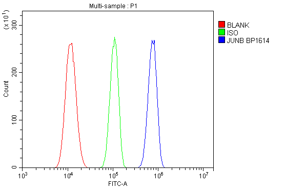 FCM (Flow Cytometry)
(Figure 7. Flow Cytometry analysis of U87 cells using anti-JUNB antibody (AAA19249)Overlay histogram showing U87 cells stained with AAA19249 (Blue line). The cells were blocked with 10% normal goat serum. And then incubated with rabbit anti-JUNB Antibody (AAA19249, 1μg/1x106 cells) for 30 min at 20 degree C. DyLight®488 conjugated goat anti-rabbit IgG (5-10μg/1x106 cells) was used as secondary antibody for 30 minutes at 20 degree C. Isotype control antibody (Green line) was rabbit IgG (1μg/1x106) used under the same conditions. Unlabelled sample (Red line) was also used as a control.)
FCM (Flow Cytometry)
(Figure 7. Flow Cytometry analysis of U87 cells using anti-JUNB antibody (AAA19249)Overlay histogram showing U87 cells stained with AAA19249 (Blue line). The cells were blocked with 10% normal goat serum. And then incubated with rabbit anti-JUNB Antibody (AAA19249, 1μg/1x106 cells) for 30 min at 20 degree C. DyLight®488 conjugated goat anti-rabbit IgG (5-10μg/1x106 cells) was used as secondary antibody for 30 minutes at 20 degree C. Isotype control antibody (Green line) was rabbit IgG (1μg/1x106) used under the same conditions. Unlabelled sample (Red line) was also used as a control.)
JUNB, Polyclonal Antibody (Cat# AAA19249)
Full Name
Anti-JUNB Antibody
Gene Names
JUNB; AP-1
Reactivity
Human
Applications
Western Blot, Immunohistochemistry, Flow Cytometry, Direct ELISA
Purity
Immunogen affinity purified.
Pricing
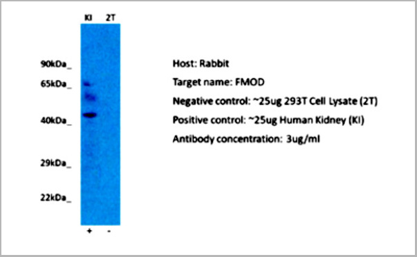
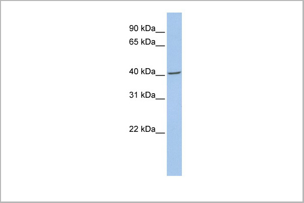
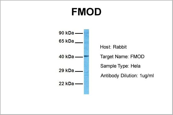
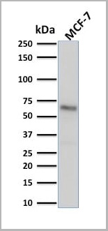
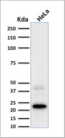
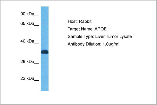
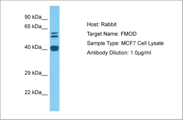 WB (Western Blot)
( Host: RabbitTarget Name: FMODSample Tissue: Human MCF7 Whole CellAntibody Dilution: 3ug/ml)
WB (Western Blot)
( Host: RabbitTarget Name: FMODSample Tissue: Human MCF7 Whole CellAntibody Dilution: 3ug/ml)
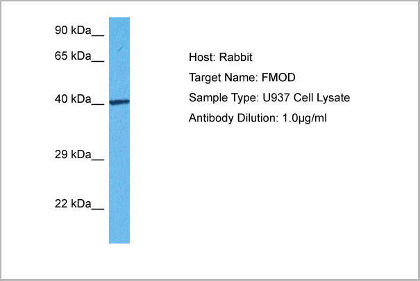
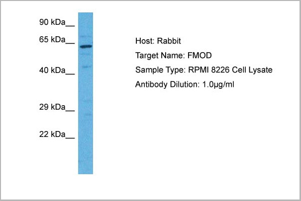
FMOD, Polyclonal Antibody (Cat# AAA23557)
Full Name
FMOD antibody - N-terminal region
Gene Names
FMOD; FM; SLRR2E
Reactivity
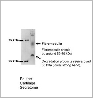
Tested Species Reactivity: Human, Horse
Predicted Species Reactivity: Human, Mouse, Rat, Cow, Dog, Guinea Pig, Horse, Rabbit
Predicted Species Reactivity: Human, Mouse, Rat, Cow, Dog, Guinea Pig, Horse, Rabbit
Applications
Western Blot
Purity
Affinity Purified
Pricing
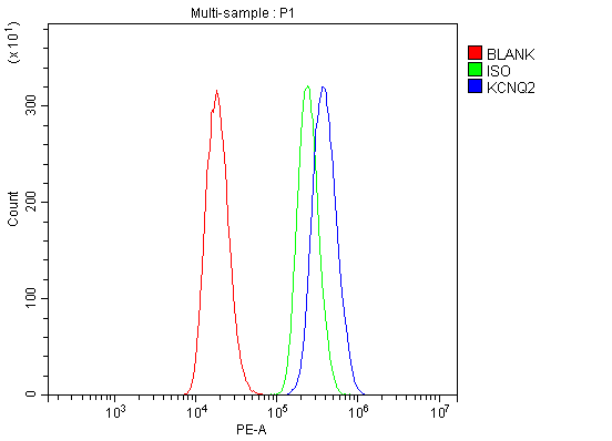
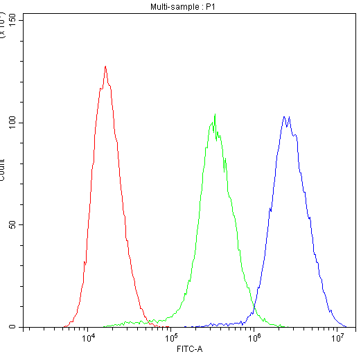
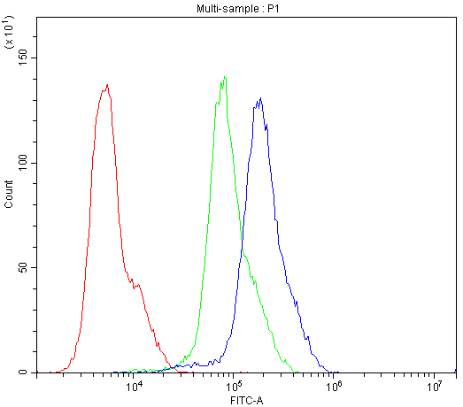
 FCM (Flow Cytometry)
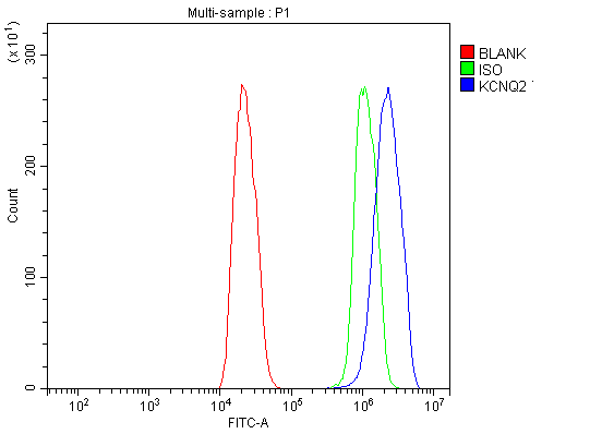
(Figure 7. Flow Cytometry analysis of C6 cells using anti-KCNQ2 antibody. Overlay histogram showing C6 cells stained with (Blue line). The cells were blocked with 10% normal goat serum. And then incubated with rabbit anti-KCNQ2 Antibody (1 ug/1x10^6 cells) for 30 min at 20°C. DyLight®488 conjugated goat anti-rabbit IgG (BA1127, 5-10 ug/1x10^6 cells) was used as secondary antibody for 30 minutes at 20°C. Isotype control antibody (Green line) was rabbit IgG (1 ug/1x10^6) used under the same conditions. Unlabelled sample (Red line) was also used as a control.)
FCM (Flow Cytometry)
(Figure 7. Flow Cytometry analysis of C6 cells using anti-KCNQ2 antibody. Overlay histogram showing C6 cells stained with (Blue line). The cells were blocked with 10% normal goat serum. And then incubated with rabbit anti-KCNQ2 Antibody (1 ug/1x10^6 cells) for 30 min at 20°C. DyLight®488 conjugated goat anti-rabbit IgG (BA1127, 5-10 ug/1x10^6 cells) was used as secondary antibody for 30 minutes at 20°C. Isotype control antibody (Green line) was rabbit IgG (1 ug/1x10^6) used under the same conditions. Unlabelled sample (Red line) was also used as a control.)
KCNQ2, Polyclonal Antibody (Cat# AAA19238)
Full Name
Anti-KCNQ2 Antibody
Gene Names
KCNQ2; EBN; BFNC; EBN1; ENB1; BFNS1; EIEE7; HNSPC; KV7.2; KCNA11; KVEBN1
Reactivity
Human, Mouse, Rat
Applications
Western Blot, Immunohistochemistry, Flow Cytometry
Purity
Immunogen affinity purified.
Pricing
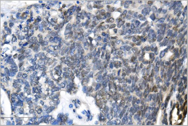
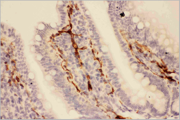
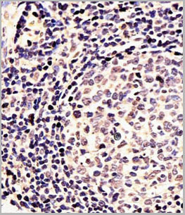
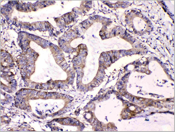
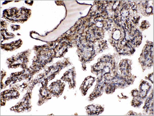
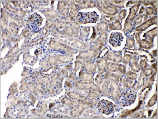
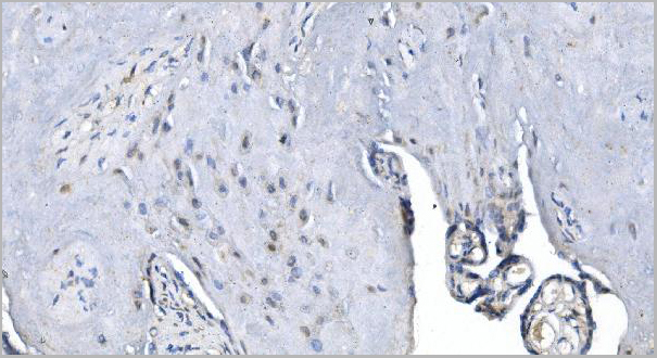
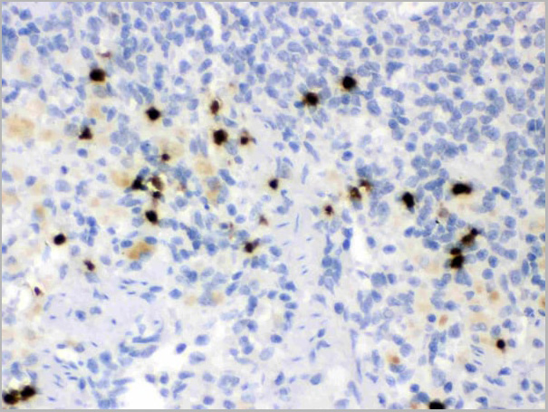
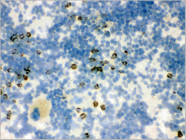
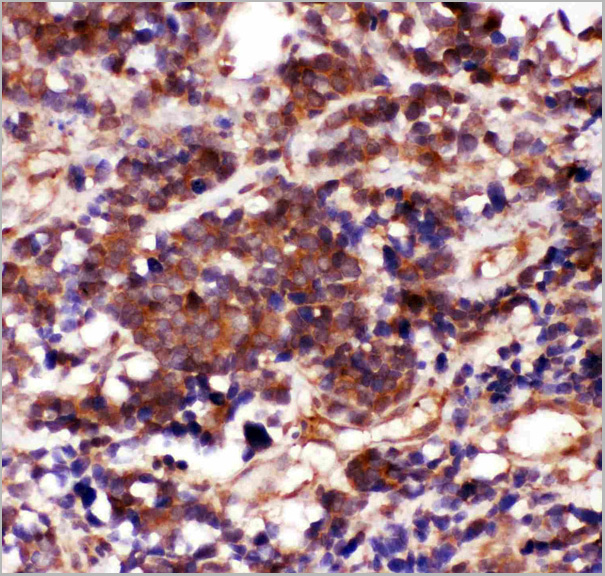
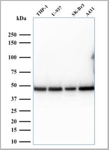
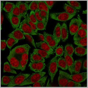
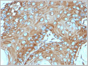
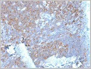
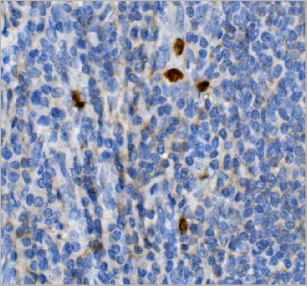
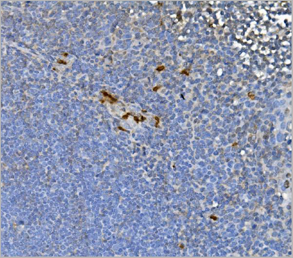
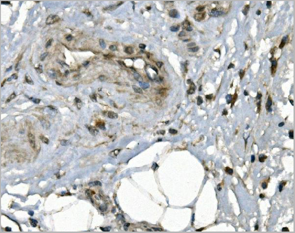
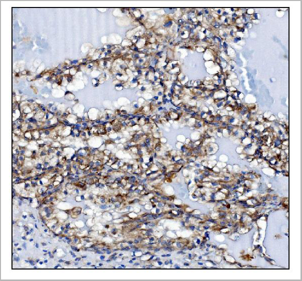
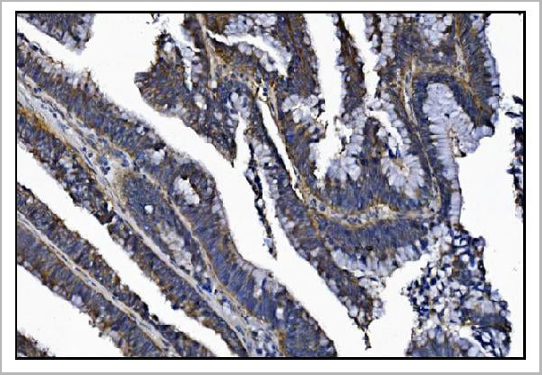
 IHC (Immunohistochemistry)
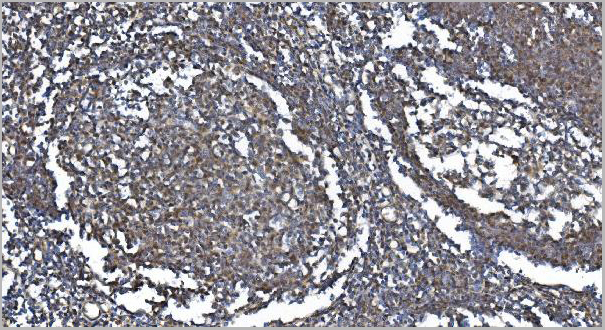
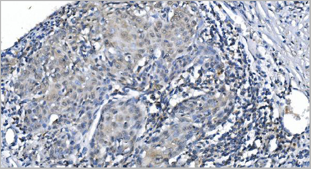
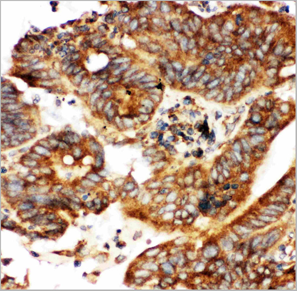
(Figure 7. IHC analysis of METTL3 using anti-METTL3 antibody (AAA19248).METTL3 was detected in paraffin-embedded section of human gastric cancer tissue. Heat mediated antigen retrieval was performed in EDTA buffer (pH8. 0, epitope retrieval solution). The tissue section was blocked with 10% goat serum. The tissue section was then incubated with 2μg/ml rabbit anti-METTL3 Antibody (AAA19248) overnight at 4 degree C. Biotinylated goat anti-rabbit IgG was used as secondary antibody and incubated for 30 minutes at 37 degree C. The tissue section was developed using Strepavidin-Biotin-Complex (SABC) (Catalog # with DAB as the chromogen.)
IHC (Immunohistochemistry)
(Figure 7. IHC analysis of METTL3 using anti-METTL3 antibody (AAA19248).METTL3 was detected in paraffin-embedded section of human gastric cancer tissue. Heat mediated antigen retrieval was performed in EDTA buffer (pH8. 0, epitope retrieval solution). The tissue section was blocked with 10% goat serum. The tissue section was then incubated with 2μg/ml rabbit anti-METTL3 Antibody (AAA19248) overnight at 4 degree C. Biotinylated goat anti-rabbit IgG was used as secondary antibody and incubated for 30 minutes at 37 degree C. The tissue section was developed using Strepavidin-Biotin-Complex (SABC) (Catalog # with DAB as the chromogen.)
METTL3, Polyclonal Antibody (Cat# AAA19248)
Full Name
Anti-METTL3 Antibody
Gene Names
METTL3; M6A; IME4; Spo8; MT-A70
Reactivity
Human
Applications
Western Blot, Immunohistochemistry, Immunocytochemistry, Immunofluorescence, Flow Cytometry, Direct ELISA
Purity
Immunogen affinity purified.
Pricing