Filters
Clonality
Type
Reactivity
Gene Name
Isotype
Host
Application
Clone
967 results for " Synthetic" - showing 600-650


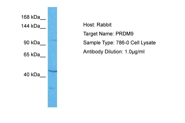


 WB (Western Blot)
(WB Suggested Anti-PRDM9 Antibody Titration: 1 ug/mlPositive Control: 293T cells lysate)
WB (Western Blot)
(WB Suggested Anti-PRDM9 Antibody Titration: 1 ug/mlPositive Control: 293T cells lysate)
PRDM9, Polyclonal Antibody (Cat# AAA23451)
Full Name
PRDM9 antibody - middle region
Gene Names
PRDM9; PFM6; KMT8B; MSBP3; ZNF899; MEISETZ
Reactivity
Human
Applications
IHC, WB
Purity
Affinity Purified
Pricing


 FCM (Flow Cytometry)
(Figure 7. Flow Cytometry analysis of RH35 cells using anti- EIF4A1 antibody (AAA19380).Overlay histogram showing RH35 cells stained with AAA19380 (Blue line). The cells were blocked with 10% normal goat serum. And then incubated with mouse anti-EIF4A1 Antibody (AAA19380, 1μg/1x106 cells) for 30 min at 20 degree C. DyLight®488 conjugated goat anti-mouse IgG (BA1126, 5-10μg/1x106 cells) was used as secondary antibody for 30 minutes at 20 degree C. Isotype control antibody (Green line) was mouse IgG (1μg/1x106) used under the same conditions. Unlabelled sample (Red line) was also used as a control.)
FCM (Flow Cytometry)
(Figure 7. Flow Cytometry analysis of RH35 cells using anti- EIF4A1 antibody (AAA19380).Overlay histogram showing RH35 cells stained with AAA19380 (Blue line). The cells were blocked with 10% normal goat serum. And then incubated with mouse anti-EIF4A1 Antibody (AAA19380, 1μg/1x106 cells) for 30 min at 20 degree C. DyLight®488 conjugated goat anti-mouse IgG (BA1126, 5-10μg/1x106 cells) was used as secondary antibody for 30 minutes at 20 degree C. Isotype control antibody (Green line) was mouse IgG (1μg/1x106) used under the same conditions. Unlabelled sample (Red line) was also used as a control.)
EIF4A1, Monoclonal Antibody (Cat# AAA19380)
Full Name
Anti-EIF4A1 Antibody (monoclonal, 11B8)
Gene Names
EIF4A1; DDX2A; EIF4A; EIF-4A; eIF4A-I; eIF-4A-I
Reactivity
Human, Mouse, Rat
Applications
WB, IHC-P, ICC, IF, FC/FACS/FCM
Purity
Immunogen affinity purified.
Pricing


 FCM (Flow Cytometry)
(Figure 13. Flow Cytometry analysis of MCF-7 cells using anti-EHD3 antibody (AAA19295).Overlay histogram showing MCF-7 cells stained with AAA19295 (Blue line). The cells were blocked with 10% normal goat serum. And then incubated with rabbit anti-EHD3 Antibody (AAA19295, 1μg/1x106 cells) for 30 min at 20 degree C. DyLight®488 conjugated goat anti-rabbit IgG (5-10μg/1x106 cells) was used as secondary antibody for 30 minutes at 20 degree C. Isotype control antibody (Green line) was rabbit IgG (1μg/1x106) used under the same conditions. Unlabelled sample (Red line) was also used as a control.)
FCM (Flow Cytometry)
(Figure 13. Flow Cytometry analysis of MCF-7 cells using anti-EHD3 antibody (AAA19295).Overlay histogram showing MCF-7 cells stained with AAA19295 (Blue line). The cells were blocked with 10% normal goat serum. And then incubated with rabbit anti-EHD3 Antibody (AAA19295, 1μg/1x106 cells) for 30 min at 20 degree C. DyLight®488 conjugated goat anti-rabbit IgG (5-10μg/1x106 cells) was used as secondary antibody for 30 minutes at 20 degree C. Isotype control antibody (Green line) was rabbit IgG (1μg/1x106) used under the same conditions. Unlabelled sample (Red line) was also used as a control.)
EHD3, Polyclonal Antibody (Cat# AAA19295)
Full Name
Anti-EHD3 Antibody
Gene Names
EHD2; PAST2
Reactivity
Human, Monkey, Mouse, Rat
Applications
WB, IHC-P, ICC, IF, FC/FACS/FCM
Purity
Immunogen affinity purified.
Pricing


 WB (Western Blot)
(WB Suggested Anti-ACTR1B Antibody Titration: 0.2-1 ug/mlPositive Control: HepG2 cell lysateACTR1B is supported by BioGPS gene expression data to be expressed in HepG2)
WB (Western Blot)
(WB Suggested Anti-ACTR1B Antibody Titration: 0.2-1 ug/mlPositive Control: HepG2 cell lysateACTR1B is supported by BioGPS gene expression data to be expressed in HepG2)
ACTR1B, Polyclonal Antibody (Cat# AAA23545)
Full Name
ACTR1B antibody - C-terminal region
Gene Names
ACTR1B; PC3; ARP1B; CTRN2
Reactivity
Cow, Dog, Guinea Pig, Horse, Human, Mouse, Rabbit, Rat, Zebrafish
Applications
WB
Purity
Affinity Purified
Pricing


 WB (Western Blot)
(WB Suggested Anti-TGFB1I1 Antibody Titration: 0.2-1 ug/mlPositive Control: Human Lung)
WB (Western Blot)
(WB Suggested Anti-TGFB1I1 Antibody Titration: 0.2-1 ug/mlPositive Control: Human Lung)
TGFB1I1, Polyclonal Antibody (Cat# AAA23425)
Full Name
TGFB1I1 Antibody - middle region
Gene Names
TGFB1I1; HIC5; ARA55; HIC-5; TSC-5
Reactivity
Cow, Dog, Goat, Guinea Pig, Horse, Human, Mouse, Rabbit, Rat
Applications
IHC, WB
Purity
Affinity Purified
Pricing


 WB (Western Blot)
(WB Suggested Anti-GJA4 Antibody Titration: 0.2-1 ug/mlELISA Titer: 1:1562500Positive Control: 721_B cell lysateGJA4 is strongly supported by BioGPS gene expression data to be expressed in Human 721_B cells)
WB (Western Blot)
(WB Suggested Anti-GJA4 Antibody Titration: 0.2-1 ug/mlELISA Titer: 1:1562500Positive Control: 721_B cell lysateGJA4 is strongly supported by BioGPS gene expression data to be expressed in Human 721_B cells)
GJA4, Polyclonal Antibody (Cat# AAA23457)
Full Name
GJA4 antibody - middle region
Gene Names
GJA4; CX37
Reactivity
Cow, Dog, Guinea Pig, Horse, Human, Mouse, Rabbit, Rat, Sheep
Applications
IHC, WB
Purity
Affinity Purified
Pricing


 WB (Western Blot)
(25 ug of the indicated Human whole cell extracts was loaded onto a 10-20% SDS-PAGE gel. 1 ug/mL of the antibody was used in this experiment. Canonical 15 kDa isoform is identified, and a second isoform of 18 kDa is also present in some samples.)
WB (Western Blot)
(25 ug of the indicated Human whole cell extracts was loaded onto a 10-20% SDS-PAGE gel. 1 ug/mL of the antibody was used in this experiment. Canonical 15 kDa isoform is identified, and a second isoform of 18 kDa is also present in some samples.)
PFN1, Polyclonal Antibody (Cat# AAA23532)
Full Name
PFN1 antibody - N-terminal region
Gene Names
PFN1; ALS18
Reactivity
Tested Species Reactivity: Human, Mouse
Predicted Species Reactivity: Cow, Guinea Pig, Horse, Human, Mouse, Rat, Zebrafish
Predicted Species Reactivity: Cow, Guinea Pig, Horse, Human, Mouse, Rat, Zebrafish
Applications
WB
Purity
Affinity Purified
Pricing


 WB (Western Blot)
(WB Suggested Anti-GRSF1 Antibody Titration: 0.2-1 ug/mlPositive Control: Hela cell lysateGRSF1 is strongly supported by BioGPS gene expression data to be expressed in Human HeLa cells)
WB (Western Blot)
(WB Suggested Anti-GRSF1 Antibody Titration: 0.2-1 ug/mlPositive Control: Hela cell lysateGRSF1 is strongly supported by BioGPS gene expression data to be expressed in Human HeLa cells)
GRSF1, Polyclonal Antibody (Cat# AAA23475)
Full Name
GRSF1 antibody - middle region
Reactivity
Cow, Dog, Guinea Pig, Horse, Human, Mouse, Rabbit, Rat
Applications
WB
Purity
Affinity Purified
Pricing


 WB (Western Blot)
(WB Suggested Anti-SLC39A6 Antibody Titration: 1.25ug/mlPositive Control: Jurkat cell lysate)
WB (Western Blot)
(WB Suggested Anti-SLC39A6 Antibody Titration: 1.25ug/mlPositive Control: Jurkat cell lysate)
SLC39A6, Polyclonal Antibody (Cat# AAA23513)
Full Name
SLC39A6 antibody - middle region
Gene Names
SLC39A6; ZIP6; LIV-1
Reactivity
Cow, Dog, Guinea Pig, Horse, Human, Mouse, Rabbit, Rat, Sheep
Applications
IHC, WB
Purity
Protein A purified
Pricing


 IP (Immunoprecipitation)
(Immunoprecipitation analysis of Hela cell lysates using Bip/GRP78 (6H7) Mouse mAb.)
IP (Immunoprecipitation)
(Immunoprecipitation analysis of Hela cell lysates using Bip/GRP78 (6H7) Mouse mAb.)
GRP78 BiP, Monoclonal Antibody (Cat# AAA23991)
Full Name
GRP78 BiP (6H7) Mouse mAb
Gene Names
HSPA5; BIP; MIF2; GRP78
Reactivity
Human, Rat
Applications
WB, IHC
Purity
Affinity purified
Pricing
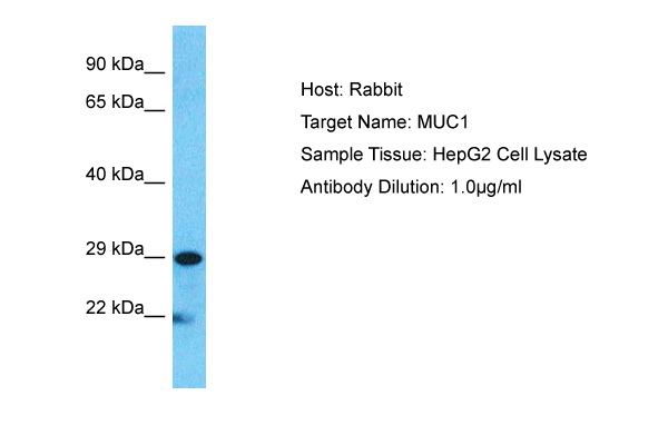


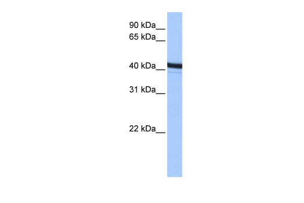
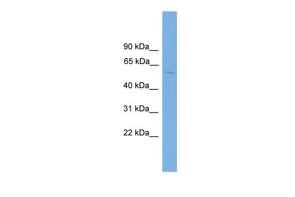
 WB (Western Blot)
(Host: RabbitTarget Name: MUC1Sample Type: HepG2Lane A: Primary AntibodyLane B: Primary Antibody + Blocking PeptidePrimary Antibody Concentration: 1ug/mlPeptide Concentration: 5ug/mlLysate Quantity: 25ug/lane/laneGel Concentration: 0.12)
WB (Western Blot)
(Host: RabbitTarget Name: MUC1Sample Type: HepG2Lane A: Primary AntibodyLane B: Primary Antibody + Blocking PeptidePrimary Antibody Concentration: 1ug/mlPeptide Concentration: 5ug/mlLysate Quantity: 25ug/lane/laneGel Concentration: 0.12)
MUC1, Polyclonal Antibody (Cat# AAA23492)
Full Name
MUC1 antibody - C-terminal region
Gene Names
MUC1; EMA; MCD; PEM; PUM; KL-6; MAM6; MCKD; PEMT; CD227; H23AG; MCKD1; MUC-1; ADMCKD; ADMCKD1; CA 15-3; MUC-1/X; MUC1/ZD; MUC-1/SEC
Reactivity
Cow, Dog, Guinea Pig, Horse, Human, Mouse, Rabbit, Rat, Pig
Applications
IHC, WB
Purity
Affinity Purified
Pricing


 DB (Dot Blot)
(Formalin-fixed and paraffin-embedded human cancer tissue reacted with the primary antibody, which was peroxidase-conjugated to the secondary antibody, followed by AEC staining. This data demonstrates the use of this antibody for immunohistochemistry; clinical relevance has not been evaluated. BC = breast carcinoma; HC = hepatocarcinoma.)
DB (Dot Blot)
(Formalin-fixed and paraffin-embedded human cancer tissue reacted with the primary antibody, which was peroxidase-conjugated to the secondary antibody, followed by AEC staining. This data demonstrates the use of this antibody for immunohistochemistry; clinical relevance has not been evaluated. BC = breast carcinoma; HC = hepatocarcinoma.)
Phospho-HIST1H3B3 (S10), Polyclonal Antibody (Cat# AAA28670)
Full Name
Phospho-HIST1H3B3 (S10) Antibody
Gene Names
HIST1H3A; H3/A; H3FA
Reactivity
Human
Applications
DB, EIA, IHC, WB
Purity
Peptide Affinity Purified Rabbit Polyclonal Antibody (Pab)
Pricing


 WB (Western Blot)
(WB Suggested Anti-SHH Antibody Titration: 0.2-1 ug/mlPositive Control: HepG2 cell lysate)
WB (Western Blot)
(WB Suggested Anti-SHH Antibody Titration: 0.2-1 ug/mlPositive Control: HepG2 cell lysate)
SHH, Polyclonal Antibody (Cat# AAA23516)
Full Name
SHH antibody - N-terminal region
Gene Names
SHH; TPT; HHG1; HLP3; HPE3; SMMCI; ShhNC; TPTPS; MCOPCB5
Reactivity
Predicted Reactivity: Cow, Dog, Goat, Guinea Pig, Horse, Human, Mouse, Rabbit, Rat, Zebrafish, Chicken (Tested Reactivity: Human, Mouse, Chicken)
Applications
IHC, WB
Purity
Affinity Purified
Pricing


 WB (Western Blot)
(WB Suggested Anti-ESR2 antibody Titration: 1 ug/mLSample Type: Human liver)
WB (Western Blot)
(WB Suggested Anti-ESR2 antibody Titration: 1 ug/mLSample Type: Human liver)
ESR2, Polyclonal Antibody (Cat# AAA23449)
Full Name
ESR2 antibody - N-terminal region
Gene Names
ESR2; Erb; ESRB; ODG8; ESTRB; NR3A2; ER-BETA; ESR-BETA
Reactivity
Human, Rat
Applications
IHC, WB
Purity
Affinity Purified
Pricing
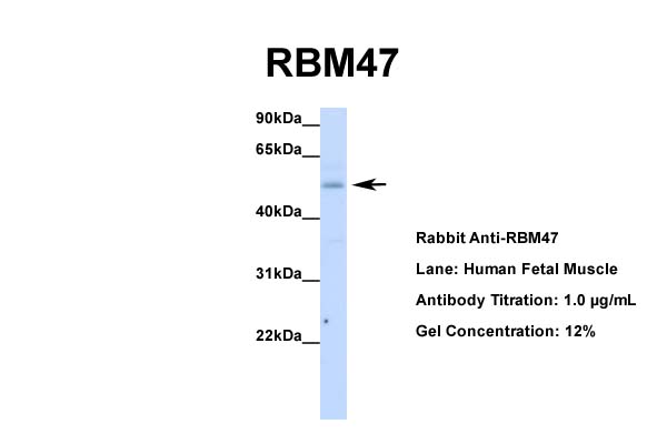


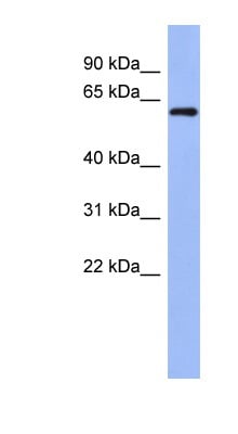 WB (Western Blot)
(WB Suggested Anti-RBM47 Antibody Titration: 0.2-1 ug/mlELISA Titer: 1:62500Positive Control: NCI-H226)
WB (Western Blot)
(WB Suggested Anti-RBM47 Antibody Titration: 0.2-1 ug/mlELISA Titer: 1:62500Positive Control: NCI-H226)
RBM47, Polyclonal Antibody (Cat# AAA23486)
Full Name
RBM47 antibody - middle region
Gene Names
RBM47; NET18
Reactivity
Cow, Dog, Guinea Pig, Horse, Human, Mouse, Rabbit, Rat, Zebrafish
Applications
IHC, WB
Purity
Affinity Purified
Pricing


 IHC (Immunohistchemistry)
(Figure 6. IHC analysis of AMD1 using anti-AMD1 antibody (AAA19177).AMD1 was detected in paraffin-embedded section of human mammary cancer tissue. Heat mediated antigen retrieval was performed in citrate buffer (pH6, epitope retrieval solution) for 20 mins. The tissue section was blocked with 10% goat serum. The tissue section was then incubated with 1ug/ml rabbit anti-AMD1 Antibody (AAA19177) overnight at 4 degree C. Biotinylated goat anti-rabbit IgG was used as secondary antibody and incubated for 30 minutes at 37 degree C. The tissue section was developed using Strepavidin-Biotin-Complex (SABC) with DAB as the chromogen.)
IHC (Immunohistchemistry)
(Figure 6. IHC analysis of AMD1 using anti-AMD1 antibody (AAA19177).AMD1 was detected in paraffin-embedded section of human mammary cancer tissue. Heat mediated antigen retrieval was performed in citrate buffer (pH6, epitope retrieval solution) for 20 mins. The tissue section was blocked with 10% goat serum. The tissue section was then incubated with 1ug/ml rabbit anti-AMD1 Antibody (AAA19177) overnight at 4 degree C. Biotinylated goat anti-rabbit IgG was used as secondary antibody and incubated for 30 minutes at 37 degree C. The tissue section was developed using Strepavidin-Biotin-Complex (SABC) with DAB as the chromogen.)
AMD1/Adometdc, Polyclonal Antibody (Cat# AAA19177)
Full Name
Anti-AMD1/Adometdc Antibody
Gene Names
AMD1; AMD; SAMDC; ADOMETDC
Reactivity
Human, Mouse, Rat
No cross reactivity with other proteins.
No cross reactivity with other proteins.
Applications
IHC, WB
Purity
Immunogen affinity purified
Pricing


 WB (Western Blot)
(WB Suggested Anti-CACNB1 Antibody Titration: 0.2-1 ug/mlELISA Titer: 1:62500Positive Control: HepG2 cell lysate)
WB (Western Blot)
(WB Suggested Anti-CACNB1 Antibody Titration: 0.2-1 ug/mlELISA Titer: 1:62500Positive Control: HepG2 cell lysate)
CACNB1, Polyclonal Antibody (Cat# AAA23439)
Full Name
CACNB1 antibody - N-terminal region
Gene Names
CACNB1; CAB1; CCHLB1; CACNLB1
Reactivity
Cow, Dog, Guinea Pig, Horse, Human, Mouse, Rabbit, Rat, Zebrafish
Applications
WB
Purity
Affinity Purified
Pricing


 WB (Western Blot)
(WB Suggested Anti-PPP3CA Antibody Titration: 0.2-1 ug/mlELISA Titer: 1:312500Positive Control: Human Muscle)
WB (Western Blot)
(WB Suggested Anti-PPP3CA Antibody Titration: 0.2-1 ug/mlELISA Titer: 1:312500Positive Control: Human Muscle)
PPP3CA, Polyclonal Antibody (Cat# AAA23542)
Full Name
PPP3CA antibody - middle region
Gene Names
PPP3CA; CALN; CCN1; CNA1; CALNA; IECEE; PPP2B; ACCIID; CALNA1; IECEE1
Reactivity
Cow, Dog, Guinea Pig, Horse, Human, Mouse, Rabbit, Rat, Zebrafish
Applications
WB
Purity
Affinity Purified
Pricing


 WB (Western Blot)
(WB Suggested Anti-PDIA6 Antibody Titration: 0.2-1 ug/mlELISA Titer: 1:1562500Positive Control: HepG2 cell lysate.PDIA6 is strongly supported by BioGPS gene expression data to be expressed in HepG2)
WB (Western Blot)
(WB Suggested Anti-PDIA6 Antibody Titration: 0.2-1 ug/mlELISA Titer: 1:1562500Positive Control: HepG2 cell lysate.PDIA6 is strongly supported by BioGPS gene expression data to be expressed in HepG2)
PDIA6, Polyclonal Antibody (Cat# AAA23546)
Full Name
PDIA6 antibody - middle region
Gene Names
PDIA6; P5; ERP5; TXNDC7
Reactivity
Cow, Dog, Guinea Pig, Horse, Human, Mouse, Pig, Rabbit, Rat
Applications
IHC, WB
Purity
Affinity Purified
Pricing


 WB (Western Blot)
(WB Suggested Anti-PANX1 Antibody Titration: 0.2-1 ug/mlPositive Control: Jurkat cell lysate)
WB (Western Blot)
(WB Suggested Anti-PANX1 Antibody Titration: 0.2-1 ug/mlPositive Control: Jurkat cell lysate)
PANX1, Polyclonal Antibody (Cat# AAA23505)
Full Name
PANX1 antibody - middle region
Gene Names
PANX1; PX1; MRS1; UNQ2529
Reactivity
Cow, Dog, Guinea Pig, Horse, Human, Mouse, Rabbit, Rat
Applications
IHC, WB
Purity
Affinity Purified
Pricing


 WB (Western Blot)
(WB Suggested Anti-DDX41 antibody Titration: 1 ug/mLSample Type: Human liver)
WB (Western Blot)
(WB Suggested Anti-DDX41 antibody Titration: 1 ug/mLSample Type: Human liver)
DDX41, Polyclonal Antibody (Cat# AAA23454)
Full Name
DDX41 antibody - N-terminal region
Gene Names
DDX41; ABS; MPLPF
Reactivity
Cow, Dog, Guinea Pig, Horse, Human, Mouse, Rabbit, Rat, Zebrafish
Applications
WB
Purity
Affinity Purified
Pricing


 WB (Western Blot)
(WB Suggested Anti-ADRB1 Antibody Titration: 0.2-1 ug/mlELISA Titer: 1:312500Positive Control: Human brain)
WB (Western Blot)
(WB Suggested Anti-ADRB1 Antibody Titration: 0.2-1 ug/mlELISA Titer: 1:312500Positive Control: Human brain)
ADRB1, Polyclonal Antibody (Cat# AAA23455)
Full Name
ADRB1 antibody - middle region
Gene Names
ADRB1; RHR; B1AR; ADRB1R; BETA1AR
Reactivity
Dog, Guinea Pig, Human, Mouse, Pig, Rabbit, Rat
Applications
WB
Purity
Affinity Purified
Pricing


 IHC (Immunohistochemistry)
(Immunohistochemistry of paraffin-embedded Human gastric cancer using p27 KIP 1 Polyclonal Antibody at dilution of 1:100 (40x lens).)
IHC (Immunohistochemistry)
(Immunohistochemistry of paraffin-embedded Human gastric cancer using p27 KIP 1 Polyclonal Antibody at dilution of 1:100 (40x lens).)
p27 KIP1, Polyclonal Antibody (Cat# AAA22188)
Full Name
p27 KIP1 Polyclonal Antibody
Gene Names
CDKN1B; KIP1; MEN4; CDKN4; MEN1B; P27KIP1
Reactivity
Human, Mouse, Rat
Applications
WB, IHC
Purity
Affinity purification
Pricing


 IHC (Immunohistchemistry)
(Figure 6. IHC analysis of LGALS3BP using anti-LGALS3BP antibody (AAA19163).LGALS3BP was detected in paraffin-embedded section of mouse small intestine tissue. Heat mediated antigen retrieval was performed in citrate buffer (pH6, epitope retrieval solution) for 20 mins. The tissue section was blocked with 10% goat serum. The tissue section was then incubated with 2ug/ml rabbit anti-LGALS3BP Antibody (AAA19163) overnight at 4 degree C. Biotinylated goat anti-rabbit IgG was used as secondary antibody and incubated for 30 minutes at 37 degree C. The tissue section was developed using Strepavidin-Biotin-Complex (SABC) with DAB as the chromogen.)
IHC (Immunohistchemistry)
(Figure 6. IHC analysis of LGALS3BP using anti-LGALS3BP antibody (AAA19163).LGALS3BP was detected in paraffin-embedded section of mouse small intestine tissue. Heat mediated antigen retrieval was performed in citrate buffer (pH6, epitope retrieval solution) for 20 mins. The tissue section was blocked with 10% goat serum. The tissue section was then incubated with 2ug/ml rabbit anti-LGALS3BP Antibody (AAA19163) overnight at 4 degree C. Biotinylated goat anti-rabbit IgG was used as secondary antibody and incubated for 30 minutes at 37 degree C. The tissue section was developed using Strepavidin-Biotin-Complex (SABC) with DAB as the chromogen.)
LGALS3BP, Polyclonal Antibody (Cat# AAA19163)
Full Name
Anti-LGALS3BP Picoband Antibody
Reactivity
Human, Mouse
No cross reactivity with other proteins.
No cross reactivity with other proteins.
Applications
IHC, WB
Pricing
 IHC (Immunohistochemistry)
(Immunohistochemistry of paraffinembedded Human esophagus cancer tissue using ERVW-1 Polyclonal Antibody at dilution 1:45)
IHC (Immunohistochemistry)
(Immunohistochemistry of paraffinembedded Human esophagus cancer tissue using ERVW-1 Polyclonal Antibody at dilution 1:45)
ERVW-1, Polyclonal Antibody (Cat# AAA21995)
Full Name
ERVW-1 Polyclonal Antibody
Reactivity
Human
Applications
EIA, IHC
Purity
Affinity purification
Pricing
 WB (Western Blot)
(WB Suggested Anti-CTRC Antibody Titration: 0.2-1 ug/mlELISA Titer: 1:312500Positive Control: Human Liver)
WB (Western Blot)
(WB Suggested Anti-CTRC Antibody Titration: 0.2-1 ug/mlELISA Titer: 1:312500Positive Control: Human Liver)
CTRC, Polyclonal Antibody (Cat# AAA23430)
Full Name
CTRC antibody - N-terminal region
Gene Names
CTRC; CLCR; ELA4
Reactivity
Tested Species Reactivity: Human!!Predicted Species Reactivity: Human, Mouse, Rat, Cow, Dog, Pig, Rabbit
Applications
WB
Purity
Affinity Purified
Pricing


 WB (Western Blot)
(Western blot analysis of MeCP2-S421* in mouse brain, cerebellum tissue and HepG2 cell line lysates (35ug/lane). CP2 (arrow) was detected using the purified Pab.)
WB (Western Blot)
(Western blot analysis of MeCP2-S421* in mouse brain, cerebellum tissue and HepG2 cell line lysates (35ug/lane). CP2 (arrow) was detected using the purified Pab.)
MeCP2, Polyclonal Antibody (Cat# AAA28666)
Full Name
MeCP2 Antibody (S421)
Reactivity
Human, mouse
Applications
WB, EIA
Purity
Peptide Affinity Purified Rabbit Polyclonal Antibody (Pab)
Pricing


 WB (Western Blot)
(WB Suggested Anti-KCNJ12 Antibody Titration: 0.2-1 ug/mlELISA Titer: 1:62500Positive Control: Human brain)
WB (Western Blot)
(WB Suggested Anti-KCNJ12 Antibody Titration: 0.2-1 ug/mlELISA Titer: 1:62500Positive Control: Human brain)
KCNJ12, Polyclonal Antibody (Cat# AAA23443)
Full Name
KCNJ12 antibody - middle region
Gene Names
KCNJ12; IRK2; hIRK; IRK-2; hIRK1; KCNJN1; Kir2.2; Kir2.2v; kcnj12x; hkir2.2x
Reactivity
Cow, Dog, Guinea Pig, Horse, Human, Mouse, Rat
Applications
WB
Purity
Affinity Purified
Pricing


 WB (Western Blot)
(Gel: 6% SDS-PAGE Lysate: 40 ug Lane: Mouse liver tissue lysate Primary antibody: (NPR2 Antibody) at dilution 1/1100 Secondary antibody: (HRP-conjugated Goat anti rabbit IgG) at 1/5000 dilution Exposure time: 40 seconds.)
WB (Western Blot)
(Gel: 6% SDS-PAGE Lysate: 40 ug Lane: Mouse liver tissue lysate Primary antibody: (NPR2 Antibody) at dilution 1/1100 Secondary antibody: (HRP-conjugated Goat anti rabbit IgG) at 1/5000 dilution Exposure time: 40 seconds.)
NPR2, Polyclonal Antibody (Cat# AAA29693)
Full Name
NPR2 Antibody
Gene Names
NPR2; AMDM; ANPb; ECDM; NPRB; SNSK; ANPRB; GUC2B; NPRBi; GUCY2B
Reactivity
Human, Mouse
Applications
IHC, WB
Purity
Antigen affinity purification.
Pricing

 IHC (Immunohistochemistry)
(Immunohistochemistry of paraffin-embedded Human colon cancer using CD192 Polyclonal Antibody at dilution of 1:20)
IHC (Immunohistochemistry)
(Immunohistochemistry of paraffin-embedded Human colon cancer using CD192 Polyclonal Antibody at dilution of 1:20)
CD192, Polyclonal Antibody (Cat# AAA22006)
Full Name
CD192 Polyclonal Antibody
Gene Names
CCR2; CKR2; CCR-2; CCR2A; CCR2B; CD192; CKR2A; CKR2B; CMKBR2; MCP-1-R; CC-CKR-2
Reactivity
Human, Mouse
Applications
EIA, WB
Purity
Affinity purification
Pricing


 IHC (Immunohistchemistry)
(Immunohistochemistry of paraffin-embedded human stomach using ALYREF antibody at dilution of 1:100 (40x lens).)
IHC (Immunohistchemistry)
(Immunohistochemistry of paraffin-embedded human stomach using ALYREF antibody at dilution of 1:100 (40x lens).)
ALYREF, Polyclonal Antibody (Cat# AAA28139)
Full Name
ALYREF Polyclonal Antibody
Gene Names
ALYREF; ALY; BEF; REF; THOC4; ALY/REF
Reactivity
Human, Mouse, Rat
Applications
WB, IP
Purity
Affinity Purification
Pricing


 WB (Western Blot)
(WB Suggested Anti-PHB Antibody Titration: 0.2-1 ug/mlPositive Control: Jurkat cell lysatePHB is supported by BioGPS gene expression data to be expressed in Jurkat)
WB (Western Blot)
(WB Suggested Anti-PHB Antibody Titration: 0.2-1 ug/mlPositive Control: Jurkat cell lysatePHB is supported by BioGPS gene expression data to be expressed in Jurkat)
PHB, Polyclonal Antibody (Cat# AAA23420)
Full Name
PHB antibody - C-terminal region
Gene Names
PHB; PHB1; HEL-215; HEL-S-54e
Reactivity
Cow, Dog, Guinea Pig, Horse, Human, Mouse, Rabbit, Rat, Zebrafish
Applications
WB, IHC
Purity
Affinity Purified
Pricing


 WB (Western Blot)
(WB Suggested Anti-YAP1 Antibody Titration: 1 ug/mlPositive Control: 721_B cell lysate)
WB (Western Blot)
(WB Suggested Anti-YAP1 Antibody Titration: 1 ug/mlPositive Control: 721_B cell lysate)
YAP1, Polyclonal Antibody (Cat# AAA23538)
Full Name
YAP1 antibody - C-terminal region
Gene Names
YAP1; YAP; YKI; COB1; YAP2; YAP65
Reactivity
Tested Reactivity: Human
Predicted Reactivity: Cow, Dog, Horse, Human, Mouse, Pig, Rabbit, Rat, Sheep, Zebrafish
Predicted Reactivity: Cow, Dog, Horse, Human, Mouse, Pig, Rabbit, Rat, Sheep, Zebrafish
Applications
IHC, WB
Purity
Affinity Purified
Pricing


 WB (Western Blot)
(WB Suggested Anti-SIRT4 antibody Titration: 1 ug/mLSample Type: Human Raji)
WB (Western Blot)
(WB Suggested Anti-SIRT4 antibody Titration: 1 ug/mLSample Type: Human Raji)
SIRT4, Polyclonal Antibody (Cat# AAA23411)
Full Name
SIRT4 antibody - middle region
Gene Names
SIRT4; SIR2L4
Reactivity
Cow, Dog, Guinea Pig, Horse, Human, Mouse, Pig, Rabbit, Rat, Sheep
Applications
IHC, WB
Purity
Affinity Purified
Pricing


 WB (Western Blot)
(WB Suggested Anti-LAS1L Antibody Titration: 0.2-1 ug/mlPositive Control: HepG2 cell lysate)
WB (Western Blot)
(WB Suggested Anti-LAS1L Antibody Titration: 0.2-1 ug/mlPositive Control: HepG2 cell lysate)
LAS1L, Polyclonal Antibody (Cat# AAA23435)
Full Name
LAS1L Antibody - middle region
Gene Names
LAS1L; WTS; Las1-like; dJ475B7.2
Reactivity
Cow, Horse, Human
Applications
WB
Purity
Affinity Purified
Pricing


 WB (Western Blot)
(WB Suggested Anti-VEZF1 Antibody Titration: 1 ug/mlPositive Control: 293T cells lysateThere is BioGPS gene expression data showing that VEZF1 is expressed in HEK293T)
WB (Western Blot)
(WB Suggested Anti-VEZF1 Antibody Titration: 1 ug/mlPositive Control: 293T cells lysateThere is BioGPS gene expression data showing that VEZF1 is expressed in HEK293T)
VEZF1, Polyclonal Antibody (Cat# AAA23448)
Full Name
VEZF1 antibody - middle region
Gene Names
VEZF1; DB1; ZNF161
Reactivity
Cow, Dog, Guinea Pig, Horse, Human, Mouse, Rabbit, Rat, Zebrafish
Applications
IHC, WB
Purity
Affinity Purified
Pricing

 IHC (Immunohistochemistry)
(Immunohistochemical analysis of Somatostatin staining in human brain formalin fixed paraffin embedded tissue section. The section was pre-treated using heat mediated antigen retrieval with sodium citrate buffer (pH 6.0). The section was then incubated with the antibody at room temperature and detected using an HRP conjugated compact polymer system. DAB was used as the chromogen. The section was then counterstained with haematoxylin and mounted with DPX.)
IHC (Immunohistochemistry)
(Immunohistochemical analysis of Somatostatin staining in human brain formalin fixed paraffin embedded tissue section. The section was pre-treated using heat mediated antigen retrieval with sodium citrate buffer (pH 6.0). The section was then incubated with the antibody at room temperature and detected using an HRP conjugated compact polymer system. DAB was used as the chromogen. The section was then counterstained with haematoxylin and mounted with DPX.)
Somatostatin, Polyclonal Antibody (Cat# AAA17844)
Full Name
Anti-Somatostatin Antibody
Gene Names
SST; SMST
Reactivity
Human, Mouse, Rat, Bovine, Chicken, Monkey, Pig, Sheep
Applications
WB, IHC
Purity
The antibody was purified by immunogen affinity chromatography.
Pricing


 WB (Western Blot)
(Western blot analysis of CD9 Antibody (Center) in HepG2 cell line lysates (35ug/lane). CD9 (arrow) was detected using the purified Pab.)
WB (Western Blot)
(Western blot analysis of CD9 Antibody (Center) in HepG2 cell line lysates (35ug/lane). CD9 (arrow) was detected using the purified Pab.)
CD9, Polyclonal Antibody (Cat# AAA28656)
Full Name
CD9 Antibody (Center)
Gene Names
CD9; MIC3; MRP-1; BTCC-1; DRAP-27; TSPAN29; TSPAN-29
Reactivity
Human
Applications
WB, EIA, IF, FC/FACS
Purity
Purified Rabbit Polyclonal Antibody (Pab)
Pricing


 IHC (Immunohistochemistry)
(Figure 8. IHC analysis of VEGF Receptor 3 using anti-VEGF Receptor 3 antibody (AAA19146).VEGF Receptor 3 was detected in paraffin-embedded section of human mammary cancer tissue. Heat mediated antigen retrieval was performed in citrate buffer (pH6, epitope retrieval solution) for 20 mins. The tissue section was blocked with 10% goat serum. The tissue section was then incubated with 1ug/ml rabbit anti-VEGF Receptor 3 Antibody (AAA19146) overnight at 4 degree C. Biotinylated goat anti-rabbit IgG was used as secondary antibody and incubated for 30 minutes at 37 degree C. The tissue section was developed using Strepavidin-Biotin-Complex (SABC) with DAB as the chromogen.)
IHC (Immunohistochemistry)
(Figure 8. IHC analysis of VEGF Receptor 3 using anti-VEGF Receptor 3 antibody (AAA19146).VEGF Receptor 3 was detected in paraffin-embedded section of human mammary cancer tissue. Heat mediated antigen retrieval was performed in citrate buffer (pH6, epitope retrieval solution) for 20 mins. The tissue section was blocked with 10% goat serum. The tissue section was then incubated with 1ug/ml rabbit anti-VEGF Receptor 3 Antibody (AAA19146) overnight at 4 degree C. Biotinylated goat anti-rabbit IgG was used as secondary antibody and incubated for 30 minutes at 37 degree C. The tissue section was developed using Strepavidin-Biotin-Complex (SABC) with DAB as the chromogen.)
VEGF Receptor 3, Polyclonal Antibody (Cat# AAA19146)
Full Name
Anti-VEGF Receptor 3 Picoband Antibody
Gene Names
FLT4; PCL; FLT-4; FLT41; LMPH1A; VEGFR3; VEGFR-3
Reactivity
Human, Mouse, Rat
No cross reactivity with other proteins.
No cross reactivity with other proteins.
Applications
IHC, WB
Purity
Immunogen affinity purified
Pricing


 FCM (Flow Cytometry)
(Figure 9. Flow Cytometry analysis of U251 cells using anti-RAB1B antibody (AAA19296).Overlay histogram showing U251 cells stained with AAA19296 (Blue line). The cells were blocked with 10% normal goat serum. And then incubated with rabbit anti-RAB1B Antibody (AAA19296, 1μg/1x106 cells) for 30 min at 20 degree C. DyLight®488 conjugated goat anti-rabbit IgG (5-10μg/1x106 cells) was used as secondary antibody for 30 minutes at 20 degree C. Isotype control antibody (Green line) was rabbit IgG (1μg/1x106) used under the same conditions. Unlabelled sample (Red line) was also used as a control.)
FCM (Flow Cytometry)
(Figure 9. Flow Cytometry analysis of U251 cells using anti-RAB1B antibody (AAA19296).Overlay histogram showing U251 cells stained with AAA19296 (Blue line). The cells were blocked with 10% normal goat serum. And then incubated with rabbit anti-RAB1B Antibody (AAA19296, 1μg/1x106 cells) for 30 min at 20 degree C. DyLight®488 conjugated goat anti-rabbit IgG (5-10μg/1x106 cells) was used as secondary antibody for 30 minutes at 20 degree C. Isotype control antibody (Green line) was rabbit IgG (1μg/1x106) used under the same conditions. Unlabelled sample (Red line) was also used as a control.)
RAB1B, Polyclonal Antibody (Cat# AAA19296)
Full Name
Anti-RAB1B Antibody
Reactivity
Human, Mouse, Rat
Applications
WB, IHC-P, ICC, IF, FC/FACS/FCM
Purity
Immunogen affinity purified.
Pricing


 WB (Western Blot)
(WB Suggested Anti-RXRA Antibody Titration: 0.2-1 ug/mlELISA Titer: 1:312500Positive Control: Transfected 293T)
WB (Western Blot)
(WB Suggested Anti-RXRA Antibody Titration: 0.2-1 ug/mlELISA Titer: 1:312500Positive Control: Transfected 293T)
RXRA, Polyclonal Antibody (Cat# AAA23431)
Full Name
RXRA antibody - N-terminal region
Gene Names
RXRA; NR2B1
Reactivity
Cow, Dog, Guinea Pig, Horse, Human, Mouse, Rat
Applications
WB
Purity
Affinity Purified
Pricing


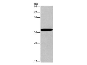
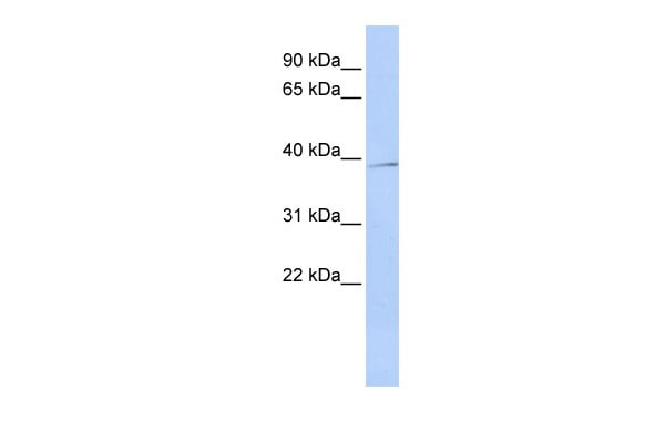
 WB (Western Blot)
(Host: RabbitTarget Name: CELF2Sample Type: JurkatAntibody Dilution: 1.0ug/mlCELF2 is strongly supported by BioGPS gene expression data to be expressed in Jurkat)
WB (Western Blot)
(Host: RabbitTarget Name: CELF2Sample Type: JurkatAntibody Dilution: 1.0ug/mlCELF2 is strongly supported by BioGPS gene expression data to be expressed in Jurkat)
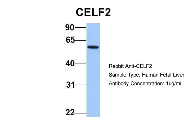
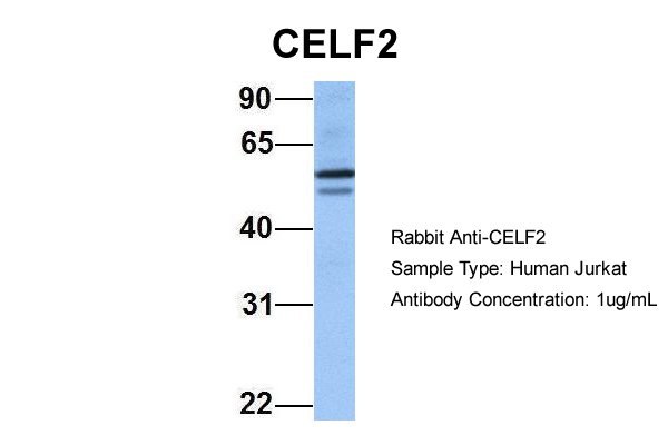
CELF2, Polyclonal Antibody (Cat# AAA23473)
Full Name
CELF2 Antibody - N-terminal region
Gene Names
CELF2; ETR3; ETR-3; NAPOR; CELF-2; CUGBP2; BRUNOL3; CUG-BP2
Reactivity
Human
Applications
WB
Purity
Affinity purified
Pricing


 WB (Western Blot)
(WB Suggested Anti-RXRA Antibody Titration: 0.2-1 ug/mlELISA Titer: 1:312500Positive Control: HepG2 cell lysateRXRA is strongly supported by BioGPS gene expression data to be expressed in Human HepG2 cells)
WB (Western Blot)
(WB Suggested Anti-RXRA Antibody Titration: 0.2-1 ug/mlELISA Titer: 1:312500Positive Control: HepG2 cell lysateRXRA is strongly supported by BioGPS gene expression data to be expressed in Human HepG2 cells)
RXRA, Polyclonal Antibody (Cat# AAA23509)
Full Name
RXRA antibody - N-terminal region
Gene Names
RXRA; NR2B1
Reactivity
Cow, Dog, Guinea Pig, Human, Mouse, Rat
Applications
WB
Purity
Affinity Purified
Pricing


 WB (Western Blot)
(WB Suggested Anti-CREB3L2 Antibody Titration: 1.25ug/mlELISA Titer: 1:12500Positive Control: Jurkat cell lysate)
WB (Western Blot)
(WB Suggested Anti-CREB3L2 Antibody Titration: 1.25ug/mlELISA Titer: 1:12500Positive Control: Jurkat cell lysate)
CREB3L2, Polyclonal Antibody (Cat# AAA23437)
Full Name
CREB3L2 antibody - N-terminal region
Gene Names
CREB3L2; BBF2H7
Reactivity
Cow, Dog, Guinea Pig, Horse, Human, Mouse, Rabbit, Rat
Applications
IHC, WB
Purity
Protein A purified
Pricing


 WB (Western Blot)
(WB Suggested Anti-CPT1B Antibody Titration: 0.2-1 ug/mlELISA Titer: 1:62500Positive Control: HT1080 cell lysateCPT1B is supported by BioGPS gene expression data to be expressed in HT1080)
WB (Western Blot)
(WB Suggested Anti-CPT1B Antibody Titration: 0.2-1 ug/mlELISA Titer: 1:62500Positive Control: HT1080 cell lysateCPT1B is supported by BioGPS gene expression data to be expressed in HT1080)
CPT1B, Polyclonal Antibody (Cat# AAA23525)
Full Name
CPT1B antibody - middle region
Gene Names
CPT1B; CPTI; CPT1M; MCPT1; CPT1-M; CPTI-M; M-CPT1; MCCPT1
Reactivity
Cow, Dog, Guinea Pig, Horse, Human, Mouse, Rabbit, Rat, Sheep, Zebrafish
Applications
IHC, WB
Purity
Affinity Purified
Pricing


 WB (Western Blot)
(WB Suggested Anti-SDHB Antibody Titration: 0.2-1 ug/mlELISA Titer: 1:62500Positive Control: Jurkat cell lysateSDHB is supported by BioGPS gene expression data to be expressed in Jurkat)
WB (Western Blot)
(WB Suggested Anti-SDHB Antibody Titration: 0.2-1 ug/mlELISA Titer: 1:62500Positive Control: Jurkat cell lysateSDHB is supported by BioGPS gene expression data to be expressed in Jurkat)
SDHB, Polyclonal Antibody (Cat# AAA23528)
Full Name
SDHB antibody - middle region
Gene Names
SDHB; IP; SDH; CWS2; PGL4; SDH1; SDH2; SDHIP
Reactivity
Tested: Human
Predicted Reactivity: Cow, Dog, Goat, Guinea Pig, Horse, Human, Mouse, Rabbit, Rat, Yeast, Zebrafish
Predicted Reactivity: Cow, Dog, Goat, Guinea Pig, Horse, Human, Mouse, Rabbit, Rat, Yeast, Zebrafish
Applications
WB
Purity
Affinity Purified
Pricing


 WB (Western Blot)
(WB Suggested Anti-OTX2 Antibody Titration: 0.2-1 ug/mlPositive Control: Jurkat cell lysate)
WB (Western Blot)
(WB Suggested Anti-OTX2 Antibody Titration: 0.2-1 ug/mlPositive Control: Jurkat cell lysate)
OTX2, Polyclonal Antibody (Cat# AAA23410)
Full Name
OTX2 antibody - N-terminal region
Gene Names
OTX2; CPHD6; MCOPS5
Reactivity
Cow, Dog, Guinea Pig, Horse, Human, Mouse, Rabbit, Rat, Sheep, Zebrafish
Applications
IHC, WB
Purity
Affinity Purified
Pricing


 WB (Western Blot)
(WB Suggested Anti-STK3 Antibody Titration: 5.0ug/mlPositive Control: HepG2 cell lysate)
WB (Western Blot)
(WB Suggested Anti-STK3 Antibody Titration: 5.0ug/mlPositive Control: HepG2 cell lysate)
STK3, Polyclonal Antibody (Cat# AAA23535)
Full Name
STK3 antibody - C-terminal region
Gene Names
STK3; KRS1; MST2
Reactivity
Cow, Dog, Guinea Pig, Horse, Human, Mouse, Rabbit, Rat, Zebrafish
Applications
IHC, WB
Purity
Protein A purified
Pricing


 WB (Western Blot)
(WB Suggested Anti-P4HB Antibody Titration: 0.5ug/mlPositive Control: HepG2 cell lysateP4HB is strongly supported by BioGPS gene expression data to be expressed in Human HepG2 cells)
WB (Western Blot)
(WB Suggested Anti-P4HB Antibody Titration: 0.5ug/mlPositive Control: HepG2 cell lysateP4HB is strongly supported by BioGPS gene expression data to be expressed in Human HepG2 cells)
P4HB, Polyclonal Antibody (Cat# AAA23529)
Full Name
P4HB antibody - N-terminal region
Gene Names
P4HB; DSI; GIT; PDI; PHDB; PDIA1; PO4DB; PO4HB; PROHB; CLCRP1; ERBA2L; P4Hbeta
Reactivity
Cow, Dog, Guinea Pig, Horse, Human, Mouse, Rabbit, Rat
Applications
IHC, WB
Purity
Affinity Purified
Pricing


 FCM (Flow Cytometry)
(Figure 7. Flow Cytometry analysis of A549 cells using anti-Hsp90 alpha antibody (AAA19359).Overlay histogram showing A549 cells stained with AAA19359 (Blue line). The cells were blocked with 10% normal goat serum. And then incubated with mouse anti-Hsp90 alpha Antibody (AAA19359, 1μg/1x106 cells) for 30 min at 20 degree C. DyLight®488 conjugated goat anti-mouse IgG (BA1126, 5-10μg/1x106 cells) was used as secondary antibody for 30 minutes at 20 degree C. Isotype control antibody (Green line) was mouse IgG (1μg/1x106) used under the same conditions. Unlabelled sample (Red line) was also used as a control.)
FCM (Flow Cytometry)
(Figure 7. Flow Cytometry analysis of A549 cells using anti-Hsp90 alpha antibody (AAA19359).Overlay histogram showing A549 cells stained with AAA19359 (Blue line). The cells were blocked with 10% normal goat serum. And then incubated with mouse anti-Hsp90 alpha Antibody (AAA19359, 1μg/1x106 cells) for 30 min at 20 degree C. DyLight®488 conjugated goat anti-mouse IgG (BA1126, 5-10μg/1x106 cells) was used as secondary antibody for 30 minutes at 20 degree C. Isotype control antibody (Green line) was mouse IgG (1μg/1x106) used under the same conditions. Unlabelled sample (Red line) was also used as a control.)
Hsp90 alpha, Monoclonal Antibody (Cat# AAA19359)
Full Name
Anti-Hsp90 alpha Antibody (monoclonal, 6B5)
Gene Names
HSP90AA1; EL52; HSPN; LAP2; HSP86; HSPC1; HSPCA; Hsp89; Hsp90; HSP89A; HSP90A; HSP90N; HSPCAL1; HSPCAL4
Reactivity
Human, Mouse, Rat, Monkey
Applications
WB, IHC-P, ICC, IF, FC/FACS/FCM
Purity
Immunogen affinity purified.
Pricing