Filters
▼Clonality
▼Type
▼Reactivity
▼Gene Name
▼Isotype
▼Host
▼Application
▼Clone
▼Recombinant Proteins
Recombinant proteins are purified laboratory reagents created through genetic engineering for advanced biomedical research, such as immunology, neuroscience, cancer research, and more.
At AAA Biotech, also known as AAA Bio or AAABio, we provide high-quality recombinant proteins. Our various protein products are carefully tested for consistent and reliable performance. With our proteins, we offer stringent quality control, lot-to-lot consistency, and precise specificity to help you achieve accurate results in your research.
Whether you're conducting research on cell expansion, polarization, differentiation, or cell processing, we can offer the exact recombinant proteins you need in order to support your work.
Viewing 5250-5300 of 14163 product results
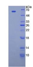
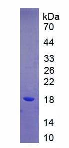
 SDS-PAGE
Slit Homolog 3 (Slit3), Recombinant Protein (Cat# AAA134229)
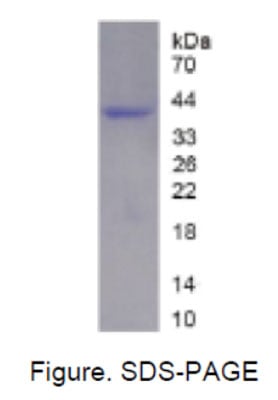
 SDS-PAGE
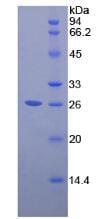
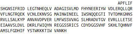
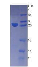
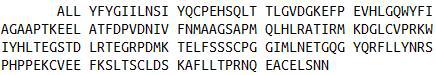
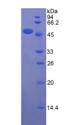
Apolipoprotein E (APOE), Recombinant Protein (Cat# AAA134231)
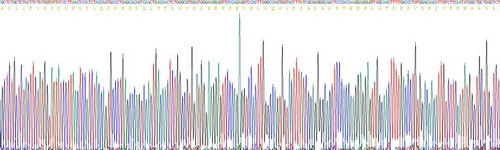
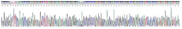
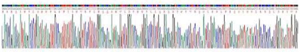
 Application Data
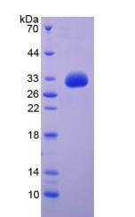
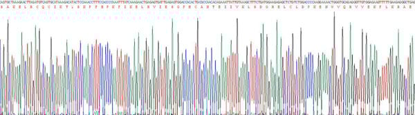
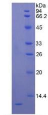
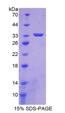
CUB And Zona Pellucida Like Domains Protein 1 (CUZD1), Recombinant Protein (Cat# AAA134243)
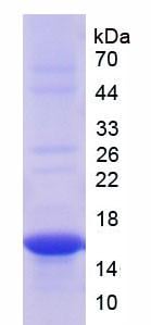
 SDS-PAGE
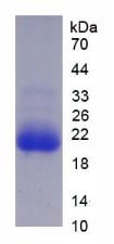
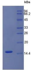
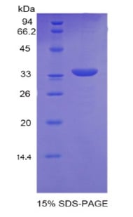
Pepsinogen A (PGA), Recombinant Protein (Cat# AAA134253)
 SDS-PAGE
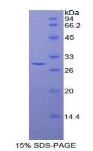
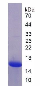
Epidermal Growth Factor (EGF), Recombinant Protein (Cat# AAA134258)

 SDS-PAGE
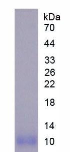
Interleukin 8 (IL8), Recombinant Protein (Cat# AAA134262)
 SDS-PAGE
Stanniocalcin 2 (STC2), Recombinant Protein (Cat# AAA134271)
 SDS-PAGE
Collagen Type XVIII (COL18), Recombinant Protein (Cat# AAA134275)
 SDS-PAGE
Vasoactive Intestinal Peptide (VIP), Recombinant Protein (Cat# AAA134280)
 SDS-PAGE
Transthyretin (TTR), Recombinant Protein (Cat# AAA134285)
 SDS-PAGE
Coagulation Factor VIII (F8), Recombinant Protein (Cat# AAA134293)
 SDS-PAGE
Diacylglycerol-O-Acyltransferase Homolog 1 (DGAT1), Recombinant Protein (Cat# AAA134301)
 Application Data
Cytochrome P450 3A7 (CYP3A7), Recombinant Protein (Cat# AAA134304)
 SDS-PAGE
Diazepam Binding Inhibitor (DBI), Recombinant Protein (Cat# AAA134310)
 SDS-PAGE
Inhibitory Subunit Of NF Kappa B Epsilon (IkBe), Recombinant Protein (Cat# AAA134315)
Single Ig IL1 Related Receptor (SIGIRR), Recombinant Protein (Cat# AAA134329)
 SDS-PAGE
Calbindin (CALB), Recombinant Protein (Cat# AAA134334)
 Application Data
Apolipoprotein M (APOM), Recombinant Protein (Cat# AAA134338)
 Application Data
Hypoxia Inducible Factor 1 Alpha (HIF1a), Recombinant Protein (Cat# AAA134348)
 SDS-PAGE
Transcription Factor A, Mitochondrial (TFAM), Recombinant Protein (Cat# AAA134511)
 SDS-PAGE
Trefoil Factor 2 (TFF2), Recombinant Protein (Cat# AAA134517)
 SDS-PAGE
Apolipoprotein D (APOD), Recombinant Protein (Cat# AAA134527)

 SDS-PAGE
Podocin (PDCN), Recombinant Protein (Cat# AAA134529)
 SDS-PAGE
Vascular Cell Adhesion Molecule 1 (VCAM1), Recombinant Protein (Cat# AAA134530)
 SDS-PAGE
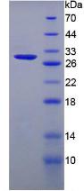
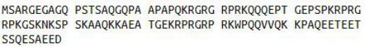
Chromogranin A (CHGA), Recombinant Protein (Cat# AAA134542)
 SDS-PAGE
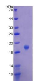
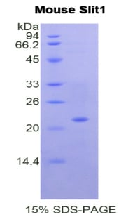
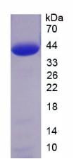
Slit Homolog 1 (Slit1), Recombinant Protein (Cat# AAA134556)
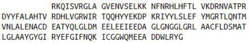
 Application Data
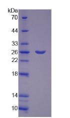
Glycogen Phosphorylase, Muscle (PYGM), Recombinant Protein (Cat# AAA134557)
 SDS-PAGE
Inducible T-Cell Co Stimulator (ICOS), Recombinant Protein (Cat# AAA134570)
 SDS-PAGE
Intelectin 1 (ITLN1), Recombinant Protein (Cat# AAA134590)
 SDS-PAGE
High Mobility Group AT Hook Protein 2 (HMGA2), Recombinant Protein (Cat# AAA134447)
 SDS-PAGE
Aquaporin 4 (AQP4), Recombinant Protein (Cat# AAA134450)
 Application Data
Keratin 5 (KRT5), Recombinant Protein (Cat# AAA134451)
 Application Data
Thy1 Cell Surface Antigen (Thy1), Recombinant Protein (Cat# AAA134461)
 SDS-PAGE
Osteonectin (ON), Recombinant Protein (Cat# AAA134481)
 SDS-PAGE
Interferon Gamma Induced Protein 10kDa (IP10), Recombinant Protein (Cat# AAA134486)
 SDS-PAGE
Noggin (NOG), Recombinant Protein (Cat# AAA134488)
 SDS-PAGE
Calpain 1, Large Subunit (CAPN1), Recombinant Protein (Cat# AAA134489)
 SDS-PAGE
Filamin C Gamma (FLNC), Recombinant Protein (Cat# AAA134503)
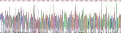
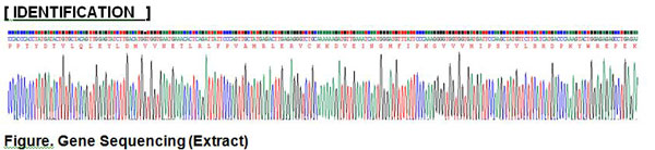
 Identification
Plexin B1 (PLXNB1), Recombinant Protein (Cat# AAA134124)
 Application Data
Melatonin Receptor 1A (MTNR1A), Recombinant Protein (Cat# AAA134125)
 SDS-PAGE
A Disintegrin And Metalloprotease 10 (ADAM10), Recombinant Protein (Cat# AAA134127)
 SDS-PAGE
Integrin Alpha M (ITGaM), Recombinant Protein (Cat# AAA134136)
 Application Data
Laminin Alpha 1 (LAMa1), Recombinant Protein (Cat# AAA134144)
 SDS-PAGE
Complement Component 3a (C3a), Recombinant Protein (Cat# AAA134149)
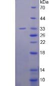
 SDS-PAGE
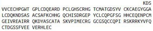
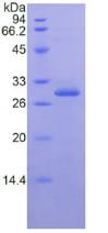
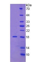
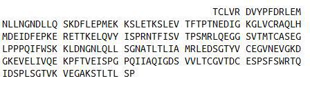
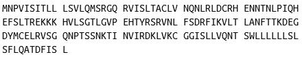
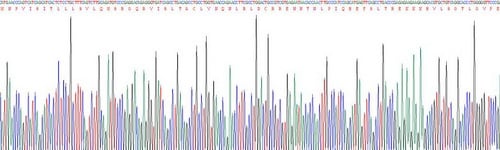
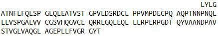
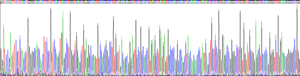
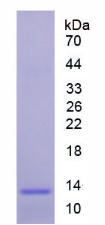
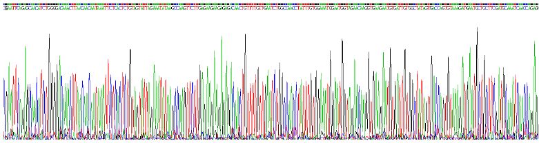
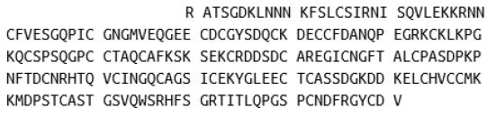
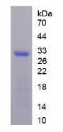
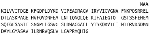
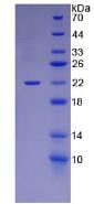
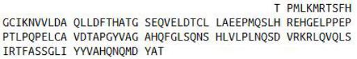
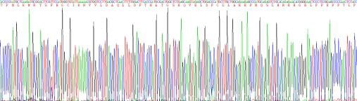
Paraneoplastic Antigen MA2 (PNMA2), Recombinant Protein (Cat# AAA134153)
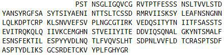
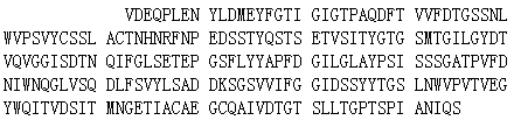
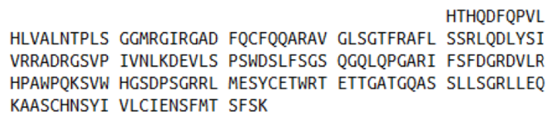
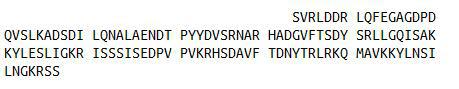
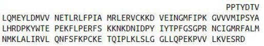
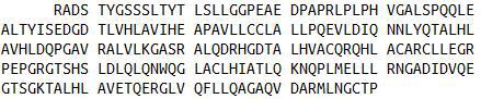
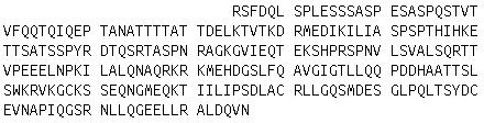
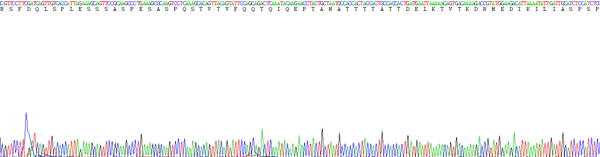
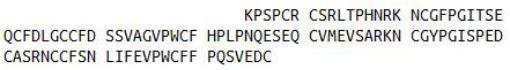
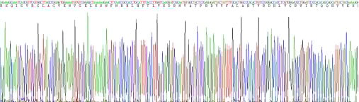
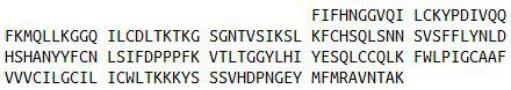
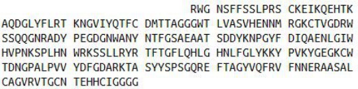
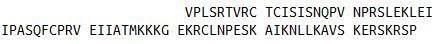
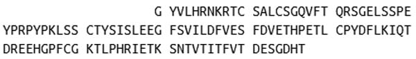 Application Data
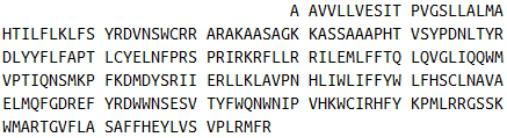
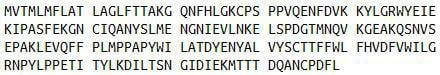
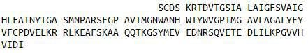
(Sequence)
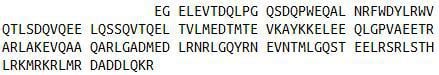
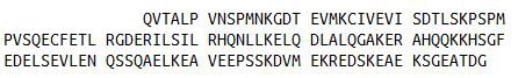
Application Data
(Sequence)
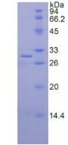
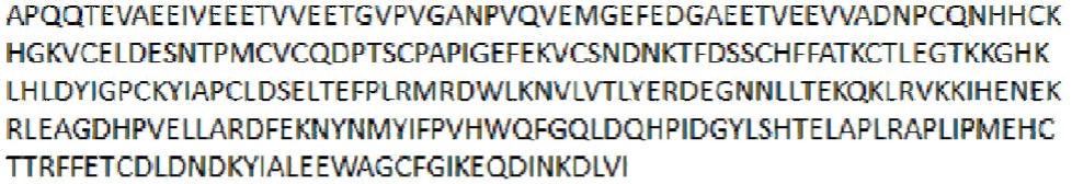
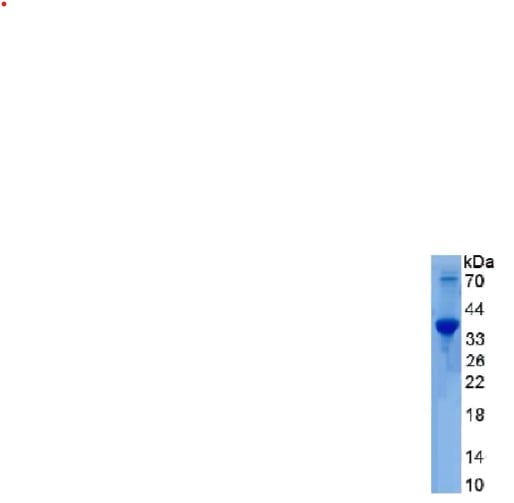
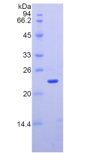
Mannose Associated Serine Protease 2 (MASP2), Recombinant Protein (Cat# AAA134180)

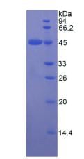
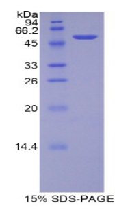
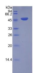
 SDS-PAGE
Nitric Oxide Synthase 2, Inducible (NOS2), Recombinant Protein (Cat# AAA134183)
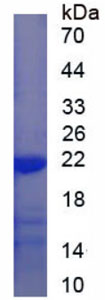
Vitamin D3 receptor (VDR), Recombinant Protein (Cat# AAA115621)
 SDS-PAGE
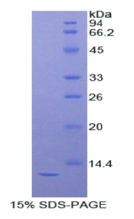
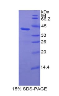
Superoxide dismutase [Mn], Recombinant Protein (Cat# AAA115625)
Carboxypeptidase D (CPD), Recombinant Protein (Cat# AAA115633)
What Are Recombinant Proteins?
A recombinant protein is created by inserting the gene of interest into a host system like E. coli, mammalian cells, or insect cells. This protein expression method results in the large-scale production of protein in a controlled environment with highly precise structures and functionality. Proteins produced via the “Recombinant” methodology have proven themselves crucial in various biological research areas, including immunology, neuroscience, cancer research, drug delivery and development, and more.
Core Recombinant Protein Product Lines:
- Human: Produced from human genes (e.g., growth factors, cytokines).
- Animal: Derived from mouse, rat, or other animals (e.g., mouse antibodies, enzymes).
- Bacterial: Produced in bacteria like E. coli (e.g., enzymes, bacterial toxins).
- Yeast: Produced in yeast cells (e.g., surface proteins, metabolic enzymes).
- Insect:Expressed in insect cells (e.g., viral proteins, glycoproteins).
- Viral: Derived from viral genomes (e.g., spike proteins for vaccine development).
How Does It Work?
- Gene Identification and Cloning: Scientists first find the exact gene that codes for the protein they are interested in producing, and then create a copy of said gene.
- Recombinant DNA Creation: The copied gene is then inserted into a vector (a vehicle-like plasmid) that can be transported safely into your intended expression host cell/system.
- Host Cell Transformation: The vector (with the foreign gene inside) is shuttled into a host cell. This host can be a bacterium (like E. coli), yeast, insect cells, or mammalian cells, depending on the type of protein or protein characteristics needed.
- Protein Expression:The host cell “reads” the copied gene that was shuttled into it, and begins to produce the gene’s coded-for protein using its own cellular machinery. The host can continue to multiply, and with each new cell, more protein is produced.
- Protein Purification:After enough protein has been made, scientists collect the host cell and separate out the target protein. The protein is then carefully purified so it is free of contaminants, ideally active, and also safe for use.
The result is a purified recombinant protein — a lab-made protein that is biologically identical (in most respects) to the natural/native version.
We take this process a step further, offering recombinant proteins that are produced with precise control over sequence modifications, expression levels, and large-scale production. This ensures high-quality, consistent results that will be able to support your research needs and empower your scientific discoveries.
Some of the expression hosts/systems used in recombinant protein generation by AAA Biotech include:
- Escherichia coli (E. coli)
- Human (Homo sapiens)
- Chinese Hamster
- Yeast
- Insect cells (e.g., Baculovirus expression system)
Advantages Of Our Recombinant Proteins:
Recombinant Protein Offerings
| S.No | Protein Expression Systems | Quality and Bioactivity | Formats and Flexibility |
|---|---|---|---|
| 01. | E. coli, HEK293, CHO, yeast, and insect cells | Validated high bioactivity and binding activity | Ready-to-use formats (lyophilized or liquid formulations available) |
| 02. | 675+ recombinant protein products | High protein purity (≥95%) | Available in flexible pack sizes |
| 03. | Custom protein production (His-tag, GST, FLAG, and Fc fusion) | Lot-to-lot consistency through QC protocols | Wide range of targets (Cytokines, growth factors, enzymes, receptors, signaling proteins, etc.) |
Applications Of Our Recombinant Proteins
- Studying protein interactions (protein–protein or protein–DNA).
- Testing biological pathways with functional/activity assays.
- Creating standard curves in ELISA and other tests.
- Making antigens/immunogens for antibody production.
- Drug discovery and screening.
- Finding and confirming biomarkers.
- Researching cell signaling and immunity.
- Developing vaccines.
Read more about the applications of the recombinant proteins here!
Why Buy Recombinant Proteins from AAA Biotech?
1. High Purity & Verified Quality:
Most of our proteins are ≥ 95% pure. We also test the biological activity of our products where it's relevant (it will be indicated on the product page).
2. Multiple Expression Systems:
We offer proteins made in multiple host systems, and this enables researchers to pick the system that gives them the best folding, post-translational modifications, or functionality for their experiment requirements.
3. Custom & Flexible Options:
Need a special tag (His, GST, FLAG, Fc fusion)? Or a custom variant? We can provide it. Our proteins come in formats that are lab-friendly: lyophilized (dry) or “ready-to-use” liquid.
4. Rigorous Quality Control & Documentation:
Every batch is backed by strong QC. This means you can rely on product consistency — two different batches will perform similarly.
5. Wide Variety & Global Availability:
We carry a large and growing catalog of recombinant proteins to cover almost all needs in research (immunology, oncology, vaccine work, etc.)
6. Researcher-Focused Support:
Technical support, clear documentation, and user-friendly product pages help you select the right protein for your work. Plus, our user-friendly packaging and handling, and global shipping are all designed to reduce delays, damage, and hassles.
Order Recombinant Proteins Today!
We are committed to supporting the research community with recombinant proteins that display exceptional performance and reliability. Our proteins are produced using industry-standard methods and are validated to meet the needs of academic, pharmaceutical, and biotechnology laboratories.
We have a track record of delivering proteins that work — large numbers of researchers rely on them in many published studies. Browse our catalog to find the perfect protein for your research applications now!
FAQ
1. What is involved in the purification of recombinant proteins?
The purification process of these recombinant proteins is performed to isolate the specific protein produced by the host cell from unwanted substances. The process generally includes techniques such as “affinity chromatography” to separate the protein from other proteins and impurities. AAA Biotech generally offers proteins ≥ 95% pure, depending on the protein type.
2. What are some examples of popular AAA Biotech recombinant proteins?
Some examples of recombinant proteins include:
Retinoblastoma Protein (Cat # AAA10852) – reportedly used in vaccine research and antibody development. Hepatitis B Surface Antigen (HBsAg) (Cat# AAA13446) – reportedly used in diagnostic tests and vaccine development for Hepatitis B. High-Mobility Group Box 1 (HMGB1) (Cat# AAA10851) – reportedly used in inflammation and immune response research.
3. How are AAA Biotech recombinant proteins validated?
Each batch undergoes stringent quality control checks, including SDS-PAGE analysis, endotoxin testing (for select products), and activity assaying (for select products). Certificates of Analysis are provided with every product (only upon request for some products).
4. Are your proteins suitable for therapeutic development or only research?
AAA Biotech recombinant proteins are strictly for research-use only and are not intended for diagnostic or therapeutic purposes in humans or animals.
5. What types of expression systems do you use for recombinant protein production?
The production labs use a variety of expression platforms, including bacterial (E. coli), yeast, insect (baculovirus), and mammalian (HEK293, CHO) systems. The expression system used depends on the complexity and intended function/use of the protein.