Filters
▼Clonality
▼Type
▼Reactivity
▼Gene Name
▼Isotype
▼Host
▼Application
▼Clone
▼Recombinant Proteins
Recombinant proteins are purified laboratory reagents created through genetic engineering for advanced biomedical research, such as immunology, neuroscience, cancer research, and more.
At AAA Biotech, also known as AAA Bio or AAABio, we provide high-quality recombinant proteins. Our various protein products are carefully tested for consistent and reliable performance. With our proteins, we offer stringent quality control, lot-to-lot consistency, and precise specificity to help you achieve accurate results in your research.
Whether you're conducting research on cell expansion, polarization, differentiation, or cell processing, we can offer the exact recombinant proteins you need in order to support your work.
Viewing 5400-5450 of 14163 product results
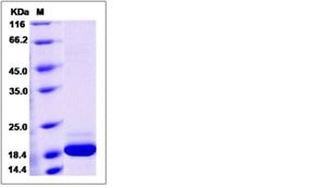
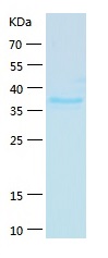
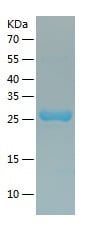
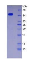
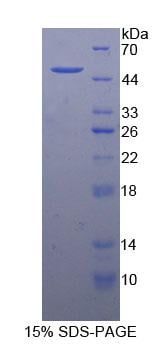
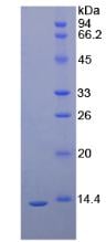
 SDS-PAGE
(SDS PAGE for Recombinant Human SLC34A2/NaPi2b protein)
SDS-PAGE
(SDS PAGE for Recombinant Human SLC34A2/NaPi2b protein)
SLC34A2/NaPi2b, Recombinant Protein (Cat# AAA120623)
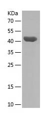
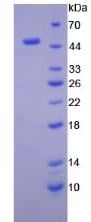
 SDS-PAGE
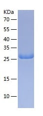
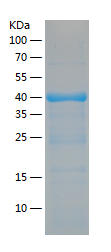
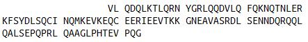
(SDS PAGE for Recombinant Human CD88/C5AR1 protein)
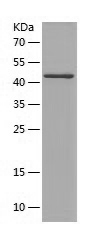
SDS-PAGE
(SDS PAGE for Recombinant Human CD88/C5AR1 protein)
CD88/C5AR1, Recombinant Protein (Cat# AAA120624)
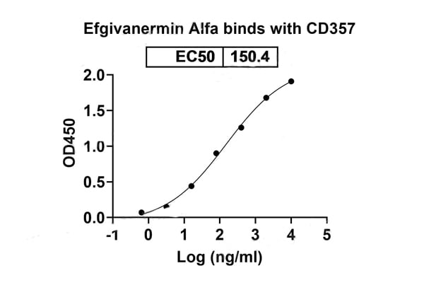
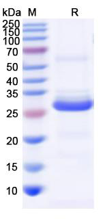
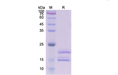
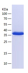
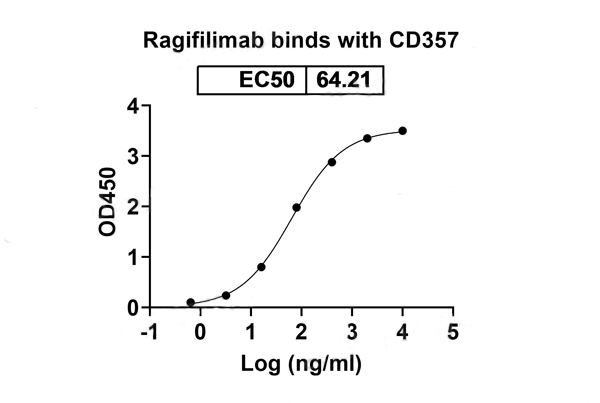 Bioactivity
(Detects Ragifilimab in indirect ELISAs.)
Bioactivity
(Detects Ragifilimab in indirect ELISAs.)
CD357/TNFRSF18/GITR, Recombinant Protein (Cat# AAA120631)
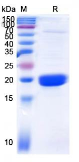
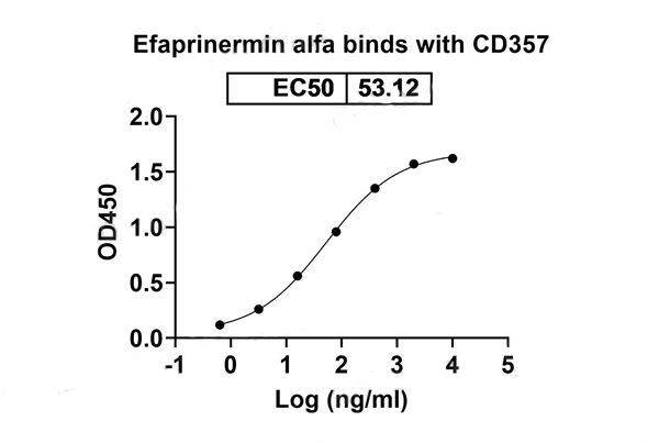
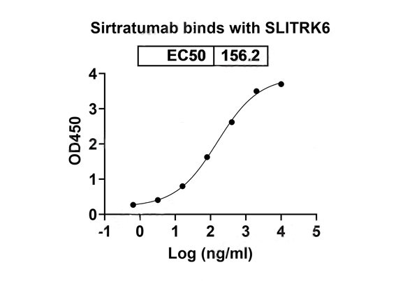 Bioactivity
(Detects Sirtratumab in indirect ELISAs.)
Bioactivity
(Detects Sirtratumab in indirect ELISAs.)
SLITRK6, Recombinant Protein (Cat# AAA120632)
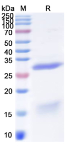
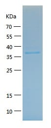
 SDS-PAGE
(SDS-PAGE for Recombinant Yersinia enterocolitica Invasin Protein)
SDS-PAGE
(SDS-PAGE for Recombinant Yersinia enterocolitica Invasin Protein)
Invasin, Recombinant Protein (Cat# AAA120639)
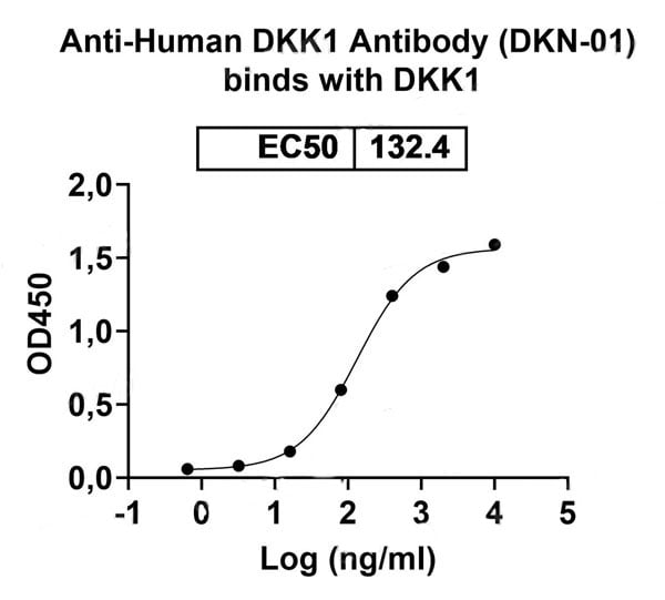 Bioactivity
(Detects Human DKK1 Antibody (DKN-01) in indirect ELISAs.)
Bioactivity
(Detects Human DKK1 Antibody (DKN-01) in indirect ELISAs.)
DKK1, Recombinant Protein (Cat# AAA120644)
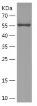
 SDS-PAGE
(SDS-PAGE for Recombinant Human PI4KB protein)
SDS-PAGE
(SDS-PAGE for Recombinant Human PI4KB protein)
PI4KB, Recombinant Protein (Cat# AAA120650)
 SDS-PAGE
(SDS-PAGE for Recombinant Human NOVA1 protein)
SDS-PAGE
(SDS-PAGE for Recombinant Human NOVA1 protein)
NOVA1, Recombinant Protein (Cat# AAA120713)
 SDS-PAGE
FHbp, Recombinant Protein (Cat# AAA120782)
 SDS-PAGE
(SDS PAGE for recombinant Human CD148/PTPRJ protein)
SDS-PAGE
(SDS PAGE for recombinant Human CD148/PTPRJ protein)
CD148/PTPRJ, Recombinant Protein (Cat# AAA120499)
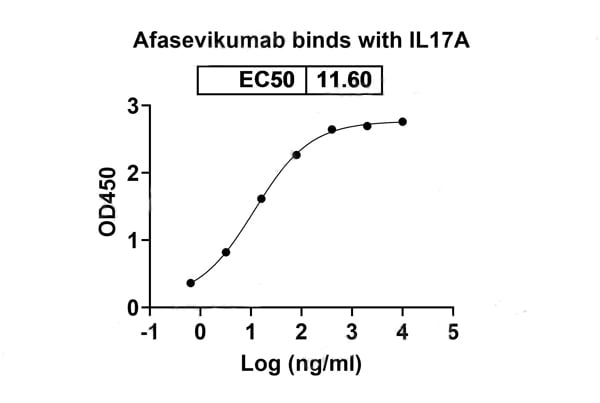
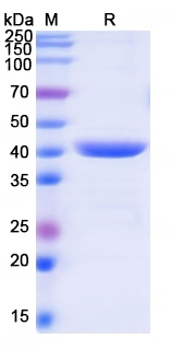
 Bioactivity
(Detects Remtolumab in indirect ELISAs.)
Bioactivity
(Detects Remtolumab in indirect ELISAs.)
IL17A, Recombinant Protein (Cat# AAA120501)
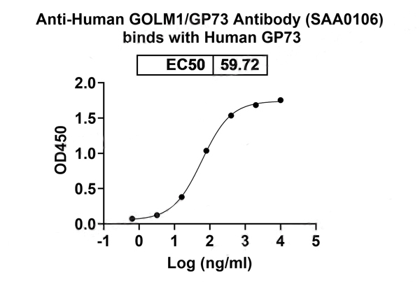 Bioactivity
(Detects Human GOLM1/GP73 Antibody in indirect ELISAs.)
Bioactivity
(Detects Human GOLM1/GP73 Antibody in indirect ELISAs.)
GOLM1/GP73, Recombinant Protein (Cat# AAA120505)
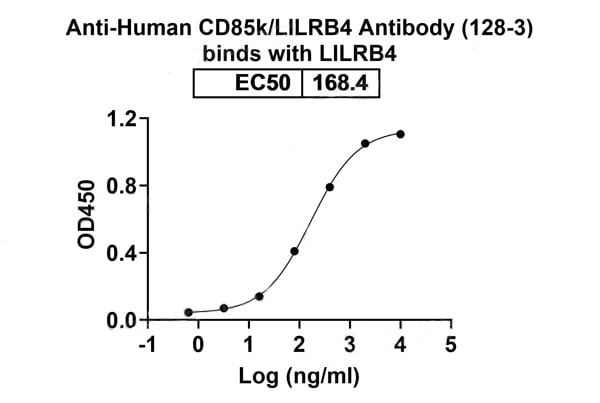
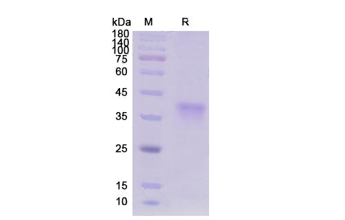
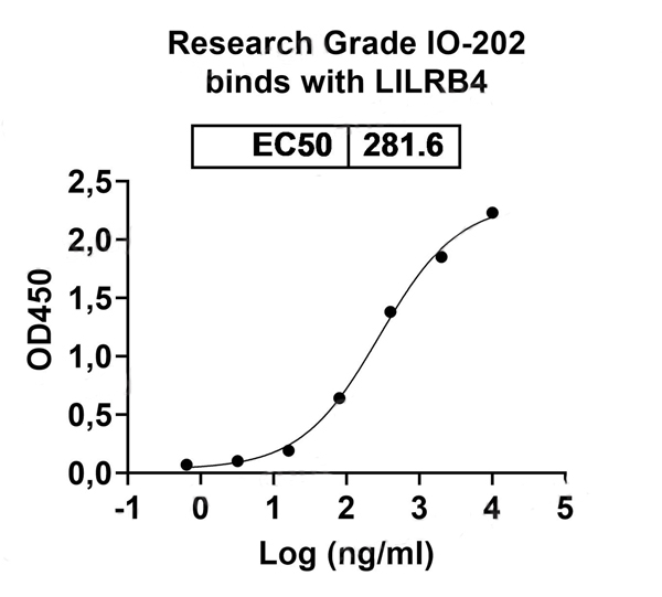 Bioactivity
(Detects IO-202 in indirect ELISAs.)
Bioactivity
(Detects IO-202 in indirect ELISAs.)
CD85k/LILRB4/ILT3, Recombinant Protein (Cat# AAA120507)
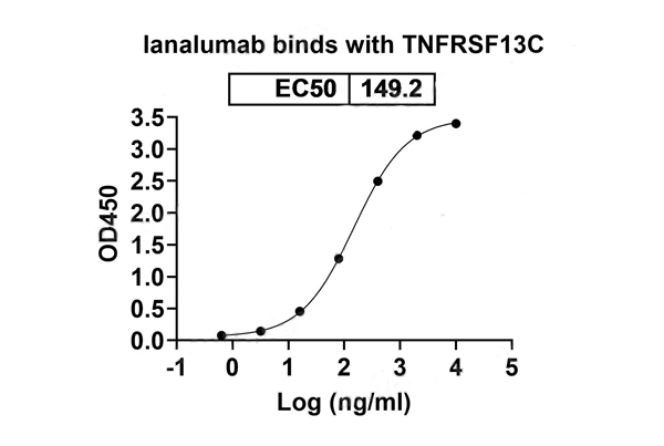
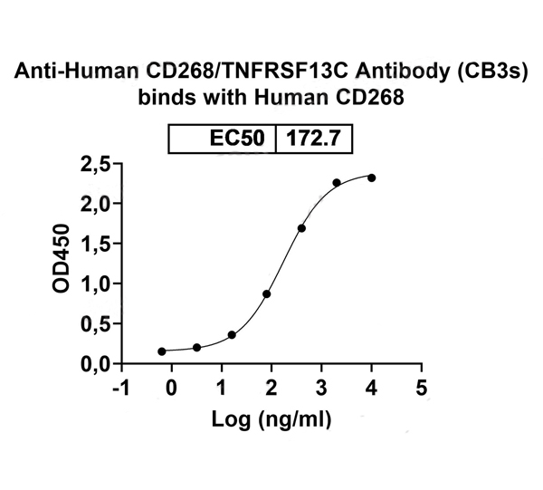 Bioactivity
(Detects Human CD268/TNFRSF13C Antibody (CB3s) in indirect ELISAs.)
Bioactivity
(Detects Human CD268/TNFRSF13C Antibody (CB3s) in indirect ELISAs.)
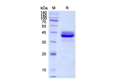
CD268/TNFRSF13C, Recombinant Protein (Cat# AAA120514)
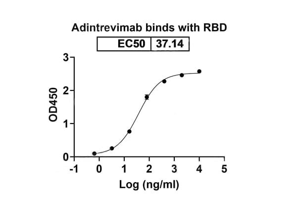
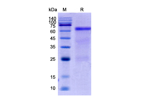
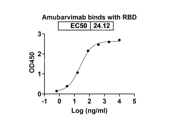 Bioactivity
(Detects Amubarvimab in indirect ELISAs.)
Bioactivity
(Detects Amubarvimab in indirect ELISAs.)
COVID 19 RBD Coronavirus, Recombinant Protein (Cat# AAA120520)
 SDS-PAGE
(SDS PAGE for recombinant Elizabethkingia miricola PNGase F)
SDS-PAGE
(SDS PAGE for recombinant Elizabethkingia miricola PNGase F)
PNGase F, Recombinant Protein (Cat# AAA120530)
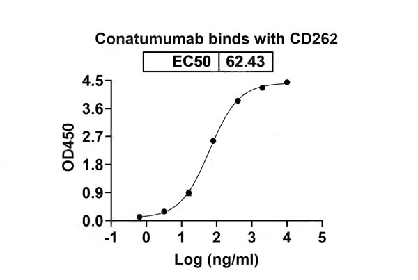
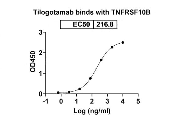
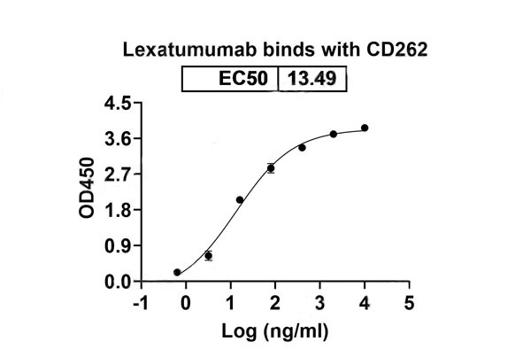 Bioactivity
(Detects Lexatumumab in indirect ELISAs.)
Bioactivity
(Detects Lexatumumab in indirect ELISAs.)
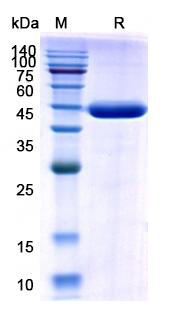
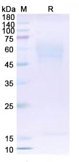
CD262/TNFRSF10B/TRAIL-R2, Recombinant Protein (Cat# AAA120536)
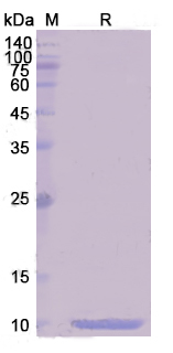
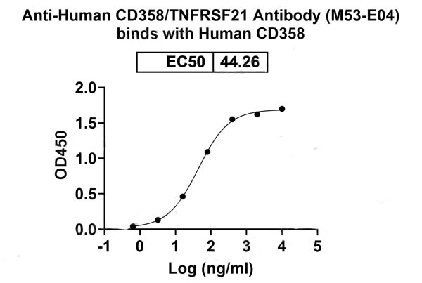 Bioactivity
(Detects Human CD358/TNFRSF21 Antibody (M53-E04) in indirect ELISAs.)
Bioactivity
(Detects Human CD358/TNFRSF21 Antibody (M53-E04) in indirect ELISAs.)
CD358/TNFRSF21, Recombinant Protein (Cat# AAA120555)
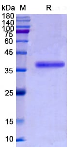
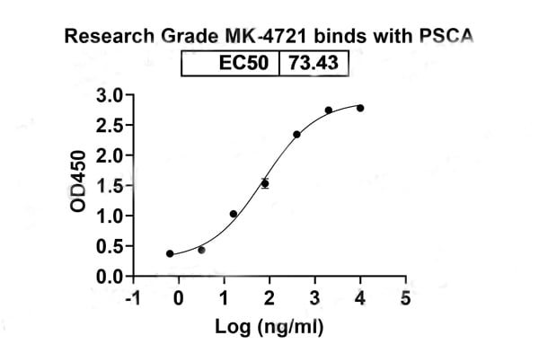 Bioactivity
(Detects MK-4721 in indirect ELISAs.)
Bioactivity
(Detects MK-4721 in indirect ELISAs.)
PSCA, Recombinant Protein (Cat# AAA120418)
 SDS-PAGE
(SDS PAGE for recombinant Human BPI)
SDS-PAGE
(SDS PAGE for recombinant Human BPI)
BPI, Recombinant Protein (Cat# AAA120460)
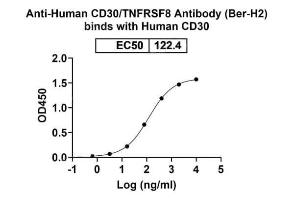
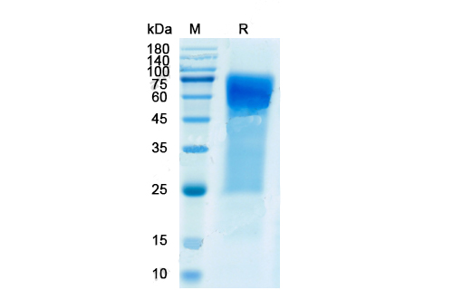
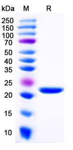
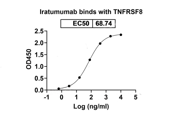 Bioactivity
(Detects Iratumumab in indirect ELISAs.)
Bioactivity
(Detects Iratumumab in indirect ELISAs.)
CD30/TNFRSF8, Recombinant Protein (Cat# AAA120470)
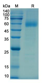
 SDS-PAGE
(SDS PAGE for recombinant Human RCVRN protein)
SDS-PAGE
(SDS PAGE for recombinant Human RCVRN protein)
RCVRN, Recombinant Protein (Cat# AAA120475)
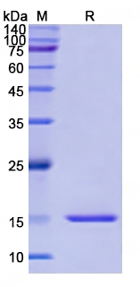
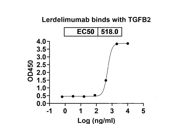 Bioactivity
(Detects Lerdelimumab in indirect ELISAs.)
Bioactivity
(Detects Lerdelimumab in indirect ELISAs.)
TGFB2, Recombinant Protein (Cat# AAA120491)
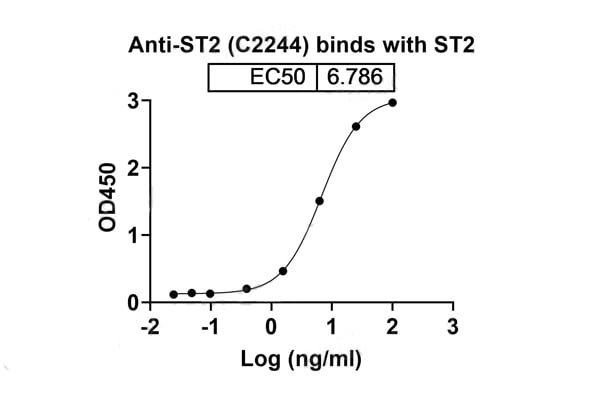
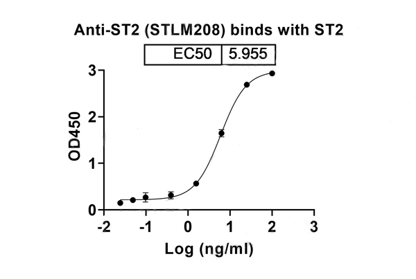 Bioactivity
(Detects Human IL1RL1/ST2 Antibody in indirect ELISAs.)
Bioactivity
(Detects Human IL1RL1/ST2 Antibody in indirect ELISAs.)
IL1RL1/ST2, Recombinant Protein (Cat# AAA120494)
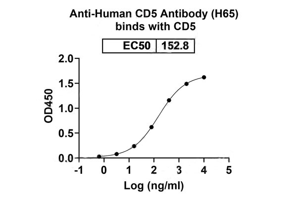
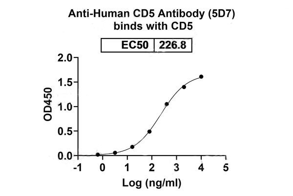 Bioactivity
(Detects Human CD5 Antibody (5D7) in indirect ELISAs.)
Bioactivity
(Detects Human CD5 Antibody (5D7) in indirect ELISAs.)
CD5, Recombinant Protein (Cat# AAA120437)
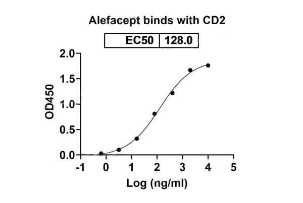
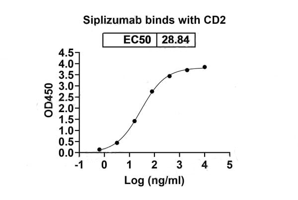
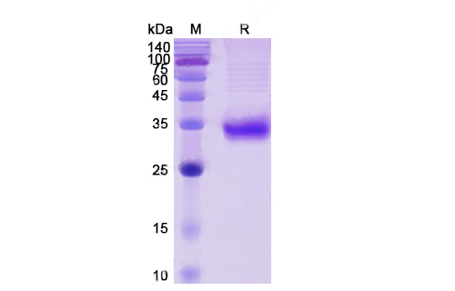
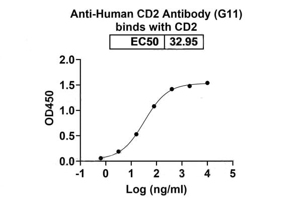 Bioactivity
(Detects Human CD2 Antibody (G11) in indirect ELISAs.)
Bioactivity
(Detects Human CD2 Antibody (G11) in indirect ELISAs.)
CD2, Recombinant Protein (Cat# AAA120438)
 Application Data
(Measured in antiviral assay using L929 cells infected with vesicular stomatitisvirus (VSV). The ED50 for this effect is typically 5-40 pg/mL.)
Application Data
(Measured in antiviral assay using L929 cells infected with vesicular stomatitisvirus (VSV). The ED50 for this effect is typically 5-40 pg/mL.)
IFNA14, Recombinant Protein (Cat# AAA257481)
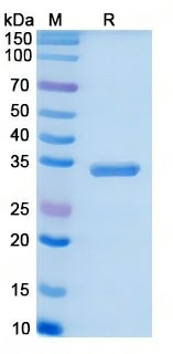
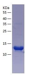
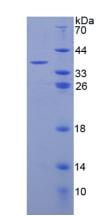
 SDS-PAGE
NUMBL, Recombinant Protein (Cat# AAA250782)
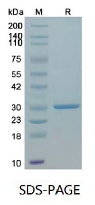
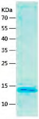
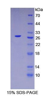
 SDS-PAGE
ACAD9, Recombinant Protein (Cat# AAA250789)
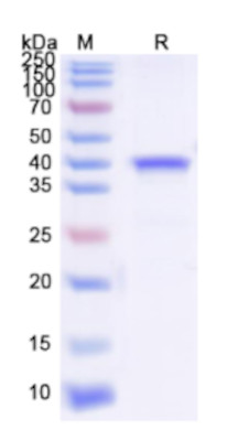
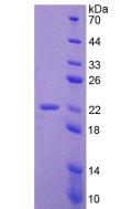
 SDS-PAGE
(SDS-Page)
SDS-PAGE
(SDS-Page)
AHSG, Recombinant Protein (Cat# AAA249457)
 SDS-PAGE
(SDS-Page)
SDS-PAGE
(SDS-Page)
Androgen Receptor, Recombinant Protein (Cat# AAA249466)
 SDS-PAGE
(SDS-Page)
SDS-PAGE
(SDS-Page)
RPS4X, Recombinant Protein (Cat# AAA249495)
 SDS-PAGE
EDA, Recombinant Protein (Cat# AAA249515)
 SDS-PAGE
(SDS-Page)
SDS-PAGE
(SDS-Page)
XAGE1, Recombinant Protein (Cat# AAA249523)
 SDS-PAGE
(SDS-Page)
SDS-PAGE
(SDS-Page)
MMACHC, Recombinant Protein (Cat# AAA249529)
 SDS-PAGE
ADCY3, Recombinant Protein (Cat# AAA249426)
 SDS-PAGE
(SDS-Page)
SDS-PAGE
(SDS-Page)
COVID 19 Spike Glycoprotein RBD Coronavirus, Recombinant Protein (Cat# AAA249428)
Nucleoprotein (NP), Recombinant Protein (Cat# AAA244042)
GYPA, Recombinant Protein (Cat# AAA250141)
 SDS-PAGE
ARHGEF4, Recombinant Protein (Cat# AAA250164)
 Application Data
Adrenomedullin (ADM), Recombinant Protein (Cat# AAA135547)
 SDS-PAGE
Complement Component 3a (C3a), Recombinant Protein (Cat# AAA135548)
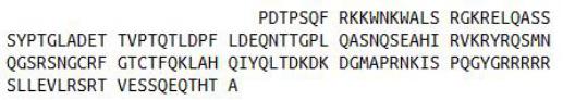
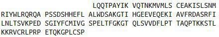
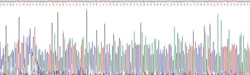
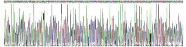 Gene Sequence
(Gene Sequencing (extract))
Gene Sequence
(Gene Sequencing (extract))
Hepatocyte Nuclear Factor 4 Alpha (HNF4a), Recombinant Protein (Cat# AAA135552)
 SDS-PAGE
Antithrombin (AT), Recombinant Protein (Cat# AAA135554)
 SDS-PAGE
Cystathionine Gamma Lyase (CSE), Recombinant Protein (Cat# AAA135566)
 SDS-PAGE
Growth Arrest Specific Protein 6 (GAS6), Recombinant Protein (Cat# AAA135568)
 SDS-PAGE
Golgi Protein 73 (GP73), Recombinant Protein (Cat# AAA135589)
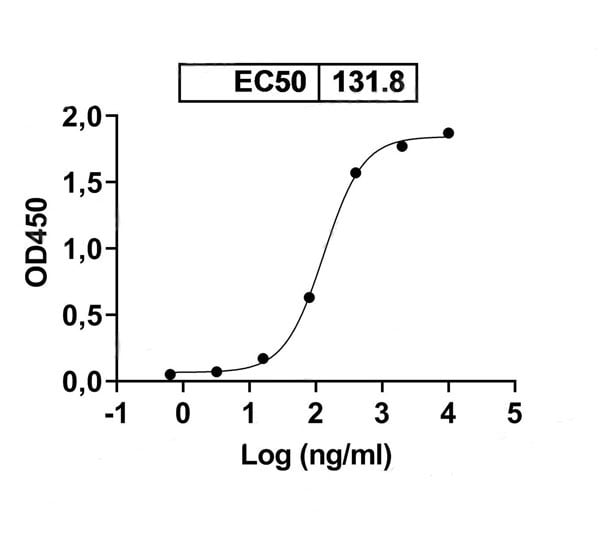
 Application Data
Cluster Of Differentiation 8b (CD8b), Recombinant Protein (Cat# AAA135598)
 SDS-PAGE
Kidney Injury Molecule 1 (Kim1), Recombinant Protein (Cat# AAA134792)
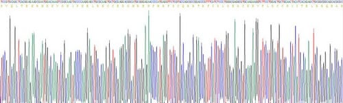
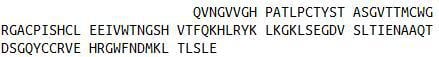
 Gene Sequence
(Gene Sequencing (extract))
Gene Sequence
(Gene Sequencing (extract))
Interferon Beta (IFNb), Recombinant Protein (Cat# AAA134648)
What Are Recombinant Proteins?
A recombinant protein is created by inserting the gene of interest into a host system like E. coli, mammalian cells, or insect cells. This protein expression method results in the large-scale production of protein in a controlled environment with highly precise structures and functionality. Proteins produced via the “Recombinant” methodology have proven themselves crucial in various biological research areas, including immunology, neuroscience, cancer research, drug delivery and development, and more.
Core Recombinant Protein Product Lines:
- Human: Produced from human genes (e.g., growth factors, cytokines).
- Animal: Derived from mouse, rat, or other animals (e.g., mouse antibodies, enzymes).
- Bacterial: Produced in bacteria like E. coli (e.g., enzymes, bacterial toxins).
- Yeast: Produced in yeast cells (e.g., surface proteins, metabolic enzymes).
- Insect:Expressed in insect cells (e.g., viral proteins, glycoproteins).
- Viral: Derived from viral genomes (e.g., spike proteins for vaccine development).
How Does It Work?
- Gene Identification and Cloning: Scientists first find the exact gene that codes for the protein they are interested in producing, and then create a copy of said gene.
- Recombinant DNA Creation: The copied gene is then inserted into a vector (a vehicle-like plasmid) that can be transported safely into your intended expression host cell/system.
- Host Cell Transformation: The vector (with the foreign gene inside) is shuttled into a host cell. This host can be a bacterium (like E. coli), yeast, insect cells, or mammalian cells, depending on the type of protein or protein characteristics needed.
- Protein Expression:The host cell “reads” the copied gene that was shuttled into it, and begins to produce the gene’s coded-for protein using its own cellular machinery. The host can continue to multiply, and with each new cell, more protein is produced.
- Protein Purification:After enough protein has been made, scientists collect the host cell and separate out the target protein. The protein is then carefully purified so it is free of contaminants, ideally active, and also safe for use.
The result is a purified recombinant protein — a lab-made protein that is biologically identical (in most respects) to the natural/native version.
We take this process a step further, offering recombinant proteins that are produced with precise control over sequence modifications, expression levels, and large-scale production. This ensures high-quality, consistent results that will be able to support your research needs and empower your scientific discoveries.
Some of the expression hosts/systems used in recombinant protein generation by AAA Biotech include:
- Escherichia coli (E. coli)
- Human (Homo sapiens)
- Chinese Hamster
- Yeast
- Insect cells (e.g., Baculovirus expression system)
Advantages Of Our Recombinant Proteins:
Recombinant Protein Offerings
| S.No | Protein Expression Systems | Quality and Bioactivity | Formats and Flexibility |
|---|---|---|---|
| 01. | E. coli, HEK293, CHO, yeast, and insect cells | Validated high bioactivity and binding activity | Ready-to-use formats (lyophilized or liquid formulations available) |
| 02. | 675+ recombinant protein products | High protein purity (≥95%) | Available in flexible pack sizes |
| 03. | Custom protein production (His-tag, GST, FLAG, and Fc fusion) | Lot-to-lot consistency through QC protocols | Wide range of targets (Cytokines, growth factors, enzymes, receptors, signaling proteins, etc.) |
Applications Of Our Recombinant Proteins
- Studying protein interactions (protein–protein or protein–DNA).
- Testing biological pathways with functional/activity assays.
- Creating standard curves in ELISA and other tests.
- Making antigens/immunogens for antibody production.
- Drug discovery and screening.
- Finding and confirming biomarkers.
- Researching cell signaling and immunity.
- Developing vaccines.
Read more about the applications of the recombinant proteins here!
Why Buy Recombinant Proteins from AAA Biotech?
1. High Purity & Verified Quality:
Most of our proteins are ≥ 95% pure. We also test the biological activity of our products where it's relevant (it will be indicated on the product page).
2. Multiple Expression Systems:
We offer proteins made in multiple host systems, and this enables researchers to pick the system that gives them the best folding, post-translational modifications, or functionality for their experiment requirements.
3. Custom & Flexible Options:
Need a special tag (His, GST, FLAG, Fc fusion)? Or a custom variant? We can provide it. Our proteins come in formats that are lab-friendly: lyophilized (dry) or “ready-to-use” liquid.
4. Rigorous Quality Control & Documentation:
Every batch is backed by strong QC. This means you can rely on product consistency — two different batches will perform similarly.
5. Wide Variety & Global Availability:
We carry a large and growing catalog of recombinant proteins to cover almost all needs in research (immunology, oncology, vaccine work, etc.)
6. Researcher-Focused Support:
Technical support, clear documentation, and user-friendly product pages help you select the right protein for your work. Plus, our user-friendly packaging and handling, and global shipping are all designed to reduce delays, damage, and hassles.
Order Recombinant Proteins Today!
We are committed to supporting the research community with recombinant proteins that display exceptional performance and reliability. Our proteins are produced using industry-standard methods and are validated to meet the needs of academic, pharmaceutical, and biotechnology laboratories.
We have a track record of delivering proteins that work — large numbers of researchers rely on them in many published studies. Browse our catalog to find the perfect protein for your research applications now!
FAQ
1. What is involved in the purification of recombinant proteins?
The purification process of these recombinant proteins is performed to isolate the specific protein produced by the host cell from unwanted substances. The process generally includes techniques such as “affinity chromatography” to separate the protein from other proteins and impurities. AAA Biotech generally offers proteins ≥ 95% pure, depending on the protein type.
2. What are some examples of popular AAA Biotech recombinant proteins?
Some examples of recombinant proteins include:
Retinoblastoma Protein (Cat # AAA10852) – reportedly used in vaccine research and antibody development. Hepatitis B Surface Antigen (HBsAg) (Cat# AAA13446) – reportedly used in diagnostic tests and vaccine development for Hepatitis B. High-Mobility Group Box 1 (HMGB1) (Cat# AAA10851) – reportedly used in inflammation and immune response research.
3. How are AAA Biotech recombinant proteins validated?
Each batch undergoes stringent quality control checks, including SDS-PAGE analysis, endotoxin testing (for select products), and activity assaying (for select products). Certificates of Analysis are provided with every product (only upon request for some products).
4. Are your proteins suitable for therapeutic development or only research?
AAA Biotech recombinant proteins are strictly for research-use only and are not intended for diagnostic or therapeutic purposes in humans or animals.
5. What types of expression systems do you use for recombinant protein production?
The production labs use a variety of expression platforms, including bacterial (E. coli), yeast, insect (baculovirus), and mammalian (HEK293, CHO) systems. The expression system used depends on the complexity and intended function/use of the protein.