Filters
▼Clonality
▼Type
▼Reactivity
▼Gene Name
▼Isotype
▼Host
▼Application
▼Clone
▼Polyclonal Antibodies
At AAA Biotech also known as AAA Bio or AAABio, we provide a broad range of purified polyclonal antibodies (pAbs) that are able to all be browsed online through our website. Due to their high specificity and strong binding affinity, these antibodies are ideal for wide swathes of research and experimental applications.
Our polyclonal antibodies can easily support your work, whether you use them for Western Blotting, Immunocytochemistry (with or without Immunofluorescence used in conjunction), Immunohistochemistry, Immunoprecipitation, and ELISA tests. We highly encourage you to browse our range of pAbs and choose the one that best suits your experimental model.
Viewing 3750-3800 of 96812 product results
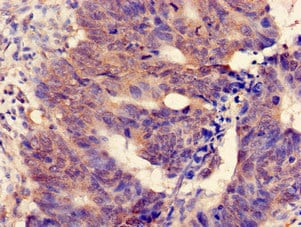
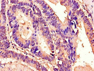
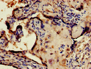
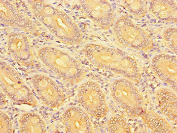
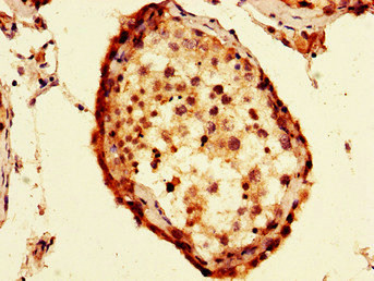 IHC (Immunohiostchemistry)
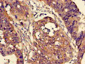
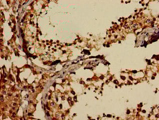
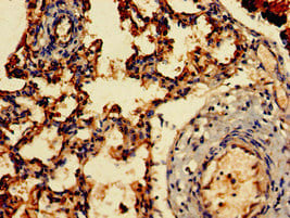
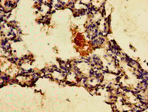
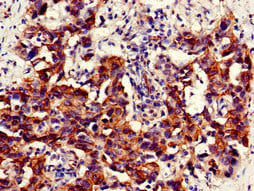
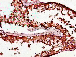
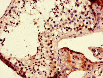
(Immunohistochemistry of paraffin-embedded human testis tissue using AAA233132 at dilution of 1:100)
IHC (Immunohiostchemistry)
(Immunohistochemistry of paraffin-embedded human testis tissue using AAA233132 at dilution of 1:100)
FAF1, Polyclonal Antibody (Cat# AAA233132)
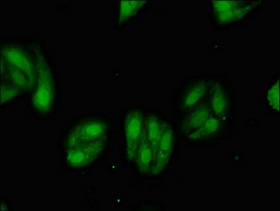
 IHC (Immunohistochemisry)
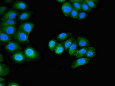
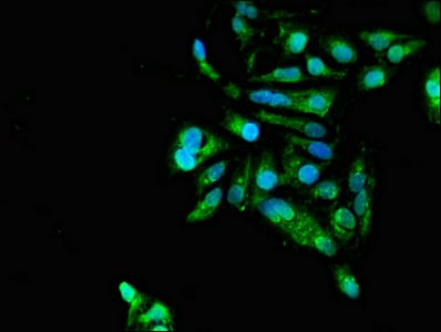
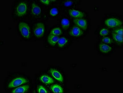
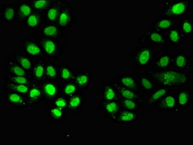
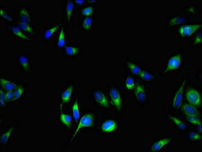
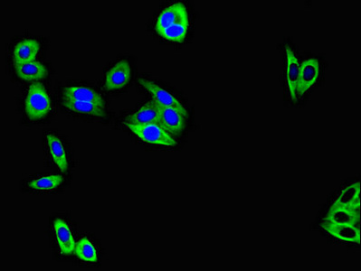
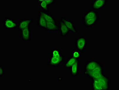
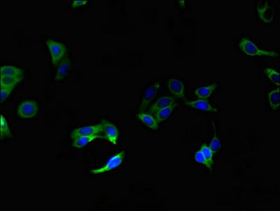
(Immunofluorescent analysis of MCF-7 cells using AAA233135 at a dilution of 1:100 and Alexa Fluor 488-congugated AffiniPure Goat Anti-Rabbit IgG(H+L))
IHC (Immunohistochemisry)
(Immunofluorescent analysis of MCF-7 cells using AAA233135 at a dilution of 1:100 and Alexa Fluor 488-congugated AffiniPure Goat Anti-Rabbit IgG(H+L))
STUB1, Polyclonal Antibody (Cat# AAA233135)
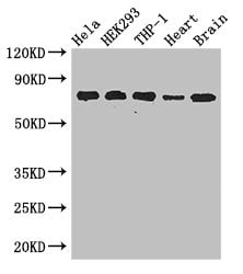
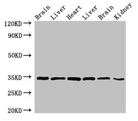
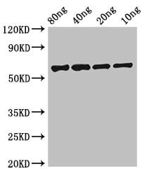
 WB (Western Blot)
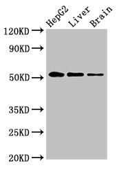
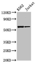
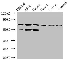
(Western BlotPositive WB detected in: K562 whole cell lysateAll lanes: CDYL antibody at 3ug/mlSecondaryGoat polyclonal to rabbit IgG at 1/50000 dilutionPredicted band size: 67,61,35,46 KDaObserved band size: 67 KDa)
WB (Western Blot)
(Western BlotPositive WB detected in: K562 whole cell lysateAll lanes: CDYL antibody at 3ug/mlSecondaryGoat polyclonal to rabbit IgG at 1/50000 dilutionPredicted band size: 67,61,35,46 KDaObserved band size: 67 KDa)
CDYL, Polyclonal Antibody (Cat# AAA233137)
 IHC (Immunohistochemisry)
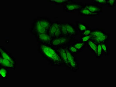
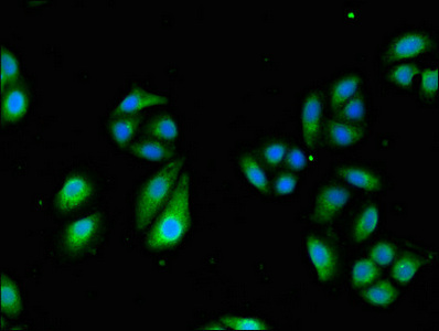
(Immunofluorescent analysis of Hela cells using AAA233172 at a dilution of 1:100 and Alexa Fluor 488-congugated AffiniPure Goat Anti-Rabbit IgG(H+L))
IHC (Immunohistochemisry)
(Immunofluorescent analysis of Hela cells using AAA233172 at a dilution of 1:100 and Alexa Fluor 488-congugated AffiniPure Goat Anti-Rabbit IgG(H+L))
C1QA, Polyclonal Antibody (Cat# AAA233172)
CACNG3, Polyclonal Antibody (Cat# AAA233174)
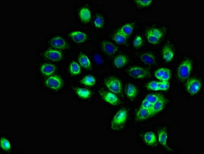
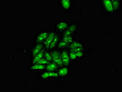
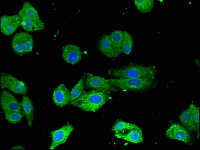
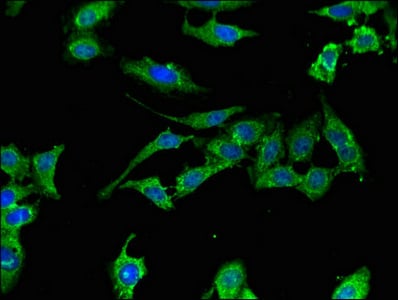
 IF (Immunofluorescence)
(Immunofluorescent analysis of Hela cells using AAA233177 at a dilution of 1:100 and Alexa Fluor 488-congugated AffiniPure Goat Anti-Rabbit IgG(H+L))
IF (Immunofluorescence)
(Immunofluorescent analysis of Hela cells using AAA233177 at a dilution of 1:100 and Alexa Fluor 488-congugated AffiniPure Goat Anti-Rabbit IgG(H+L))
CAPN14, Polyclonal Antibody (Cat# AAA233177)
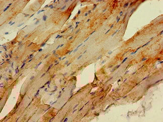
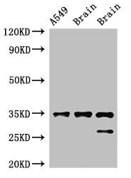
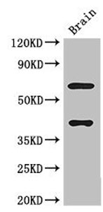
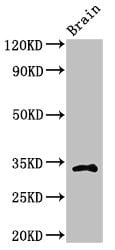 WB (Western Blot)
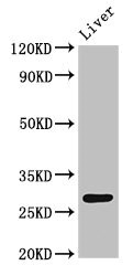
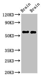
(Western BlotPositive WB detected in: Mouse brain tissueAll lanes: CASP6 antibody at 2.4ug/mlSecondaryGoat polyclonal to rabbit IgG at 1/50000 dilutionPredicted band size: 34,23 KDaObserved band size: 34 KDa)
WB (Western Blot)
(Western BlotPositive WB detected in: Mouse brain tissueAll lanes: CASP6 antibody at 2.4ug/mlSecondaryGoat polyclonal to rabbit IgG at 1/50000 dilutionPredicted band size: 34,23 KDaObserved band size: 34 KDa)
CASP6, Polyclonal Antibody (Cat# AAA233178)
 IF (Immunofluorescence)
(Immunofluorescent analysis of A549 cells using AAA233181 at a dilution of 1:100 and Alexa Fluor 488-congugated AffiniPure Goat Anti-Rabbit IgG(H+L))
IF (Immunofluorescence)
(Immunofluorescent analysis of A549 cells using AAA233181 at a dilution of 1:100 and Alexa Fluor 488-congugated AffiniPure Goat Anti-Rabbit IgG(H+L))
CD1C, Polyclonal Antibody (Cat# AAA233181)
 IF (Immunofluorescence)
(Immunofluorescent analysis of Hela cells using AAA233185 at a dilution of 1:100 and Alexa Fluor 488-congugated AffiniPure Goat Anti-Rabbit IgG(H+L))
IF (Immunofluorescence)
(Immunofluorescent analysis of Hela cells using AAA233185 at a dilution of 1:100 and Alexa Fluor 488-congugated AffiniPure Goat Anti-Rabbit IgG(H+L))
CDX1, Polyclonal Antibody (Cat# AAA233185)
 IHC (Immunohistochemisry)
(Immunofluorescent analysis of A549 cells using AAA233187 at a dilution of 1:100 and Alexa Fluor 488-congugated AffiniPure Goat Anti-Rabbit IgG(H+L))
IHC (Immunohistochemisry)
(Immunofluorescent analysis of A549 cells using AAA233187 at a dilution of 1:100 and Alexa Fluor 488-congugated AffiniPure Goat Anti-Rabbit IgG(H+L))
CHRM5, Polyclonal Antibody (Cat# AAA233187)
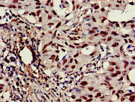
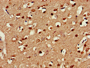
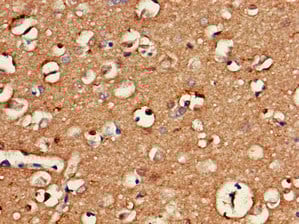
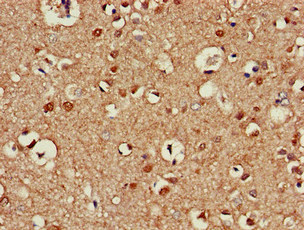
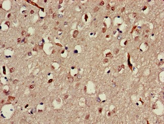
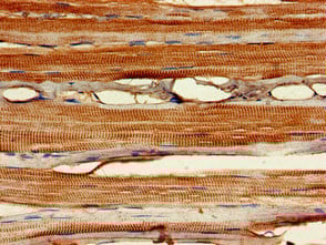
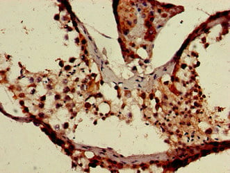
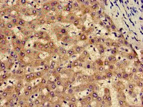
 IHC (Immunohiostchemistry)
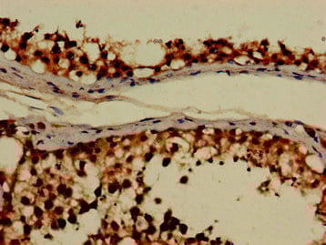
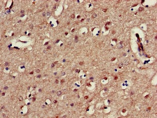
(Immunohistochemistry of paraffin-embedded human brain tissue using AAA233188 at dilution of 1:100)
IHC (Immunohiostchemistry)
(Immunohistochemistry of paraffin-embedded human brain tissue using AAA233188 at dilution of 1:100)
CHST1, Polyclonal Antibody (Cat# AAA233188)
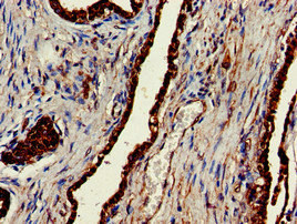
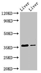
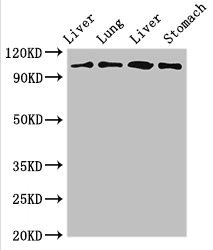
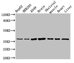
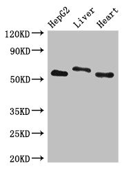
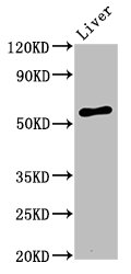
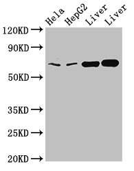 WB (Western Blot)
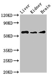
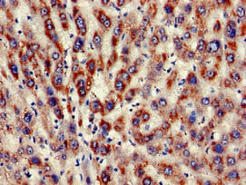
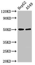
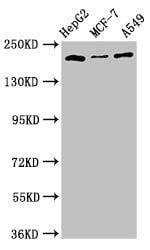
(Western BlotPositive WB detected in: Hela whole cell lysate,HepG2 whole cell lysate,Rat liver tissue,Mouse liver tissueAll lanes: CLINT1 antibody at 2ug/mlSecondaryGoat polyclonal to rabbit IgG at 1/50000 dilutionPredicted band size: 69,71 KDaObserved band size: 69 KDa)
WB (Western Blot)
(Western BlotPositive WB detected in: Hela whole cell lysate,HepG2 whole cell lysate,Rat liver tissue,Mouse liver tissueAll lanes: CLINT1 antibody at 2ug/mlSecondaryGoat polyclonal to rabbit IgG at 1/50000 dilutionPredicted band size: 69,71 KDaObserved band size: 69 KDa)
CLINT1, Polyclonal Antibody (Cat# AAA233189)
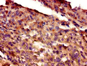
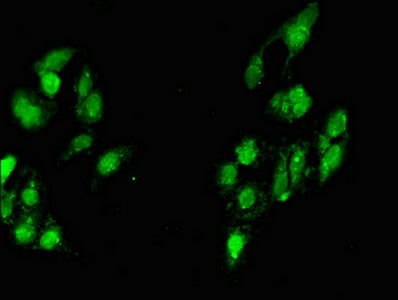
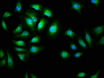
 IHC (Immunohistochemisry)
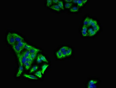
(Immunofluorescent analysis of PC3 cells using AAA233191 at a dilution of 1:100 and Alexa Fluor 488-congugated AffiniPure Goat Anti-Rabbit IgG(H+L))
IHC (Immunohistochemisry)
(Immunofluorescent analysis of PC3 cells using AAA233191 at a dilution of 1:100 and Alexa Fluor 488-congugated AffiniPure Goat Anti-Rabbit IgG(H+L))
CPB2, Polyclonal Antibody (Cat# AAA233191)
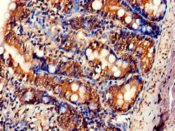
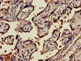
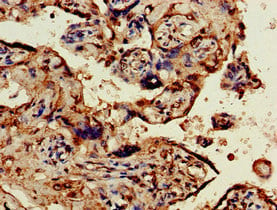
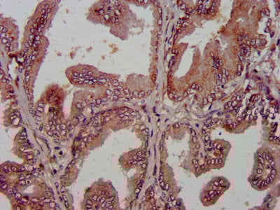
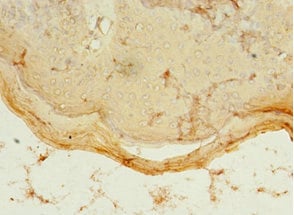
 IHC (Immunohiostchemistry)
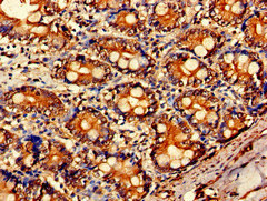
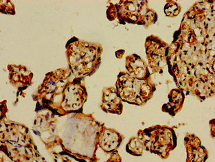
(Immunohistochemistry of paraffin-embedded human placenta tissue using AAA233193 at dilution of 1:100)
IHC (Immunohiostchemistry)
(Immunohistochemistry of paraffin-embedded human placenta tissue using AAA233193 at dilution of 1:100)
CSH2, Polyclonal Antibody (Cat# AAA233193)
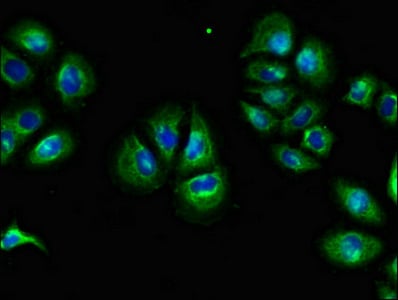
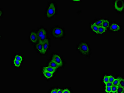
 IHC (Immunohistochemisry)
(Immunofluorescent analysis of A549 cells using AAA233195 at a dilution of 1:100 and Alexa Fluor 488-congugated AffiniPure Goat Anti-Rabbit IgG(H+L))
IHC (Immunohistochemisry)
(Immunofluorescent analysis of A549 cells using AAA233195 at a dilution of 1:100 and Alexa Fluor 488-congugated AffiniPure Goat Anti-Rabbit IgG(H+L))
CUTA, Polyclonal Antibody (Cat# AAA233195)
 IF (Immunofluorescence)
(Immunofluorescent analysis of Hela cells using AAA233198 at a dilution of 1:100 and Alexa Fluor 488-congugated AffiniPure Goat Anti-Rabbit IgG(H+L))
IF (Immunofluorescence)
(Immunofluorescent analysis of Hela cells using AAA233198 at a dilution of 1:100 and Alexa Fluor 488-congugated AffiniPure Goat Anti-Rabbit IgG(H+L))
EGR2, Polyclonal Antibody (Cat# AAA233198)
 IF (Immunofluorescence)
(Immunofluorescent analysis of HepG2 cells using AAA233199 at a dilution of 1:100 and Alexa Fluor 488-congugated AffiniPure Goat Anti-Rabbit IgG(H+L))
IF (Immunofluorescence)
(Immunofluorescent analysis of HepG2 cells using AAA233199 at a dilution of 1:100 and Alexa Fluor 488-congugated AffiniPure Goat Anti-Rabbit IgG(H+L))
EIF2AK1, Polyclonal Antibody (Cat# AAA233199)
 IF (Immunofluorescence)
(Immunofluorescent analysis of A549 cells using AAA233200 at a dilution of 1:100 and Alexa Fluor 488-congugated AffiniPure Goat Anti-Rabbit IgG(H+L))
IF (Immunofluorescence)
(Immunofluorescent analysis of A549 cells using AAA233200 at a dilution of 1:100 and Alexa Fluor 488-congugated AffiniPure Goat Anti-Rabbit IgG(H+L))
F2RL1, Polyclonal Antibody (Cat# AAA233200)
 IF (Immunofluorescence)
(Immunofluorescent analysis of Hela cells using AAA233209 at a dilution of 1:100 and Alexa Fluor 488-congugated AffiniPure Goat Anti-Rabbit IgG(H+L))
IF (Immunofluorescence)
(Immunofluorescent analysis of Hela cells using AAA233209 at a dilution of 1:100 and Alexa Fluor 488-congugated AffiniPure Goat Anti-Rabbit IgG(H+L))
GAS7, Polyclonal Antibody (Cat# AAA233209)
 IHC (Immunohistochemisry)
(Immunofluorescent analysis of Hela cells using AAA233210 at a dilution of 1:100 and Alexa Fluor 488-congugated AffiniPure Goat Anti-Rabbit IgG(H+L))
IHC (Immunohistochemisry)
(Immunofluorescent analysis of Hela cells using AAA233210 at a dilution of 1:100 and Alexa Fluor 488-congugated AffiniPure Goat Anti-Rabbit IgG(H+L))
ARF1, Polyclonal Antibody (Cat# AAA233210)
 WB (Western Blot)
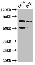
(Western BlotPositive WB detected in: Hela whole cell lysate,PC3 whole cell lysateAll lanes: GLB1 antibody at 4ug/mlSecondaryGoat polyclonal to rabbit IgG at 1/50000 dilutionPredicted band size: 77,61,73 KDaObserved band size: 77 KDa)
WB (Western Blot)
(Western BlotPositive WB detected in: Hela whole cell lysate,PC3 whole cell lysateAll lanes: GLB1 antibody at 4ug/mlSecondaryGoat polyclonal to rabbit IgG at 1/50000 dilutionPredicted band size: 77,61,73 KDaObserved band size: 77 KDa)
GLB1, Polyclonal Antibody (Cat# AAA233214)
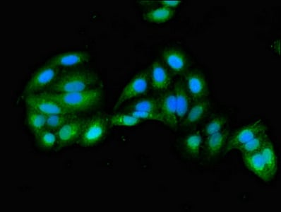
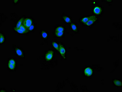
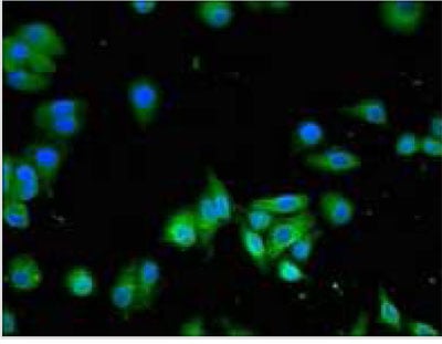
 IHC (Immunohistochemisry)
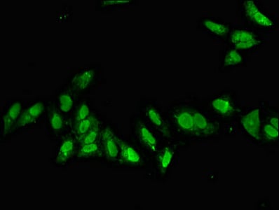
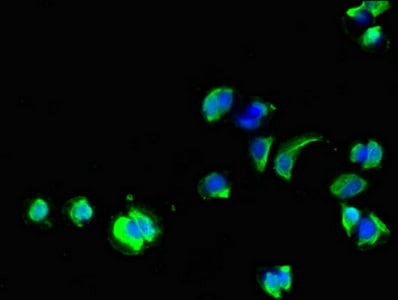
(Immunofluorescent analysis of MCF-7 cells using AAA233218 at a dilution of 1:100 and Alexa Fluor 488-congugated AffiniPure Goat Anti-Rabbit IgG(H+L))
IHC (Immunohistochemisry)
(Immunofluorescent analysis of MCF-7 cells using AAA233218 at a dilution of 1:100 and Alexa Fluor 488-congugated AffiniPure Goat Anti-Rabbit IgG(H+L))
GPC1, Polyclonal Antibody (Cat# AAA233218)
 IF (Immunofluorescence)
(Immunofluorescent analysis of A549 cells using AAA233219 at a dilution of 1:100 and Alexa Fluor 488-congugated AffiniPure Goat Anti-Rabbit IgG(H+L))
IF (Immunofluorescence)
(Immunofluorescent analysis of A549 cells using AAA233219 at a dilution of 1:100 and Alexa Fluor 488-congugated AffiniPure Goat Anti-Rabbit IgG(H+L))
GPR4, Polyclonal Antibody (Cat# AAA233219)
 IF (Immunofluorescence)
(Immunofluorescent analysis of HepG2 cells using AAA233221 at a dilution of 1:100 and Alexa Fluor 488-congugated AffiniPure Goat Anti-Rabbit IgG(H+L))
IF (Immunofluorescence)
(Immunofluorescent analysis of HepG2 cells using AAA233221 at a dilution of 1:100 and Alexa Fluor 488-congugated AffiniPure Goat Anti-Rabbit IgG(H+L))
GRHPR, Polyclonal Antibody (Cat# AAA233221)
 IF (Immunofluorescence)
(Immunofluorescent analysis of HepG2 cells using AAA233224 at a dilution of 1:100 and Alexa Fluor 488-congugated AffiniPure Goat Anti-Rabbit IgG(H+L))
IF (Immunofluorescence)
(Immunofluorescent analysis of HepG2 cells using AAA233224 at a dilution of 1:100 and Alexa Fluor 488-congugated AffiniPure Goat Anti-Rabbit IgG(H+L))
HAT1, Polyclonal Antibody (Cat# AAA233224)
 IF (Immunofluorescence)
(Immunofluorescent analysis of PC3 cells using AAA233225 at a dilution of 1:100 and Alexa Fluor 488-congugated AffiniPure Goat Anti-Rabbit IgG (H+L))
IF (Immunofluorescence)
(Immunofluorescent analysis of PC3 cells using AAA233225 at a dilution of 1:100 and Alexa Fluor 488-congugated AffiniPure Goat Anti-Rabbit IgG (H+L))
HK2, Polyclonal Antibody (Cat# AAA233225)
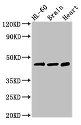
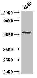 WB (Western Blot)
(Western BlotPositive WB detected in: A549 whole cell lysateAll lanes: ATG13 antibody at 4ug/mlSecondaryGoat polyclonal to rabbit IgG at 1/50000 dilutionPredicted band size: 57,53,45,61 KDaObserved band size: 57 KDa)
WB (Western Blot)
(Western BlotPositive WB detected in: A549 whole cell lysateAll lanes: ATG13 antibody at 4ug/mlSecondaryGoat polyclonal to rabbit IgG at 1/50000 dilutionPredicted band size: 57,53,45,61 KDaObserved band size: 57 KDa)
ATG13, Polyclonal Antibody (Cat# AAA233236)
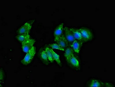
 IF (Immunofluorescence)
(Immunofluorescent analysis of PC3 cells using AAA233238 at a dilution of 1:100 and Alexa Fluor 488-congugated AffiniPure Goat Anti-Rabbit IgG(H+L))
IF (Immunofluorescence)
(Immunofluorescent analysis of PC3 cells using AAA233238 at a dilution of 1:100 and Alexa Fluor 488-congugated AffiniPure Goat Anti-Rabbit IgG(H+L))
KIR3DL1, Polyclonal Antibody (Cat# AAA233238)
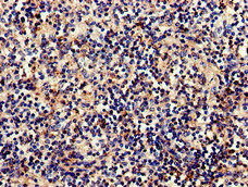
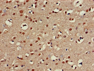
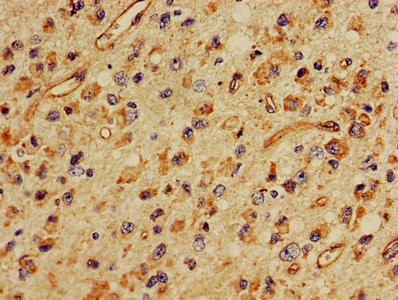
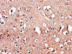
 IHC (Immunohiostchemistry)
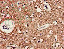
(Immunohistochemistry of paraffin-embedded human brain tissue using AAA233239 at dilution of 1:100)
IHC (Immunohiostchemistry)
(Immunohistochemistry of paraffin-embedded human brain tissue using AAA233239 at dilution of 1:100)
MAP3K13, Polyclonal Antibody (Cat# AAA233239)
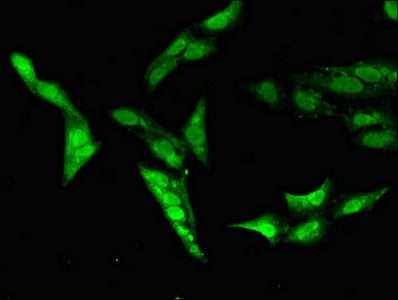
 IF (Immunofluorescence)
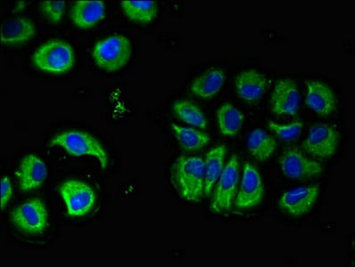
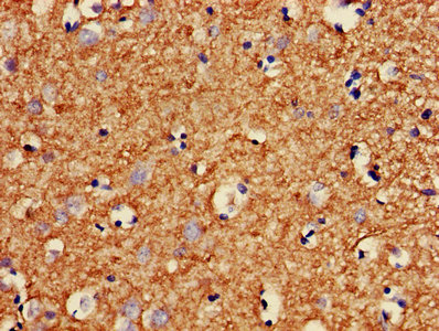
(Immunofluorescent analysis of Hela cells using AAA233241 at a dilution of 1:100 and Alexa Fluor 488-congugated AffiniPure Goat Anti-Rabbit IgG(H+L))
IF (Immunofluorescence)
(Immunofluorescent analysis of Hela cells using AAA233241 at a dilution of 1:100 and Alexa Fluor 488-congugated AffiniPure Goat Anti-Rabbit IgG(H+L))
MBP, Polyclonal Antibody (Cat# AAA233241)
 IF (Immunofluorescence)
(Immunofluorescent analysis of HepG2 cells using AAA233242 at a dilution of 1:100 and Alexa Fluor 488-congugated AffiniPure Goat Anti-Rabbit IgG(H+L))
IF (Immunofluorescence)
(Immunofluorescent analysis of HepG2 cells using AAA233242 at a dilution of 1:100 and Alexa Fluor 488-congugated AffiniPure Goat Anti-Rabbit IgG(H+L))
MGST2, Polyclonal Antibody (Cat# AAA233242)
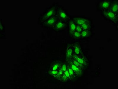
 IHC (Immunohistochemisry)
(Immunofluorescent analysis of Hela cells using AAA233246 at a dilution of 1:100 and Alexa Fluor 488-congugated AffiniPure Goat Anti-Rabbit IgG(H+L))
IHC (Immunohistochemisry)
(Immunofluorescent analysis of Hela cells using AAA233246 at a dilution of 1:100 and Alexa Fluor 488-congugated AffiniPure Goat Anti-Rabbit IgG(H+L))
MYF6, Polyclonal Antibody (Cat# AAA233246)
 IF (Immunofluorescence)
(Immunofluorescent analysis of A549 cells using AAA233247 at a dilution of 1:100 and Alexa Fluor 488-congugated AffiniPure Goat Anti-Rabbit IgG(H+L))
IF (Immunofluorescence)
(Immunofluorescent analysis of A549 cells using AAA233247 at a dilution of 1:100 and Alexa Fluor 488-congugated AffiniPure Goat Anti-Rabbit IgG(H+L))
NAMPT, Polyclonal Antibody (Cat# AAA233247)
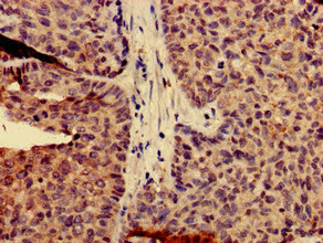
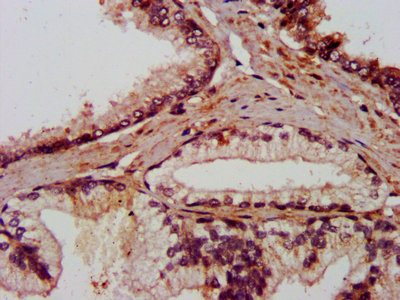
 IHC (Immunohiostchemistry)
(Immunohistochemistry of paraffin-embedded human placenta tissue using AAA233255 at dilution of 1:100)
IHC (Immunohiostchemistry)
(Immunohistochemistry of paraffin-embedded human placenta tissue using AAA233255 at dilution of 1:100)
OBSL1, Polyclonal Antibody (Cat# AAA233255)
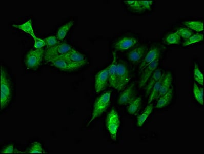
 IHC (Immunohistochemisry)
(Immunofluorescent analysis of Hela cells using AAA232892 at a dilution of 1:100 and Alexa Fluor 488-congugated AffiniPure Goat Anti-Rabbit IgG(H+L))
IHC (Immunohistochemisry)
(Immunofluorescent analysis of Hela cells using AAA232892 at a dilution of 1:100 and Alexa Fluor 488-congugated AffiniPure Goat Anti-Rabbit IgG(H+L))
C1QTNF9B-AS1, Polyclonal Antibody (Cat# AAA232892)
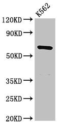
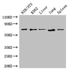
 WB (Western Blot)
(Western BlotPositive WB detected in Recombinant proteinAll lanes: FSIP2 antibody at 0.8ug/mlSecondaryGoat polyclonal to rabbit IgG at 1/50000 dilutionpredicted band size: 62 KDaobserved band size: 62 KDa)
WB (Western Blot)
(Western BlotPositive WB detected in Recombinant proteinAll lanes: FSIP2 antibody at 0.8ug/mlSecondaryGoat polyclonal to rabbit IgG at 1/50000 dilutionpredicted band size: 62 KDaobserved band size: 62 KDa)
FSIP2, Polyclonal Antibody (Cat# AAA232893)
nosZ, Polyclonal Antibody (Cat# AAA232897)
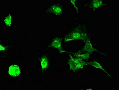
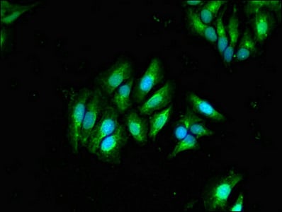
 IF (Immunofluorescence)
(Immunofluorescent analysis of HepG2 cells using AAA232900 at a dilution of 1:100 and Alexa Fluor 488-congugated AffiniPure Goat Anti-Rabbit IgG(H+L))
IF (Immunofluorescence)
(Immunofluorescent analysis of HepG2 cells using AAA232900 at a dilution of 1:100 and Alexa Fluor 488-congugated AffiniPure Goat Anti-Rabbit IgG(H+L))
TM4SF20, Polyclonal Antibody (Cat# AAA232900)
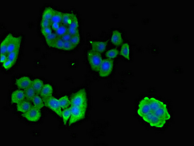
 IF (Immunofluorescence)
(Immunofluorescent analysis of MCF-7 cells using AAA232902 at a dilution of 1:100 and Alexa Fluor 488-congugated AffiniPure Goat Anti-Rabbit IgG(H+L))
IF (Immunofluorescence)
(Immunofluorescent analysis of MCF-7 cells using AAA232902 at a dilution of 1:100 and Alexa Fluor 488-congugated AffiniPure Goat Anti-Rabbit IgG(H+L))
IL23R, Polyclonal Antibody (Cat# AAA232902)
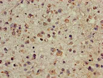
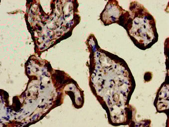
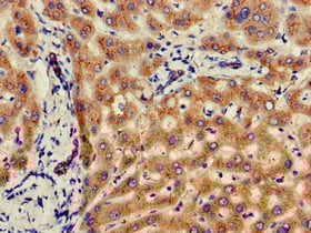
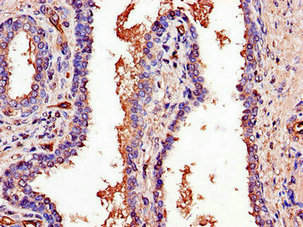
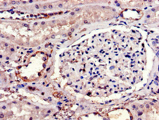
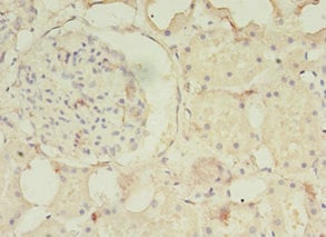
 IHC (Immunohistochemistry)
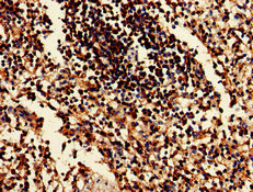
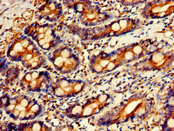
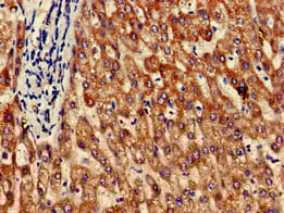
(Immunohistochemistry of paraffin-embedded human kidney tissue using AAA232904 at dilution of 1:100)
IHC (Immunohistochemistry)
(Immunohistochemistry of paraffin-embedded human kidney tissue using AAA232904 at dilution of 1:100)
RPL22L1, Polyclonal Antibody (Cat# AAA232904)
 IF (Immunofluorescence)
(Immunofluorescent analysis of HepG2 cells using AAA232906 at a dilution of 1:100 and Alexa Fluor 488-congugated AffiniPure Goat Anti-Rabbit IgG(H+L))
IF (Immunofluorescence)
(Immunofluorescent analysis of HepG2 cells using AAA232906 at a dilution of 1:100 and Alexa Fluor 488-congugated AffiniPure Goat Anti-Rabbit IgG(H+L))
LIN28B, Polyclonal Antibody (Cat# AAA232906)
 IF (Immunofluorescence)
(Immunofluorescent analysis of HepG2 cells using AAA232913 at a dilution of 1:100 and Alexa Fluor 488-congugated AffiniPure Goat Anti-Rabbit IgG(H+L))
IF (Immunofluorescence)
(Immunofluorescent analysis of HepG2 cells using AAA232913 at a dilution of 1:100 and Alexa Fluor 488-congugated AffiniPure Goat Anti-Rabbit IgG(H+L))
TCTN3, Polyclonal Antibody (Cat# AAA232913)
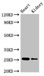
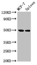
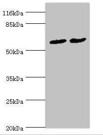
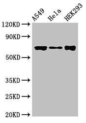 WB (Western Blot)
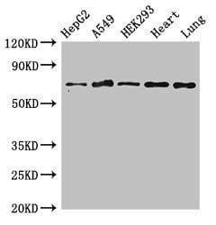
(Western BlotPositive WB detected in: A549 whole cell lysate,Hela whole cell lysate,HEK293 whole cell lysateAll lanes: CRTC1 antibody at 3.5ug/mlSecondaryGoat polyclonal to rabbit IgG at 1/50000 dilutionPredicted band size: 68,69,63 KDaObserved band size: 68 KDa)
WB (Western Blot)
(Western BlotPositive WB detected in: A549 whole cell lysate,Hela whole cell lysate,HEK293 whole cell lysateAll lanes: CRTC1 antibody at 3.5ug/mlSecondaryGoat polyclonal to rabbit IgG at 1/50000 dilutionPredicted band size: 68,69,63 KDaObserved band size: 68 KDa)
CRTC1, Polyclonal Antibody (Cat# AAA232922)
 IF (Immunofluorescence)
(Immunofluorescent analysis of Hela cells using AAA232924 at a dilution of 1:100 and Alexa Fluor 488-congugated AffiniPure Goat Anti-Rabbit IgG(H+L))
IF (Immunofluorescence)
(Immunofluorescent analysis of Hela cells using AAA232924 at a dilution of 1:100 and Alexa Fluor 488-congugated AffiniPure Goat Anti-Rabbit IgG(H+L))
XAF1, Polyclonal Antibody (Cat# AAA232924)
 IF (Immunofluorescence)
(Immunofluorescent analysis of HepG2 cells using AAA232928 at a dilution of 1:100 and Alexa Fluor 488-congugated AffiniPure Goat Anti-Rabbit IgG(H+L))
IF (Immunofluorescence)
(Immunofluorescent analysis of HepG2 cells using AAA232928 at a dilution of 1:100 and Alexa Fluor 488-congugated AffiniPure Goat Anti-Rabbit IgG(H+L))
SLC51A, Polyclonal Antibody (Cat# AAA232928)
 IF (Immunofluorescence)
(Immunofluorescent analysis of HepG2 cells using AAA232929 at dilution of 1:100 and Alexa Fluor 488-congugated AffiniPure Goat Anti-Rabbit IgG(H+L))
IF (Immunofluorescence)
(Immunofluorescent analysis of HepG2 cells using AAA232929 at dilution of 1:100 and Alexa Fluor 488-congugated AffiniPure Goat Anti-Rabbit IgG(H+L))
SLC13A5, Polyclonal Antibody (Cat# AAA232929)
 IF (Immunofluorescence)
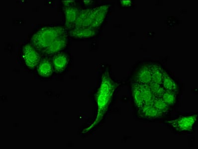
(Immunofluorescence staining of A549 cells with AAA232934 at 1:266,counter-stained with DAPI. The cells were fixed in 4% formaldehyde, permeabilized using 0.2% Triton X-100 and blocked in 10% normal Goat Serum. The cells were then incubated with the antibody overnight at 4 degree C.The secondary antibody was Alexa Fluor 488-congugated AffiniPure Goat Anti-Rabbit IgG (H+L).)
IF (Immunofluorescence)
(Immunofluorescence staining of A549 cells with AAA232934 at 1:266,counter-stained with DAPI. The cells were fixed in 4% formaldehyde, permeabilized using 0.2% Triton X-100 and blocked in 10% normal Goat Serum. The cells were then incubated with the antibody overnight at 4 degree C.The secondary antibody was Alexa Fluor 488-congugated AffiniPure Goat Anti-Rabbit IgG (H+L).)
AMIGO2, Polyclonal Antibody (Cat# AAA232934)
 IF (Immunofluorescence)
(Immunofluorescent analysis of HepG2 cells using AAA232935 at a dilution of 1:100 and Alexa Fluor 488-congugated AffiniPure Goat Anti-Rabbit IgG(H+L))
IF (Immunofluorescence)
(Immunofluorescent analysis of HepG2 cells using AAA232935 at a dilution of 1:100 and Alexa Fluor 488-congugated AffiniPure Goat Anti-Rabbit IgG(H+L))
SLC51B, Polyclonal Antibody (Cat# AAA232935)
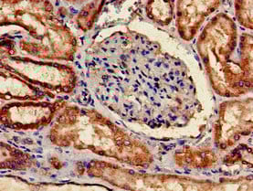
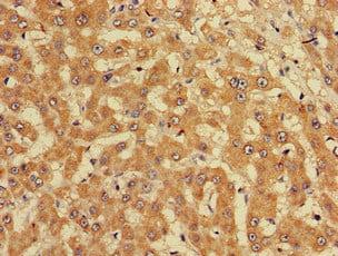
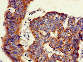
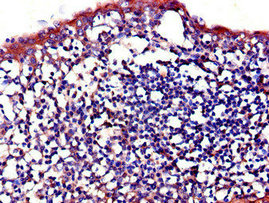
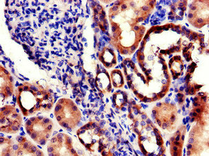
 IHC (Immunohistochemisry)
(Immunohistochemistry of paraffin-embedded human kidney using AAA232940 at dilution 1:100)
IHC (Immunohistochemisry)
(Immunohistochemistry of paraffin-embedded human kidney using AAA232940 at dilution 1:100)
PDZD3, Polyclonal Antibody (Cat# AAA232940)
 IHC (Immunohistochemisry)
(Immunofluorescent analysis of HepG2 cells using AAA232942 at a dilution of 1:100 and Alexa Fluor 488-congugated AffiniPure Goat Anti-Rabbit IgG(H+L))
IHC (Immunohistochemisry)
(Immunofluorescent analysis of HepG2 cells using AAA232942 at a dilution of 1:100 and Alexa Fluor 488-congugated AffiniPure Goat Anti-Rabbit IgG(H+L))
LRRTM4, Polyclonal Antibody (Cat# AAA232942)
What are Polyclonal Antibodies?
Polyclonal antibodies are antibodies that come from multiple B cell clones of a host animal. The typical hosts used for the majority of polyclonal antibody production are rabbits, goats, sheep, and donkeys. These polyclonal antibodies, once having identified their target, will bind to different epitopes located at different regions or sequences on the same protein/antigen. As a result, they are ideal at locating and binding to the target, even if the target is in very low concentrations (due to many different antibodies being able to bind to the same target molecule, which allows for significant amplification of a downstream signal).
Polyclonal antibodies are typically produced by injecting an antigen into a host animal, which causes the animal’s immune system to attack the foreign antigen by mass generating antibodies against it. After a period of time, serum is collected from the animal and purified using physicochemical fractionation, class-specific affinity purification, and/or antigen-affinity purification.
Key Uses of Polyclonal Antibodies
- Western Blotting: This method is used to find specific proteins in biological samples after separating them by size.
- Immunohistochemistry: IHC helps visualize the location of proteins in tissue sections using various staining techniques.
- ELISA: (Enzyme-Linked Immunosorbent Assay) is typically used to identify specific protein quantities in a sample. ELISAs can be either “Quantitative” or “Qualitative”.
- Flow Cytometry: technique that identifies and measures the specific protein on the surface or inside the cells in a fluid suspension.
- Immunoprecipitation: IP isolates and studies a specific protein from a complex mixture using antibodies.
Why Buy Polyclonal Antibodies from AAA Biotech?
1. Ideal for Various Applications
Our antibodies are generally going to be validated for use in multiple types of assays, including ELISA, Western Blotting, Immunohistochemistry, Immunoprecipitation, amongst others. They are ideal for a wide range of research applications.
2. Rigorous Quality Control
All of the antibodies in our catalog undergo strict quality testing to ensure specificity, sensitivity, and consistent performance. We are confident in the ability of our antibodies to provide you with accurate results.
3. Wide Assortment of Antibodies
Antibodies in are catalog can be found for both common and exotic species, and these antibodies are also available in both conjugated and recombinant forms to suit many diverse experimental needs.
4. Highly Purified
Our antibodies are available in purified forms with over 85% purity, as confirmed by SDS-PAGE. They are also available with tags such as His, Flag, GST, or MBP. We cater to customers worldwide.
FAQ
1. How are polyclonal antibodies produced?
Traditionally, polyclonal antibodies are produced by injecting an antigen into a host animal (such as a rabbit or goat), which then triggers an immune response from the host animal. The animal’s B cells produce antibodies that will recognize different parts of the injected antigen. These antibodies are then collected from the animal’s blood and purified for use.
2. How do polyclonal antibodies differ from monoclonal antibodies?
Polyclonal antibodies are a mix of antibodies that bind to different locations (epitopes) of the same antigen, while monoclonal antibodies are identical and bind to just one specific epitope. This makes polyclonal antibodies more versatile and better at detecting proteins that may be present in low quantities or in altered/modified forms.
3. How should I store polyclonal antibodies?
Polyclonal antibodies should be stored at 4°C for short-term use (up to a few weeks) and at -20°C or -80°C for long-term storage. Avoid repeated freeze-thaw cycles by dividing them into small aliquots. Always check the datasheet for specific storage instructions.