Filters
▼Clonality
▼Type
▼Reactivity
▼Gene Name
▼Isotype
▼Host
▼Application
▼Clone
▼Polyclonal Antibodies
At AAA Biotech also known as AAA Bio or AAABio, we provide a broad range of purified polyclonal antibodies (pAbs) that are able to all be browsed online through our website. Due to their high specificity and strong binding affinity, these antibodies are ideal for wide swathes of research and experimental applications.
Our polyclonal antibodies can easily support your work, whether you use them for Western Blotting, Immunocytochemistry (with or without Immunofluorescence used in conjunction), Immunohistochemistry, Immunoprecipitation, and ELISA tests. We highly encourage you to browse our range of pAbs and choose the one that best suits your experimental model.
Viewing 2650-2700 of 96805 product results
 FCM/FACS (Flow Cytometry)
(Figure 2. Flow Cytometry analysis of THP-1 cells using anti-TAF1 antibody (AAA126204).Overlay histogram showing THP-1 cells stained with AAA126204 (Blue line). The cells were blocked with 10% normal goat serum. And then incubated with rabbit anti-TAF1 Antibody (AAA126204, 1 ug/1x10^6 cells) for 30 min at 20 degree C. DyLight488 conjugated goat anti-rabbit IgG was used as secondary antibody for 30 minutes at 20 degree C. Isotype control antibody (Green line) was rabbit IgG (1 ug/1x10^6) used under the same conditions. Unlabelled sample (Red line) was also used as a control.)
FCM/FACS (Flow Cytometry)
(Figure 2. Flow Cytometry analysis of THP-1 cells using anti-TAF1 antibody (AAA126204).Overlay histogram showing THP-1 cells stained with AAA126204 (Blue line). The cells were blocked with 10% normal goat serum. And then incubated with rabbit anti-TAF1 Antibody (AAA126204, 1 ug/1x10^6 cells) for 30 min at 20 degree C. DyLight488 conjugated goat anti-rabbit IgG was used as secondary antibody for 30 minutes at 20 degree C. Isotype control antibody (Green line) was rabbit IgG (1 ug/1x10^6) used under the same conditions. Unlabelled sample (Red line) was also used as a control.)
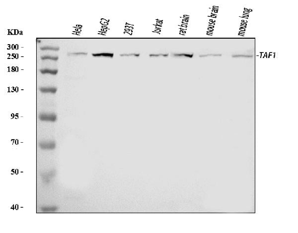
TAF1, Polyclonal Antibody (Cat# AAA126204)
 FCM/FACS (Flow Cytometry)
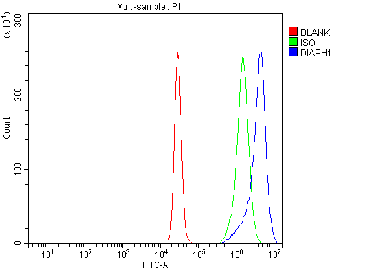
(Figure 3. Flow Cytometry analysis of K562 cells using anti-DIAPH1 antibody (AAA126212).Overlay histogram showing K562 cells stained with AAA126212 (Blue line). The cells were blocked with 10% normal goat serum. And then incubated with rabbit anti-DIAPH1 Antibody (AAA126212, 1 ug/1x10^6 cells) for 30 min at 20 degree C. DyLight488 conjugated goat anti-rabbit IgG was used as secondary antibody for 30 minutes at 20 degree C. Isotype control antibody (Green line) was rabbit IgG (1 ug/1x10^6) used under the same conditions. Unlabelled sample (Red line) was also used as a control.)
FCM/FACS (Flow Cytometry)
(Figure 3. Flow Cytometry analysis of K562 cells using anti-DIAPH1 antibody (AAA126212).Overlay histogram showing K562 cells stained with AAA126212 (Blue line). The cells were blocked with 10% normal goat serum. And then incubated with rabbit anti-DIAPH1 Antibody (AAA126212, 1 ug/1x10^6 cells) for 30 min at 20 degree C. DyLight488 conjugated goat anti-rabbit IgG was used as secondary antibody for 30 minutes at 20 degree C. Isotype control antibody (Green line) was rabbit IgG (1 ug/1x10^6) used under the same conditions. Unlabelled sample (Red line) was also used as a control.)
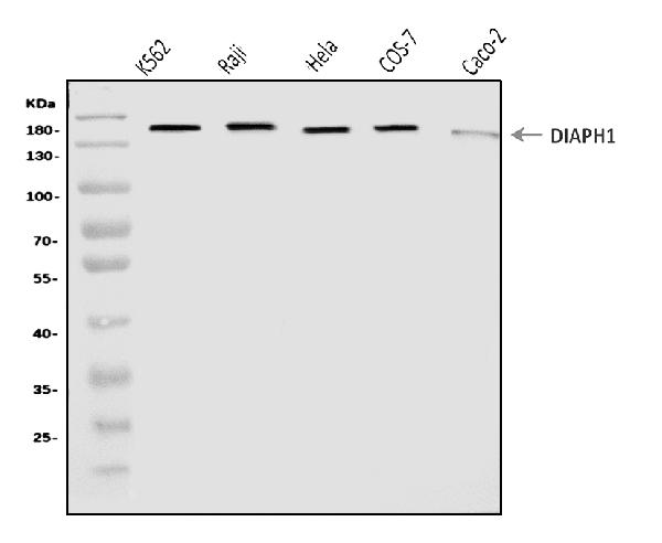
DIAPH1, Polyclonal Antibody (Cat# AAA126212)
 FCM/FACS (Flow Cytometry)
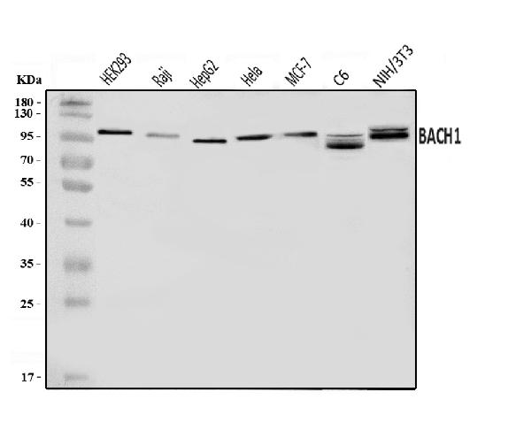
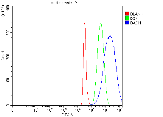
(Figure 2. Flow Cytometry analysis of HepG2 cells using anti-BACH1.3/BACH1 antibody (AAA126217).Overlay histogram showing HepG2 cells stained with AAA126217 (Blue line). The cells were blocked with 10% normal goat serum. And then incubated with rabbit anti-BACH1.3/BACH1 Antibody (AAA126217, 1 ug/1x10^6 cells) for 30 min at 20 degree C. DyLight488 conjugated goat anti-rabbit IgG was used as secondary antibody for 30 minutes at 20 degree C. Isotype control antibody (Green line) was rabbit IgG (1 ug/1x10^6) used under the same conditions. Unlabelled sample (Red line) was also used as a control.)
FCM/FACS (Flow Cytometry)
(Figure 2. Flow Cytometry analysis of HepG2 cells using anti-BACH1.3/BACH1 antibody (AAA126217).Overlay histogram showing HepG2 cells stained with AAA126217 (Blue line). The cells were blocked with 10% normal goat serum. And then incubated with rabbit anti-BACH1.3/BACH1 Antibody (AAA126217, 1 ug/1x10^6 cells) for 30 min at 20 degree C. DyLight488 conjugated goat anti-rabbit IgG was used as secondary antibody for 30 minutes at 20 degree C. Isotype control antibody (Green line) was rabbit IgG (1 ug/1x10^6) used under the same conditions. Unlabelled sample (Red line) was also used as a control.)
BACH1.3/BACH1, Polyclonal Antibody (Cat# AAA126217)
 FCM/FACS (Flow Cytometry)
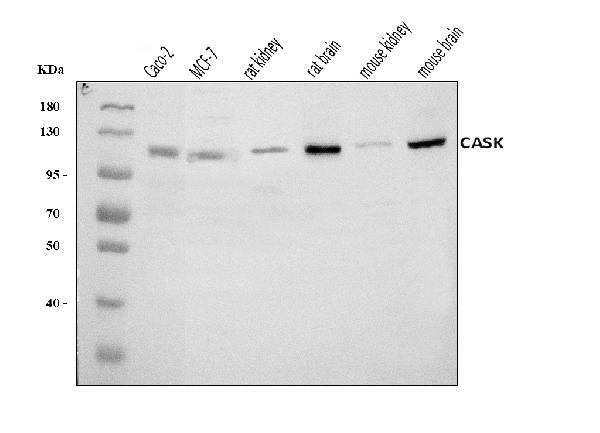
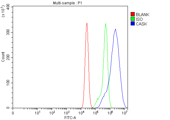
(Figure 2. Flow Cytometry analysis of CACO-2 cells using anti-CASK antibody (AAA126230).Overlay histogram showing CACO-2 cells stained with AAA126230 (Blue line). The cells were blocked with 10% normal goat serum. And then incubated with rabbit anti-CASK Antibody (AAA126230, 1 ug/1x10^6 cells) for 30 min at 20 degree C. DyLight488 conjugated goat anti-rabbit IgG was used as secondary antibody for 30 minutes at 20 degree C. Isotype control antibody (Green line) was rabbit IgG (1 ug/1x10^6) used under the same conditions. Unlabelled sample (Red line) was also used as a control.)
FCM/FACS (Flow Cytometry)
(Figure 2. Flow Cytometry analysis of CACO-2 cells using anti-CASK antibody (AAA126230).Overlay histogram showing CACO-2 cells stained with AAA126230 (Blue line). The cells were blocked with 10% normal goat serum. And then incubated with rabbit anti-CASK Antibody (AAA126230, 1 ug/1x10^6 cells) for 30 min at 20 degree C. DyLight488 conjugated goat anti-rabbit IgG was used as secondary antibody for 30 minutes at 20 degree C. Isotype control antibody (Green line) was rabbit IgG (1 ug/1x10^6) used under the same conditions. Unlabelled sample (Red line) was also used as a control.)
CASK, Polyclonal Antibody (Cat# AAA126230)
 FCM/FACS (Flow Cytometry)
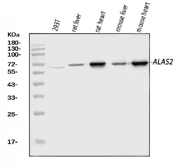
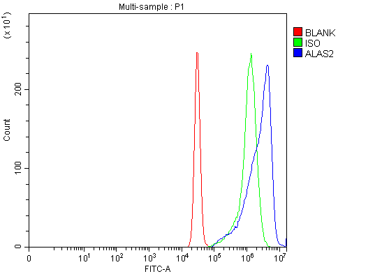
(Figure 3. Flow Cytometry analysis of K562 cells using anti-ALAS2/ASB antibody (AAA126232).Overlay histogram showing K562 cells stained with AAA126232 (Blue line). The cells were blocked with 10% normal goat serum. And then incubated with rabbit anti-ALAS2/ASB Antibody (AAA126232, 1 ug/1x10^6 cells) for 30 min at 20 degree C. DyLight488 conjugated goat anti-rabbit IgG was used as secondary antibody for 30 minutes at 20 degree C. Isotype control antibody (Green line) was rabbit IgG (1 ug/1x10^6) used under the same conditions. Unlabelled sample (Red line) was also used as a control.)
FCM/FACS (Flow Cytometry)
(Figure 3. Flow Cytometry analysis of K562 cells using anti-ALAS2/ASB antibody (AAA126232).Overlay histogram showing K562 cells stained with AAA126232 (Blue line). The cells were blocked with 10% normal goat serum. And then incubated with rabbit anti-ALAS2/ASB Antibody (AAA126232, 1 ug/1x10^6 cells) for 30 min at 20 degree C. DyLight488 conjugated goat anti-rabbit IgG was used as secondary antibody for 30 minutes at 20 degree C. Isotype control antibody (Green line) was rabbit IgG (1 ug/1x10^6) used under the same conditions. Unlabelled sample (Red line) was also used as a control.)
ALAS2/ASB, Polyclonal Antibody (Cat# AAA126232)
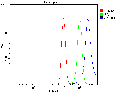
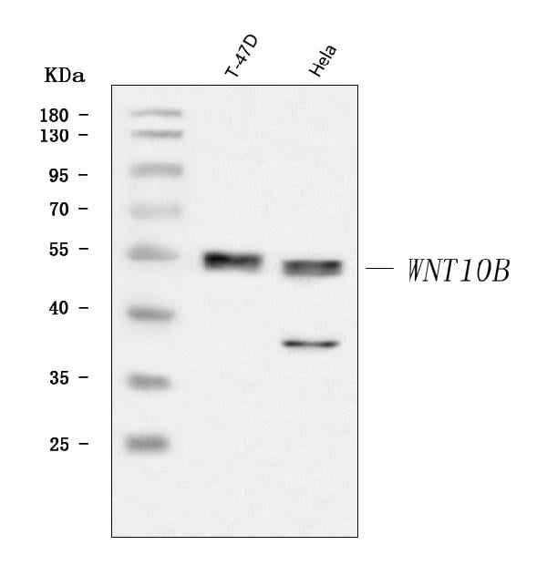
 FCM/FACS (Flow Cytometry)
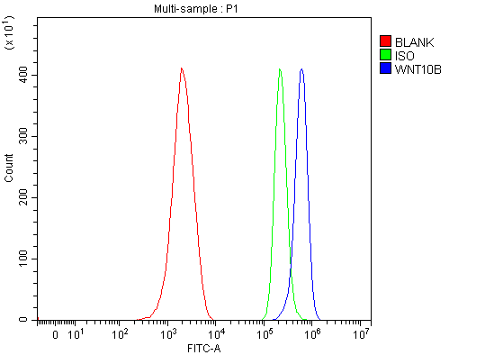
(Figure 4. Flow Cytometry analysis of NRK cells using anti-WNT10B antibody (AAA126236).Overlay histogram showing NRK cells stained with AAA126236 (Blue line). The cells were blocked with 10% normal goat serum. And then incubated with rabbit anti-WNT10B Antibody (AAA126236, 1 ug/1x10^6 cells) for 30 min at 20 degree C. DyLight488 conjugated goat anti-rabbit IgG was used as secondary antibody for 30 minutes at 20 degree C. Isotype control antibody (Green line) was rabbit IgG (1 ug/1x10^6) used under the same conditions. Unlabelled sample (Red line) was also used as a control.)
FCM/FACS (Flow Cytometry)
(Figure 4. Flow Cytometry analysis of NRK cells using anti-WNT10B antibody (AAA126236).Overlay histogram showing NRK cells stained with AAA126236 (Blue line). The cells were blocked with 10% normal goat serum. And then incubated with rabbit anti-WNT10B Antibody (AAA126236, 1 ug/1x10^6 cells) for 30 min at 20 degree C. DyLight488 conjugated goat anti-rabbit IgG was used as secondary antibody for 30 minutes at 20 degree C. Isotype control antibody (Green line) was rabbit IgG (1 ug/1x10^6) used under the same conditions. Unlabelled sample (Red line) was also used as a control.)
WNT10B, Polyclonal Antibody (Cat# AAA126236)
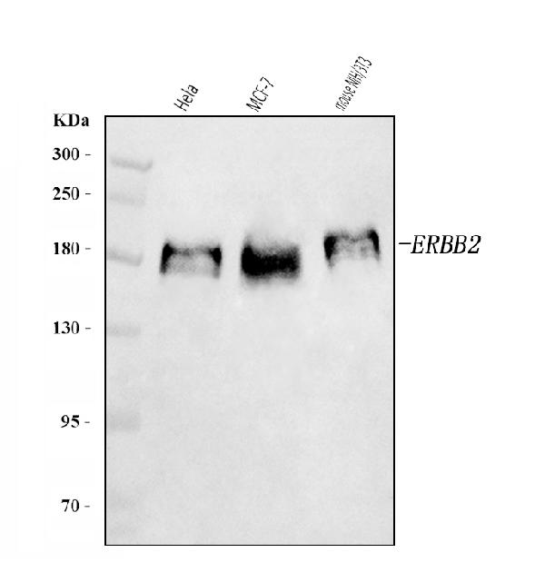
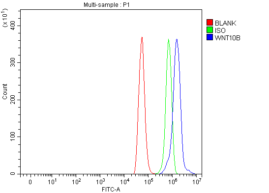
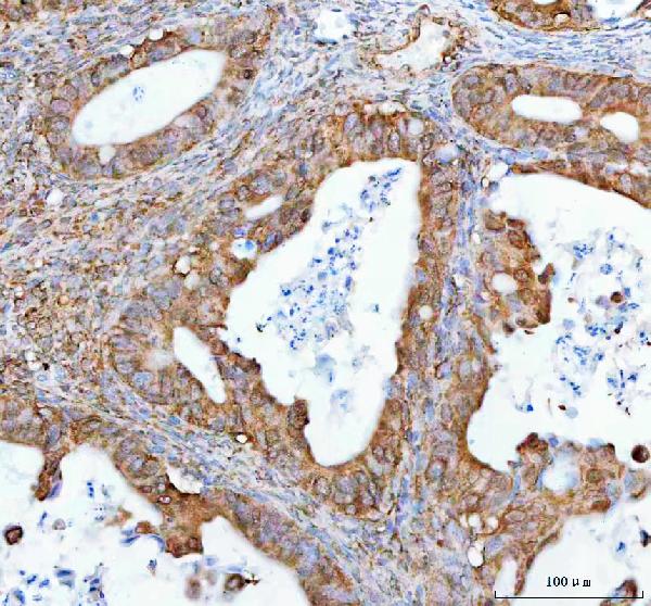
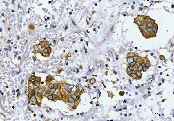 IHC (Immunohiostchemistry)
(Figure 2. IHC analysis of HER2/ERBB2 using anti-HER2/ERBB2 antibody (AAA125940).HER2/ERBB2 was detected in a paraffin-embedded section of human breast cancer tissue. Heat mediated antigen retrieval was performed in EDTA buffer (pH 8.0, epitope retrieval solution). The tissue section was blocked with 10% goat serum. The tissue section was then incubated with 2 ug/ml rabbit anti-HER2/ERBB2 Antibody (AAA125940) overnight at 4 degree C. Peroxidase Conjugated Goat Anti-rabbit IgG was used as secondary antibody and incubated for 30 minutes at 37 degree C. The tissue section was developed using HRP Conjugated Rabbit IgG Super Vision Assay Kit with DAB as the chromogen.)
IHC (Immunohiostchemistry)
(Figure 2. IHC analysis of HER2/ERBB2 using anti-HER2/ERBB2 antibody (AAA125940).HER2/ERBB2 was detected in a paraffin-embedded section of human breast cancer tissue. Heat mediated antigen retrieval was performed in EDTA buffer (pH 8.0, epitope retrieval solution). The tissue section was blocked with 10% goat serum. The tissue section was then incubated with 2 ug/ml rabbit anti-HER2/ERBB2 Antibody (AAA125940) overnight at 4 degree C. Peroxidase Conjugated Goat Anti-rabbit IgG was used as secondary antibody and incubated for 30 minutes at 37 degree C. The tissue section was developed using HRP Conjugated Rabbit IgG Super Vision Assay Kit with DAB as the chromogen.)
HER2/ERBB2, Polyclonal Antibody (Cat# AAA125940)
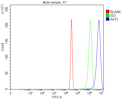 FCM/FACS (Flow Cytometry)
(Figure 3. Flow Cytometry analysis of HeLa cells using anti-AKT1,2,3 antibody (AAA125943).Overlay histogram showing HeLa cells stained with AAA125943 (Blue line). The cells were blocked with 10% normal goat serum. And then incubated with rabbit anti-AKT1,2,3 Antibody (AAA125943, 1 ug/1x10^6 cells) for 30 min at 20 degree C. DyLight488 conjugated goat anti-rabbit IgG was used as secondary antibody for 30 minutes at 20 degree C. Isotype control antibody (Green line) was rabbit IgG (1 ug/1x10^6) used under the same conditions. Unlabelled sample (Red line) was also used as a control.)
FCM/FACS (Flow Cytometry)
(Figure 3. Flow Cytometry analysis of HeLa cells using anti-AKT1,2,3 antibody (AAA125943).Overlay histogram showing HeLa cells stained with AAA125943 (Blue line). The cells were blocked with 10% normal goat serum. And then incubated with rabbit anti-AKT1,2,3 Antibody (AAA125943, 1 ug/1x10^6 cells) for 30 min at 20 degree C. DyLight488 conjugated goat anti-rabbit IgG was used as secondary antibody for 30 minutes at 20 degree C. Isotype control antibody (Green line) was rabbit IgG (1 ug/1x10^6) used under the same conditions. Unlabelled sample (Red line) was also used as a control.)
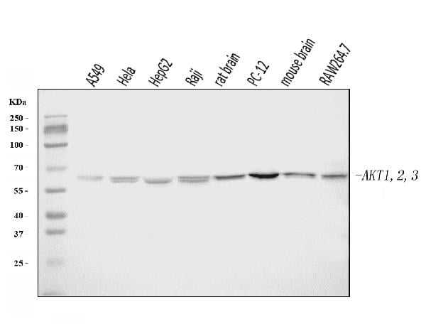
AKT1,2,3, Polyclonal Antibody (Cat# AAA125943)
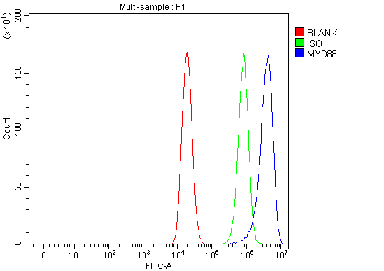 FCM/FACS (Flow Cytometry)
(Figure 3. Flow Cytometry analysis of Caco-2 cells using anti-MYD88 antibody (AAA125944).Overlay histogram showing Caco-2 cells stained with AAA125944 (Blue line). The cells were blocked with 10% normal goat serum. And then incubated with rabbit anti-MYD88 Antibody (AAA125944, 1 ug/1x10^6 cells) for 30 min at 20 degree C. DyLight488 conjugated goat anti-rabbit IgG was used as secondary antibody for 30 minutes at 20 degree C. Isotype control antibody (Green line) was rabbit IgG (1 ug/1x10^6) used under the same conditions. Unlabelled sample (Red line) was also used as a control.)
FCM/FACS (Flow Cytometry)
(Figure 3. Flow Cytometry analysis of Caco-2 cells using anti-MYD88 antibody (AAA125944).Overlay histogram showing Caco-2 cells stained with AAA125944 (Blue line). The cells were blocked with 10% normal goat serum. And then incubated with rabbit anti-MYD88 Antibody (AAA125944, 1 ug/1x10^6 cells) for 30 min at 20 degree C. DyLight488 conjugated goat anti-rabbit IgG was used as secondary antibody for 30 minutes at 20 degree C. Isotype control antibody (Green line) was rabbit IgG (1 ug/1x10^6) used under the same conditions. Unlabelled sample (Red line) was also used as a control.)
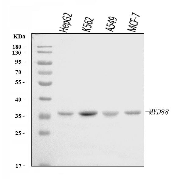
MYD88, Polyclonal Antibody (Cat# AAA125944)
 FCM/FACS (Flow Cytometry)
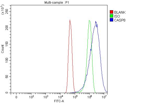
(Figure 2. Flow Cytometry analysis of U251 cells using anti-Caspase-8(P18)/CASP8 antibody (AAA125946).Overlay histogram showing U251 cells stained with AAA125946 (Blue line). The cells were blocked with 10% normal goat serum. And then incubated with rabbit anti-Caspase-8(P18)/CASP8 Antibody (AAA125946, 1 ug/1x10^6 cells) for 30 min at 20 degree C. DyLight488 conjugated goat anti-rabbit IgG was used as secondary antibody for 30 minutes at 20 degree C. Isotype control antibody (Green line) was rabbit IgG (1 ug/1x10^6) used under the same conditions. Unlabelled sample (Red line) was also used as a control.)
FCM/FACS (Flow Cytometry)
(Figure 2. Flow Cytometry analysis of U251 cells using anti-Caspase-8(P18)/CASP8 antibody (AAA125946).Overlay histogram showing U251 cells stained with AAA125946 (Blue line). The cells were blocked with 10% normal goat serum. And then incubated with rabbit anti-Caspase-8(P18)/CASP8 Antibody (AAA125946, 1 ug/1x10^6 cells) for 30 min at 20 degree C. DyLight488 conjugated goat anti-rabbit IgG was used as secondary antibody for 30 minutes at 20 degree C. Isotype control antibody (Green line) was rabbit IgG (1 ug/1x10^6) used under the same conditions. Unlabelled sample (Red line) was also used as a control.)
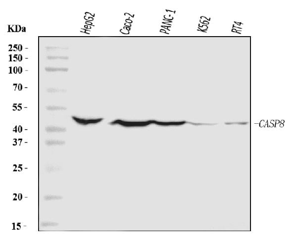
Caspase-8(p18)/CASP8, Polyclonal Antibody (Cat# AAA125946)
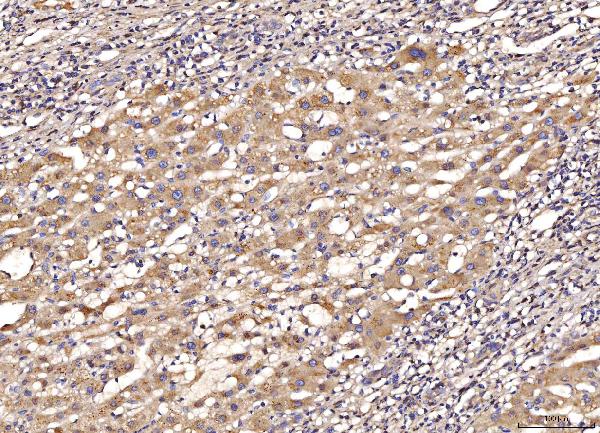
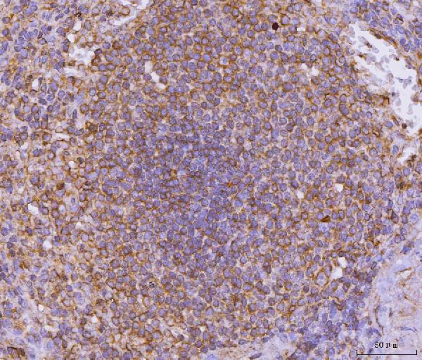
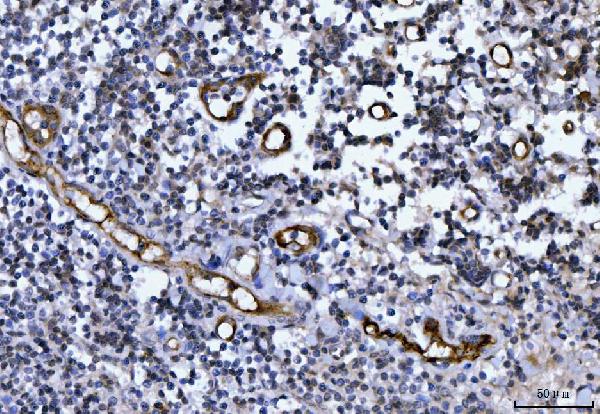
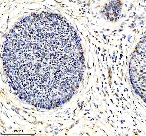
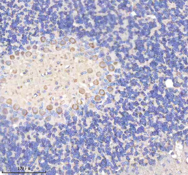
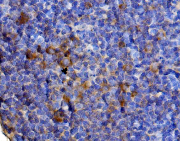 IHC (Immunohistochemisry)
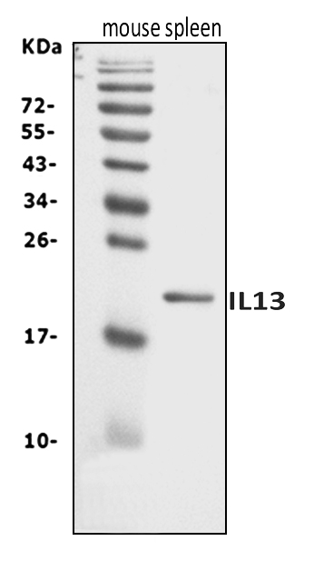
(Figure 3. IHC analysis of Il13 using anti-Il13 antibody (AAA125951).Il13 was detected in a paraffin-embedded section of rat lymph node tissue. Heat mediated antigen retrieval was performed in EDTA buffer (pH 8.0, epitope retrieval solution). The tissue section was blocked with 10% goat serum. The tissue section was then incubated with 2 ug/ml rabbit anti-Il13 Antibody (AAA125951) overnight at 4 degree C. Biotinylated goat anti-rabbit IgG was used as secondary antibody and incubated for 30 minutes at 37 degree C. The tissue section was developed using Strepavidin-Biotin-Complex (SABC) with DAB as the chromogen.)
IHC (Immunohistochemisry)
(Figure 3. IHC analysis of Il13 using anti-Il13 antibody (AAA125951).Il13 was detected in a paraffin-embedded section of rat lymph node tissue. Heat mediated antigen retrieval was performed in EDTA buffer (pH 8.0, epitope retrieval solution). The tissue section was blocked with 10% goat serum. The tissue section was then incubated with 2 ug/ml rabbit anti-Il13 Antibody (AAA125951) overnight at 4 degree C. Biotinylated goat anti-rabbit IgG was used as secondary antibody and incubated for 30 minutes at 37 degree C. The tissue section was developed using Strepavidin-Biotin-Complex (SABC) with DAB as the chromogen.)
Il13, Polyclonal Antibody (Cat# AAA125951)
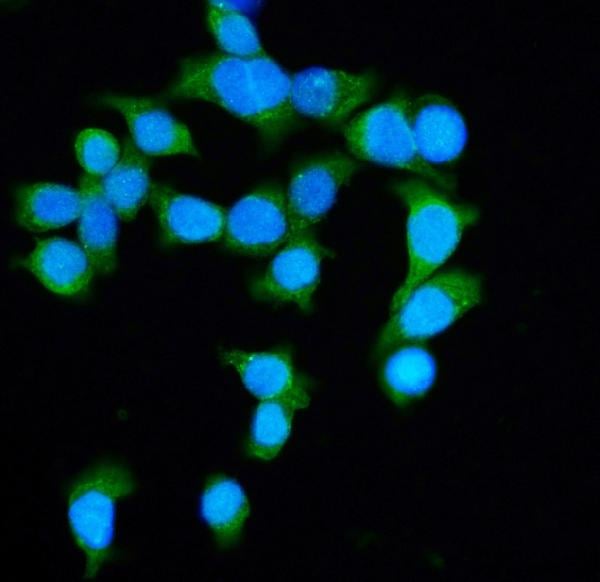
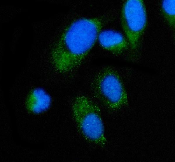
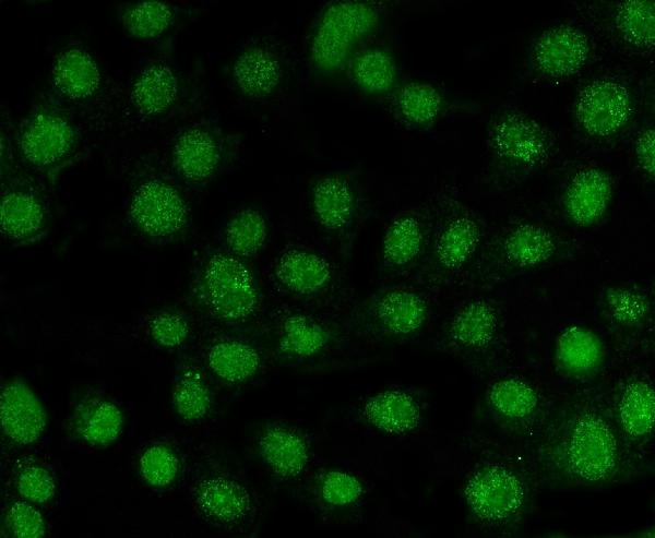
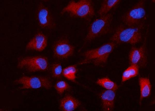
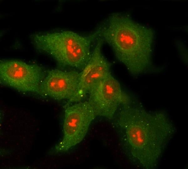
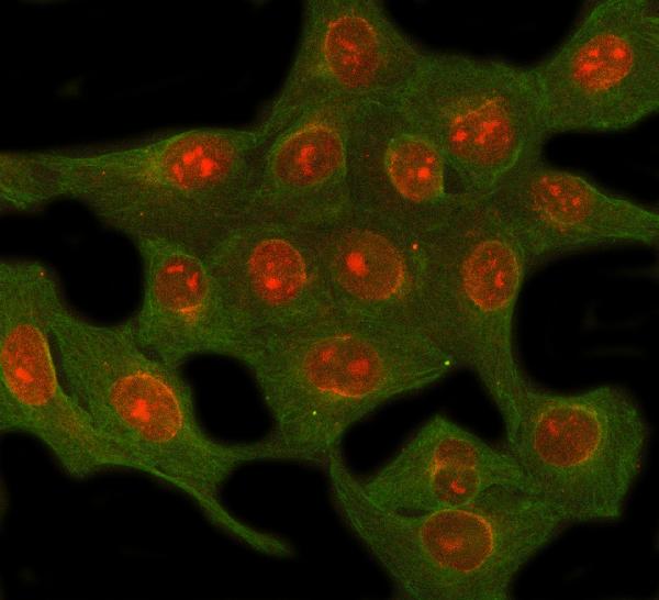
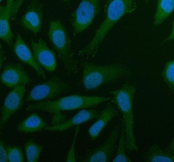
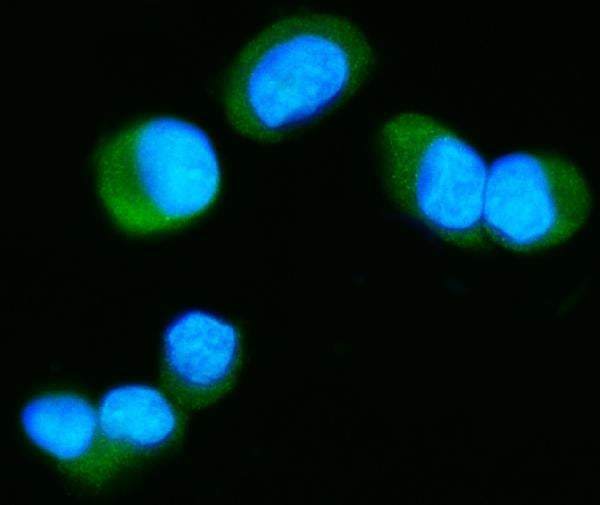 IF (Immunofluorescence)
(Figure 2. IF analysis of Tau/MAPT using anti-Tau/MAPT antibody (AAA125953).Tau/MAPT was detected in an immunocytochemical section of T-47D cells. Enzyme antigen retrieval was performed using IHC enzyme antigen retrieval reagent (AR0022) for 15 mins. The cells were blocked with 10% goat serum. And then incubated with 5 ug/mL rabbit anti-Tau/MAPT Antibody (AAA125953) overnight at 4 degree C. DyLight488 Conjugated Goat Anti-Rabbit IgG was used as secondary antibody at 1:100 dilution and incubated for 30 minutes at 37 degree C. The section was counterstained with DAPI. Visualize using a fluorescence microscope and filter sets appropriate for the label used.)
IF (Immunofluorescence)
(Figure 2. IF analysis of Tau/MAPT using anti-Tau/MAPT antibody (AAA125953).Tau/MAPT was detected in an immunocytochemical section of T-47D cells. Enzyme antigen retrieval was performed using IHC enzyme antigen retrieval reagent (AR0022) for 15 mins. The cells were blocked with 10% goat serum. And then incubated with 5 ug/mL rabbit anti-Tau/MAPT Antibody (AAA125953) overnight at 4 degree C. DyLight488 Conjugated Goat Anti-Rabbit IgG was used as secondary antibody at 1:100 dilution and incubated for 30 minutes at 37 degree C. The section was counterstained with DAPI. Visualize using a fluorescence microscope and filter sets appropriate for the label used.)
Tau/MAPT, Polyclonal Antibody (Cat# AAA125953)
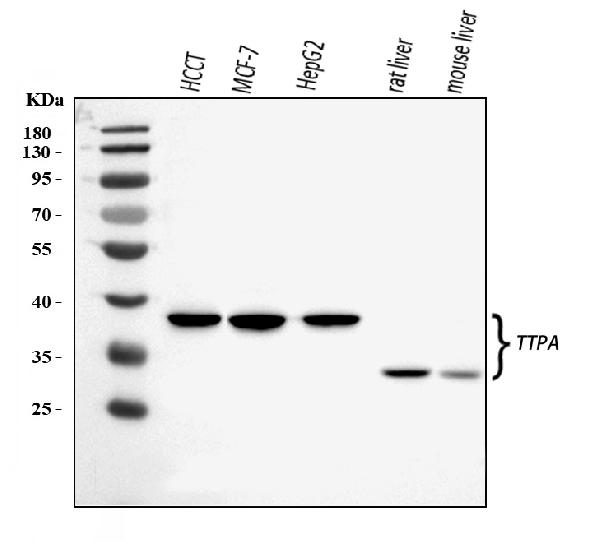
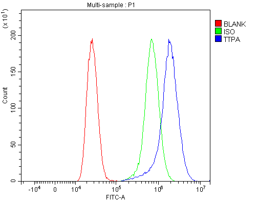 FCM/FACS (Flow Cytometry)
(Figure 2. Flow Cytometry analysis of MCF-7 cells using anti-TTPA/TPP1 antibody (AAA125954).Overlay histogram showing MCF-7 cells stained with AAA125954 (Blue line). The cells were blocked with 10% normal goat serum. And then incubated with rabbit anti-TTPA/TPP1 Antibody (AAA125954, 1 ug/1x10^6 cells) for 30 min at 20 degree C. DyLight488 conjugated goat anti-rabbit IgG was used as secondary antibody for 30 minutes at 20 degree C. Isotype control antibody (Green line) was rabbit IgG (1 ug/1x10^6) used under the same conditions. Unlabelled sample (Red line) was also used as a control.)
FCM/FACS (Flow Cytometry)
(Figure 2. Flow Cytometry analysis of MCF-7 cells using anti-TTPA/TPP1 antibody (AAA125954).Overlay histogram showing MCF-7 cells stained with AAA125954 (Blue line). The cells were blocked with 10% normal goat serum. And then incubated with rabbit anti-TTPA/TPP1 Antibody (AAA125954, 1 ug/1x10^6 cells) for 30 min at 20 degree C. DyLight488 conjugated goat anti-rabbit IgG was used as secondary antibody for 30 minutes at 20 degree C. Isotype control antibody (Green line) was rabbit IgG (1 ug/1x10^6) used under the same conditions. Unlabelled sample (Red line) was also used as a control.)
TTPA/TPP1, Polyclonal Antibody (Cat# AAA125954)
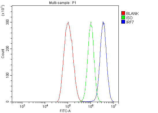 FCM/FACS (Flow Cytometry)
(Figure 2. Flow Cytometry analysis of THP-1 cells using anti-IRF7 antibody (AAA125956).Overlay histogram showing THP-1 cells stained with AAA125956 (Blue line). The cells were blocked with 10% normal goat serum. And then incubated with rabbit anti-IRF7 Antibody (AAA125956, 1 ug/1x10^6 cells) for 30 min at 20 degree C. DyLight488 conjugated goat anti-rabbit IgG was used as secondary antibody for 30 minutes at 20 degree C. Isotype control antibody (Green line) was rabbit IgG (1 ug/1x10^6) used under the same conditions. Unlabelled sample (Red line) was also used as a control.)
FCM/FACS (Flow Cytometry)
(Figure 2. Flow Cytometry analysis of THP-1 cells using anti-IRF7 antibody (AAA125956).Overlay histogram showing THP-1 cells stained with AAA125956 (Blue line). The cells were blocked with 10% normal goat serum. And then incubated with rabbit anti-IRF7 Antibody (AAA125956, 1 ug/1x10^6 cells) for 30 min at 20 degree C. DyLight488 conjugated goat anti-rabbit IgG was used as secondary antibody for 30 minutes at 20 degree C. Isotype control antibody (Green line) was rabbit IgG (1 ug/1x10^6) used under the same conditions. Unlabelled sample (Red line) was also used as a control.)
IRF7, Polyclonal Antibody (Cat# AAA125956)
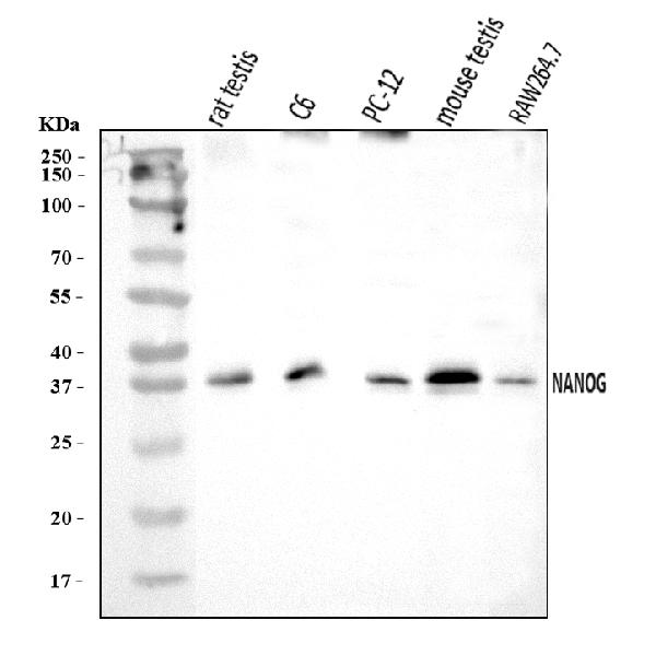
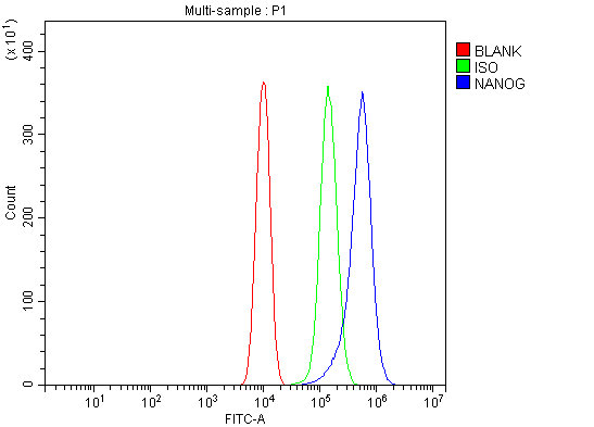 FCM/FACS (Flow Cytometry)
(Figure 3. Flow Cytometry analysis of RAW264.7 cells using anti-Nanog antibody (AAA125966).Overlay histogram showing RAW264.7 cells stained with AAA125966 (Blue line). The cells were blocked with 10% normal goat serum. And then incubated with rabbit anti-Nanog Antibody (AAA125966, 1 ug/1x10^6 cells) for 30 min at 20 degree C. DyLight488 conjugated goat anti-rabbit IgG was used as secondary antibody for 30 minutes at 20 degree C. Isotype control antibody (Green line) was rabbit IgG (1 ug/1x10^6) used under the same conditions. Unlabelled sample (Red line) was also used as a control.)
FCM/FACS (Flow Cytometry)
(Figure 3. Flow Cytometry analysis of RAW264.7 cells using anti-Nanog antibody (AAA125966).Overlay histogram showing RAW264.7 cells stained with AAA125966 (Blue line). The cells were blocked with 10% normal goat serum. And then incubated with rabbit anti-Nanog Antibody (AAA125966, 1 ug/1x10^6 cells) for 30 min at 20 degree C. DyLight488 conjugated goat anti-rabbit IgG was used as secondary antibody for 30 minutes at 20 degree C. Isotype control antibody (Green line) was rabbit IgG (1 ug/1x10^6) used under the same conditions. Unlabelled sample (Red line) was also used as a control.)
Nanog, Polyclonal Antibody (Cat# AAA125966)
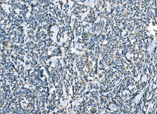
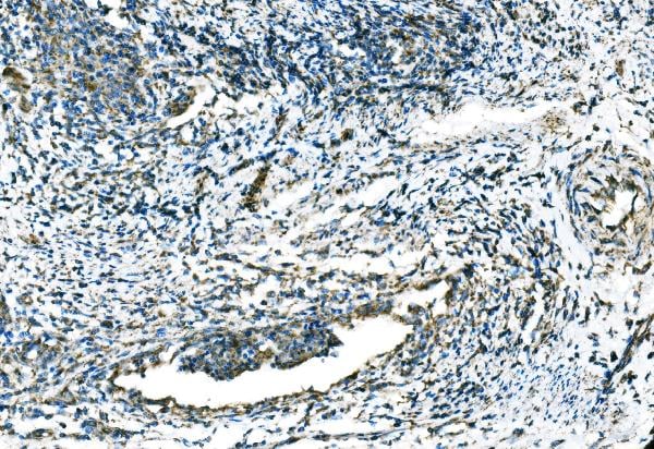
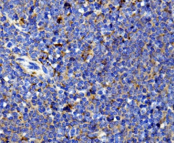
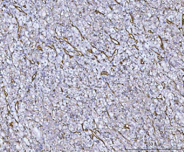
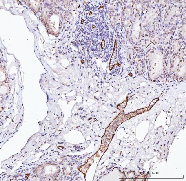
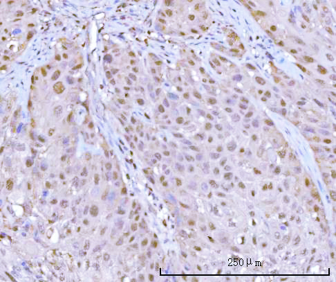
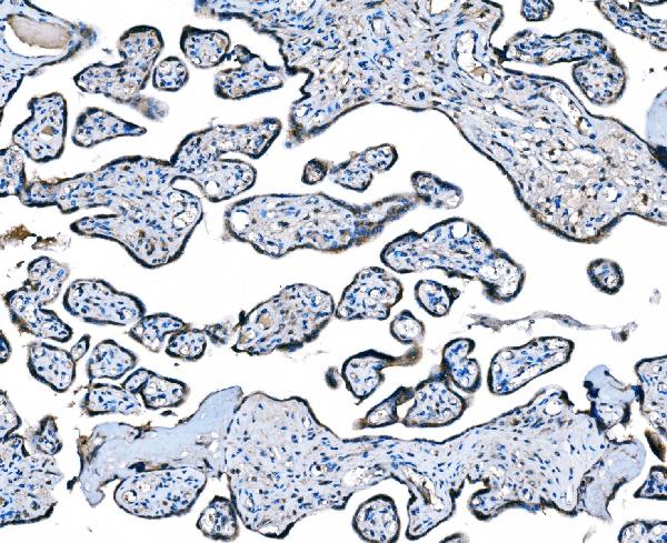 IHC (Immunohistochemistry)
(Figure 4. IHC analysis of CCR2 using anti-CCR2 antibody (AAA125969).CCR2 was detected in a paraffin-embedded section of human placenta tissue. Heat mediated antigen retrieval was performed in EDTA buffer (pH 8.0, epitope retrieval solution). The tissue section was blocked with 10% goat serum. The tissue section was then incubated with 2 ug/ml rabbit anti-CCR2 Antibody (AAA125969) overnight at 4 degree C. Peroxidase Conjugated Goat Anti-rabbit IgG was used as secondary antibody and incubated for 30 minutes at 37 degree C. The tissue section was developed using HRP Conjugated Rabbit IgG Super Vision Assay Kit with DAB as the chromogen.)
IHC (Immunohistochemistry)
(Figure 4. IHC analysis of CCR2 using anti-CCR2 antibody (AAA125969).CCR2 was detected in a paraffin-embedded section of human placenta tissue. Heat mediated antigen retrieval was performed in EDTA buffer (pH 8.0, epitope retrieval solution). The tissue section was blocked with 10% goat serum. The tissue section was then incubated with 2 ug/ml rabbit anti-CCR2 Antibody (AAA125969) overnight at 4 degree C. Peroxidase Conjugated Goat Anti-rabbit IgG was used as secondary antibody and incubated for 30 minutes at 37 degree C. The tissue section was developed using HRP Conjugated Rabbit IgG Super Vision Assay Kit with DAB as the chromogen.)
CCR2, Polyclonal Antibody (Cat# AAA125969)
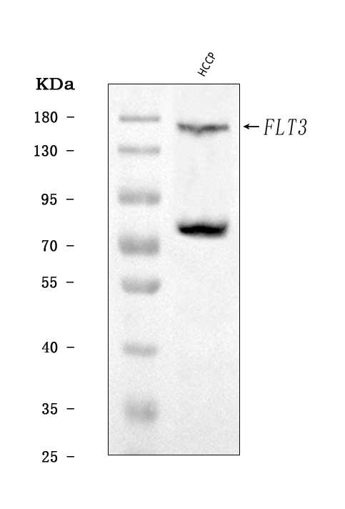
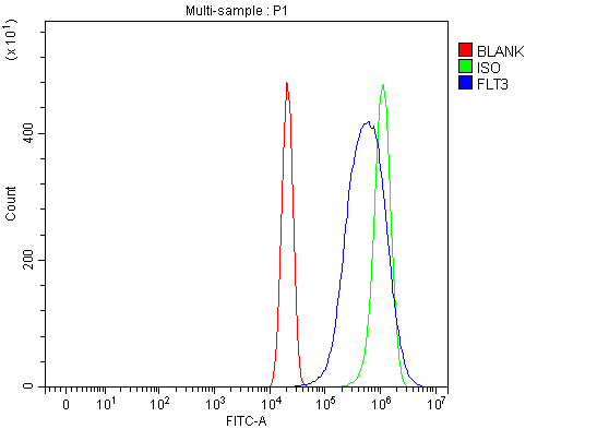 FCM/FACS (Flow Cytometry)
(Figure 3. Flow Cytometry analysis of U2OS cells using anti-CD135/FLT3 antibody (AAA125976).Overlay histogram showing U2OS cells stained with AAA125976 (Blue line). The cells were blocked with 10% normal goat serum. And then incubated with rabbit anti-CD135/FLT3 Antibody (AAA125976, 1 ug/1x10^6 cells) for 30 min at 20 degree C. DyLight488 conjugated goat anti-rabbit IgG was used as secondary antibody for 30 minutes at 20 degree C. Isotype control antibody (Green line) was rabbit IgG (1 ug/1x10^6) used under the same conditions. Unlabelled sample (Red line) was also used as a control.)
FCM/FACS (Flow Cytometry)
(Figure 3. Flow Cytometry analysis of U2OS cells using anti-CD135/FLT3 antibody (AAA125976).Overlay histogram showing U2OS cells stained with AAA125976 (Blue line). The cells were blocked with 10% normal goat serum. And then incubated with rabbit anti-CD135/FLT3 Antibody (AAA125976, 1 ug/1x10^6 cells) for 30 min at 20 degree C. DyLight488 conjugated goat anti-rabbit IgG was used as secondary antibody for 30 minutes at 20 degree C. Isotype control antibody (Green line) was rabbit IgG (1 ug/1x10^6) used under the same conditions. Unlabelled sample (Red line) was also used as a control.)
CD135/FLT3, Polyclonal Antibody (Cat# AAA125976)
 FCM/FACS (Flow Cytometry)
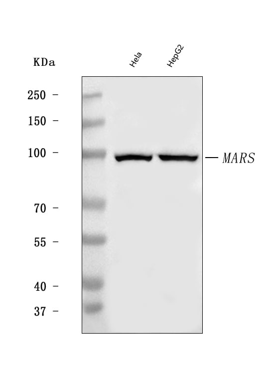
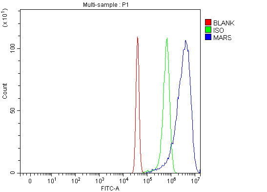
(Figure 2. Flow Cytometry analysis of SiHa cells using anti-MARS1 antibody (AAA125985).Overlay histogram showing SiHa cells stained with AAA125985 (Blue line). The cells were blocked with 10% normal goat serum. And then incubated with rabbit anti-MARS1 Antibody (AAA125985, 1 ug/1x10^6 cells) for 30 min at 20 degree C. DyLight488 conjugated goat anti-rabbit IgG was used as secondary antibody for 30 minutes at 20 degree C. Isotype control antibody (Green line) was rabbit IgG (1 ug/1x10^6) used under the same conditions. Unlabelled sample (Red line) was also used as a control.)
FCM/FACS (Flow Cytometry)
(Figure 2. Flow Cytometry analysis of SiHa cells using anti-MARS1 antibody (AAA125985).Overlay histogram showing SiHa cells stained with AAA125985 (Blue line). The cells were blocked with 10% normal goat serum. And then incubated with rabbit anti-MARS1 Antibody (AAA125985, 1 ug/1x10^6 cells) for 30 min at 20 degree C. DyLight488 conjugated goat anti-rabbit IgG was used as secondary antibody for 30 minutes at 20 degree C. Isotype control antibody (Green line) was rabbit IgG (1 ug/1x10^6) used under the same conditions. Unlabelled sample (Red line) was also used as a control.)
MARS1, Polyclonal Antibody (Cat# AAA125985)
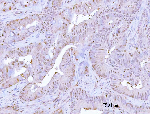
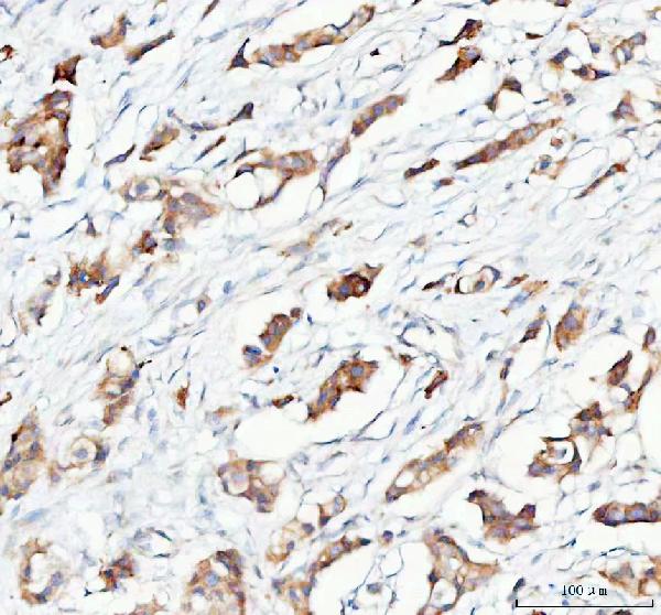
 IHC (Immunohistochemisry)
(Figure 3. IHC analysis of Von Willebrand Factor/VWF using anti-Von Willebrand Factor/VWF antibody (AAA125999).Von Willebrand Factor/VWF was detected in a paraffin-embedded section of human thyroid cancer tissue. Heat mediated antigen retrieval was performed in EDTA buffer (pH 8.0, epitope retrieval solution). The tissue section was blocked with 10% goat serum. The tissue section was then incubated with 2 ug/ml rabbit anti-Von Willebrand Factor/VWF Antibody (AAA125999) overnight at 4 degree C. Peroxidase Conjugated Goat Anti-rabbit IgG was used as secondary antibody and incubated for 30 minutes at 37 degree C. The tissue section was developed using HRP Conjugated Rabbit IgG Super Vision Assay Kit with DAB as the chromogen.)
IHC (Immunohistochemisry)
(Figure 3. IHC analysis of Von Willebrand Factor/VWF using anti-Von Willebrand Factor/VWF antibody (AAA125999).Von Willebrand Factor/VWF was detected in a paraffin-embedded section of human thyroid cancer tissue. Heat mediated antigen retrieval was performed in EDTA buffer (pH 8.0, epitope retrieval solution). The tissue section was blocked with 10% goat serum. The tissue section was then incubated with 2 ug/ml rabbit anti-Von Willebrand Factor/VWF Antibody (AAA125999) overnight at 4 degree C. Peroxidase Conjugated Goat Anti-rabbit IgG was used as secondary antibody and incubated for 30 minutes at 37 degree C. The tissue section was developed using HRP Conjugated Rabbit IgG Super Vision Assay Kit with DAB as the chromogen.)
Von Willebrand Factor/VWF, Polyclonal Antibody (Cat# AAA125999)
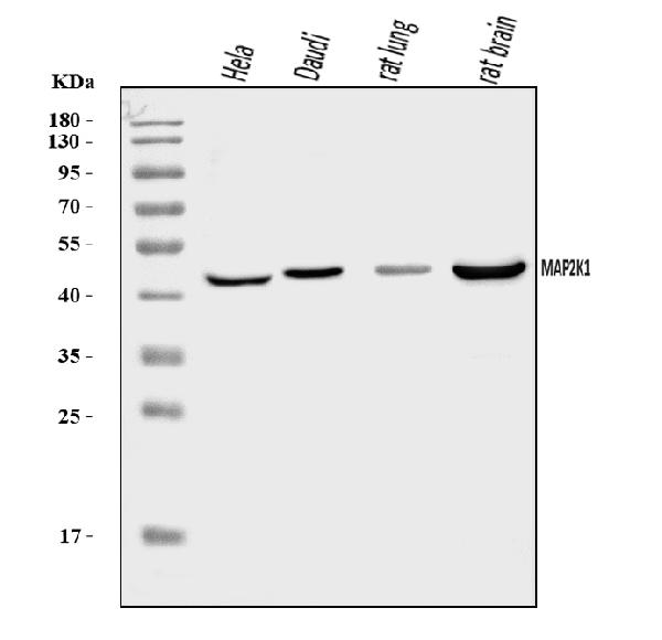
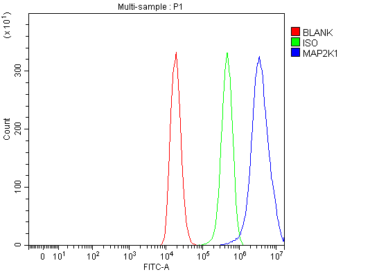 FCM/FACS (Flow Cytometry)
(Figure 2. Flow Cytometry analysis of 293T cells using anti-MEK1/MAP2K1 antibody (AAA126001).Overlay histogram showing 293T cells stained with AAA126001 (Blue line). The cells were blocked with 10% normal goat serum. And then incubated with rabbit anti-MEK1/MAP2K1 Antibody (AAA126001, 1 ug/1x10^6 cells) for 30 min at 20 degree C. DyLight488 conjugated goat anti-rabbit IgG was used as secondary antibody for 30 minutes at 20 degree C. Isotype control antibody (Green line) was rabbit IgG (1 ug/1x10^6) used under the same conditions. Unlabelled sample (Red line) was also used as a control.)
FCM/FACS (Flow Cytometry)
(Figure 2. Flow Cytometry analysis of 293T cells using anti-MEK1/MAP2K1 antibody (AAA126001).Overlay histogram showing 293T cells stained with AAA126001 (Blue line). The cells were blocked with 10% normal goat serum. And then incubated with rabbit anti-MEK1/MAP2K1 Antibody (AAA126001, 1 ug/1x10^6 cells) for 30 min at 20 degree C. DyLight488 conjugated goat anti-rabbit IgG was used as secondary antibody for 30 minutes at 20 degree C. Isotype control antibody (Green line) was rabbit IgG (1 ug/1x10^6) used under the same conditions. Unlabelled sample (Red line) was also used as a control.)
MEK1/MAP2K1, Polyclonal Antibody (Cat# AAA126001)
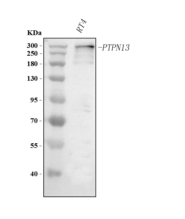
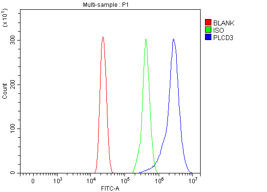
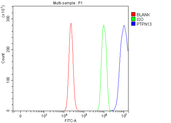 FCM/FACS (Flow Cytometry)
(Figure 2. Flow Cytometry analysis of RT4 cells using anti-PTPN13 antibody (AAA127121).Overlay histogram showing RT4 cells stained with AAA127121 (Blue line). To facilitate intracellular staining, cells were fixed with 4% paraformaldehyde and permeabilized with permeabilization buffer. The cells were blocked with 10% normal goat serum. And then incubated with rabbit anti-PTPN13 Antibody (AAA127121, 1ug/1x106 cells) for 30 min at 20 degree C. DyLight488 conjugated goat anti-rabbit IgG was used as secondary antibody for 30 minutes at 20 degree C. Isotype control antibody (Green line) was rabbit IgG (1ug/1x106) used under the same conditions. Unlabelled sample (Red line) was also used as a control.)
FCM/FACS (Flow Cytometry)
(Figure 2. Flow Cytometry analysis of RT4 cells using anti-PTPN13 antibody (AAA127121).Overlay histogram showing RT4 cells stained with AAA127121 (Blue line). To facilitate intracellular staining, cells were fixed with 4% paraformaldehyde and permeabilized with permeabilization buffer. The cells were blocked with 10% normal goat serum. And then incubated with rabbit anti-PTPN13 Antibody (AAA127121, 1ug/1x106 cells) for 30 min at 20 degree C. DyLight488 conjugated goat anti-rabbit IgG was used as secondary antibody for 30 minutes at 20 degree C. Isotype control antibody (Green line) was rabbit IgG (1ug/1x106) used under the same conditions. Unlabelled sample (Red line) was also used as a control.)
PTPN13, Polyclonal Antibody (Cat# AAA127121)
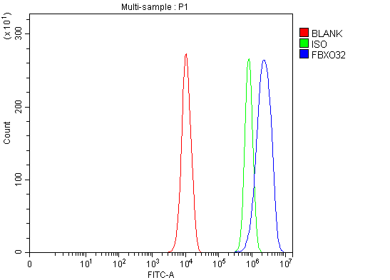 FCM/FACS (Flow Cytometry)
(Figure 3. Flow Cytometry analysis of PC-3 cells using anti-Fbx32/FBXO32 antibody (AAA127124).Overlay histogram showing PC-3 cells stained with AAA127124 (Blue line). To facilitate intracellular staining, cells were fixed with 4% paraformaldehyde and permeabilized with permeabilization buffer. The cells were blocked with 10% normal goat serum. And then incubated with rabbit anti-Fbx32/FBXO32 Antibody (AAA127124, 1ug/1x106 cells) for 30 min at 20 degree C. DyLight488 conjugated goat anti-rabbit IgG was used as secondary antibody for 30 minutes at 20 degree C. Isotype control antibody (Green line) was rabbit IgG (1ug/1x106) used under the same conditions. Unlabelled sample (Red line) was also used as a control.)
FCM/FACS (Flow Cytometry)
(Figure 3. Flow Cytometry analysis of PC-3 cells using anti-Fbx32/FBXO32 antibody (AAA127124).Overlay histogram showing PC-3 cells stained with AAA127124 (Blue line). To facilitate intracellular staining, cells were fixed with 4% paraformaldehyde and permeabilized with permeabilization buffer. The cells were blocked with 10% normal goat serum. And then incubated with rabbit anti-Fbx32/FBXO32 Antibody (AAA127124, 1ug/1x106 cells) for 30 min at 20 degree C. DyLight488 conjugated goat anti-rabbit IgG was used as secondary antibody for 30 minutes at 20 degree C. Isotype control antibody (Green line) was rabbit IgG (1ug/1x106) used under the same conditions. Unlabelled sample (Red line) was also used as a control.)
FBXO32, Polyclonal Antibody (Cat# AAA127124)
 FCM/FACS (Flow Cytometry)
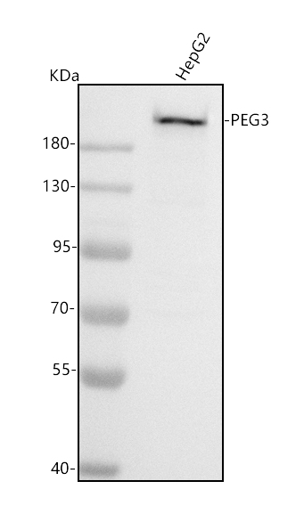
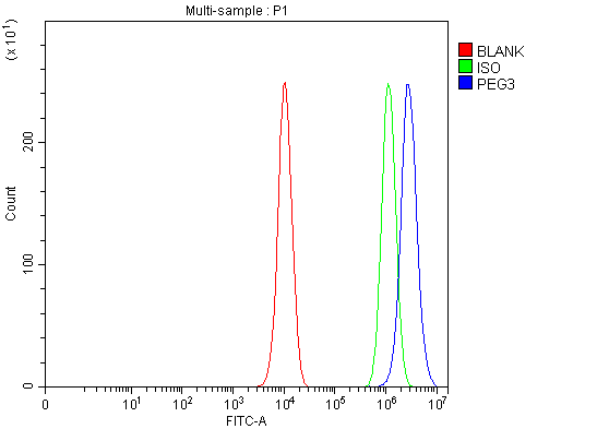
(Figure 3. Flow Cytometry analysis of PC-3 cells using anti-PEG3 antibody (AAA127125).Overlay histogram showing PC-3 cells stained with AAA127125 (Blue line). To facilitate intracellular staining, cells were fixed with 4% paraformaldehyde and permeabilized with permeabilization buffer. The cells were blocked with 10% normal goat serum. And then incubated with rabbit anti-PEG3 Antibody (AAA127125, 1ug/1x106 cells) for 30 min at 20 degree C. DyLight488 conjugated goat anti-rabbit IgG was used as secondary antibody for 30 minutes at 20 degree C. Isotype control antibody (Green line) was rabbit IgG (1ug/1x106) used under the same conditions. Unlabelled sample (Red line) was also used as a control.)
FCM/FACS (Flow Cytometry)
(Figure 3. Flow Cytometry analysis of PC-3 cells using anti-PEG3 antibody (AAA127125).Overlay histogram showing PC-3 cells stained with AAA127125 (Blue line). To facilitate intracellular staining, cells were fixed with 4% paraformaldehyde and permeabilized with permeabilization buffer. The cells were blocked with 10% normal goat serum. And then incubated with rabbit anti-PEG3 Antibody (AAA127125, 1ug/1x106 cells) for 30 min at 20 degree C. DyLight488 conjugated goat anti-rabbit IgG was used as secondary antibody for 30 minutes at 20 degree C. Isotype control antibody (Green line) was rabbit IgG (1ug/1x106) used under the same conditions. Unlabelled sample (Red line) was also used as a control.)
PEG3, Polyclonal Antibody (Cat# AAA127125)
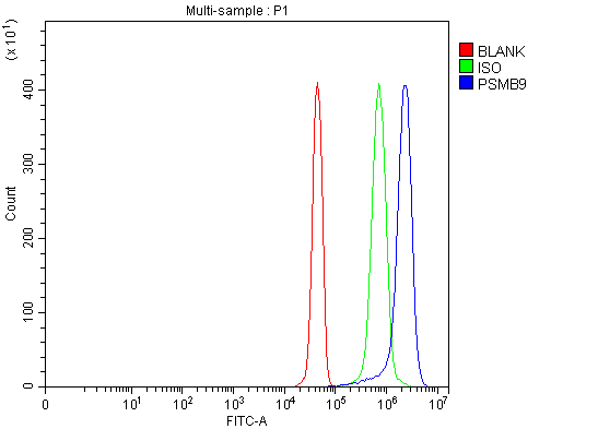 FCM/FACS (Flow Cytometry)
(Figure 5. Flow Cytometry analysis of JK cells using anti-PSMB9 antibody (AAA127146).Overlay histogram showing JK cells stained with AAA127146 (Blue line). To facilitate intracellular staining, cells were fixed with 4% paraformaldehyde and permeabilized with permeabilization buffer. The cells were blocked with 10% normal goat serum. And then incubated with rabbit anti-PSMB9 Antibody (AAA127146, 1ug/1x106 cells) for 30 min at 20 degree C. DyLight488 conjugated goat anti-rabbit IgG was used as secondary antibody for 30 minutes at 20 degree C. Isotype control antibody (Green line) was rabbit IgG (1ug/1x106) used under the same conditions. Unlabelled sample (Red line) was also used as a control.)
FCM/FACS (Flow Cytometry)
(Figure 5. Flow Cytometry analysis of JK cells using anti-PSMB9 antibody (AAA127146).Overlay histogram showing JK cells stained with AAA127146 (Blue line). To facilitate intracellular staining, cells were fixed with 4% paraformaldehyde and permeabilized with permeabilization buffer. The cells were blocked with 10% normal goat serum. And then incubated with rabbit anti-PSMB9 Antibody (AAA127146, 1ug/1x106 cells) for 30 min at 20 degree C. DyLight488 conjugated goat anti-rabbit IgG was used as secondary antibody for 30 minutes at 20 degree C. Isotype control antibody (Green line) was rabbit IgG (1ug/1x106) used under the same conditions. Unlabelled sample (Red line) was also used as a control.)
PSMB9, Polyclonal Antibody (Cat# AAA127146)
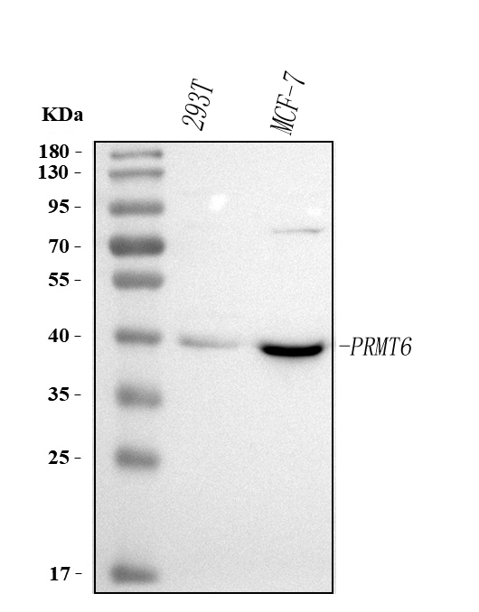
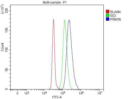 FCM/FACS (Flow Cytometry)
(Figure 2. Flow Cytometry analysis of SH-SY5Y cells using anti-PRMT6 antibody (AAA127150).Overlay histogram showing SH-SY5Y cells stained with AAA127150 (Blue line). To facilitate intracellular staining, cells were fixed with 4% paraformaldehyde and permeabilized with permeabilization buffer. The cells were blocked with 10% normal goat serum. And then incubated with rabbit anti-PRMT6 Antibody (AAA127150, 1ug/1x106 cells) for 30 min at 20 degree C. DyLight488 conjugated goat anti-rabbit IgG was used as secondary antibody for 30 minutes at 20 degree C. Isotype control antibody (Green line) was rabbit IgG (1ug/1x106) used under the same conditions. Unlabelled sample (Red line) was also used as a control.)
FCM/FACS (Flow Cytometry)
(Figure 2. Flow Cytometry analysis of SH-SY5Y cells using anti-PRMT6 antibody (AAA127150).Overlay histogram showing SH-SY5Y cells stained with AAA127150 (Blue line). To facilitate intracellular staining, cells were fixed with 4% paraformaldehyde and permeabilized with permeabilization buffer. The cells were blocked with 10% normal goat serum. And then incubated with rabbit anti-PRMT6 Antibody (AAA127150, 1ug/1x106 cells) for 30 min at 20 degree C. DyLight488 conjugated goat anti-rabbit IgG was used as secondary antibody for 30 minutes at 20 degree C. Isotype control antibody (Green line) was rabbit IgG (1ug/1x106) used under the same conditions. Unlabelled sample (Red line) was also used as a control.)
PRMT6, Polyclonal Antibody (Cat# AAA127150)
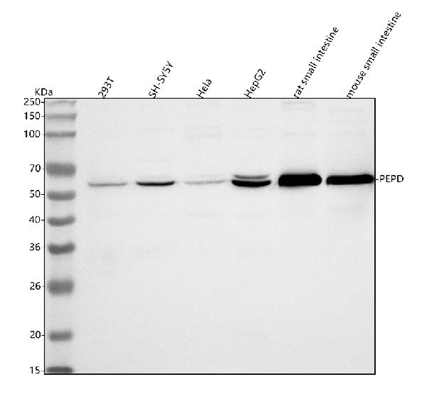
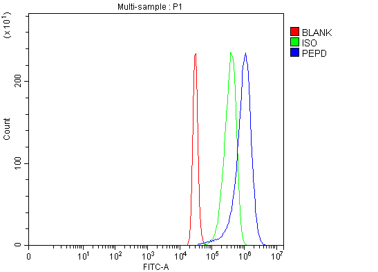 FCM/FACS (Flow Cytometry)
(Figure 3. Flow Cytometry analysis of 293T cells using anti-PEPD antibody (AAA127175).Overlay histogram showing 293T cells stained with AAA127175 (Blue line). The cells were fixed with 4% paraformaldehyde and blocked with 10% normal goat serum. And then incubated with rabbit anti-PEPD Antibody (AAA127175, 1ug/1x106 cells) for 30 min at 20 degree C. DyLight488 conjugated goat anti-rabbit IgG was used as secondary antibody for 30 minutes at 20 degree C. Isotype control antibody (Green line) was rabbit IgG (1ug/1x106) used under the same conditions. Unlabelled sample (Red line) was also used as a control.)
FCM/FACS (Flow Cytometry)
(Figure 3. Flow Cytometry analysis of 293T cells using anti-PEPD antibody (AAA127175).Overlay histogram showing 293T cells stained with AAA127175 (Blue line). The cells were fixed with 4% paraformaldehyde and blocked with 10% normal goat serum. And then incubated with rabbit anti-PEPD Antibody (AAA127175, 1ug/1x106 cells) for 30 min at 20 degree C. DyLight488 conjugated goat anti-rabbit IgG was used as secondary antibody for 30 minutes at 20 degree C. Isotype control antibody (Green line) was rabbit IgG (1ug/1x106) used under the same conditions. Unlabelled sample (Red line) was also used as a control.)
PEPD, Polyclonal Antibody (Cat# AAA127175)
 FCM/FACS (Flow Cytometry)
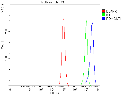
(Figure 4. Flow Cytometry analysis of PC-3 cells using anti-POMGNT1 antibody (AAA127182).Overlay histogram showing PC-3 cells stained with AAA127182 (Blue line). To facilitate intracellular staining, cells were fixed with 4% paraformaldehyde and permeabilized with permeabilization buffer. The cells were blocked with 10% normal goat serum. And then incubated with rabbit anti-POMGNT1 Antibody (AAA127182, 1ug/1x106 cells) for 30 min at 20 degree C. DyLight488 conjugated goat anti-rabbit IgG was used as secondary antibody for 30 minutes at 20 degree C. Isotype control antibody (Green line) was rabbit IgG (1ug/1x106) used under the same conditions. Unlabelled sample (Red line) was also used as a control.)
FCM/FACS (Flow Cytometry)
(Figure 4. Flow Cytometry analysis of PC-3 cells using anti-POMGNT1 antibody (AAA127182).Overlay histogram showing PC-3 cells stained with AAA127182 (Blue line). To facilitate intracellular staining, cells were fixed with 4% paraformaldehyde and permeabilized with permeabilization buffer. The cells were blocked with 10% normal goat serum. And then incubated with rabbit anti-POMGNT1 Antibody (AAA127182, 1ug/1x106 cells) for 30 min at 20 degree C. DyLight488 conjugated goat anti-rabbit IgG was used as secondary antibody for 30 minutes at 20 degree C. Isotype control antibody (Green line) was rabbit IgG (1ug/1x106) used under the same conditions. Unlabelled sample (Red line) was also used as a control.)
POMGNT1, Polyclonal Antibody (Cat# AAA127182)
 FCM/FACS (Flow Cytometry)
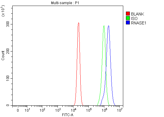
(Figure 2. Flow Cytometry analysis of U20S cells using anti-RNASE1 antibody (AAA127187).Overlay histogram showing U20S cells stained with AAA127187 (Blue line). The cells were fixed with 4% paraformaldehyde and blocked with 10% normal goat serum. And then incubated with rabbit anti-RNASE1 Antibody (AAA127187, 1ug/1x106 cells) for 30 min at 20 degree C. DyLight488 conjugated goat anti-rabbit IgG was used as secondary antibody for 30 minutes at 20 degree C. Isotype control antibody (Green line) was rabbit IgG (1ug/1x106) used under the same conditions. Unlabelled sample (Red line) was also used as a control.)
FCM/FACS (Flow Cytometry)
(Figure 2. Flow Cytometry analysis of U20S cells using anti-RNASE1 antibody (AAA127187).Overlay histogram showing U20S cells stained with AAA127187 (Blue line). The cells were fixed with 4% paraformaldehyde and blocked with 10% normal goat serum. And then incubated with rabbit anti-RNASE1 Antibody (AAA127187, 1ug/1x106 cells) for 30 min at 20 degree C. DyLight488 conjugated goat anti-rabbit IgG was used as secondary antibody for 30 minutes at 20 degree C. Isotype control antibody (Green line) was rabbit IgG (1ug/1x106) used under the same conditions. Unlabelled sample (Red line) was also used as a control.)
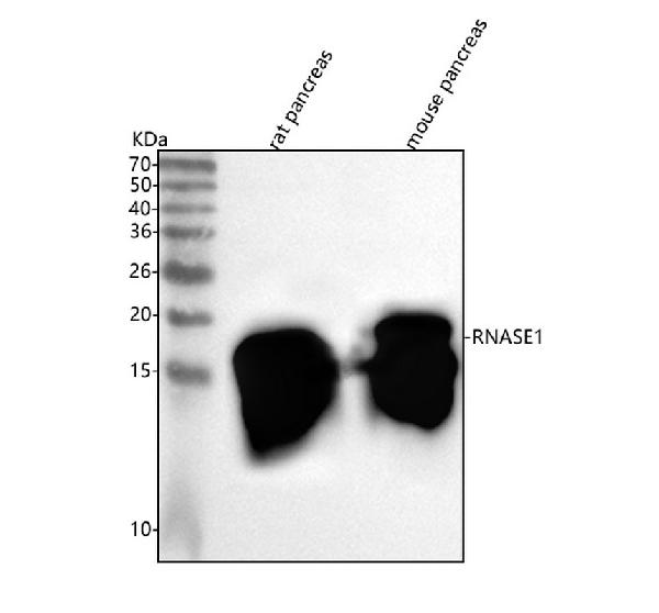
RNASE1, Polyclonal Antibody (Cat# AAA127187)
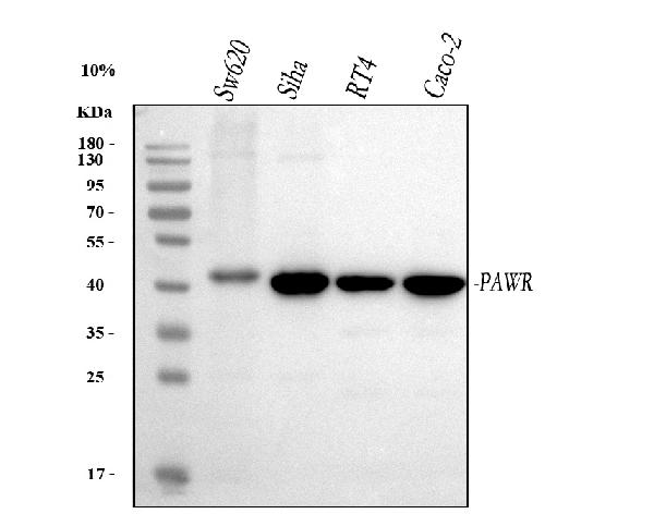
 FCM/FACS (Flow Cytometry)
(Figure 3. Flow Cytometry analysis of Caco-2 cells using anti-PAWR antibody (AAA127191).Overlay histogram showing Caco-2 cells stained with AAA127191 (Blue line). To facilitate intracellular staining, cells were fixed with 4% paraformaldehyde and permeabilized with permeabilization buffer. The cells were blocked with 10% normal goat serum. And then incubated with rabbit anti-PAWR Antibody (AAA127191, 1ug/1x106 cells) for 30 min at 20 degree C. DyLight488 conjugated goat anti-rabbit IgG was used as secondary antibody for 30 minutes at 20 degree C. Isotype control antibody (Green line) was rabbit IgG (1ug/1x106) used under the same conditions. Unlabelled sample (Red line) was also used as a control.)
FCM/FACS (Flow Cytometry)
(Figure 3. Flow Cytometry analysis of Caco-2 cells using anti-PAWR antibody (AAA127191).Overlay histogram showing Caco-2 cells stained with AAA127191 (Blue line). To facilitate intracellular staining, cells were fixed with 4% paraformaldehyde and permeabilized with permeabilization buffer. The cells were blocked with 10% normal goat serum. And then incubated with rabbit anti-PAWR Antibody (AAA127191, 1ug/1x106 cells) for 30 min at 20 degree C. DyLight488 conjugated goat anti-rabbit IgG was used as secondary antibody for 30 minutes at 20 degree C. Isotype control antibody (Green line) was rabbit IgG (1ug/1x106) used under the same conditions. Unlabelled sample (Red line) was also used as a control.)
PAWR, Polyclonal Antibody (Cat# AAA127191)
 FCM/FACS (Flow Cytometry)
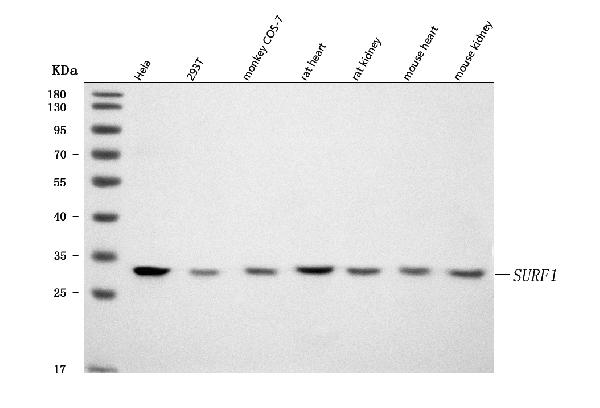
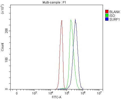
(Figure 3. Flow Cytometry analysis of Jurkat cells using anti-SURF1 antibody (AAA127193).Overlay histogram showing Jurkat cells stained with AAA127193 (Blue line). To facilitate intracellular staining, cells were fixed with 4% paraformaldehyde and permeabilized with permeabilization buffer. The cells were blocked with 10% normal goat serum. And then incubated with rabbit anti-SURF1 Antibody (AAA127193, 1ug/1x106 cells) for 30 min at 20 degree C. DyLight488 conjugated goat anti-rabbit IgG was used as secondary antibody for 30 minutes at 20 degree C. Isotype control antibody (Green line) was rabbit IgG (1ug/1x106) used under the same conditions. Unlabelled sample without incubation with primary antibody and secondary antibody (Red line) was used as a blank control.)
FCM/FACS (Flow Cytometry)
(Figure 3. Flow Cytometry analysis of Jurkat cells using anti-SURF1 antibody (AAA127193).Overlay histogram showing Jurkat cells stained with AAA127193 (Blue line). To facilitate intracellular staining, cells were fixed with 4% paraformaldehyde and permeabilized with permeabilization buffer. The cells were blocked with 10% normal goat serum. And then incubated with rabbit anti-SURF1 Antibody (AAA127193, 1ug/1x106 cells) for 30 min at 20 degree C. DyLight488 conjugated goat anti-rabbit IgG was used as secondary antibody for 30 minutes at 20 degree C. Isotype control antibody (Green line) was rabbit IgG (1ug/1x106) used under the same conditions. Unlabelled sample without incubation with primary antibody and secondary antibody (Red line) was used as a blank control.)
SURF1, Polyclonal Antibody (Cat# AAA127193)
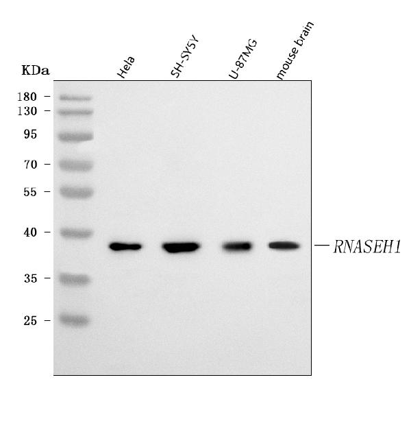
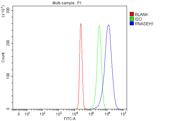 FCM/FACS (Flow Cytometry)
(Figure 3. Flow Cytometry analysis of HepG2 cells using anti-RNASEH1 antibody (AAA127358).Overlay histogram showing HepG2 cells stained with AAA127358 (Blue line). To facilitate intracellular staining, cells were fixed with 4% paraformaldehyde and permeabilized with permeabilization buffer. The cells were blocked with 10% normal goat serum. And then incubated with rabbit anti-RNASEH1 Antibody (AAA127358, 1ug/1x106 cells) for 30 min at 20 degree C. DyLight488 conjugated goat anti-rabbit IgG was used as secondary antibody for 30 minutes at 20 degree C. Isotype control antibody (Green line) was rabbit IgG (1ug/1x106) used under the same conditions. Unlabelled sample without incubation with primary antibody and secondary antibody (Red line) was used as a blank control.)
FCM/FACS (Flow Cytometry)
(Figure 3. Flow Cytometry analysis of HepG2 cells using anti-RNASEH1 antibody (AAA127358).Overlay histogram showing HepG2 cells stained with AAA127358 (Blue line). To facilitate intracellular staining, cells were fixed with 4% paraformaldehyde and permeabilized with permeabilization buffer. The cells were blocked with 10% normal goat serum. And then incubated with rabbit anti-RNASEH1 Antibody (AAA127358, 1ug/1x106 cells) for 30 min at 20 degree C. DyLight488 conjugated goat anti-rabbit IgG was used as secondary antibody for 30 minutes at 20 degree C. Isotype control antibody (Green line) was rabbit IgG (1ug/1x106) used under the same conditions. Unlabelled sample without incubation with primary antibody and secondary antibody (Red line) was used as a blank control.)
RNASEH1, Polyclonal Antibody (Cat# AAA127358)
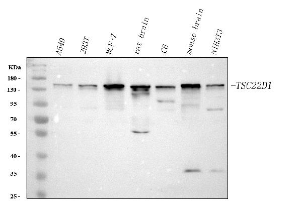
 FCM/FACS (Flow Cytometry)
(Figure 2. Flow Cytometry analysis of SH-SY5Y cells using anti-TSC22D1 antibody (AAA127365).Overlay histogram showing SH-SY5Y cells stained with AAA127365 (Blue line). To facilitate intracellular staining, cells were fixed with 4% paraformaldehyde and permeabilized with permeabilization buffer. The cells were blocked with 10% normal goat serum. And then incubated with rabbit anti-TSC22D1 Antibody (AAA127365, 1ug/1x106 cells) for 30 min at 20 degree C. DyLight488 conjugated goat anti-rabbit IgG was used as secondary antibody for 30 minutes at 20 degree C. Isotype control antibody (Green line) was rabbit IgG (1ug/1x106) used under the same conditions. Unlabelled sample (Red line) was also used as a control.)
FCM/FACS (Flow Cytometry)
(Figure 2. Flow Cytometry analysis of SH-SY5Y cells using anti-TSC22D1 antibody (AAA127365).Overlay histogram showing SH-SY5Y cells stained with AAA127365 (Blue line). To facilitate intracellular staining, cells were fixed with 4% paraformaldehyde and permeabilized with permeabilization buffer. The cells were blocked with 10% normal goat serum. And then incubated with rabbit anti-TSC22D1 Antibody (AAA127365, 1ug/1x106 cells) for 30 min at 20 degree C. DyLight488 conjugated goat anti-rabbit IgG was used as secondary antibody for 30 minutes at 20 degree C. Isotype control antibody (Green line) was rabbit IgG (1ug/1x106) used under the same conditions. Unlabelled sample (Red line) was also used as a control.)
TSC22D1, Polyclonal Antibody (Cat# AAA127365)
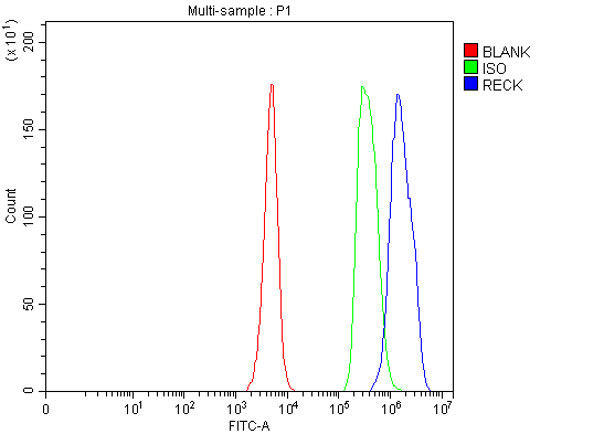 FCM/FACS (Flow Cytometry)
(Figure 2. Flow Cytometry analysis of JK cells using anti-RECK antibody (AAA127371).Overlay histogram showing JK cells stained with AAA127371 (Blue line). The cells were fixed with 4% paraformaldehyde and blocked with 10% normal goat serum. And then incubated with rabbit anti-RECK Antibody (AAA127371, 1ug/1x106 cells) for 30 min at 20 degree C. DyLight488 conjugated goat anti-rabbit IgG was used as secondary antibody for 30 minutes at 20 degree C. Isotype control antibody (Green line) was rabbit IgG (1ug/1x106) used under the same conditions. Unlabelled sample (Red line) was also used as a control.)
FCM/FACS (Flow Cytometry)
(Figure 2. Flow Cytometry analysis of JK cells using anti-RECK antibody (AAA127371).Overlay histogram showing JK cells stained with AAA127371 (Blue line). The cells were fixed with 4% paraformaldehyde and blocked with 10% normal goat serum. And then incubated with rabbit anti-RECK Antibody (AAA127371, 1ug/1x106 cells) for 30 min at 20 degree C. DyLight488 conjugated goat anti-rabbit IgG was used as secondary antibody for 30 minutes at 20 degree C. Isotype control antibody (Green line) was rabbit IgG (1ug/1x106) used under the same conditions. Unlabelled sample (Red line) was also used as a control.)
RECK, Polyclonal Antibody (Cat# AAA127371)
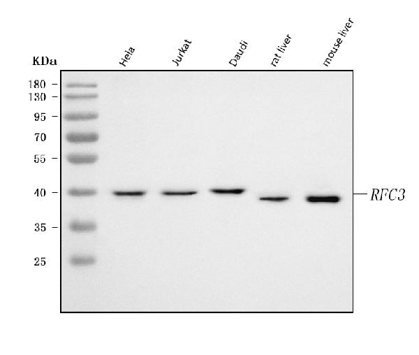
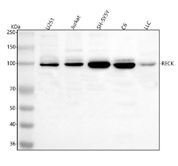
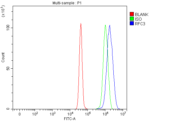 FCM/FACS (Flow Cytometry)
(Figure 3. Flow Cytometry analysis of U251 cells using anti-RFC3 antibody (AAA127372).Overlay histogram showing U251 cells stained with AAA127372 (Blue line). To facilitate intracellular staining, cells were fixed with 4% paraformaldehyde and permeabilized with permeabilization buffer. The cells were blocked with 10% normal goat serum. And then incubated with rabbit anti-RFC3 Antibody (AAA127372, 1ug/1x106 cells) for 30 min at 20 degree C. DyLight488 conjugated goat anti-rabbit IgG was used as secondary antibody for 30 minutes at 20 degree C. Isotype control antibody (Green line) was rabbit IgG (1ug/1x106) used under the same conditions. Unlabelled sample without incubation with primary antibody and secondary antibody (Red line) was used as a blank control.)
FCM/FACS (Flow Cytometry)
(Figure 3. Flow Cytometry analysis of U251 cells using anti-RFC3 antibody (AAA127372).Overlay histogram showing U251 cells stained with AAA127372 (Blue line). To facilitate intracellular staining, cells were fixed with 4% paraformaldehyde and permeabilized with permeabilization buffer. The cells were blocked with 10% normal goat serum. And then incubated with rabbit anti-RFC3 Antibody (AAA127372, 1ug/1x106 cells) for 30 min at 20 degree C. DyLight488 conjugated goat anti-rabbit IgG was used as secondary antibody for 30 minutes at 20 degree C. Isotype control antibody (Green line) was rabbit IgG (1ug/1x106) used under the same conditions. Unlabelled sample without incubation with primary antibody and secondary antibody (Red line) was used as a blank control.)
RFC3, Polyclonal Antibody (Cat# AAA127372)
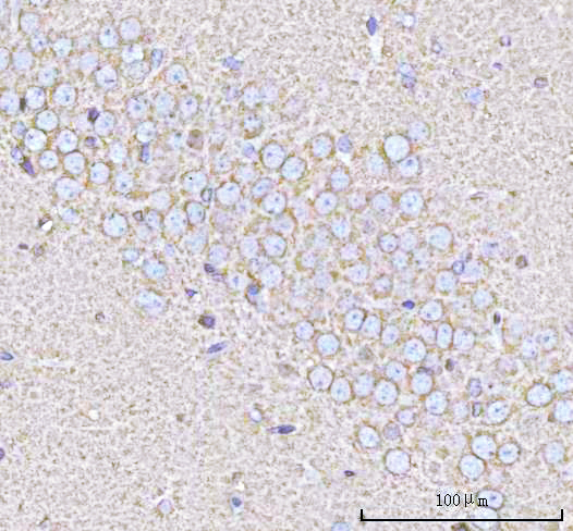
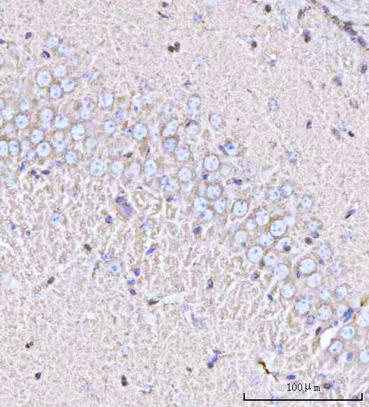
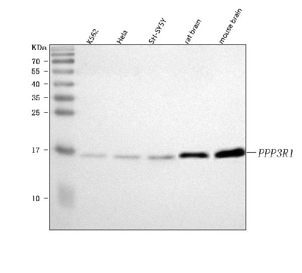
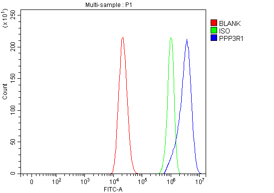 FCM/FACS (Flow Cytometry)
(Figure 4. Flow Cytometry analysis of Caco-2 cells using anti-PPP3R1 antibody (AAA127374).Overlay histogram showing Caco-2 cells stained with AAA127374 (Blue line). To facilitate intracellular staining, cells were fixed with 4% paraformaldehyde and permeabilized with permeabilization buffer. The cells were blocked with 10% normal goat serum. And then incubated with rabbit anti-PPP3R1 Antibody (AAA127374, 1ug/1x106 cells) for 30 min at 20 degree C. DyLight488 conjugated goat anti-rabbit IgG was used as secondary antibody for 30 minutes at 20 degree C. Isotype control antibody (Green line) was rabbit IgG (1ug/1x106) used under the same conditions. Unlabelled sample (Red line) was also used as a control.)
FCM/FACS (Flow Cytometry)
(Figure 4. Flow Cytometry analysis of Caco-2 cells using anti-PPP3R1 antibody (AAA127374).Overlay histogram showing Caco-2 cells stained with AAA127374 (Blue line). To facilitate intracellular staining, cells were fixed with 4% paraformaldehyde and permeabilized with permeabilization buffer. The cells were blocked with 10% normal goat serum. And then incubated with rabbit anti-PPP3R1 Antibody (AAA127374, 1ug/1x106 cells) for 30 min at 20 degree C. DyLight488 conjugated goat anti-rabbit IgG was used as secondary antibody for 30 minutes at 20 degree C. Isotype control antibody (Green line) was rabbit IgG (1ug/1x106) used under the same conditions. Unlabelled sample (Red line) was also used as a control.)
PPP3R1, Polyclonal Antibody (Cat# AAA127374)
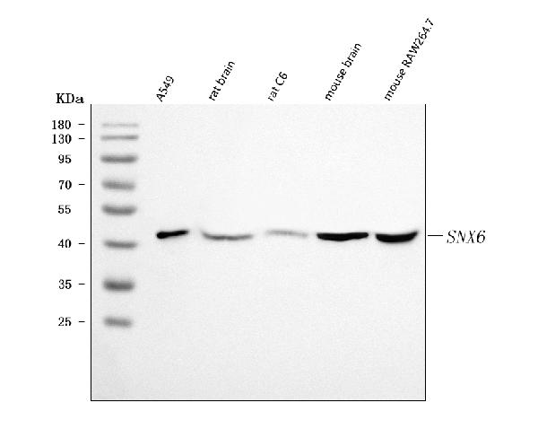
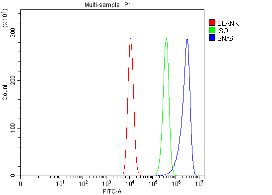 FCM/FACS (Flow Cytometry)
(Figure 3. Flow Cytometry analysis of HCT116 cells using anti-SNX6 antibody (AAA127382).Overlay histogram showing HCT116 cells stained with AAA127382 (Blue line). To facilitate intracellular staining, cells were fixed with 4% paraformaldehyde and permeabilized with permeabilization buffer. The cells were blocked with 10% normal goat serum. And then incubated with rabbit anti-SNX6 Antibody (AAA127382, 1ug/1x106 cells) for 30 min at 20 degree C. DyLight488 conjugated goat anti-rabbit IgG was used as secondary antibody for 30 minutes at 20 degree C. Isotype control antibody (Green line) was rabbit IgG (1ug/1x106) used under the same conditions. Unlabelled sample without incubation with primary antibody and secondary antibody (Red line) was used as a blank control.)
FCM/FACS (Flow Cytometry)
(Figure 3. Flow Cytometry analysis of HCT116 cells using anti-SNX6 antibody (AAA127382).Overlay histogram showing HCT116 cells stained with AAA127382 (Blue line). To facilitate intracellular staining, cells were fixed with 4% paraformaldehyde and permeabilized with permeabilization buffer. The cells were blocked with 10% normal goat serum. And then incubated with rabbit anti-SNX6 Antibody (AAA127382, 1ug/1x106 cells) for 30 min at 20 degree C. DyLight488 conjugated goat anti-rabbit IgG was used as secondary antibody for 30 minutes at 20 degree C. Isotype control antibody (Green line) was rabbit IgG (1ug/1x106) used under the same conditions. Unlabelled sample without incubation with primary antibody and secondary antibody (Red line) was used as a blank control.)
SNX6, Polyclonal Antibody (Cat# AAA127382)
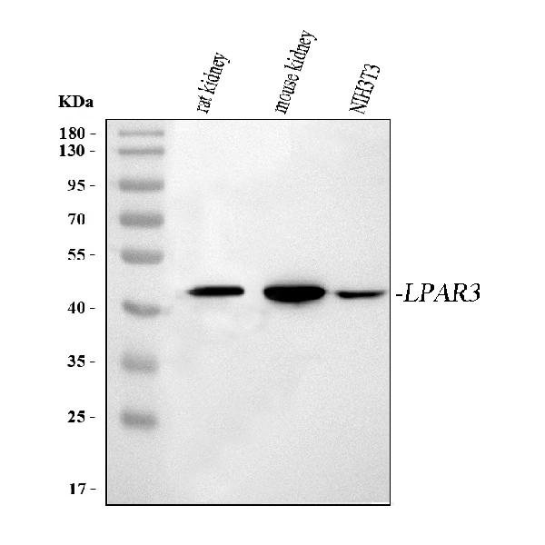
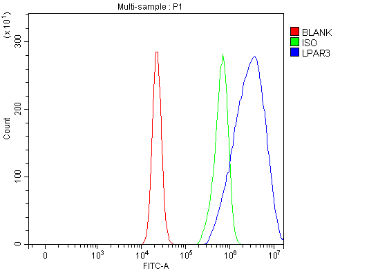 FCM/FACS (Flow Cytometry)
(Figure 2. Flow Cytometry analysis of RT4 cells using anti-EDG7/LPAR3 antibody (AAA127387).Overlay histogram showing RT4 cells stained with AAA127387 (Blue line). The cells were fixed with 4% paraformaldehyde and blocked with 10% normal goat serum. And then incubated with rabbit anti-EDG7/LPAR3 Antibody (AAA127387, 1ug/1x106 cells) for 30 min at 20 degree C. DyLight488 conjugated goat anti-rabbit IgG was used as secondary antibody for 30 minutes at 20 degree C. Isotype control antibody (Green line) was rabbit IgG (1ug/1x106) used under the same conditions. Unlabelled sample (Red line) was also used as a control.)
FCM/FACS (Flow Cytometry)
(Figure 2. Flow Cytometry analysis of RT4 cells using anti-EDG7/LPAR3 antibody (AAA127387).Overlay histogram showing RT4 cells stained with AAA127387 (Blue line). The cells were fixed with 4% paraformaldehyde and blocked with 10% normal goat serum. And then incubated with rabbit anti-EDG7/LPAR3 Antibody (AAA127387, 1ug/1x106 cells) for 30 min at 20 degree C. DyLight488 conjugated goat anti-rabbit IgG was used as secondary antibody for 30 minutes at 20 degree C. Isotype control antibody (Green line) was rabbit IgG (1ug/1x106) used under the same conditions. Unlabelled sample (Red line) was also used as a control.)
LPAR3, Polyclonal Antibody (Cat# AAA127387)
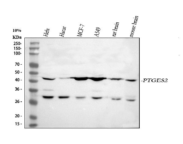
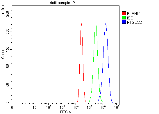 FCM/FACS (Flow Cytometry)
(Figure 5. Flow Cytometry analysis of MCF-7 cells using anti-PTGES2/Gbf1 antibody (AAA127397).Overlay histogram showing MCF-7 cells stained with AAA127397 (Blue line). To facilitate intracellular staining, cells were fixed with 4% paraformaldehyde and permeabilized with permeabilization buffer. The cells were blocked with 10% normal goat serum. And then incubated with rabbit anti-PTGES2/Gbf1 Antibody (AAA127397, 1ug/1x106 cells) for 30 min at 20 degree C. DyLight488 conjugated goat anti-rabbit IgG was used as secondary antibody for 30 minutes at 20 degree C. Isotype control antibody (Green line) was rabbit IgG (1ug/1x106) used under the same conditions. Unlabelled sample (Red line) was also used as a control.)
FCM/FACS (Flow Cytometry)
(Figure 5. Flow Cytometry analysis of MCF-7 cells using anti-PTGES2/Gbf1 antibody (AAA127397).Overlay histogram showing MCF-7 cells stained with AAA127397 (Blue line). To facilitate intracellular staining, cells were fixed with 4% paraformaldehyde and permeabilized with permeabilization buffer. The cells were blocked with 10% normal goat serum. And then incubated with rabbit anti-PTGES2/Gbf1 Antibody (AAA127397, 1ug/1x106 cells) for 30 min at 20 degree C. DyLight488 conjugated goat anti-rabbit IgG was used as secondary antibody for 30 minutes at 20 degree C. Isotype control antibody (Green line) was rabbit IgG (1ug/1x106) used under the same conditions. Unlabelled sample (Red line) was also used as a control.)
PTGES2/Gbf1, Polyclonal Antibody (Cat# AAA127397)
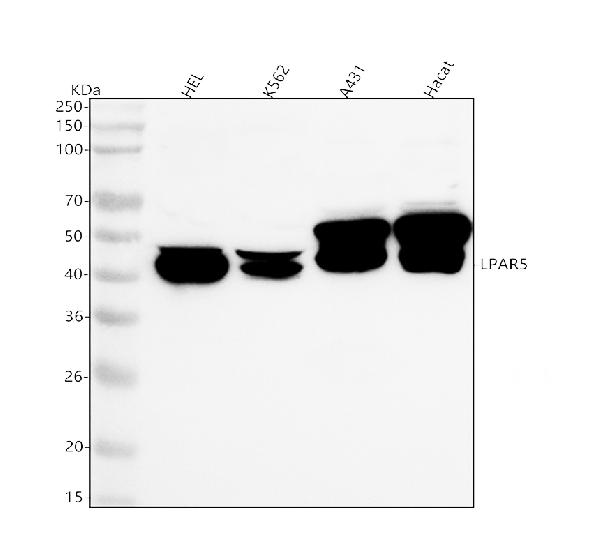
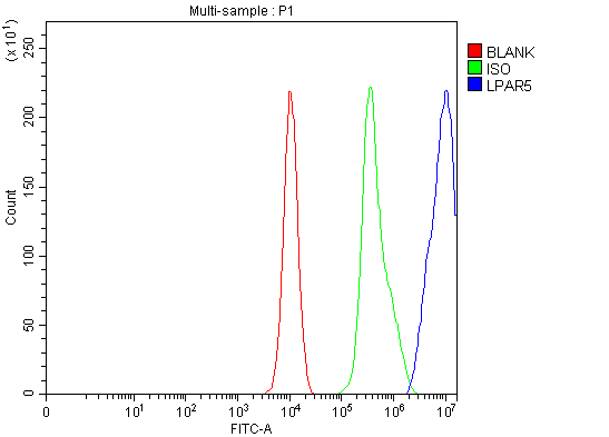 FCM/FACS (Flow Cytometry)
(Figure 2. Flow Cytometry analysis of K562 cells using anti-GPR92/LPAR5 antibody (AAA127398).Overlay histogram showing K562 cells stained with AAA127398 (Blue line). The cells were fixed with 4% paraformaldehyde and blocked with 10% normal goat serum. And then incubated with rabbit anti-GPR92/LPAR5 Antibody (AAA127398, 1ug/1x106 cells) for 30 min at 20 degree C. DyLight488 conjugated goat anti-rabbit IgG was used as secondary antibody for 30 minutes at 20 degree C. Isotype control antibody (Green line) was rabbit IgG (1ug/1x106) used under the same conditions. Unlabelled sample (Red line) was also used as a control.)
FCM/FACS (Flow Cytometry)
(Figure 2. Flow Cytometry analysis of K562 cells using anti-GPR92/LPAR5 antibody (AAA127398).Overlay histogram showing K562 cells stained with AAA127398 (Blue line). The cells were fixed with 4% paraformaldehyde and blocked with 10% normal goat serum. And then incubated with rabbit anti-GPR92/LPAR5 Antibody (AAA127398, 1ug/1x106 cells) for 30 min at 20 degree C. DyLight488 conjugated goat anti-rabbit IgG was used as secondary antibody for 30 minutes at 20 degree C. Isotype control antibody (Green line) was rabbit IgG (1ug/1x106) used under the same conditions. Unlabelled sample (Red line) was also used as a control.)
LPAR5, Polyclonal Antibody (Cat# AAA127398)
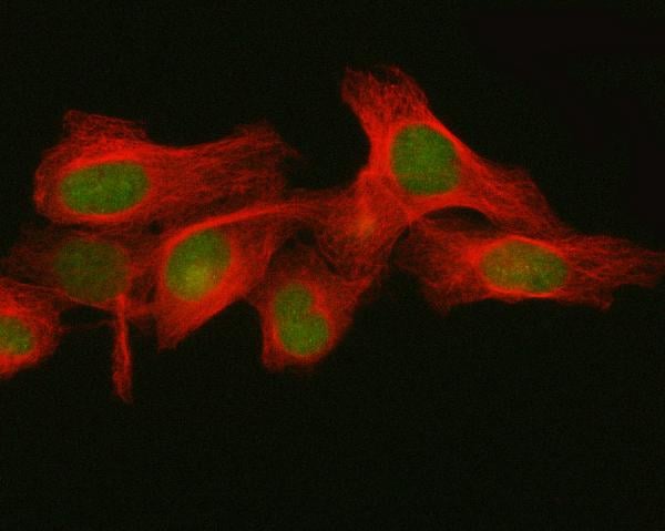
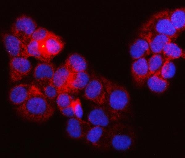
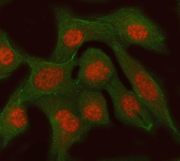
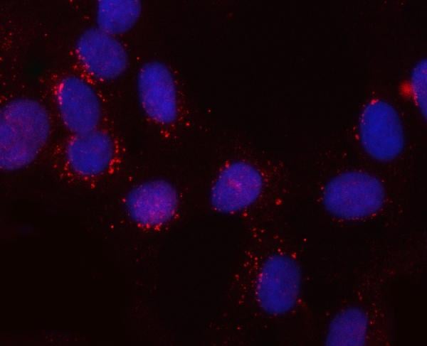
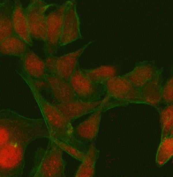
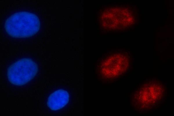
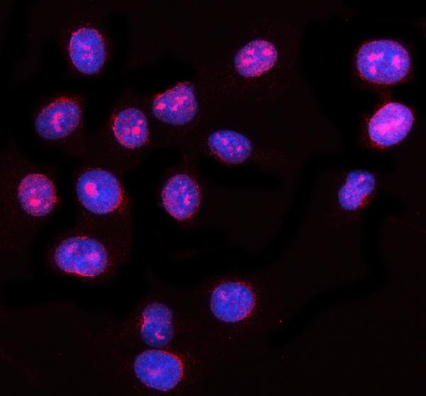
 IF (Immunofluorescence)
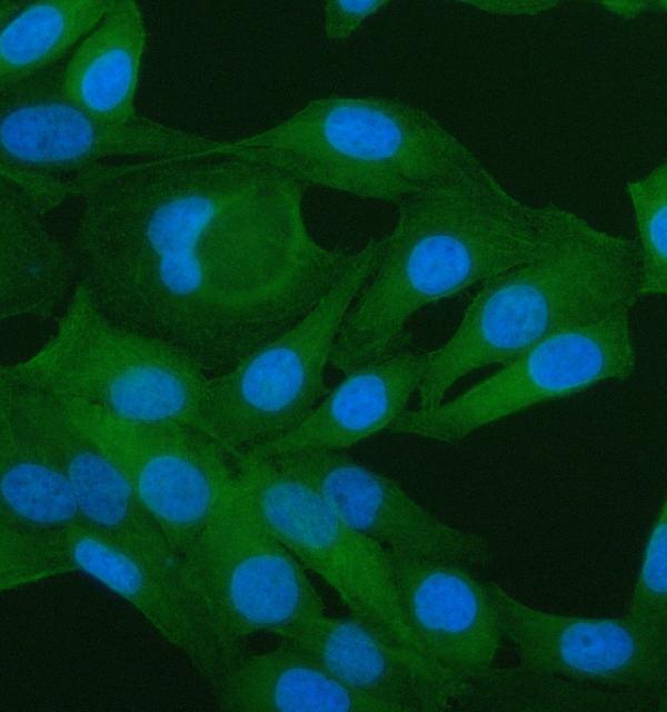
(Figure 2. IF analysis of RBMY1A1 and Tubulin beta using anti-RBMY1A1 antibody (AAA127401) and anti-Tubulin beta antibody (M05613-4).RBMY1A1 and Tubulin beta was detected in an immunocytochemical section of Hela cells. Enzyme antigen retrieval was performed using IHC enzyme antigen retrieval reagent for 15 mins. The cells were blocked with 10% goat serum. And then incubated with 5ug/mL rabbit anti-RBMY1A1 Antibody (AAA127401) and mouse anti-Tubulin beta Antibody (M05613-4) overnight at 4 degree C. Cy3 Conjugated Goat Anti-Rabbit IgG (BA1032) and DyLight488 Conjugated Goat Anti-Mouse IgG (BA1126) were used as secondary antibody at 1:500 dilution and incubated for 30 minutes at 37 degree C. Visualize using a fluorescence microscope and filter sets appropriate for the label used.)
IF (Immunofluorescence)
(Figure 2. IF analysis of RBMY1A1 and Tubulin beta using anti-RBMY1A1 antibody (AAA127401) and anti-Tubulin beta antibody (M05613-4).RBMY1A1 and Tubulin beta was detected in an immunocytochemical section of Hela cells. Enzyme antigen retrieval was performed using IHC enzyme antigen retrieval reagent for 15 mins. The cells were blocked with 10% goat serum. And then incubated with 5ug/mL rabbit anti-RBMY1A1 Antibody (AAA127401) and mouse anti-Tubulin beta Antibody (M05613-4) overnight at 4 degree C. Cy3 Conjugated Goat Anti-Rabbit IgG (BA1032) and DyLight488 Conjugated Goat Anti-Mouse IgG (BA1126) were used as secondary antibody at 1:500 dilution and incubated for 30 minutes at 37 degree C. Visualize using a fluorescence microscope and filter sets appropriate for the label used.)
RBMY1A1, Polyclonal Antibody (Cat# AAA127401)
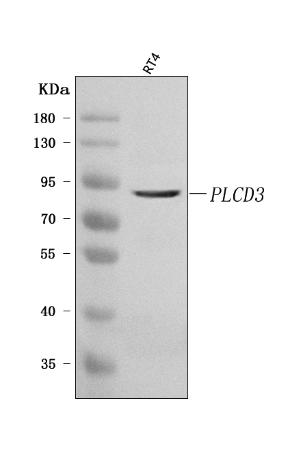
 FCM/FACS (Flow Cytometry)
(Figure 3. Flow Cytometry analysis of HL-60 cells using anti-PLCD3 antibody (AAA127403).Overlay histogram showing HL-60 cells stained with AAA127403 (Blue line). To facilitate intracellular staining, cells were fixed with 4% paraformaldehyde and permeabilized with permeabilization buffer. The cells were blocked with 10% normal goat serum. And then incubated with rabbit anti-PLCD3 Antibody (AAA127403, 1ug/1x106 cells) for 30 min at 20 degree C. DyLight488 conjugated goat anti-rabbit IgG was used as secondary antibody for 30 minutes at 20 degree C. Isotype control antibody (Green line) was rabbit IgG (1ug/1x106) used under the same conditions. Unlabelled sample without incubation with primary antibody and secondary antibody (Red line) was used as a blank control.)
FCM/FACS (Flow Cytometry)
(Figure 3. Flow Cytometry analysis of HL-60 cells using anti-PLCD3 antibody (AAA127403).Overlay histogram showing HL-60 cells stained with AAA127403 (Blue line). To facilitate intracellular staining, cells were fixed with 4% paraformaldehyde and permeabilized with permeabilization buffer. The cells were blocked with 10% normal goat serum. And then incubated with rabbit anti-PLCD3 Antibody (AAA127403, 1ug/1x106 cells) for 30 min at 20 degree C. DyLight488 conjugated goat anti-rabbit IgG was used as secondary antibody for 30 minutes at 20 degree C. Isotype control antibody (Green line) was rabbit IgG (1ug/1x106) used under the same conditions. Unlabelled sample without incubation with primary antibody and secondary antibody (Red line) was used as a blank control.)
PLCD3, Polyclonal Antibody (Cat# AAA127403)
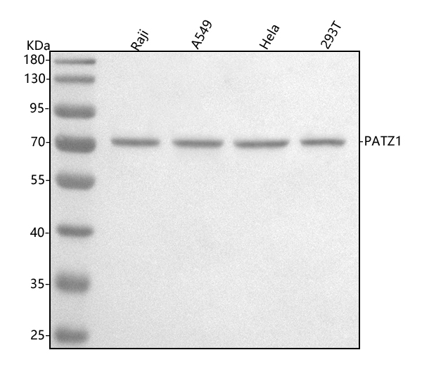
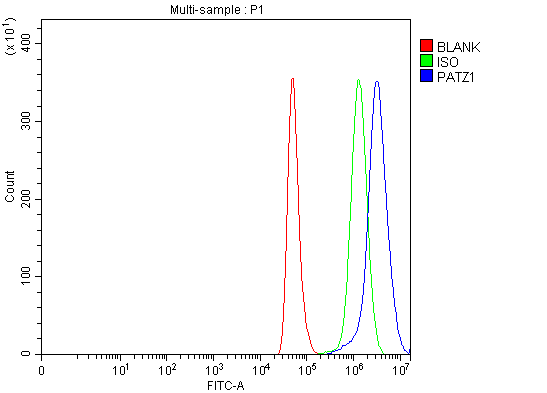 FCM/FACS (Flow Cytometry)
(Figure 3. Flow Cytometry analysis of Hela cells using anti-PATZ1 antibody (AAA127406).Overlay histogram showing Hela cells stained with AAA127406 (Blue line). To facilitate intracellular staining, cells were fixed with 4% paraformaldehyde and permeabilized with permeabilization buffer. The cells were blocked with 10% normal goat serum. And then incubated with rabbit anti-PATZ1 Antibody (AAA127406, 1ug/1x106 cells) for 30 min at 20 degree C. DyLight488 conjugated goat anti-rabbit IgG was used as secondary antibody for 30 minutes at 20 degree C. Isotype control antibody (Green line) was rabbit IgG (1ug/1x106) used under the same conditions. Unlabelled sample (Red line) was also used as a control.)
FCM/FACS (Flow Cytometry)
(Figure 3. Flow Cytometry analysis of Hela cells using anti-PATZ1 antibody (AAA127406).Overlay histogram showing Hela cells stained with AAA127406 (Blue line). To facilitate intracellular staining, cells were fixed with 4% paraformaldehyde and permeabilized with permeabilization buffer. The cells were blocked with 10% normal goat serum. And then incubated with rabbit anti-PATZ1 Antibody (AAA127406, 1ug/1x106 cells) for 30 min at 20 degree C. DyLight488 conjugated goat anti-rabbit IgG was used as secondary antibody for 30 minutes at 20 degree C. Isotype control antibody (Green line) was rabbit IgG (1ug/1x106) used under the same conditions. Unlabelled sample (Red line) was also used as a control.)
PATZ1, Polyclonal Antibody (Cat# AAA127406)
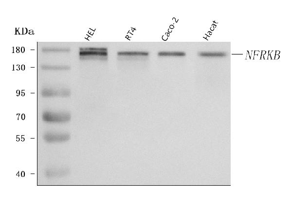
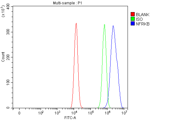 FCM/FACS (Flow Cytometry)
(Figure 3. Flow Cytometry analysis of HEL cells using anti-NFRKB antibody (AAA127411).Overlay histogram showing HEL cells stained with AAA127411 (Blue line). To facilitate intracellular staining, cells were fixed with 4% paraformaldehyde and permeabilized with permeabilization buffer. The cells were blocked with 10% normal goat serum. And then incubated with rabbit anti-NFRKB Antibody (AAA127411, 1ug/1x106 cells) for 30 min at 20 degree C. DyLight488 conjugated goat anti-rabbit IgG was used as secondary antibody for 30 minutes at 20 degree C. Isotype control antibody (Green line) was rabbit IgG (1ug/1x106) used under the same conditions. Unlabelled sample (Red line) was also used as a control.)
FCM/FACS (Flow Cytometry)
(Figure 3. Flow Cytometry analysis of HEL cells using anti-NFRKB antibody (AAA127411).Overlay histogram showing HEL cells stained with AAA127411 (Blue line). To facilitate intracellular staining, cells were fixed with 4% paraformaldehyde and permeabilized with permeabilization buffer. The cells were blocked with 10% normal goat serum. And then incubated with rabbit anti-NFRKB Antibody (AAA127411, 1ug/1x106 cells) for 30 min at 20 degree C. DyLight488 conjugated goat anti-rabbit IgG was used as secondary antibody for 30 minutes at 20 degree C. Isotype control antibody (Green line) was rabbit IgG (1ug/1x106) used under the same conditions. Unlabelled sample (Red line) was also used as a control.)
NFRKB, Polyclonal Antibody (Cat# AAA127411)
 FCM/FACS (Flow Cytometry)
(Figure 2. Flow Cytometry analysis of MCF-7 cells using anti-MYO1B antibody (AAA127416).Overlay histogram showing MCF-7 cells stained with AAA127416 (Blue line). To facilitate intracellular staining, cells were fixed with 4% paraformaldehyde and permeabilized with permeabilization buffer. The cells were blocked with 10% normal goat serum. And then incubated with rabbit anti-MYO1B Antibody (AAA127416, 1ug/1x106 cells) for 30 min at 20 degree C. DyLight488 conjugated goat anti-rabbit IgG was used as secondary antibody for 30 minutes at 20 degree C. Isotype control antibody (Green line) was rabbit IgG (1ug/1x106) used under the same conditions. Unlabelled sample (Red line) was also used as a control.)
FCM/FACS (Flow Cytometry)
(Figure 2. Flow Cytometry analysis of MCF-7 cells using anti-MYO1B antibody (AAA127416).Overlay histogram showing MCF-7 cells stained with AAA127416 (Blue line). To facilitate intracellular staining, cells were fixed with 4% paraformaldehyde and permeabilized with permeabilization buffer. The cells were blocked with 10% normal goat serum. And then incubated with rabbit anti-MYO1B Antibody (AAA127416, 1ug/1x106 cells) for 30 min at 20 degree C. DyLight488 conjugated goat anti-rabbit IgG was used as secondary antibody for 30 minutes at 20 degree C. Isotype control antibody (Green line) was rabbit IgG (1ug/1x106) used under the same conditions. Unlabelled sample (Red line) was also used as a control.)
MYO1B, Polyclonal Antibody (Cat# AAA127416)
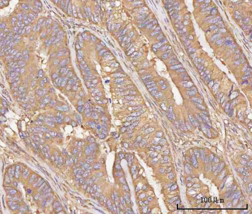
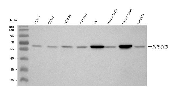
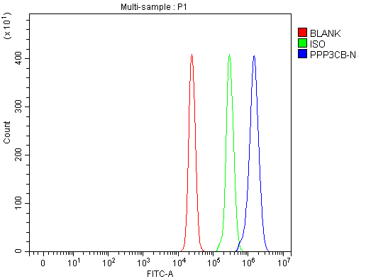 FCM/FACS (Flow Cytometry)
(Figure 5. Flow Cytometry analysis of U937 cells using anti-PPP3CB antibody (AAA127434).Overlay histogram showing U937 cells stained with AAA127434 (Blue line). To facilitate intracellular staining, cells were fixed with 4% paraformaldehyde and permeabilized with permeabilization buffer. The cells were blocked with 10% normal goat serum. And then incubated with rabbit anti-PPP3CB Antibody (AAA127434, 1ug/1x106 cells) for 30 min at 20 degree C. DyLight488 conjugated goat anti-rabbit IgG was used as secondary antibody for 30 minutes at 20 degree C. Isotype control antibody (Green line) was rabbit IgG (1ug/1x106) used under the same conditions. Unlabelled sample (Red line) was also used as a control.)
FCM/FACS (Flow Cytometry)
(Figure 5. Flow Cytometry analysis of U937 cells using anti-PPP3CB antibody (AAA127434).Overlay histogram showing U937 cells stained with AAA127434 (Blue line). To facilitate intracellular staining, cells were fixed with 4% paraformaldehyde and permeabilized with permeabilization buffer. The cells were blocked with 10% normal goat serum. And then incubated with rabbit anti-PPP3CB Antibody (AAA127434, 1ug/1x106 cells) for 30 min at 20 degree C. DyLight488 conjugated goat anti-rabbit IgG was used as secondary antibody for 30 minutes at 20 degree C. Isotype control antibody (Green line) was rabbit IgG (1ug/1x106) used under the same conditions. Unlabelled sample (Red line) was also used as a control.)
PPP3CB, Polyclonal Antibody (Cat# AAA127434)
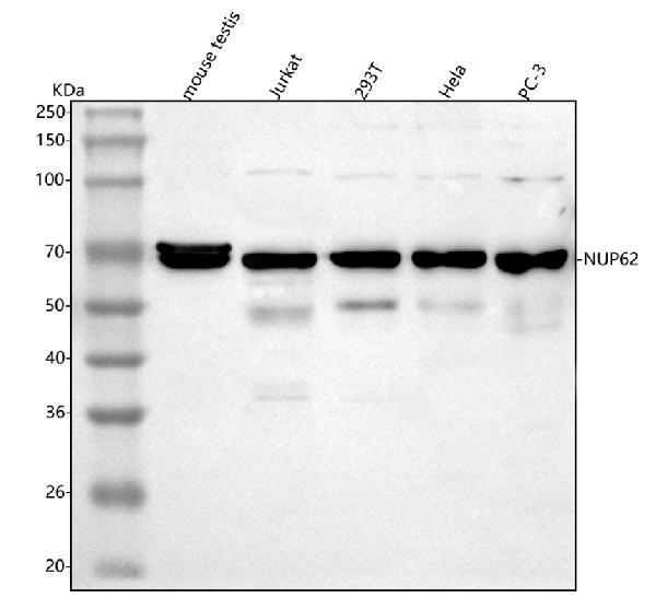
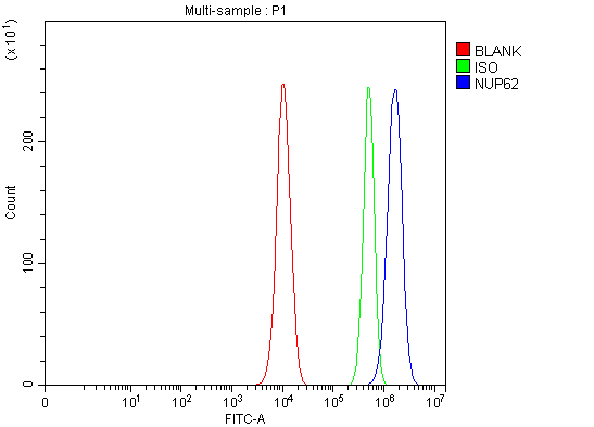 FCM/FACS (Flow Cytometry)
(Figure 5. Flow Cytometry analysis of PC-3 cells using anti-NUP62 antibody (AAA127207).Overlay histogram showing PC-3 cells stained with AAA127207 (Blue line). To facilitate intracellular staining, cells were fixed with 4% paraformaldehyde and permeabilized with permeabilization buffer. The cells were blocked with 10% normal goat serum. And then incubated with rabbit anti-NUP62 Antibody (AAA127207, 1ug/1x106 cells) for 30 min at 20 degree C. DyLight488 conjugated goat anti-rabbit IgG was used as secondary antibody for 30 minutes at 20 degree C. Isotype control antibody (Green line) was rabbit IgG (1ug/1x106) used under the same conditions. Unlabelled sample (Red line) was also used as a control.)
FCM/FACS (Flow Cytometry)
(Figure 5. Flow Cytometry analysis of PC-3 cells using anti-NUP62 antibody (AAA127207).Overlay histogram showing PC-3 cells stained with AAA127207 (Blue line). To facilitate intracellular staining, cells were fixed with 4% paraformaldehyde and permeabilized with permeabilization buffer. The cells were blocked with 10% normal goat serum. And then incubated with rabbit anti-NUP62 Antibody (AAA127207, 1ug/1x106 cells) for 30 min at 20 degree C. DyLight488 conjugated goat anti-rabbit IgG was used as secondary antibody for 30 minutes at 20 degree C. Isotype control antibody (Green line) was rabbit IgG (1ug/1x106) used under the same conditions. Unlabelled sample (Red line) was also used as a control.)
NUP62, Polyclonal Antibody (Cat# AAA127207)
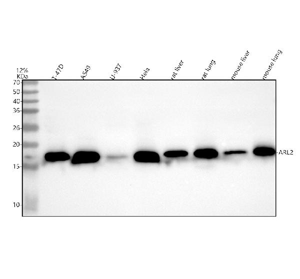
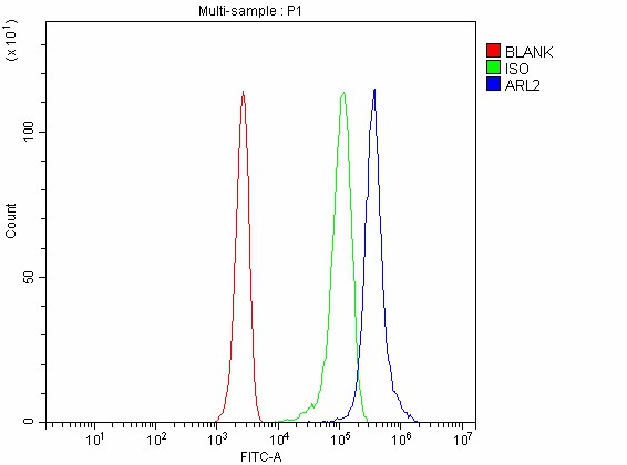 FCM/FACS (Flow Cytometry)
(Figure 4. Flow Cytometry analysis of JK cells using anti-ARL2 antibody (AAA127217).Overlay histogram showing JK cells stained with AAA127217 (Blue line). To facilitate intracellular staining, cells were fixed with 4% paraformaldehyde and permeabilized with permeabilization buffer. The cells were blocked with 10% normal goat serum. And then incubated with rabbit anti-ARL2 Antibody (AAA127217, 1ug/1x106 cells) for 30 min at 20 degree C. DyLight488 conjugated goat anti-rabbit IgG was used as secondary antibody for 30 minutes at 20 degree C. Isotype control antibody (Green line) was rabbit IgG (1ug/1x106) used under the same conditions. Unlabelled sample without incubation with primary antibody and secondary antibody (Red line) was used as a blank control.)
FCM/FACS (Flow Cytometry)
(Figure 4. Flow Cytometry analysis of JK cells using anti-ARL2 antibody (AAA127217).Overlay histogram showing JK cells stained with AAA127217 (Blue line). To facilitate intracellular staining, cells were fixed with 4% paraformaldehyde and permeabilized with permeabilization buffer. The cells were blocked with 10% normal goat serum. And then incubated with rabbit anti-ARL2 Antibody (AAA127217, 1ug/1x106 cells) for 30 min at 20 degree C. DyLight488 conjugated goat anti-rabbit IgG was used as secondary antibody for 30 minutes at 20 degree C. Isotype control antibody (Green line) was rabbit IgG (1ug/1x106) used under the same conditions. Unlabelled sample without incubation with primary antibody and secondary antibody (Red line) was used as a blank control.)
ARL2, Polyclonal Antibody (Cat# AAA127217)
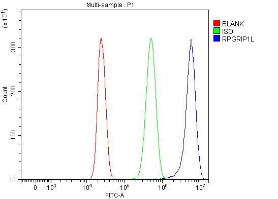 FCM/FACS (Flow Cytometry)
(Figure 2. Flow Cytometry analysis of HepG2 cells using anti-RPGRIP1L antibody (AAA127218).Overlay histogram showing HepG2 cells stained with AAA127218 (Blue line). To facilitate intracellular staining, cells were fixed with 4% paraformaldehyde and permeabilized with permeabilization buffer. The cells were blocked with 10% normal goat serum. And then incubated with rabbit anti-RPGRIP1L Antibody (AAA127218, 1ug/1x106 cells) for 30 min at 20 degree C. DyLight488 conjugated goat anti-rabbit IgG was used as secondary antibody for 30 minutes at 20 degree C. Isotype control antibody (Green line) was rabbit IgG (1ug/1x106) used under the same conditions. Unlabelled sample without incubation with primary antibody and secondary antibody (Red line) was used as a blank control.)
FCM/FACS (Flow Cytometry)
(Figure 2. Flow Cytometry analysis of HepG2 cells using anti-RPGRIP1L antibody (AAA127218).Overlay histogram showing HepG2 cells stained with AAA127218 (Blue line). To facilitate intracellular staining, cells were fixed with 4% paraformaldehyde and permeabilized with permeabilization buffer. The cells were blocked with 10% normal goat serum. And then incubated with rabbit anti-RPGRIP1L Antibody (AAA127218, 1ug/1x106 cells) for 30 min at 20 degree C. DyLight488 conjugated goat anti-rabbit IgG was used as secondary antibody for 30 minutes at 20 degree C. Isotype control antibody (Green line) was rabbit IgG (1ug/1x106) used under the same conditions. Unlabelled sample without incubation with primary antibody and secondary antibody (Red line) was used as a blank control.)
RPGRIP1L, Polyclonal Antibody (Cat# AAA127218)
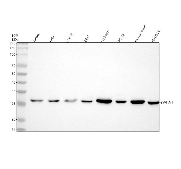
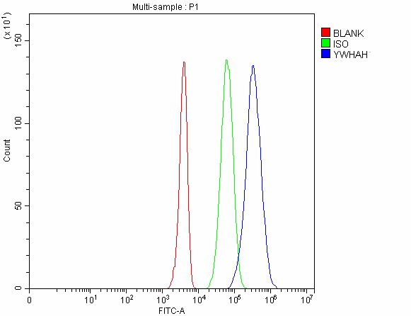 FCM/FACS (Flow Cytometry)
(Figure 5. Flow Cytometry analysis of 293T cells using anti-YWHAH antibody (AAA127220).Overlay histogram showing 293T cells stained with AAA127220(Blue line). The cells were fixed with 4% paraformaldehyde and blocked with 10% normal goat serum. And then incubated with rabbit anti-YWHAH Antibody (AAA127220, 1ug/1x106 cells) for 30 min at 20 degree C. DyLight488 conjugated goat anti-rabbit IgG was used as secondary antibody for 30 minutes at 20 degree C. Isotype control antibody (Green line) was rabbit IgG (1ug/1x106) used under the same conditions. Unlabelled sample without incubation with primary antibody and secondary antibody (Red line) was used as a blank control.)
FCM/FACS (Flow Cytometry)
(Figure 5. Flow Cytometry analysis of 293T cells using anti-YWHAH antibody (AAA127220).Overlay histogram showing 293T cells stained with AAA127220(Blue line). The cells were fixed with 4% paraformaldehyde and blocked with 10% normal goat serum. And then incubated with rabbit anti-YWHAH Antibody (AAA127220, 1ug/1x106 cells) for 30 min at 20 degree C. DyLight488 conjugated goat anti-rabbit IgG was used as secondary antibody for 30 minutes at 20 degree C. Isotype control antibody (Green line) was rabbit IgG (1ug/1x106) used under the same conditions. Unlabelled sample without incubation with primary antibody and secondary antibody (Red line) was used as a blank control.)
YWHAH, Polyclonal Antibody (Cat# AAA127220)
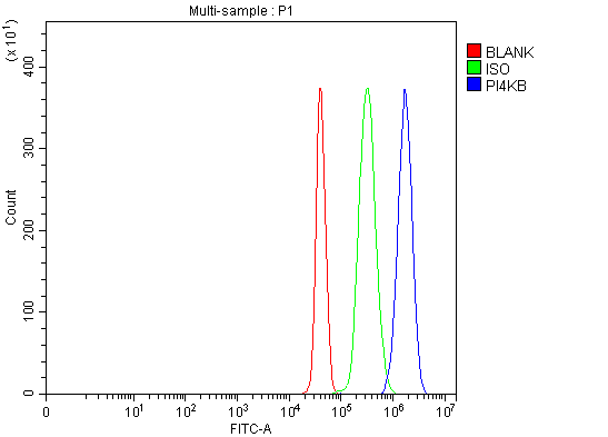
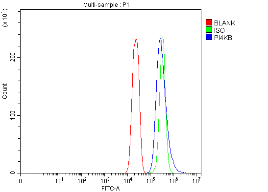 FCM/FACS (Flow Cytometry)
(Figure 4. Flow Cytometry analysis of U251 cells using anti-PI4KB antibody (AAA127222).Overlay histogram showing U251 cells stained with AAA127222 (Blue line). To facilitate intracellular staining, cells were fixed with 4% paraformaldehyde and permeabilized with permeabilization buffer. The cells were blocked with 10% normal goat serum. And then incubated with rabbit anti-PI4KB Antibody (AAA127222, 1ug/1x106 cells) for 30 min at 20 degree C. DyLight488 conjugated goat anti-rabbit IgG was used as secondary antibody for 30 minutes at 20 degree C. Isotype control antibody (Green line) was rabbit IgG (1ug/1x106) used under the same conditions. Unlabelled sample (Red line) was also used as a control.)
FCM/FACS (Flow Cytometry)
(Figure 4. Flow Cytometry analysis of U251 cells using anti-PI4KB antibody (AAA127222).Overlay histogram showing U251 cells stained with AAA127222 (Blue line). To facilitate intracellular staining, cells were fixed with 4% paraformaldehyde and permeabilized with permeabilization buffer. The cells were blocked with 10% normal goat serum. And then incubated with rabbit anti-PI4KB Antibody (AAA127222, 1ug/1x106 cells) for 30 min at 20 degree C. DyLight488 conjugated goat anti-rabbit IgG was used as secondary antibody for 30 minutes at 20 degree C. Isotype control antibody (Green line) was rabbit IgG (1ug/1x106) used under the same conditions. Unlabelled sample (Red line) was also used as a control.)
PI4KB, Polyclonal Antibody (Cat# AAA127222)
What are Polyclonal Antibodies?
Polyclonal antibodies are antibodies that come from multiple B cell clones of a host animal. The typical hosts used for the majority of polyclonal antibody production are rabbits, goats, sheep, and donkeys. These polyclonal antibodies, once having identified their target, will bind to different epitopes located at different regions or sequences on the same protein/antigen. As a result, they are ideal at locating and binding to the target, even if the target is in very low concentrations (due to many different antibodies being able to bind to the same target molecule, which allows for significant amplification of a downstream signal).
Polyclonal antibodies are typically produced by injecting an antigen into a host animal, which causes the animal’s immune system to attack the foreign antigen by mass generating antibodies against it. After a period of time, serum is collected from the animal and purified using physicochemical fractionation, class-specific affinity purification, and/or antigen-affinity purification.
Key Uses of Polyclonal Antibodies
- Western Blotting: This method is used to find specific proteins in biological samples after separating them by size.
- Immunohistochemistry: IHC helps visualize the location of proteins in tissue sections using various staining techniques.
- ELISA: (Enzyme-Linked Immunosorbent Assay) is typically used to identify specific protein quantities in a sample. ELISAs can be either “Quantitative” or “Qualitative”.
- Flow Cytometry: technique that identifies and measures the specific protein on the surface or inside the cells in a fluid suspension.
- Immunoprecipitation: IP isolates and studies a specific protein from a complex mixture using antibodies.
Why Buy Polyclonal Antibodies from AAA Biotech?
1. Ideal for Various Applications
Our antibodies are generally going to be validated for use in multiple types of assays, including ELISA, Western Blotting, Immunohistochemistry, Immunoprecipitation, amongst others. They are ideal for a wide range of research applications.
2. Rigorous Quality Control
All of the antibodies in our catalog undergo strict quality testing to ensure specificity, sensitivity, and consistent performance. We are confident in the ability of our antibodies to provide you with accurate results.
3. Wide Assortment of Antibodies
Antibodies in are catalog can be found for both common and exotic species, and these antibodies are also available in both conjugated and recombinant forms to suit many diverse experimental needs.
4. Highly Purified
Our antibodies are available in purified forms with over 85% purity, as confirmed by SDS-PAGE. They are also available with tags such as His, Flag, GST, or MBP. We cater to customers worldwide.
FAQ
1. How are polyclonal antibodies produced?
Traditionally, polyclonal antibodies are produced by injecting an antigen into a host animal (such as a rabbit or goat), which then triggers an immune response from the host animal. The animal’s B cells produce antibodies that will recognize different parts of the injected antigen. These antibodies are then collected from the animal’s blood and purified for use.
2. How do polyclonal antibodies differ from monoclonal antibodies?
Polyclonal antibodies are a mix of antibodies that bind to different locations (epitopes) of the same antigen, while monoclonal antibodies are identical and bind to just one specific epitope. This makes polyclonal antibodies more versatile and better at detecting proteins that may be present in low quantities or in altered/modified forms.
3. How should I store polyclonal antibodies?
Polyclonal antibodies should be stored at 4°C for short-term use (up to a few weeks) and at -20°C or -80°C for long-term storage. Avoid repeated freeze-thaw cycles by dividing them into small aliquots. Always check the datasheet for specific storage instructions.