Filters
▼Clonality
▼Type
▼Reactivity
▼Gene Name
▼Isotype
▼Host
▼Application
▼Clone
▼Phospho Antibodies
Phospho-specific antibodies’ typical purpose is to enable researchers to detect changes in proteins. They will exclusively bind to the amino acid sequence on a protein that has been phosphorylated (which is both a physical & chemical change) and do not bind to the same amino acid sequence on said protein if it lacks said phosphorylation. This aids in being able to clearly see and understand the data produced from this particular protein modification.
Viewing 400-450 of 5298 product results
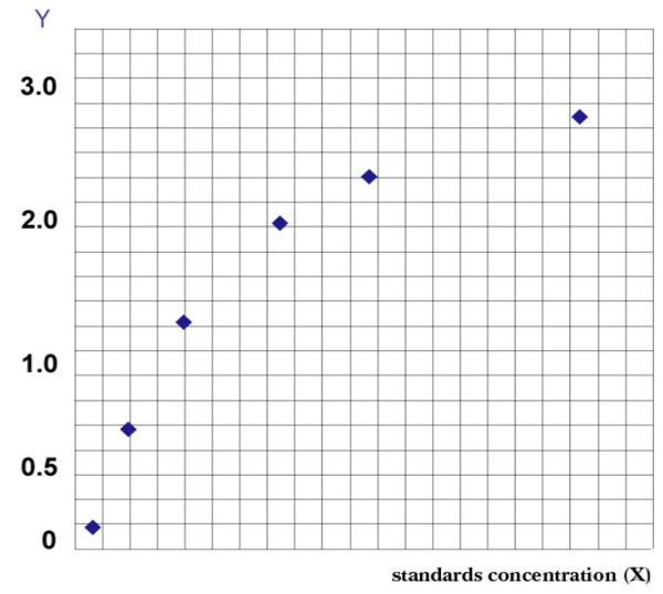
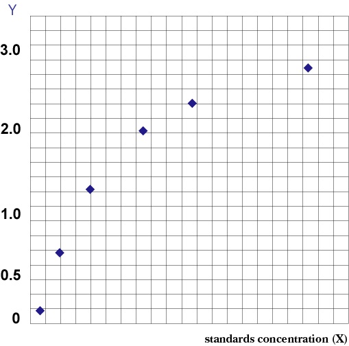 Standard Curve (Sample)
Phospho-Disabled Homolog 1, ELISA Kit (Cat# AAA209999)
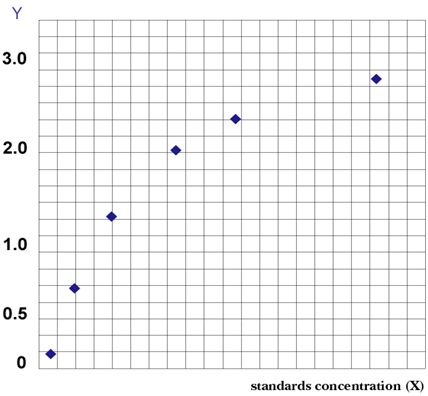 Standard Curve (Sample)
Phospho-Acetyl CoA Carboxylase, ELISA Kit (Cat# AAA209875)
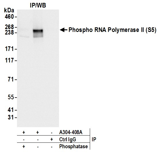
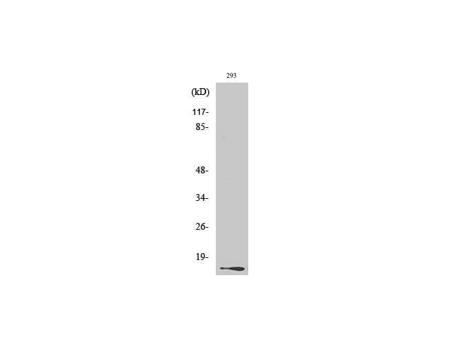
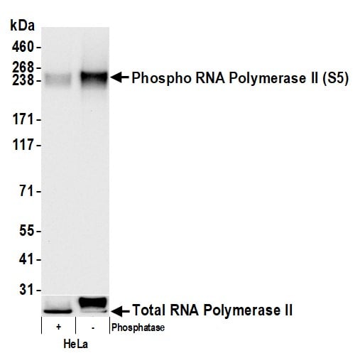 WB (Western Blot)
(Detection of human Phospho RNA Polymerase II (S5) by western blot. Samples: Whole cell lysate (2 ug) from HeLa cells prepared using NETN lysis buffer. Lysates were either mock treated (-) or treated with phosphatases (+). Antibody: Affinity purified rabbit anti-Phospho RNA Polymerase II (S5) antibody (AAA212675 lot 3) used for WB at 0.04 ug/ml. Detection: Chemiluminescence with an exposure time of 3 seconds. Lower Panel: Rabbit anti-RNA Polymerase II antibody .)
WB (Western Blot)
(Detection of human Phospho RNA Polymerase II (S5) by western blot. Samples: Whole cell lysate (2 ug) from HeLa cells prepared using NETN lysis buffer. Lysates were either mock treated (-) or treated with phosphatases (+). Antibody: Affinity purified rabbit anti-Phospho RNA Polymerase II (S5) antibody (AAA212675 lot 3) used for WB at 0.04 ug/ml. Detection: Chemiluminescence with an exposure time of 3 seconds. Lower Panel: Rabbit anti-RNA Polymerase II antibody .)
RNA Polymerase II, Polyclonal Antibody (Cat# AAA212675)
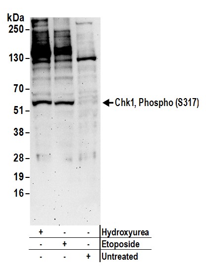 WB (Western Blot)
(Detection of human Chk1, Phospho (S317) by western blot. Samples: Whole cell lysate (50 ug) from HeLa cells prepared using NETN lysis buffer treated with hydroxyurea (2mM for 24 hours), etoposide (100uM for 4 hours), or untreated. Antibodies: Affinity purified rabbit anti-Chk1, Phospho (S317) antibody AAA212853 (lot AAA212853-1) used for WB at 0.2 ug/ml. Detection: Chemiluminescence with an exposure time of 3 minutes.)
WB (Western Blot)
(Detection of human Chk1, Phospho (S317) by western blot. Samples: Whole cell lysate (50 ug) from HeLa cells prepared using NETN lysis buffer treated with hydroxyurea (2mM for 24 hours), etoposide (100uM for 4 hours), or untreated. Antibodies: Affinity purified rabbit anti-Chk1, Phospho (S317) antibody AAA212853 (lot AAA212853-1) used for WB at 0.2 ug/ml. Detection: Chemiluminescence with an exposure time of 3 minutes.)
Chk1, Polyclonal Antibody (Cat# AAA212853)
 Standard Curve (Sample)
Phospho-C-Jun N-Terminal Kinases (p-JNK), ELISA Kit (Cat# AAA209002)
 Standard Curve (Sample)
NFKBIA, ELISA Kit (Cat# AAA209003)
 Standard Curve (Sample)
Phospho-P38 Mitogen Activated Protein Kinase (p-P38MAPK), ELISA Kit (Cat# AAA209004)
 Standard Curve (Sample)
Phospho-Extracellular Signal-Regulated Kinase (PERK), ELISA Kit (Cat# AAA209009)
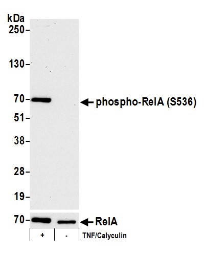 WB (Western Blot)
(Detection of human phospho RelA-S536 by western blot. Samples: Whole cell lysate (15 ug) from Jurkat cells treated with TNF alpha and Calyculin A (+) or mock treated (-). Antibodies: Affinity purified rabbit anti-Phospho RelA (S536) antibody AAA211745 (lot AAA211745-12) used for WB at 0.1 ug/ml. Detection: Chemiluminescence with an exposure time of 75 seconds. Lower panel: Western blot for total RelA using rabbit anti-RelA antibody at 0.1 ug/ml with exposure time of 30 seconds.)
WB (Western Blot)
(Detection of human phospho RelA-S536 by western blot. Samples: Whole cell lysate (15 ug) from Jurkat cells treated with TNF alpha and Calyculin A (+) or mock treated (-). Antibodies: Affinity purified rabbit anti-Phospho RelA (S536) antibody AAA211745 (lot AAA211745-12) used for WB at 0.1 ug/ml. Detection: Chemiluminescence with an exposure time of 75 seconds. Lower panel: Western blot for total RelA using rabbit anti-RelA antibody at 0.1 ug/ml with exposure time of 30 seconds.)
RelA, Polyclonal Antibody (Cat# AAA211745)
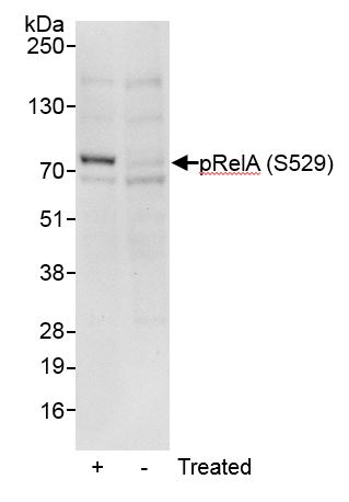 WB (Western Blot)
(Detection of human Phospho RelA (S529) by western blot. Samples: Whole cell lysate (50 ug) from Jurkat cells that were treated with TNF alpha and Calyculin A (+) or mock treated (-). Antibody: Affinity purified rabbit anti-Phospho RelA (S529) antibody AAA211746 (lot AAA211746-1) used at 0.1 ug/ml. Detection: Chemiluminescence with an exposure time of 30 seconds.)
WB (Western Blot)
(Detection of human Phospho RelA (S529) by western blot. Samples: Whole cell lysate (50 ug) from Jurkat cells that were treated with TNF alpha and Calyculin A (+) or mock treated (-). Antibody: Affinity purified rabbit anti-Phospho RelA (S529) antibody AAA211746 (lot AAA211746-1) used at 0.1 ug/ml. Detection: Chemiluminescence with an exposure time of 30 seconds.)
RelA, Polyclonal Antibody (Cat# AAA211746)
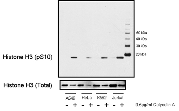 WB (Western Blot)
(Detection of human Phospho Histone H3 (pS10) by western blot. A-549, HeLa, K-562, and Jurkat cell lines were stimulated with 0.5 ug/ml of Calyculin A at 37 degree C, 5% CO? for 15 minutes. Cells were subsequently lysed and 1 ug of appropriate cell lysates were loaded into respective wells. Western blot was performed using affinity purified rabbit anti-Histone H3 (pS10) antibody AAA211652 (20 ng/ml) and affinity purified rabbit anti-Histone H3 antibody (1 ug/ml; Cat. No. Western blots were developed utilizing chemiluminescence with an exposure time of 30 seconds.)
WB (Western Blot)
(Detection of human Phospho Histone H3 (pS10) by western blot. A-549, HeLa, K-562, and Jurkat cell lines were stimulated with 0.5 ug/ml of Calyculin A at 37 degree C, 5% CO? for 15 minutes. Cells were subsequently lysed and 1 ug of appropriate cell lysates were loaded into respective wells. Western blot was performed using affinity purified rabbit anti-Histone H3 (pS10) antibody AAA211652 (20 ng/ml) and affinity purified rabbit anti-Histone H3 antibody (1 ug/ml; Cat. No. Western blots were developed utilizing chemiluminescence with an exposure time of 30 seconds.)
Histone H3, Polyclonal Antibody (Cat# AAA211652)
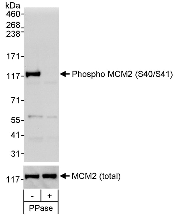 WB (Western Blot)
(Detection of human Phospho MCM2 (S40/S41) by western blot. Samples: Whole cell lysate (50 ug) from asynchronous HEK293T cells that was mock treated (-) or treated (+) with phosphatases (PPase). Antibody: Affinity purified rabbit anti-phospho MCM2 (S40/S41) antibody (Cat. No. AAA211140) used at 0.1 ug/ml. To examine total MCM2, the blot was stripped and then blotted with rabbit anti-MCM2 antibody (Cat. No. at 0.1 ug/ml. Detection: Chemiluminescence with exposure times of 30 seconds (upper and lower panels).)
WB (Western Blot)
(Detection of human Phospho MCM2 (S40/S41) by western blot. Samples: Whole cell lysate (50 ug) from asynchronous HEK293T cells that was mock treated (-) or treated (+) with phosphatases (PPase). Antibody: Affinity purified rabbit anti-phospho MCM2 (S40/S41) antibody (Cat. No. AAA211140) used at 0.1 ug/ml. To examine total MCM2, the blot was stripped and then blotted with rabbit anti-MCM2 antibody (Cat. No. at 0.1 ug/ml. Detection: Chemiluminescence with exposure times of 30 seconds (upper and lower panels).)
MCM2, Polyclonal Antibody (Cat# AAA211140)
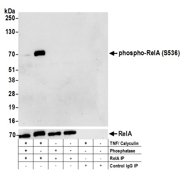
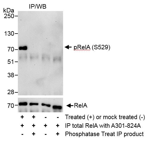
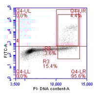
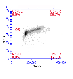
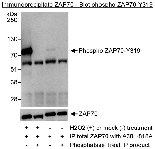 IP (Immunoprecipitation)
(Detection of Phosphorylation of human ZAP70 on Y319 by western blot of immunoprecipitates. Samples: Whole cell lysate (1 mg for IP, 20% of IP loaded) from Jurkat cells that had been treated with hydrogen peroxide (+) or mock treated (-). Antibodies: ZAP70 antibody was used at 3 ug/mg lysate to immunoprecipitate ZAP70. The immunoprecipitates were treated with phosphatase (+) or mock (-) treated. For blotting immunoprecipitated Phospho ZAP70, Phospho ZAP70 (Y319) antibody AAA211718 was used at 1 ug/ml. Detection: Chemiluminescence with an exposure time of 30 seconds.)
IP (Immunoprecipitation)
(Detection of Phosphorylation of human ZAP70 on Y319 by western blot of immunoprecipitates. Samples: Whole cell lysate (1 mg for IP, 20% of IP loaded) from Jurkat cells that had been treated with hydrogen peroxide (+) or mock treated (-). Antibodies: ZAP70 antibody was used at 3 ug/mg lysate to immunoprecipitate ZAP70. The immunoprecipitates were treated with phosphatase (+) or mock (-) treated. For blotting immunoprecipitated Phospho ZAP70, Phospho ZAP70 (Y319) antibody AAA211718 was used at 1 ug/ml. Detection: Chemiluminescence with an exposure time of 30 seconds.)
ZAP70, Polyclonal Antibody (Cat# AAA211718)
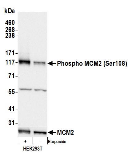
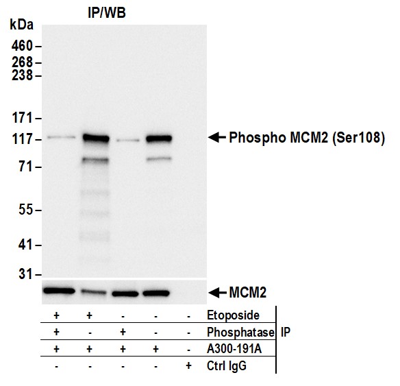
 WB (Western Blot)
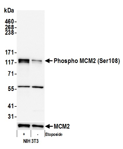
(Detection of mouse Phospho MCM2 (Ser108) by western blot. Samples: Whole cell lysate (10 ug) from NIH 3T3 cells prepared using NETN lysis buffer. Antibody: Affinity purified rabbit anti-Phospho MCM2 (Ser108) antibody (AAA210759 lot 5) used for WB at 1 ug/ml. Detection: Chemiluminescence with an exposure time of 3 seconds. Lower Panel: Rabbit anti-MCM2 antibody .)
WB (Western Blot)
(Detection of mouse Phospho MCM2 (Ser108) by western blot. Samples: Whole cell lysate (10 ug) from NIH 3T3 cells prepared using NETN lysis buffer. Antibody: Affinity purified rabbit anti-Phospho MCM2 (Ser108) antibody (AAA210759 lot 5) used for WB at 1 ug/ml. Detection: Chemiluminescence with an exposure time of 3 seconds. Lower Panel: Rabbit anti-MCM2 antibody .)
MCM2, Polyclonal Antibody (Cat# AAA210759)
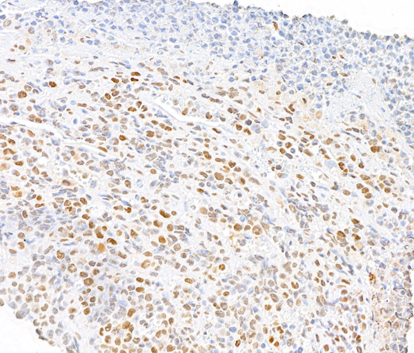
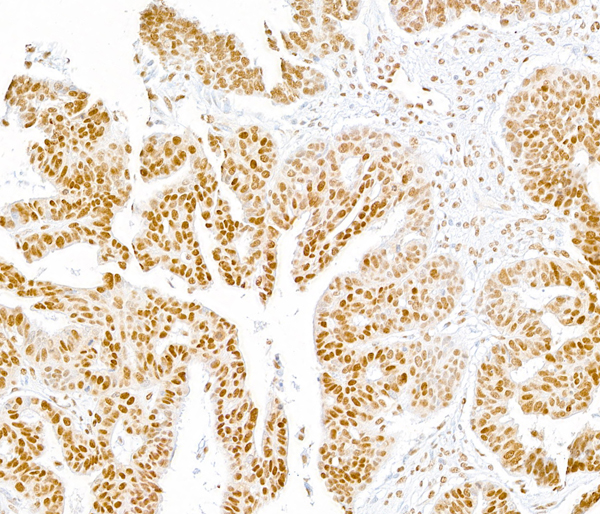
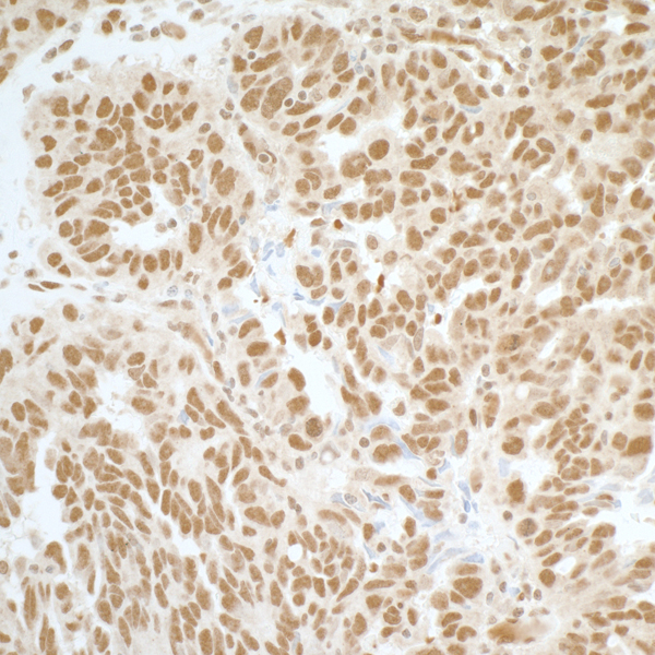
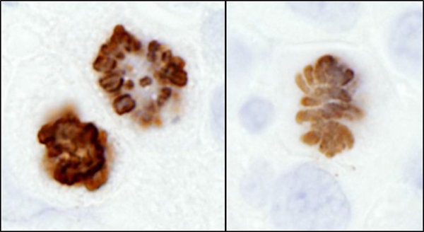
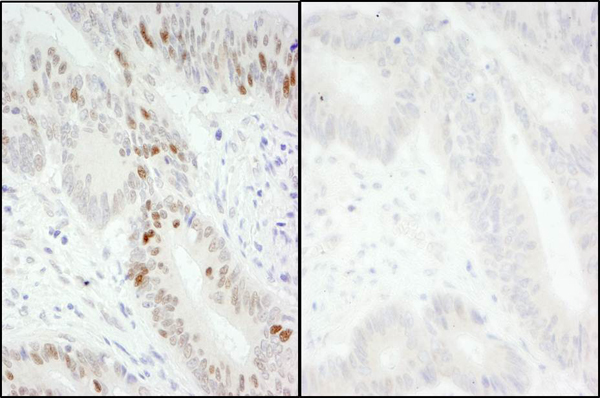
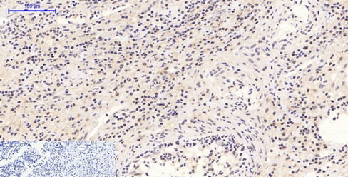
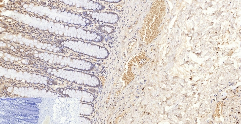
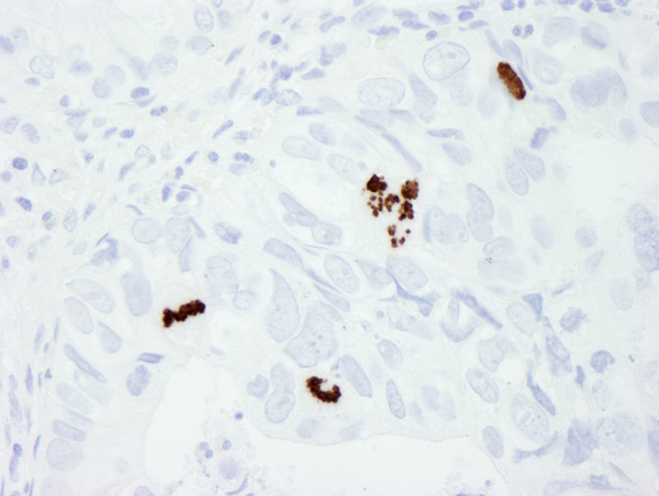 IHC (Immunohiostchemistry)
(Detection of human Phospho Histone H3 (S10) by immunohistochemistry. Sample: FFPE section of human colon adenocarcinoma. Antibody: Affinity purified rabbit anti-Phospho Histone H3 (S10) (Cat. No. AAA213763) used at a dilution of 1:250. Detection: DAB)
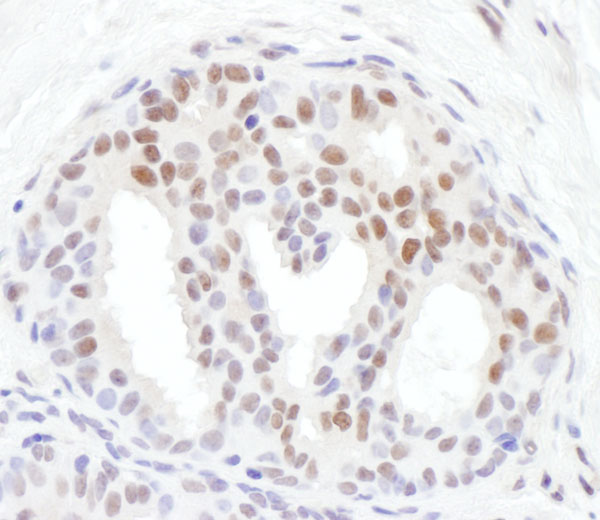
IHC (Immunohiostchemistry)
(Detection of human Phospho Histone H3 (S10) by immunohistochemistry. Sample: FFPE section of human colon adenocarcinoma. Antibody: Affinity purified rabbit anti-Phospho Histone H3 (S10) (Cat. No. AAA213763) used at a dilution of 1:250. Detection: DAB)
Histone H3, Polyclonal Antibody (Cat# AAA213763)
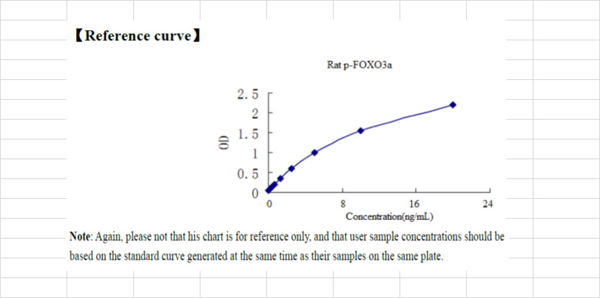 Standard Curve (Sample)
phospho Forkhead Box Protein O3a (p-FOXO3a), ELISA Kit (Cat# AAA181816)
 Standard Curve (Sample)
Phospho-Tyrosine Kinase, ELISA Kit (Cat# AAA206061)
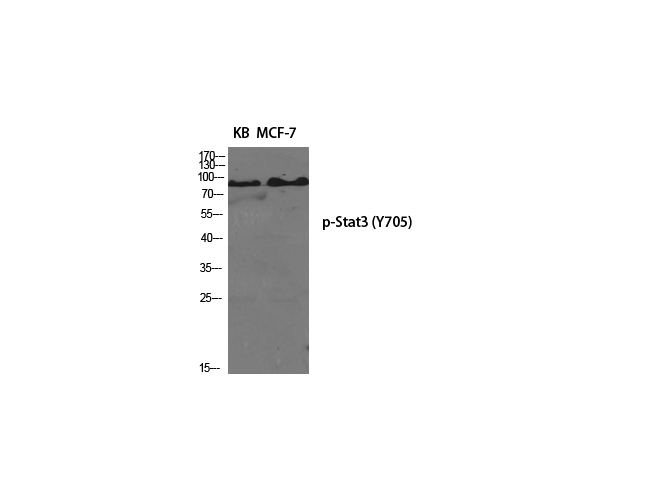
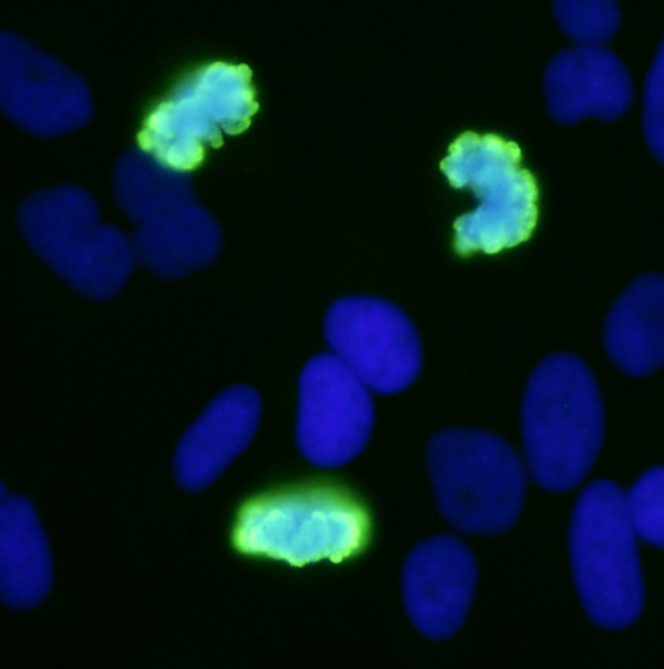
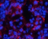
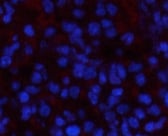 IF (Immunofluorescence)
(Immunofluorescence analysis of Mouse spleen tissue with Phospho-Stat3 (Tyr705) Polyclonal Antibody at dilution of 1:200)
IF (Immunofluorescence)
(Immunofluorescence analysis of Mouse spleen tissue with Phospho-Stat3 (Tyr705) Polyclonal Antibody at dilution of 1:200)
Stat3, Polyclonal Antibody (Cat# AAA171642)
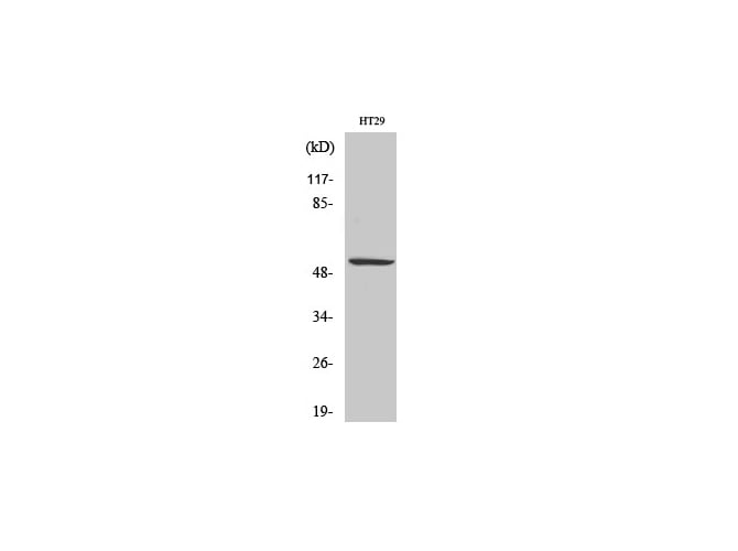
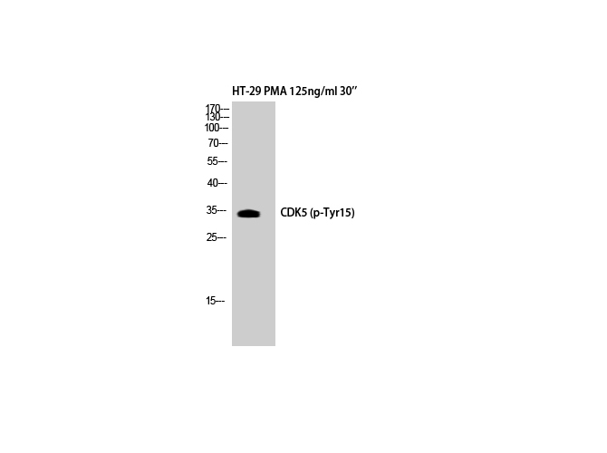 WB (Western Blot)
(Western Blot analysis of HT-29+PMA cells with Phospho-Cdk5 (Tyr15) Polyclonal Antibody)
WB (Western Blot)
(Western Blot analysis of HT-29+PMA cells with Phospho-Cdk5 (Tyr15) Polyclonal Antibody)
Cdk5, Polyclonal Antibody (Cat# AAA171669)
 IF (Immunofluorescence)
(Immunofluorescence analysis of Human kidney tissue with Phospho-Catenin-? (Ser37) Polyclonal Antibody at dilution of 1:200)
IF (Immunofluorescence)
(Immunofluorescence analysis of Human kidney tissue with Phospho-Catenin-? (Ser37) Polyclonal Antibody at dilution of 1:200)
Catenin-beta, Polyclonal Antibody (Cat# AAA171712)

 IF (Immunofluorescence)
(Immunofluorescence analysis of Mouse spleen tissue with Phospho-Tau (Ser396) Polyclonal Antibody at dilution of 1:200)
IF (Immunofluorescence)
(Immunofluorescence analysis of Mouse spleen tissue with Phospho-Tau (Ser396) Polyclonal Antibody at dilution of 1:200)
Tau, Polyclonal Antibody (Cat# AAA171627)
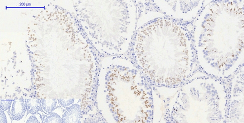 IHC (Immunohiostchemistry)
(Immunohistochemistry of paraffin-embedded Rat testis tissue with Phospho-Histone H2AX (Ser139) Polyclonal Antibody at dilution of 1:200)
IHC (Immunohiostchemistry)
(Immunohistochemistry of paraffin-embedded Rat testis tissue with Phospho-Histone H2AX (Ser139) Polyclonal Antibody at dilution of 1:200)
Histone H2A.X, Polyclonal Antibody (Cat# AAA171631)
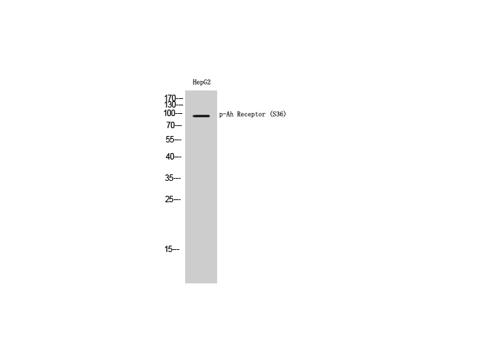 WB (Western Blot)
(Western Blot analysis of HepG2 cells with Phospho-Ah Receptor (Ser36) Polyclonal Antibody)
WB (Western Blot)
(Western Blot analysis of HepG2 cells with Phospho-Ah Receptor (Ser36) Polyclonal Antibody)
Ah Receptor, Polyclonal Antibody (Cat# AAA171901)
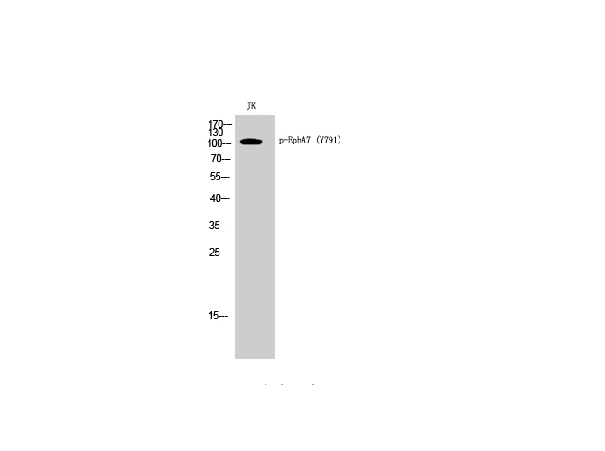 WB (Western Blot)
(Western Blot analysis of Jurkat cells with Phospho-EphA7 (Tyr791) Polyclonal Antibody)
WB (Western Blot)
(Western Blot analysis of Jurkat cells with Phospho-EphA7 (Tyr791) Polyclonal Antibody)
EphA7, Polyclonal Antibody (Cat# AAA171913)
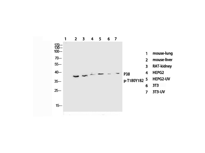
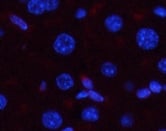 IF (Immunofluorescence)
(Immunofluorescence analysis of Mouse liver tissue with Phospho-p38 (Thr180/Tyr182) Polyclonal Antibody at dilution of 1:200)
IF (Immunofluorescence)
(Immunofluorescence analysis of Mouse liver tissue with Phospho-p38 (Thr180/Tyr182) Polyclonal Antibody at dilution of 1:200)
p38, Polyclonal Antibody (Cat# AAA171964)
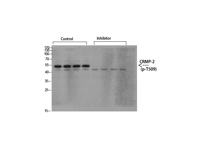
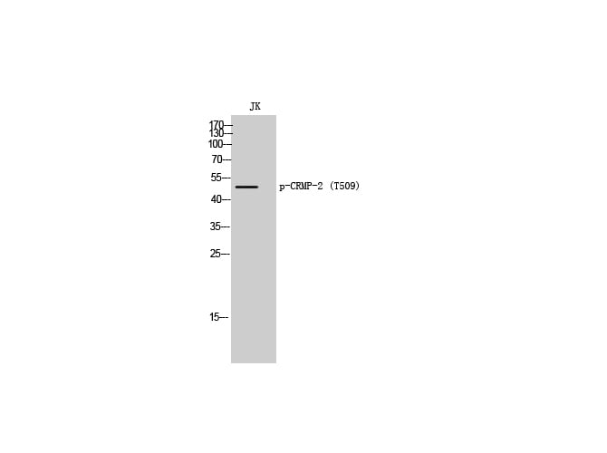 WB (Western Blot)
(Western Blot analysis of Jurkat cells with Phospho-CRMP-2 (Thr509) Polyclonal Antibody)
WB (Western Blot)
(Western Blot analysis of Jurkat cells with Phospho-CRMP-2 (Thr509) Polyclonal Antibody)
CRMP-2, Polyclonal Antibody (Cat# AAA171972)
 IF (Immunofluorescence)
(Immunofluorescence analysis of Human lung tissue with Phospho-NF?B-p65 (Ser529) Polyclonal Antibody at dilution of 1:200)
IF (Immunofluorescence)
(Immunofluorescence analysis of Human lung tissue with Phospho-NF?B-p65 (Ser529) Polyclonal Antibody at dilution of 1:200)
NFkappaB-p65, Polyclonal Antibody (Cat# AAA171974)
 IF (Immunofluorescence)
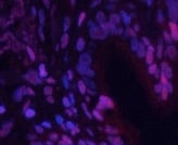
(Immunofluorescence analysis of Rat spleen tissue with Phospho-CaMKII?/?/? (Thr287) Polyclonal Antibody at dilution of 1:200)
IF (Immunofluorescence)
(Immunofluorescence analysis of Rat spleen tissue with Phospho-CaMKII?/?/? (Thr287) Polyclonal Antibody at dilution of 1:200)
CaMKIIbeta/gamma/delta, Polyclonal Antibody (Cat# AAA172019)
Tau, Polyclonal Antibody (Cat# AAA172026)

 IF (Immunofluorescence)
(Immunofluorescence analysis of Rat lung tissue with Phospho-p53 (Thr18) Polyclonal Antibody at dilution of 1:200)
IF (Immunofluorescence)
(Immunofluorescence analysis of Rat lung tissue with Phospho-p53 (Thr18) Polyclonal Antibody at dilution of 1:200)
p53, Polyclonal Antibody (Cat# AAA172225)
 IF (Immunofluorescence)
(Immunofluorescence analysis of Rat spleen tissue with Phospho-p53 (Ser15) Polyclonal Antibody at dilution of 1:200)
IF (Immunofluorescence)
(Immunofluorescence analysis of Rat spleen tissue with Phospho-p53 (Ser15) Polyclonal Antibody at dilution of 1:200)
p53, Polyclonal Antibody (Cat# AAA172248)
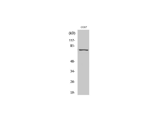
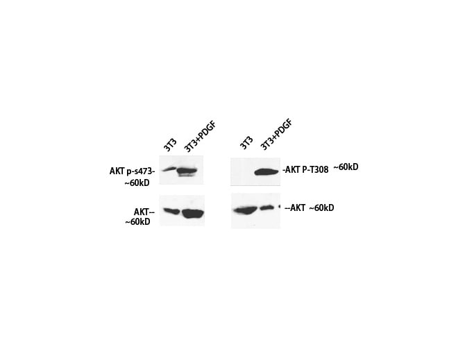
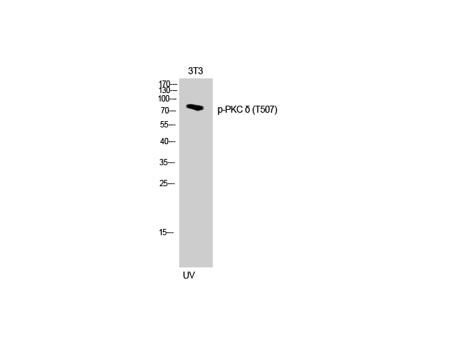 WB (Western Blot)
(Western Blot analysis of 3T3 cells with Phospho-PKC delta (Thr507) Polyclonal Antibody)
WB (Western Blot)
(Western Blot analysis of 3T3 cells with Phospho-PKC delta (Thr507) Polyclonal Antibody)
PKC delta, Polyclonal Antibody (Cat# AAA172273)
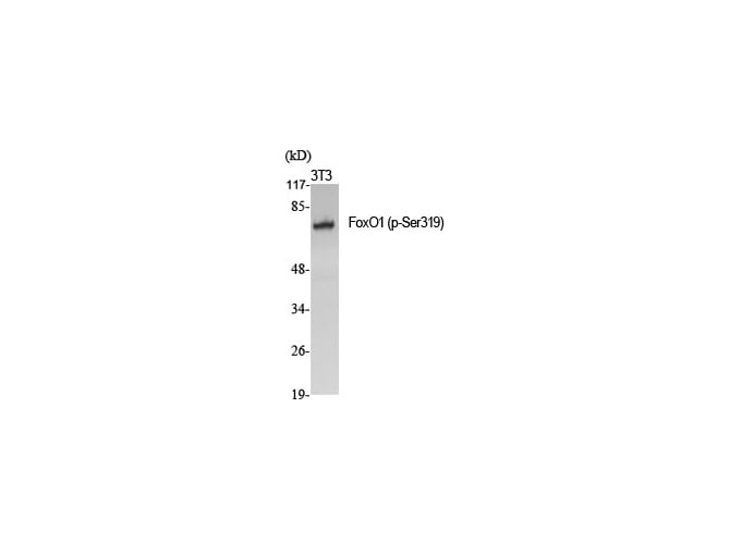 WB (Western Blot)
(Western Blot analysis of various cells with Phospho-FoxO1 (Ser319) Polyclonal Antibody)
WB (Western Blot)
(Western Blot analysis of various cells with Phospho-FoxO1 (Ser319) Polyclonal Antibody)
FoxO1, Polyclonal Antibody (Cat# AAA172278)
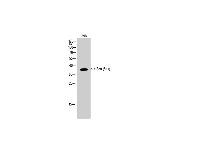
 IF (Immunofluorescence)
(Immunofluorescence analysis of Rat heart tissue with Phospho-eIF2? (Ser51) Polyclonal Antibody at dilution of 1:200)
IF (Immunofluorescence)
(Immunofluorescence analysis of Rat heart tissue with Phospho-eIF2? (Ser51) Polyclonal Antibody at dilution of 1:200)
eIF2alpha, Polyclonal Antibody (Cat# AAA171881)
 IF (Immunofluorescence)
(Immunofluorescence analysis of Rat lung tissue with Phospho-NF?B-p65 (Ser536) Polyclonal Antibody at dilution of 1:200)
IF (Immunofluorescence)
(Immunofluorescence analysis of Rat lung tissue with Phospho-NF?B-p65 (Ser536) Polyclonal Antibody at dilution of 1:200)
NFkappaB-p65, Polyclonal Antibody (Cat# AAA171756)

 IF (Immunofluorescence)
(Immunofluorescence analysis of Mouse liver tissue with Phospho-JAK1 (Tyr1022) Polyclonal Antibody at dilution of 1:200)
IF (Immunofluorescence)
(Immunofluorescence analysis of Mouse liver tissue with Phospho-JAK1 (Tyr1022) Polyclonal Antibody at dilution of 1:200)
JAK1, Polyclonal Antibody (Cat# AAA172298)
 IF (Immunofluorescence)
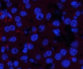
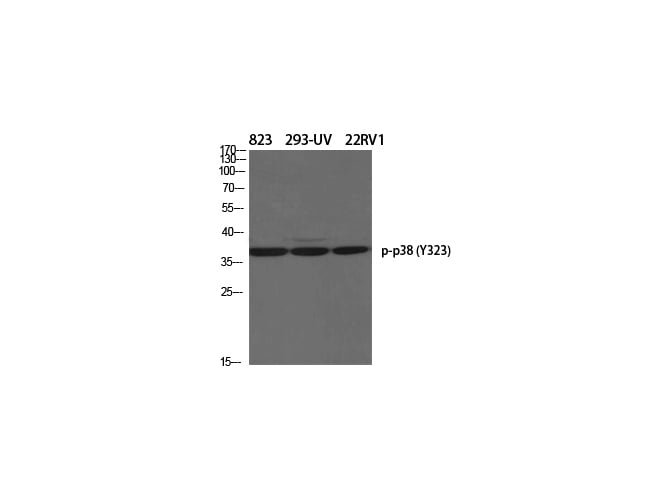
(Immunofluorescence analysis of Human stomach tissue with Phospho-p38 (Tyr323) Polyclonal Antibody at dilution of 1:200)
IF (Immunofluorescence)
(Immunofluorescence analysis of Human stomach tissue with Phospho-p38 (Tyr323) Polyclonal Antibody at dilution of 1:200)
p38, Polyclonal Antibody (Cat# AAA172301)
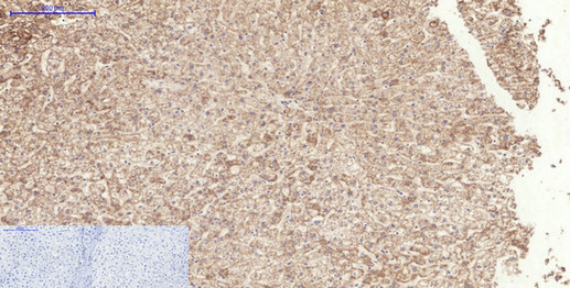
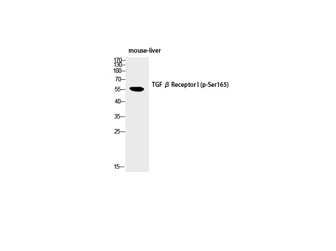
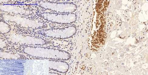
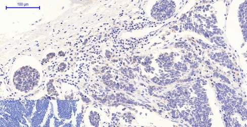
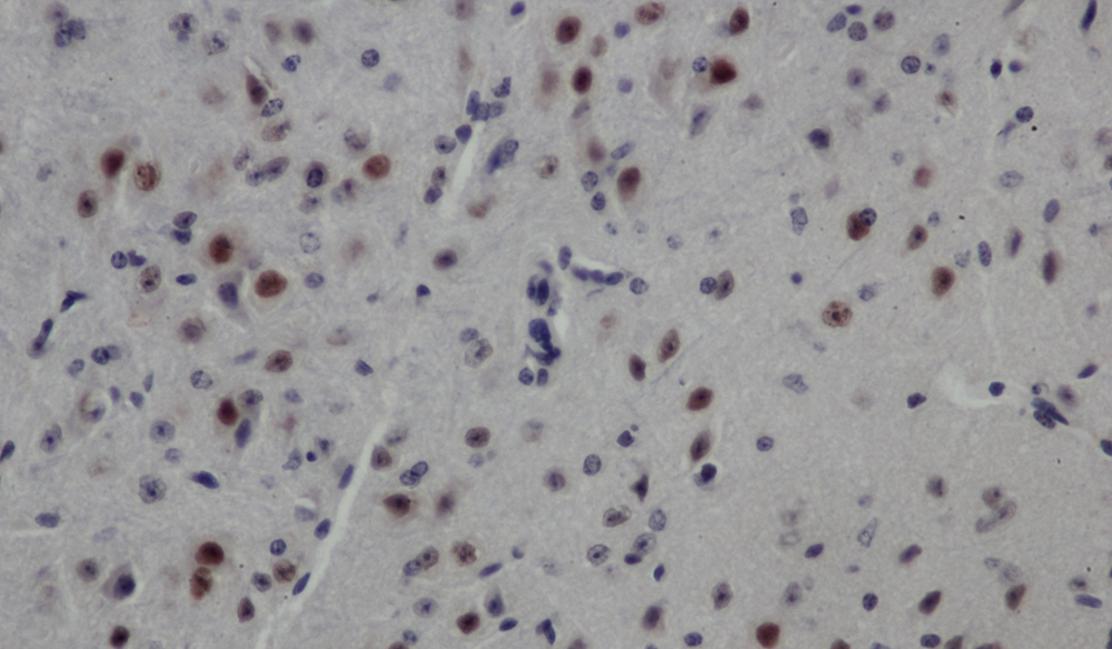
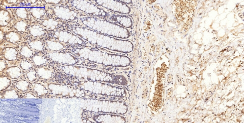
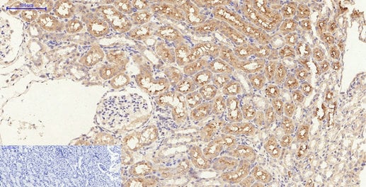 IHC (Immunohistochemisry)
(Immunohistochemistry of paraffin-embedded Rat kidney tissue with Phospho-TGF? RI (Ser165) Polyclonal Antibody at dilution of 1:200)
IHC (Immunohistochemisry)
(Immunohistochemistry of paraffin-embedded Rat kidney tissue with Phospho-TGF? RI (Ser165) Polyclonal Antibody at dilution of 1:200)
TGFbeta RI, Polyclonal Antibody (Cat# AAA172307)
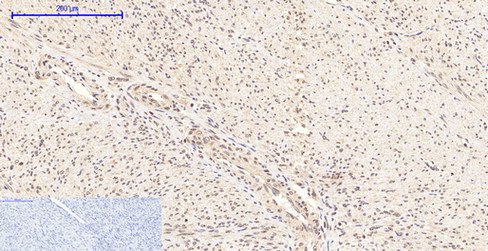
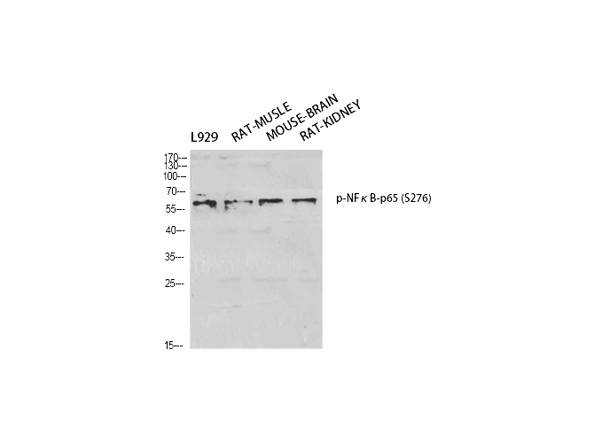
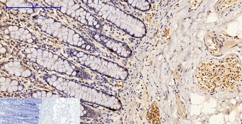
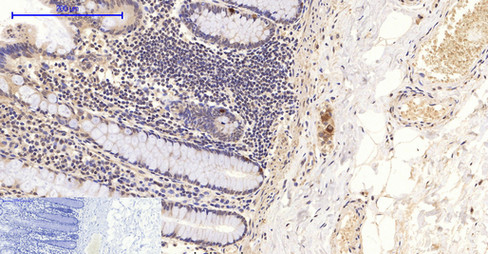 IHC (Immunohistochemisry)
(Immunohistochemistry of paraffin-embedded Human colon tissue with Phospho-NF?B-p65 (Ser276) Polyclonal Antibody at dilution of 1:200)
IHC (Immunohistochemisry)
(Immunohistochemistry of paraffin-embedded Human colon tissue with Phospho-NF?B-p65 (Ser276) Polyclonal Antibody at dilution of 1:200)
NFkappaB-p65, Polyclonal Antibody (Cat# AAA172335)
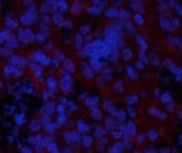 IF (Immunofluorescence)
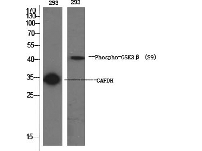
(Immunofluorescence analysis of Rat spleen tissue with Phospho-GSK3? (Ser9) Polyclonal Antibody at dilution of 1:200)
IF (Immunofluorescence)
(Immunofluorescence analysis of Rat spleen tissue with Phospho-GSK3? (Ser9) Polyclonal Antibody at dilution of 1:200)
GSK3beta, Polyclonal Antibody (Cat# AAA172097)
 IF (Immunofluorescence)
(Immunofluorescence analysis of Rat liver tissue with Phospho-Akt (Ser473) Polyclonal Antibody at dilution of 1:200)
IF (Immunofluorescence)
(Immunofluorescence analysis of Rat liver tissue with Phospho-Akt (Ser473) Polyclonal Antibody at dilution of 1:200)
Akt, Polyclonal Antibody (Cat# AAA172111)
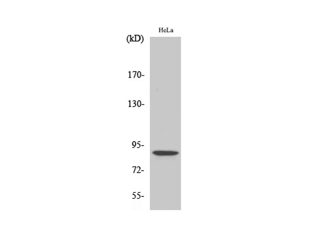 WB (Western Blot)
(Western Blot analysis of Hela cells using Phospho-Stat5 (Ser726/731) Polyclonal Antibody at dilution of 1:500.)
WB (Western Blot)
(Western Blot analysis of Hela cells using Phospho-Stat5 (Ser726/731) Polyclonal Antibody at dilution of 1:500.)
Stat5, Polyclonal Antibody (Cat# AAA172131)
 IF (Immunofluorescence)
(Immunofluorescence analysis of Mouse testis tissue with Phospho-AP-1 (Ser63) Polyclonal Antibody at dilution of 1:200)
IF (Immunofluorescence)
(Immunofluorescence analysis of Mouse testis tissue with Phospho-AP-1 (Ser63) Polyclonal Antibody at dilution of 1:200)
AP-1, Polyclonal Antibody (Cat# AAA172505)
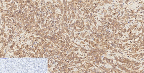
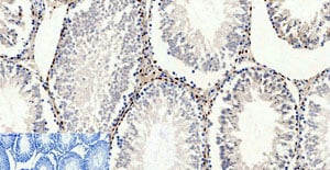
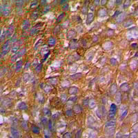
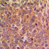 IHC (Immunohiostchemistry)
(Immunohistochemical analysis of EIF2S1 (pS51) staining in human breast cancer formalin fixed paraffin embedded tissue section. The section was pre-treated using heat mediated antigen retrieval with sodium citrate buffer (pH 6.0). The section was then incubated with the antibody at room temperature and detected using an HRP conjugated compact polymer system. DAB was used as the chromogen. The section was then counterstained with haematoxylin and mounted with DPX.)
IHC (Immunohiostchemistry)
(Immunohistochemical analysis of EIF2S1 (pS51) staining in human breast cancer formalin fixed paraffin embedded tissue section. The section was pre-treated using heat mediated antigen retrieval with sodium citrate buffer (pH 6.0). The section was then incubated with the antibody at room temperature and detected using an HRP conjugated compact polymer system. DAB was used as the chromogen. The section was then counterstained with haematoxylin and mounted with DPX.)
EIF2S1 (pS51), Polyclonal Antibody (Cat# AAA105048)
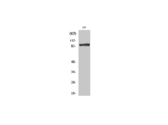
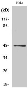
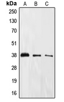
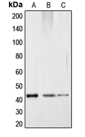
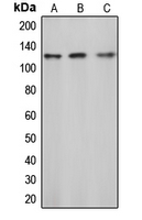
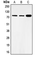
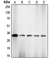
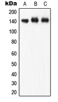
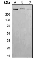 WB (Western Blot)
(Western blot analysis of ATM (pS1981) expression in Hela (A), Raw264.7 (B), H9C2 (C) whole cell lysates.)
WB (Western Blot)
(Western blot analysis of ATM (pS1981) expression in Hela (A), Raw264.7 (B), H9C2 (C) whole cell lysates.)
ATM (pS1981), Polyclonal Antibody (Cat# AAA104820)
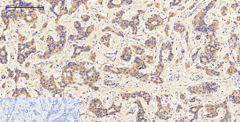
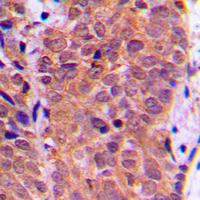
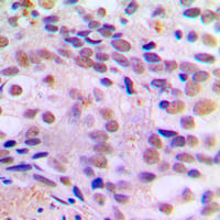 IHC (Immunohiostchemistry)
(Immunohistochemical analysis of CREB (pS121) staining in human breast cancer formalin fixed paraffin embedded tissue section. The section was pre-treated using heat mediated antigen retrieval with sodium citrate buffer (pH 6.0). The section was then incubated with the antibody at room temperature and detected using an HRP conjugated compact polymer system. DAB was used as the chromogen. The section was then counterstained with haematoxylin and mounted with DPX.)
IHC (Immunohiostchemistry)
(Immunohistochemical analysis of CREB (pS121) staining in human breast cancer formalin fixed paraffin embedded tissue section. The section was pre-treated using heat mediated antigen retrieval with sodium citrate buffer (pH 6.0). The section was then incubated with the antibody at room temperature and detected using an HRP conjugated compact polymer system. DAB was used as the chromogen. The section was then counterstained with haematoxylin and mounted with DPX.)
CREB (pS121), Polyclonal Antibody (Cat# AAA104831)
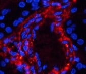
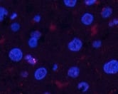
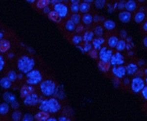
 IF (Immunofluorescence)
(Immunofluorescent analysis of c-CBL (pY774) staining in Jurkat cells. Formalin-fixed cells were permeabilized with 0.1% Triton X-100 in TBS for 5-10 minutes and blocked with 3% BSA-PBS for 30 minutes at room temperature. Cells were probed with the primary antibody in 3% BSA-PBS and incubated overnight at 4 °C in a humidified chamber. Cells were washed with PBST and incubated with a DyLight 594-conjugated secondary antibody (red) in PBS at room temperature in the dark. DAPI was used to stain the cell nuclei (blue).)
IF (Immunofluorescence)
(Immunofluorescent analysis of c-CBL (pY774) staining in Jurkat cells. Formalin-fixed cells were permeabilized with 0.1% Triton X-100 in TBS for 5-10 minutes and blocked with 3% BSA-PBS for 30 minutes at room temperature. Cells were probed with the primary antibody in 3% BSA-PBS and incubated overnight at 4 °C in a humidified chamber. Cells were washed with PBST and incubated with a DyLight 594-conjugated secondary antibody (red) in PBS at room temperature in the dark. DAPI was used to stain the cell nuclei (blue).)
c-CBL (pY774), Polyclonal Antibody (Cat# AAA104867)
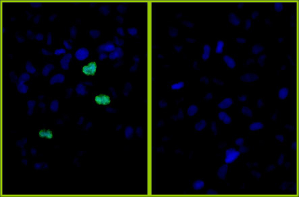
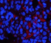
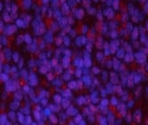
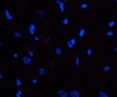
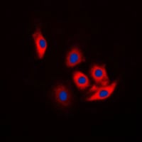
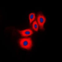
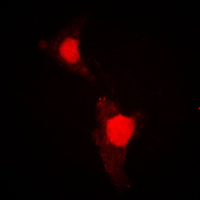 IF (Immunofluorescence)
(Immunofluorescent analysis of FOXO1 (pS319) staining in HeLa cells. Formalin-fixed cells were permeabilized with 0.1% Triton X-100 in TBS for 5-10 minutes and blocked with 3% BSA-PBS for 30 minutes at room temperature. Cells were probed with the primary antibody in 3% BSA-PBS and incubated overnight at 4 °C in a humidified chamber. Cells were washed with PBST and incubated with a DyLight 594-conjugated secondary antibody (red) in PBS at room temperature in the dark.)
IF (Immunofluorescence)
(Immunofluorescent analysis of FOXO1 (pS319) staining in HeLa cells. Formalin-fixed cells were permeabilized with 0.1% Triton X-100 in TBS for 5-10 minutes and blocked with 3% BSA-PBS for 30 minutes at room temperature. Cells were probed with the primary antibody in 3% BSA-PBS and incubated overnight at 4 °C in a humidified chamber. Cells were washed with PBST and incubated with a DyLight 594-conjugated secondary antibody (red) in PBS at room temperature in the dark.)
FOXO1 (pS319), Polyclonal Antibody (Cat# AAA104744)
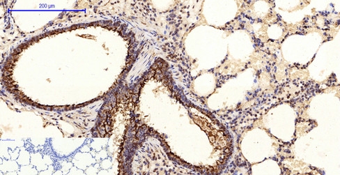
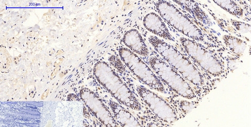
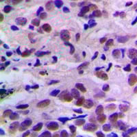 IHC (Immunohiostchemistry)
(Immunohistochemical analysis of CDK2 (pT160) staining in human breast cancer formalin fixed paraffin embedded tissue section. The section was pre-treated using heat mediated antigen retrieval with sodium citrate buffer (pH 6.0). The section was then incubated with the antibody at room temperature and detected using an HRP conjugated compact polymer system. DAB was used as the chromogen. The section was then counterstained with haematoxylin and mounted with DPX.)
IHC (Immunohiostchemistry)
(Immunohistochemical analysis of CDK2 (pT160) staining in human breast cancer formalin fixed paraffin embedded tissue section. The section was pre-treated using heat mediated antigen retrieval with sodium citrate buffer (pH 6.0). The section was then incubated with the antibody at room temperature and detected using an HRP conjugated compact polymer system. DAB was used as the chromogen. The section was then counterstained with haematoxylin and mounted with DPX.)
CDK2 (pT160), Polyclonal Antibody (Cat# AAA104926)
 IF (Immunofluorescence)
(Immunofluorescent analysis of eNOS (pS1177) staining in HuvEc cells. Formalin-fixed cells were permeabilized with 0.1% Triton X-100 in TBS for 5-10 minutes and blocked with 3% BSA-PBS for 30 minutes at room temperature. Cells were probed with the primary antibody in 3% BSA-PBS and incubated overnight at 4 °C in a humidified chamber. Cells were washed with PBST and incubated with a DyLight 594-conjugated secondary antibody (red) in PBS at room temperature in the dark. DAPI was used to stain the cell nuclei (blue).)
IF (Immunofluorescence)
(Immunofluorescent analysis of eNOS (pS1177) staining in HuvEc cells. Formalin-fixed cells were permeabilized with 0.1% Triton X-100 in TBS for 5-10 minutes and blocked with 3% BSA-PBS for 30 minutes at room temperature. Cells were probed with the primary antibody in 3% BSA-PBS and incubated overnight at 4 °C in a humidified chamber. Cells were washed with PBST and incubated with a DyLight 594-conjugated secondary antibody (red) in PBS at room temperature in the dark. DAPI was used to stain the cell nuclei (blue).)
eNOS (pS1177), Polyclonal Antibody (Cat# AAA104941)
What Are Phospho Antibodies?
Protein phosphorylation is a process where a phosphate group is added to certain amino acid residues of a protein – usually serine (S), threonine (T), or tyrosine (Y) - by enzymes called kinases. This process is integral in controlling cellular signaling, cellular growth, and other biological functions.
Our catalog includes a wide range of phospho-specific antibodies that can accurately detect this important marker. They perform strongly in widely-used laboratory applications such as Western blot, flow cytometry, immunohistochemistry, and immunofluorescence microscopy. We value your trust in us and are committed to providing top-quality products and services. All of our antibodies are guaranteed to work for the applications and species indicated on our website & associated product pages.
What Are The Key Applications of Phospho Antibodies?
1. Western Blotting
One of the first steps a researcher can take in utilizing these phospho-specific antibodies, is to check if the antibody works using a technique referred to as “Western blot”. For those unfamiliar, Western Blot aids in showing whether the protein that the antibody recognizes is appearing at the correct/expected size. These phospho-specific antibodies should also be able to detect changes in the target protein’s phosphorylation (on/off state) when cells are stimulated in certain ways.
2. Staining of Fixed Cells (Immunocytochemistry)
Another routine use of these phospho-specific antibodies, is to test if the antibody is able to demonstrate similar performance when used on fixed cells (intact cells that have been preserved) as it did in the Western blot tests. It is an important aspect in many cases to confirm that the antibody works in actual intact cell samples. Ideally, the method used for cellular fixation should be the same as what is used in pathology labs (like using 10% formalin). To check if the antibody works well in tissue sections (FFPE), researchers will often test it on fixed cells that are processed similar to tissue samples.
3. Specificity Tests Using Peptides
In order to make sure that the antibody is only binding to the right target:
- Laboratory technicians will mix the antibody with phospho-peptides (short segments of the protein containing the phosphate group modification).
- If the antibody signal disappears, it is confirmation that it is binding to the correct phosphorylated location.
- A more robust test is to use both the phosphorylated and non-phosphorylated (dephosphorylated) versions of the protein. The antibody should react only with the phosphorylated one.
- Another method sometimes utilized is to treat the sample with an enzyme, such as alkaline phosphatase, that specifically removes phosphate groups. If the antibody signal disappears after this, it also confirms specificity.
4. Genetic Confirmation
As a final step, scientists can genetically manipulate the nucleotide sequence and alter the target protein by removing the exact site where phosphorylation happens. If the antibody no longer appears to detect the modified protein, it is strong evidence supporting the antibody being specific for that phosphorylated site.
Why Buy Phospho Antibodies Through Us?
- The production laboratory adheres to strict and consistent protocols prior to releasing any of these phospho-specific antibodies:
- Standard methods and proper controls in all tests to ensure high quality.
- These antibodies are tested and validated in different cell types and species.
- High quality control criterion to ensure each batch is consistent, so you will obtain reliable results every time.
FAQ
1. What Are Phospho-Specific Antibodies?
Phospho-specific antibodies are made to detect proteins only when they have a phosphate group linked to a specific amino acid residue. This empowers scientists understand if a protein is "turned on" or active, based on its phosphorylation state.
2. How to Detect Phosphorylated Proteins in a Western Blot?
To find out if a protein is phosphorylated using Western blot:
- Use a phospho-specific antibody that binds only to the phosphorylated form of the protein.
- You can also use a “regular” antibody for the same amino acid sequence of the protein that the phospho-specific antibody is binding to (but in this case, this antibody will not bind if there is a phosphate group present) in order to compare how much of it is phosphorylated versus how much is non-phosphorylated (or “total” protein, if the “normal” antibody’s epitopes are non-phospho-site-specific).
3. How to Choose the Best Antibody?
Here are some simple tips to help you pick the right antibody:
- Know your target
- Match your sample characteristics
- Confirm the intended use is appropriate
- Check “host” and “type”
- Check the “quality” of the presented data/images
- Appraise whether the available validation meets your needs