Filters
▼Clonality
▼Type
▼Reactivity
▼Gene Name
▼Isotype
▼Host
▼Application
▼Clone
▼Active Proteins
AAA Biotech also known as AAA Bio or AAABio provides a variety of high-quality recombinant and natural/native proteins that are proven to work in a wide range of experiments. Explore our products to find the active protein that best fits your needs or experimental model.
Viewing 50-100 of 2567 product results
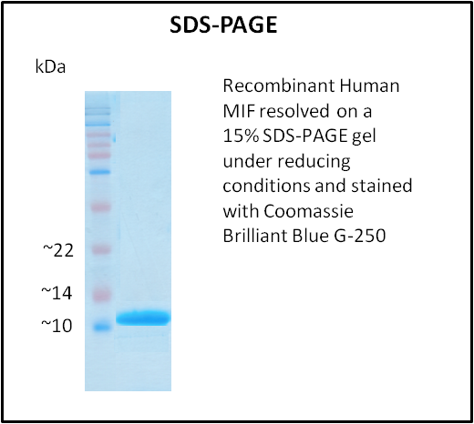
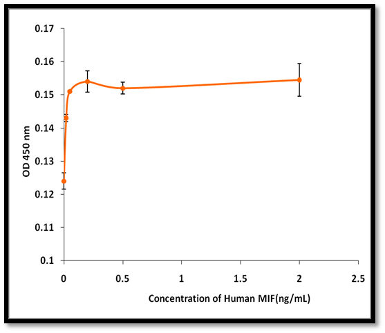 Application Data
MIF, Active Protein (Cat# AAA214256)
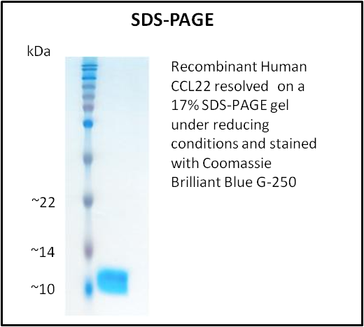
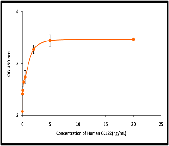 Application Data
CCL22, Active Protein (Cat# AAA214259)
 Application Data
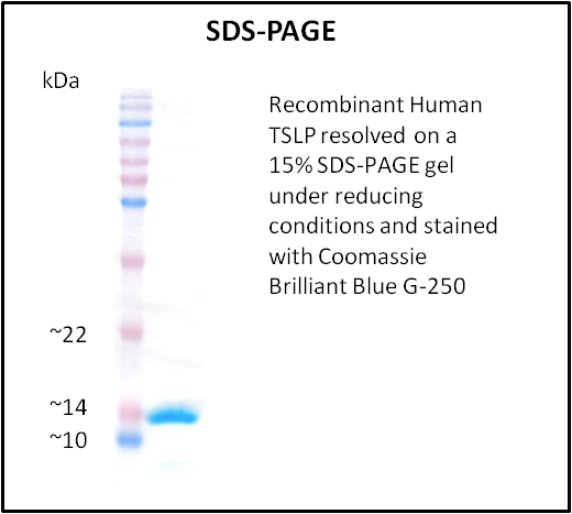
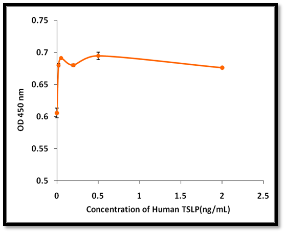
TSLP, Active Protein (Cat# AAA214262)

 Application Data
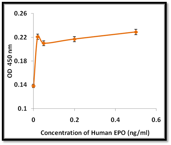
EPO, Active Protein (Cat# AAA214265)
 Application Data
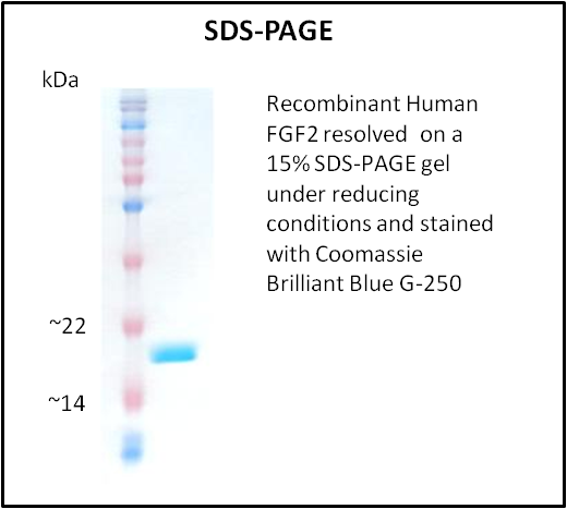
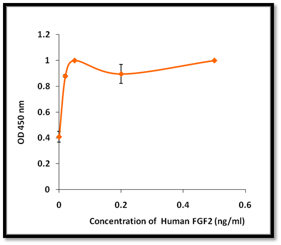
FGF2, Active Protein (Cat# AAA214267)
 Application Data
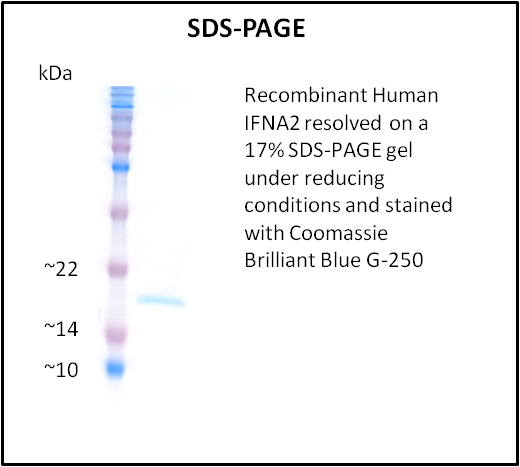
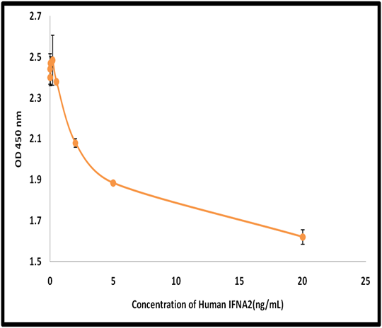
IFNA2, Active Protein (Cat# AAA214269)
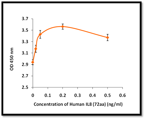 Application Data
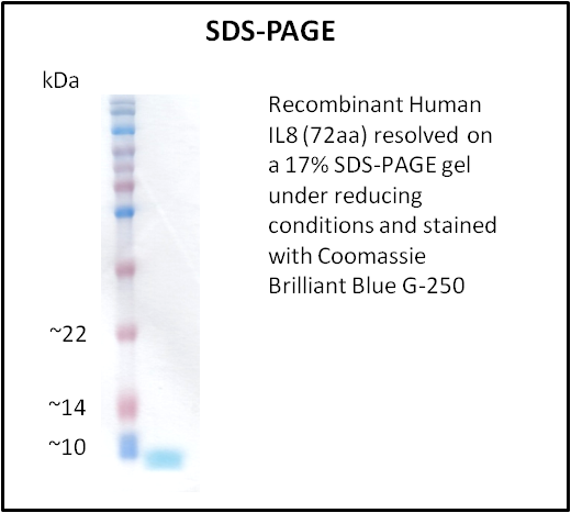
IL8, Active Protein (Cat# AAA214270)
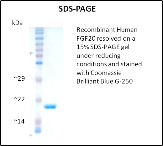
 Application Data
FGF20, Active Protein (Cat# AAA214273)
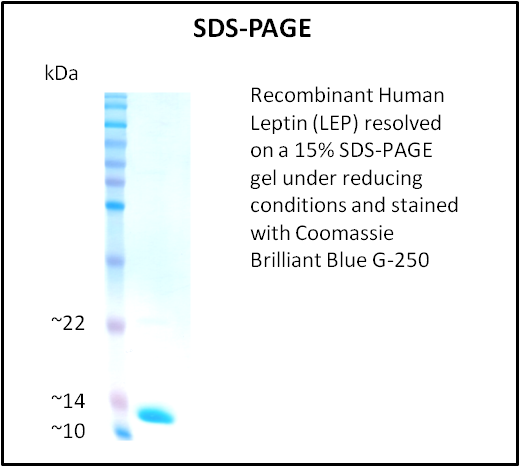
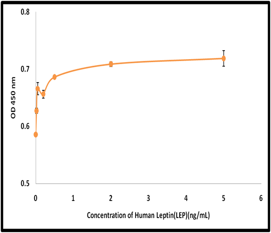 Application Data
Leptin (LEP), Active Protein (Cat# AAA214276)
 Application Data
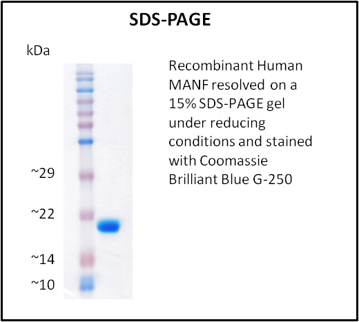
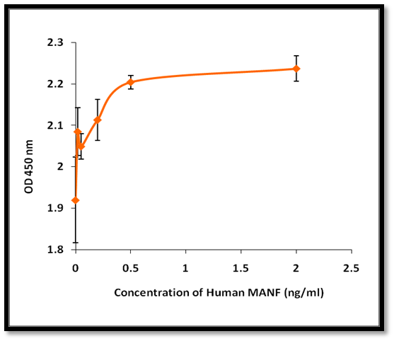
MANF, Active Protein (Cat# AAA214286)
 Application Data
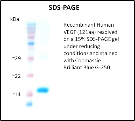
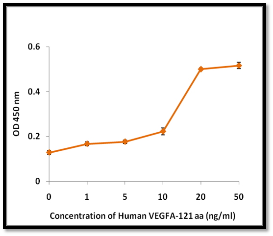
VEGF, Active Protein (Cat# AAA214288)
 Application Data
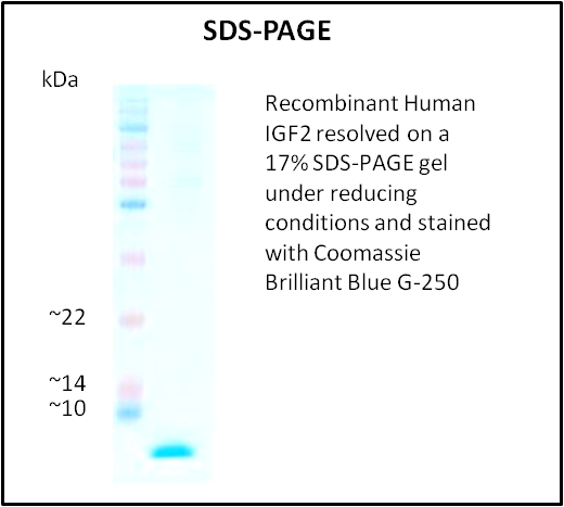
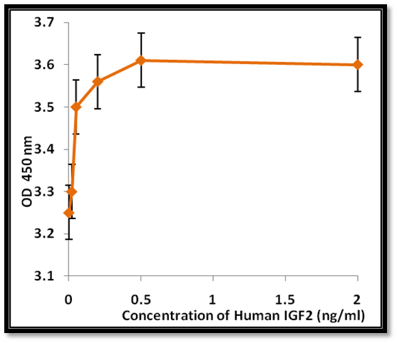
IGF2, Active Protein (Cat# AAA214298)
 Application Data
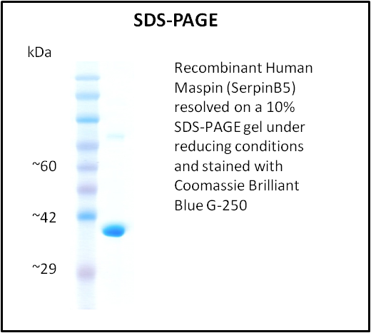
Maspin (SerpinB5), Active Protein (Cat# AAA214304)
 Application Data
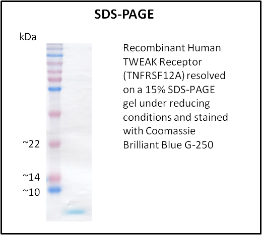
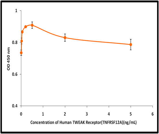
TWEAK Receptor (TNFRSF12A), Active Protein (Cat# AAA214305)
 Application Data
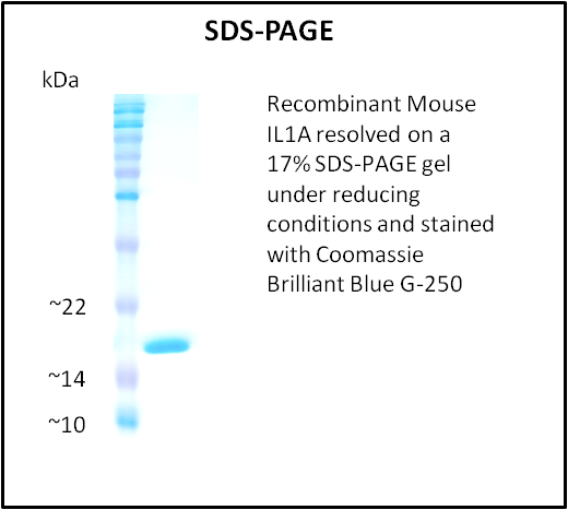
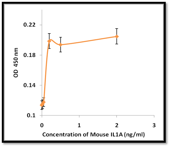
IL1A, Active Protein (Cat# AAA214312)
 Application Data
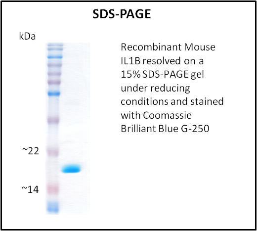
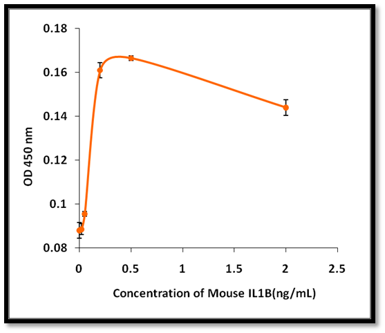
IL1B, Active Protein (Cat# AAA214313)
 Application Data
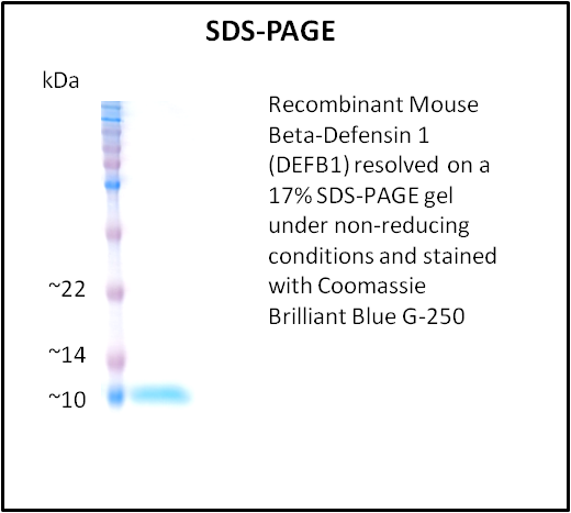
Beta-Defensin 1 (DEFB1), Active Protein (Cat# AAA214324)
 Application Data
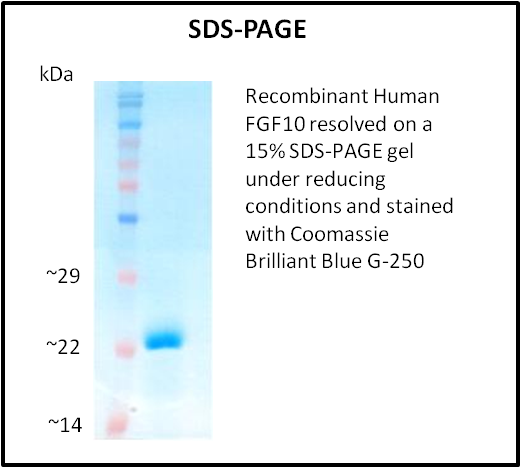
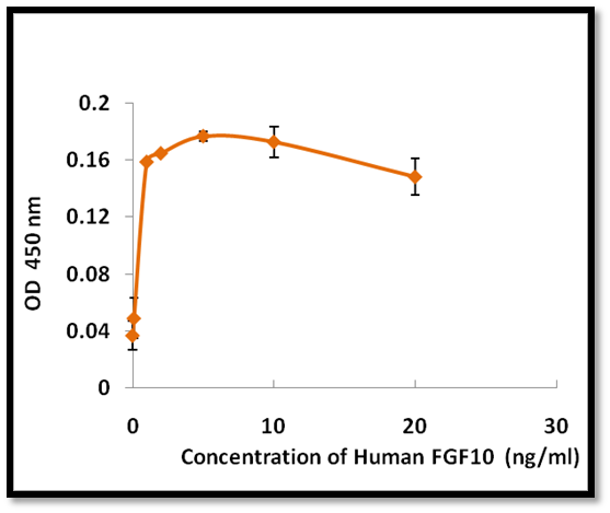
FGF10, Active Protein (Cat# AAA214219)
 Application Data
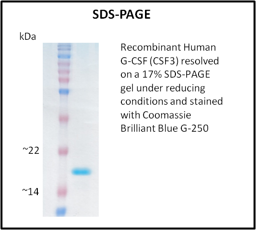
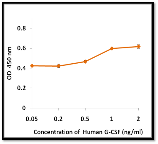
G-CSF (CSF3), Active Protein (Cat# AAA214220)
 Application Data
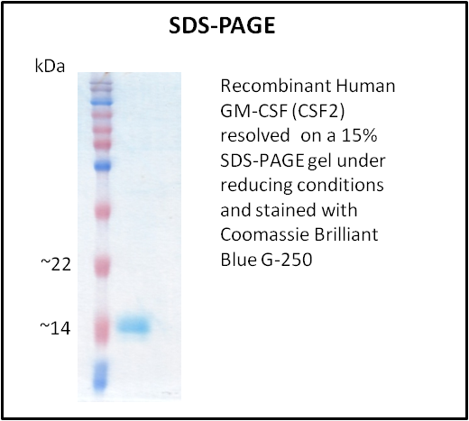
GM-CSF (CSF2), Active Protein (Cat# AAA214222)
 Application Data
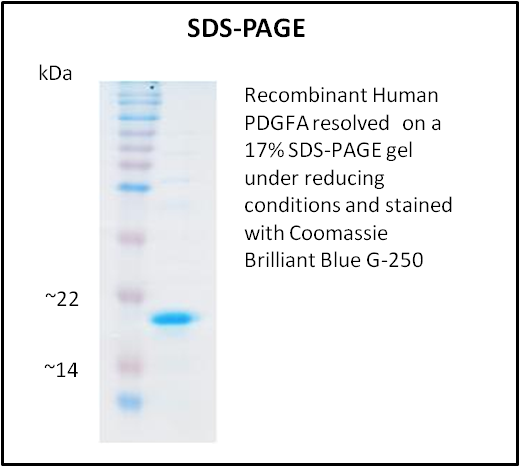
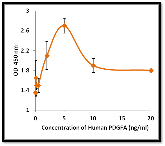
PDGFA, Active Protein (Cat# AAA214228)
 Application Data
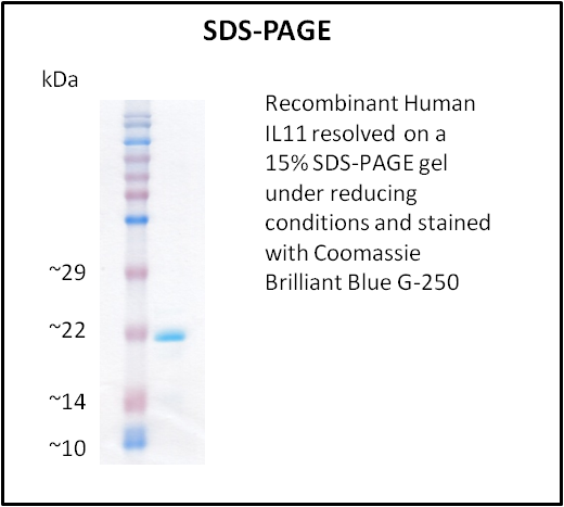
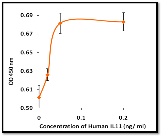
IL11, Active Protein (Cat# AAA214230)
 Application Data
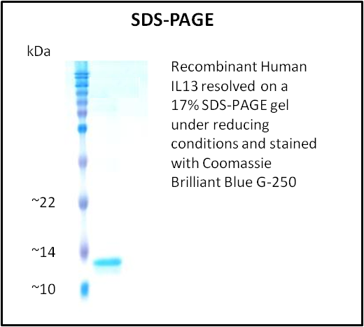
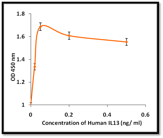
IL13, Active Protein (Cat# AAA214231)
 Application Data
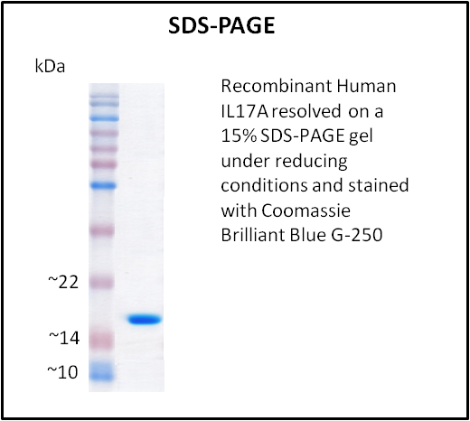
IL17A, Active Protein (Cat# AAA214233)
 Application Data
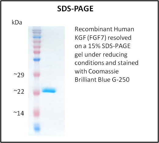
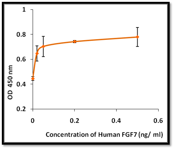
KGF (FGF7), Active Protein (Cat# AAA214239)
Lymphotactin (XCL1), Active Protein (Cat# AAA214241)
 Application Data
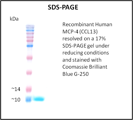
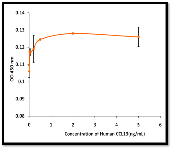
MCP-4 (CCL13), Active Protein (Cat# AAA214244)
 Application Data
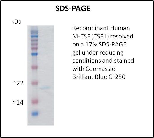
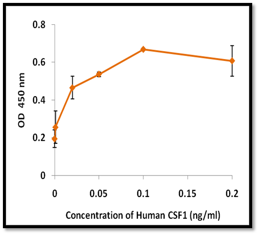
M-CSF (CSF1), Active Protein (Cat# AAA214245)
 Application Data
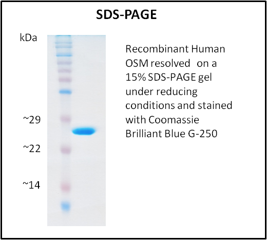
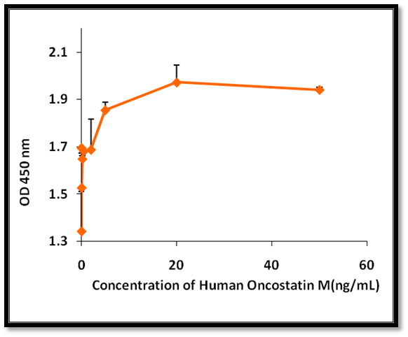
OSM, Active Protein (Cat# AAA214249)
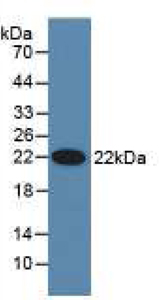
 WB (Western Blot)
(Western BlotSample: Recombinant PON3, Human;Antibody: Rabbit Anti-Human PON3 Ab)
WB (Western Blot)
(Western BlotSample: Recombinant PON3, Human;Antibody: Rabbit Anti-Human PON3 Ab)
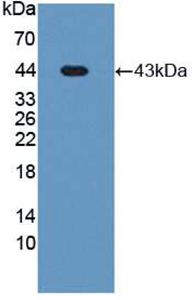
Paraoxonase 3 (PON3), Active Protein (Cat# AAA147870)
 WB (Western Blot)
(Sample: Recombinant PON1, Human;Antibody: Rabbit Anti-Human PON1 Ab)
WB (Western Blot)
(Sample: Recombinant PON1, Human;Antibody: Rabbit Anti-Human PON1 Ab)
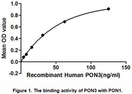
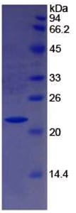
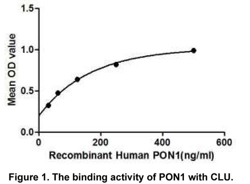
Paraoxonase 1 (PON1), Active Protein (Cat# AAA147871)
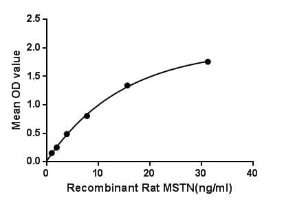
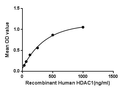
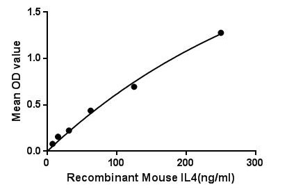 Bioactivity
(Figure. The binding activity of IL4 with IL2g.The interleukin 4 (IL4, IL-4) is a cytokine that induces differentiation of naive helper T cells (Th0 cells) to Th2 cells. Interleukin 4 has many biological roles, including the stimulation of activated B-cell and T-cell proliferation, and the differentiation of B cells into plasma cells. It is a key regulator in humoral and adaptive immunity. IL-4 induces B-cell class switching to IgE, and up-regulates MHC class II production. IL-4 decreases the production of Th1 cells, macrophages, IFN-gamma, and dendritic cell IL-12. Besides, Interleukin 2 Receptor Gamma (IL2Rg) has been identified as an interactor of IL4, thus a binding ELISA assay was conducted to detect the interaction of recombinant mouse IL4 and recombinant mouse IL2Rg. Briefly, IL4 were diluted serially in PBS, with 0.01% BSA (pH 7.4). Duplicate samples of 100uL were then transferred to IL2Rg-coated microtiter wells and incubated for 2h at 37. Wells were washed with PBST and incubated for 1h with anti-IL4 pAb, then aspirated and washed 3 times. After incubation with HRP labelled secondary antibody, wells were aspirated and washed 3 times. With the addition of substrate solution, wells were incubated 15-25 minutes at 37. Finally, add 50uL stop solution to the wells and read at 450nm immediately. The binding activity of IL4 and IL2Rg was shown in Figure 1, and this effect was in a dose dependent manner.)
Bioactivity
(Figure. The binding activity of IL4 with IL2g.The interleukin 4 (IL4, IL-4) is a cytokine that induces differentiation of naive helper T cells (Th0 cells) to Th2 cells. Interleukin 4 has many biological roles, including the stimulation of activated B-cell and T-cell proliferation, and the differentiation of B cells into plasma cells. It is a key regulator in humoral and adaptive immunity. IL-4 induces B-cell class switching to IgE, and up-regulates MHC class II production. IL-4 decreases the production of Th1 cells, macrophages, IFN-gamma, and dendritic cell IL-12. Besides, Interleukin 2 Receptor Gamma (IL2Rg) has been identified as an interactor of IL4, thus a binding ELISA assay was conducted to detect the interaction of recombinant mouse IL4 and recombinant mouse IL2Rg. Briefly, IL4 were diluted serially in PBS, with 0.01% BSA (pH 7.4). Duplicate samples of 100uL were then transferred to IL2Rg-coated microtiter wells and incubated for 2h at 37. Wells were washed with PBST and incubated for 1h with anti-IL4 pAb, then aspirated and washed 3 times. After incubation with HRP labelled secondary antibody, wells were aspirated and washed 3 times. With the addition of substrate solution, wells were incubated 15-25 minutes at 37. Finally, add 50uL stop solution to the wells and read at 450nm immediately. The binding activity of IL4 and IL2Rg was shown in Figure 1, and this effect was in a dose dependent manner.)
Interleukin 4, Active Protein (Cat# AAA150060)

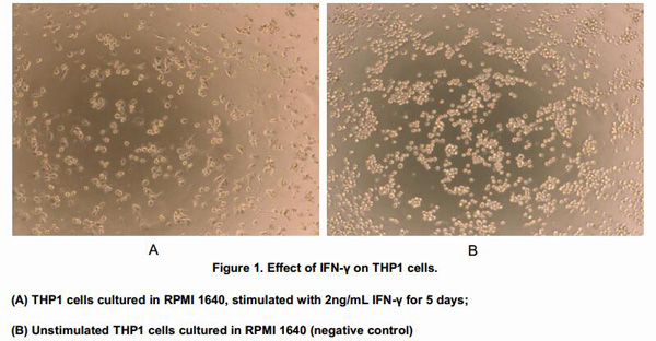
 Application Data
Interferon Gamma (IFNg), Active Protein (Cat# AAA146600)
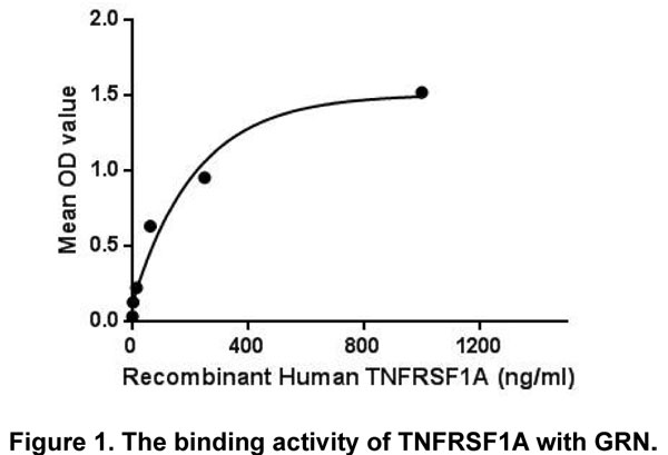
 WB (Western Blot)
(Figure 3. Western BlotSample: Recombinant TNFRSF1A, Human;Antibody: Rabbit Anti-Human TNFRSF1A Ab)
WB (Western Blot)
(Figure 3. Western BlotSample: Recombinant TNFRSF1A, Human;Antibody: Rabbit Anti-Human TNFRSF1A Ab)
Tumor Necrosis Factor Receptor 1 (TNFR1), Active Protein (Cat# AAA149245)
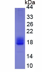
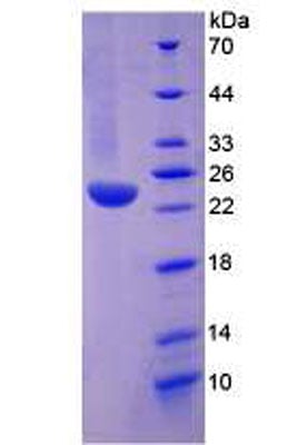
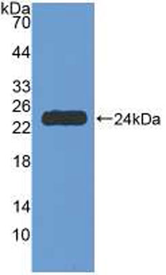
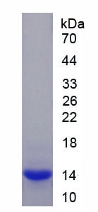 SDS-PAGE
Myostatin (MSTN), Active Protein (Cat# AAA149249)


 WB (Western Blot)
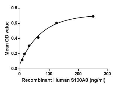
(Sample: Recombinant S100A8, Human;Antibody: Rabbit Anti-Human S100A8 Ab)
WB (Western Blot)
(Sample: Recombinant S100A8, Human;Antibody: Rabbit Anti-Human S100A8 Ab)
S100 Calcium Binding Protein A8 (S100A8), Active Protein (Cat# AAA149250)
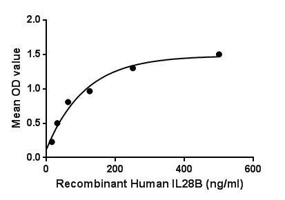
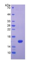
 SDS-PAGE
Interleukin 28B (IL28B), Active Protein (Cat# AAA149254)
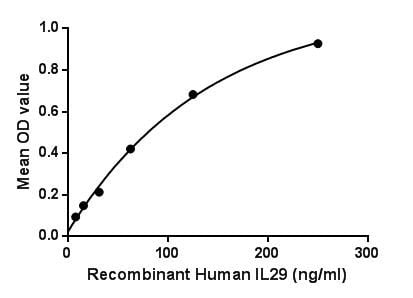
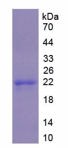
 SDS-PAGE
Interleukin 29 (IL29), Active Protein (Cat# AAA149255)
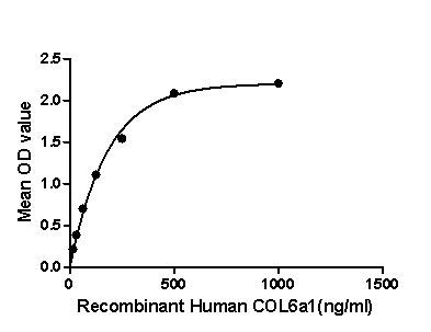
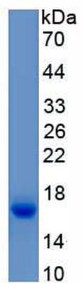
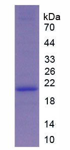 SDS-PAGE
Collagen Type VI Alpha 1 (COL6a1), Active Protein (Cat# AAA149257)
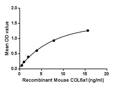
 WB (Western Blot)
(Western BlotSample: Recombinant COL6a1, Mouse;Antibody: Rabbit Anti-Mouse COL6a1 Ab)
WB (Western Blot)
(Western BlotSample: Recombinant COL6a1, Mouse;Antibody: Rabbit Anti-Mouse COL6a1 Ab)
Collagen Type VI Alpha 1 (COL6a1), Active Protein (Cat# AAA149258)

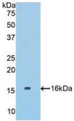
 WB (Western Blot)
(Sample: Recombinant HDAC1, Human;Antibody: Rabbit Anti-Human HDAC1 Ab)
WB (Western Blot)
(Sample: Recombinant HDAC1, Human;Antibody: Rabbit Anti-Human HDAC1 Ab)
Histone Deacetylase 1 (HDAC1), Active Protein (Cat# AAA149259)
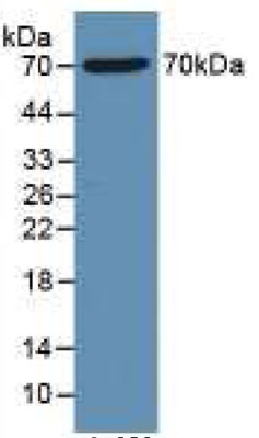
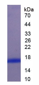 SDS-PAGE
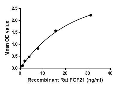
Fibroblast Growth Factor 21 (FGF21), Active Protein (Cat# AAA149261)

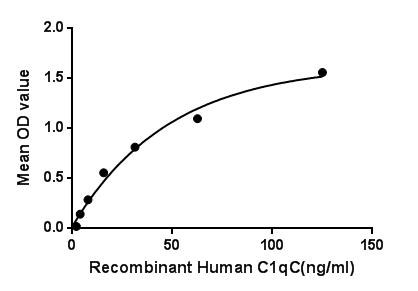

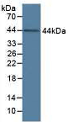 WB (Western Blot)
(Figure 3. Western BlotSample: Recombinant C1qC, Human;Antibody: Rabbit Anti-Human C1qC Ab)
WB (Western Blot)
(Figure 3. Western BlotSample: Recombinant C1qC, Human;Antibody: Rabbit Anti-Human C1qC Ab)
Complement Component 1, Q Subcomponent C (C1qC), Active Protein (Cat# AAA149263)

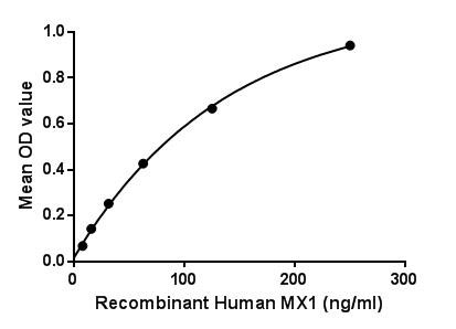
 WB (Western Blot)
(Figure 4. Western BlotSample: Recombinant MX1, Human;Antibody: Rabbit Anti-Human MX1 Ab)
WB (Western Blot)
(Figure 4. Western BlotSample: Recombinant MX1, Human;Antibody: Rabbit Anti-Human MX1 Ab)
Myxovirus Resistance 1 (MX1), Active Protein (Cat# AAA149266)
 SDS-PAGE
Chemokine C-X3-C-Motif Ligand 1 (CX3CL1), Active Protein (Cat# AAA149223)
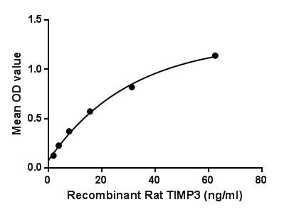
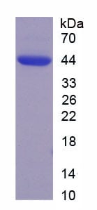
 SDS-PAGE
Tissue Inhibitors Of Metalloproteinase 3 (TIMP3), Active Protein (Cat# AAA149228)
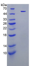
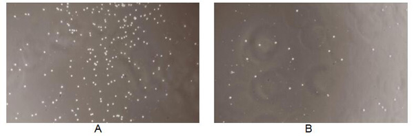
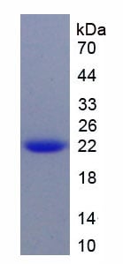
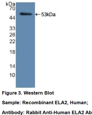 WB (Western Blot)
Elastase 2, Neutrophil, Active Protein (Cat# AAA149230)
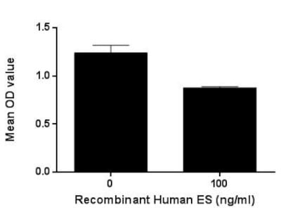

 WB (Western Blot)
(Figure 4. Western BlotSample: Recombinant ES, Human;Antibody: Rabbit Anti-Human ES Ab)
WB (Western Blot)
(Figure 4. Western BlotSample: Recombinant ES, Human;Antibody: Rabbit Anti-Human ES Ab)
Endostatin (ES), Active Protein (Cat# AAA149235)
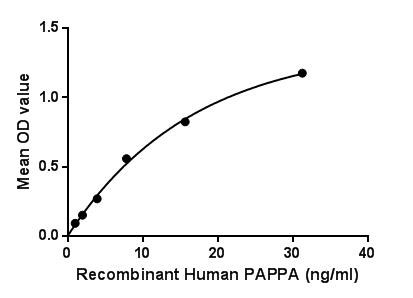
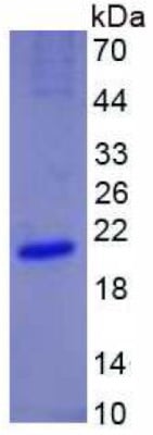
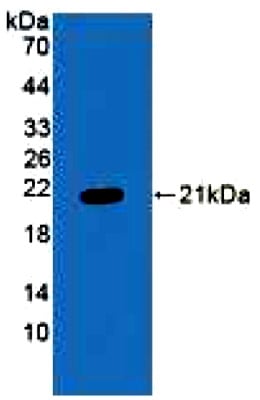
 SDS-PAGE
Pregnancy Associated Plasma Protein A (PAPPA), Active Protein (Cat# AAA149241)
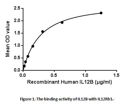
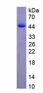
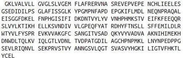
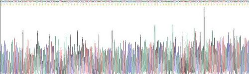
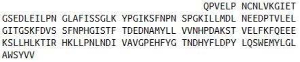
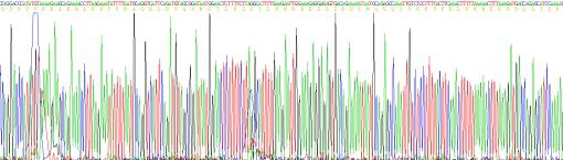
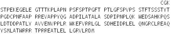
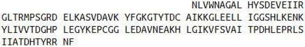
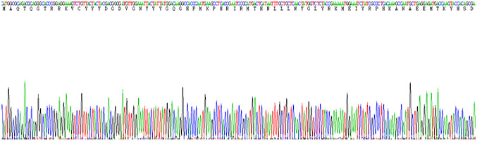
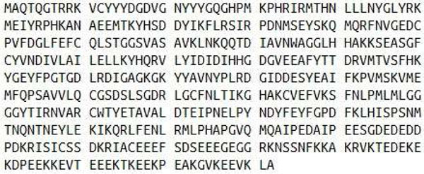
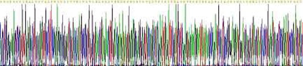
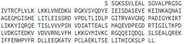
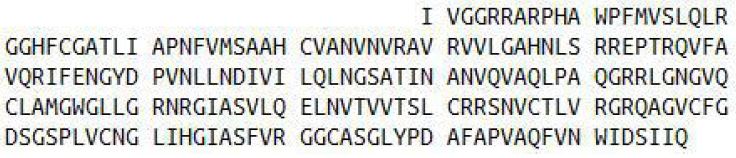
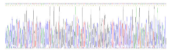
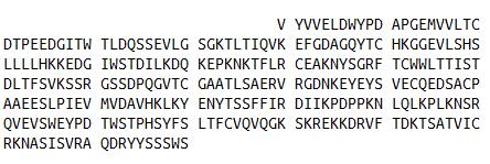 Sequence
Interleukin 12B (IL12B), Active Protein (Cat# AAA150970)
What Are Active Proteins?
Proteins are large molecules made up of long chains of amino acids.
They will typically fold into a very particular 3-dimensional shape/conformation, that is sometimes referred to as their “native” form, which allows them to work properly in the body. For the purposes of product categorization, AAA Biotech will typically refer to proteins purified from their original animal host as being “native” proteins (this is to signify their difference compared to their “recombinant” or “synthetic” protein counterparts).
If a protein successfully folds into the correct shape, it is will typically display high fidelity characteristics to its original protein in its original animal host, and be classified as an active protein, as it will be able to function “normally” in most enzymatic or binding capacities. If it loses this shape, due to factors such as heat or strong chemicals (such as detergents), it becomes inactive and is no longer able to perform its basic functions. All of the proteins in this category are made under strict quality control, and they are active, pure, low in contaminants, and stable.
Most are stored as freeze-dried powders and come without extra tags, so they’re very close to the actual natural/native form.
Key Applications of Active Proteins
1. Scientific Research
- Aid in the study of how proteins function in the body
- Aid in understanding various disease processes
2. Drug Development
- Powerful tools to investigate how potential drugs interact with specific proteins
- Ideal for identifying drug targets
3. Cell Culture
- Are routinely utilized to support cell growth and function (e.g., using exogenous growth factors)
- Can be used to promote cellular development into specific types (differentiation)
4. Diagnostics
- Regularly utilized in tests to detect diseases or infections (e.g., COVID-19, cancer)
- Note: All products are strictly for research-use only (RUO).
5. Therapeutics
- Some active proteins are used directly as treatments (e.g., insulin, enzymes)
- Note: All products are strictly for research-use only (RUO).
6. Vaccine Development
- Used to create or test vaccines by mimicking parts of viruses or bacteria
7. Biochemical Assays
- They can facilitate the characterization of enzyme activity, binding strength, or protein interactions in lab tests
Why Buy Active Proteins from AAA Biotech?
- High biological activity – Verified to perform as expected or indicated on datasheet
- Strict quality control – We are confident in our active proteins’ reliability and consistency
- High purity & low endotoxin – Ideal for applications involving sensitive or precious samples/components
- Freeze-dried for stability – Long shelf life and straightforward storage
- Mostly tag-free – Closer to natural/native protein form
FAQ
1. What are active proteins used for in research?
Active proteins are used primarily in the study of how proteins function, in characterizing/discovering drug interactions, supporting cell growth, running biochemical assays, and in development of diagnostics or therapeutics.
2. How are AAA Biotech's active proteins validated?
AAA Biotech’s active proteins are validated through strict quality control and functional assays to ensure they are properly folded and active. “Active”, though, can be an ambiguous term, so if a specific “activity” or “binding” capability of a protein is of crucial interest to you, please inquire with us prior to purchase, and we will provide further details on how the “Active” modifier was determined to be applicable.
3. Are these proteins tested for biological activity?
Yes, all active proteins from AAA Biotech are tested to confirm they have the expected biological activity before being offered for use. Though, said “biological activity” can be either “enzymatic”, “binding”, or both.