Filters
▼Clonality
▼Type
▼Reactivity
▼Gene Name
▼Isotype
▼Host
▼Application
▼Clone
▼Recombinant Proteins
Recombinant proteins are purified laboratory reagents created through genetic engineering for advanced biomedical research, such as immunology, neuroscience, cancer research, and more.
At AAA Biotech, also known as AAA Bio or AAABio, we provide high-quality recombinant proteins. Our various protein products are carefully tested for consistent and reliable performance. With our proteins, we offer stringent quality control, lot-to-lot consistency, and precise specificity to help you achieve accurate results in your research.
Whether you're conducting research on cell expansion, polarization, differentiation, or cell processing, we can offer the exact recombinant proteins you need in order to support your work.
Viewing 6250-6300 of 14163 product results
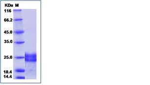
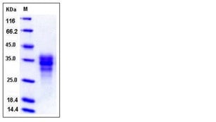
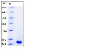
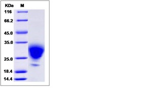
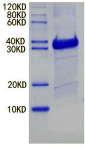 SDS-PAGE
(12% SDS-PAGE:)
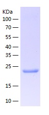
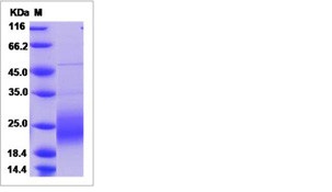
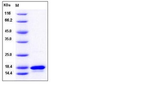
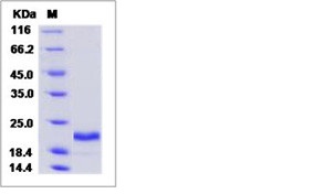
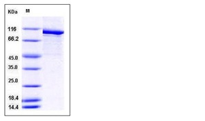
SDS-PAGE
(12% SDS-PAGE:)
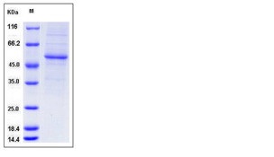
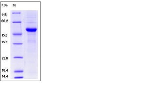
UBE3A / Ubiquitin-Protein Ligase E3A, Recombinant Protein (Cat# AAA196944)
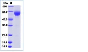
HIV1 gp160, Recombinant Protein (Cat# AAA224574)
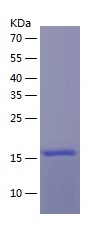
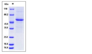
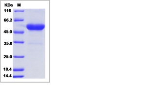
 SDS-PAGE
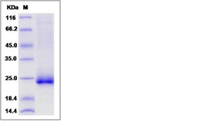
Lassa virus Nucleoprotein (N), Recombinant Protein (Cat# AAA235207)
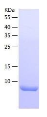
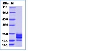
 SDS-PAGE
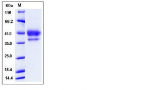
IGF2BP2, Recombinant Protein (Cat# AAA249531)
 SDS-PAGE
SLUG, Recombinant Protein (Cat# AAA249439)
 SDS-PAGE
VILIP3, Recombinant Protein (Cat# AAA249480)
 SDS-PAGE
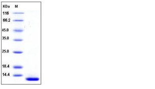
beta Defensin 1, Recombinant Protein (Cat# AAA249490)
 SDS-PAGE
(SDS-Page)
SDS-PAGE
(SDS-Page)
Triosephosphate isomerase, Recombinant Protein (Cat# AAA249491)
 Application Data
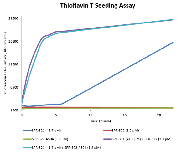
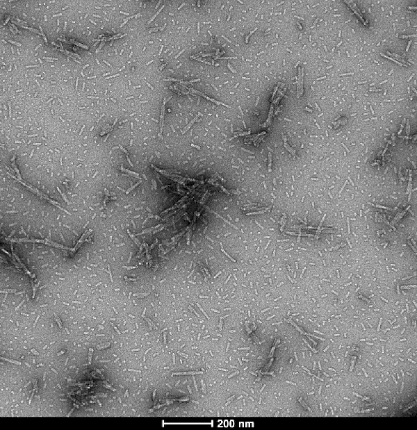
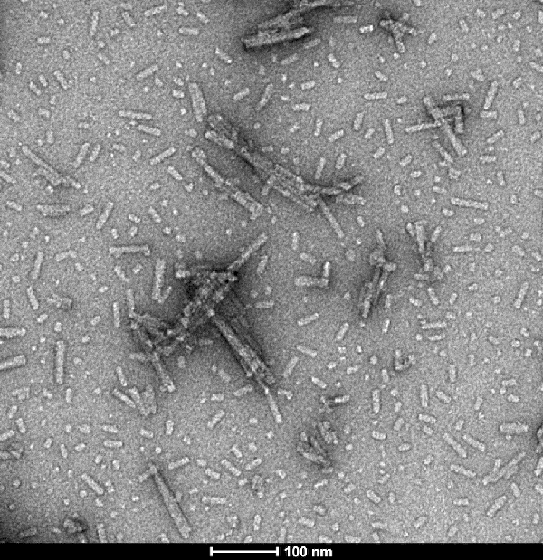
(ThioflavinTisafluorescentdyethatbindstobetasheet-richstructures,suchasthoseinalphasynucleinfibrils.ATTO594-conjugatedalphasynucleinpre-formedfibrils(AAA254080)showsimilarseedingcapabilitiesasuncojugatedalphasynucleinpre-formedfibrils whencombinedwithmonomers.PlateUsed:HTRF96-welllowvolumewhiteplate(Cisbio).ReactionVolume:15uL.ThioflavinTConcentration:40uM.SDSCatalystconcentration:0.73mM.Incubation:37C,600rpm.)
Application Data
(ThioflavinTisafluorescentdyethatbindstobetasheet-richstructures,suchasthoseinalphasynucleinfibrils.ATTO594-conjugatedalphasynucleinpre-formedfibrils(AAA254080)showsimilarseedingcapabilitiesasuncojugatedalphasynucleinpre-formedfibrils whencombinedwithmonomers.PlateUsed:HTRF96-welllowvolumewhiteplate(Cisbio).ReactionVolume:15uL.ThioflavinTConcentration:40uM.SDSCatalystconcentration:0.73mM.Incubation:37C,600rpm.)
Alpha Synuclein: ATTO 594 (Type 1), Recombinant Protein (Cat# AAA254080)
 Application Data
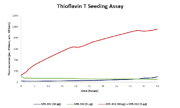
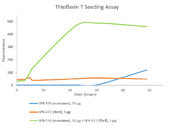
(ThioflavinTassayforN-terminalacetylatedalphasynucleinmonomers andpre-formedfibrils(PFFs)(AAA254082).Increasedfluorescence,indicativeofbeta-sheetformation,wasseenwhen50ugN-acetylatedmonomerand5ugN-acetylatedfibrilwerecombined,comparedtomonomerorfibrilalone.)
Application Data
(ThioflavinTassayforN-terminalacetylatedalphasynucleinmonomers andpre-formedfibrils(PFFs)(AAA254082).Increasedfluorescence,indicativeofbeta-sheetformation,wasseenwhen50ugN-acetylatedmonomerand5ugN-acetylatedfibrilwerecombined,comparedtomonomerorfibrilalone.)
Acetylated Alpha Synuclein Pre-formed Fibrils (Type 1), Recombinant Protein (Cat# AAA254082)
 Application Data
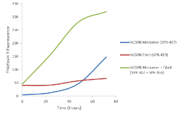
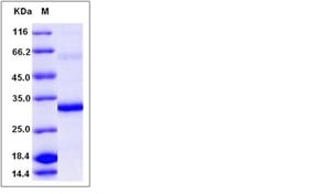
(CoomassiegelstainofHumanGammaSynucleinMonomer(AAA254084).Load:2ug.)
Application Data
(CoomassiegelstainofHumanGammaSynucleinMonomer(AAA254084).Load:2ug.)
Gamma Synuclein Protein Monomers, Recombinant Protein (Cat# AAA254084)
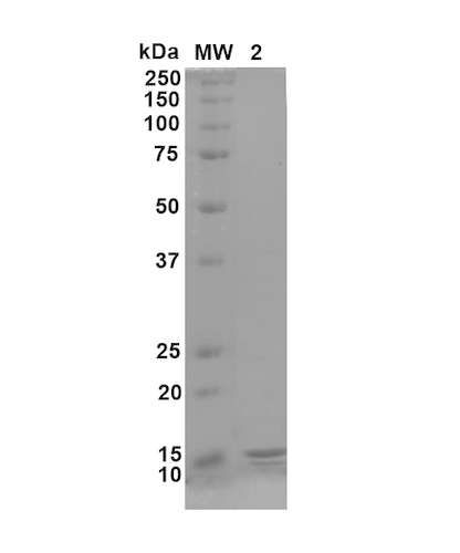
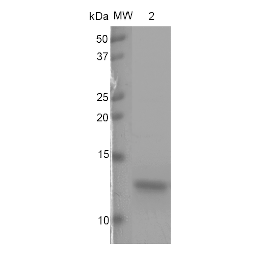 Application Data
Truncated Tau Fragment (AA297-391)(dGAE C322A) Protein Monomers, Recombinant Protein (Cat# AAA254087)
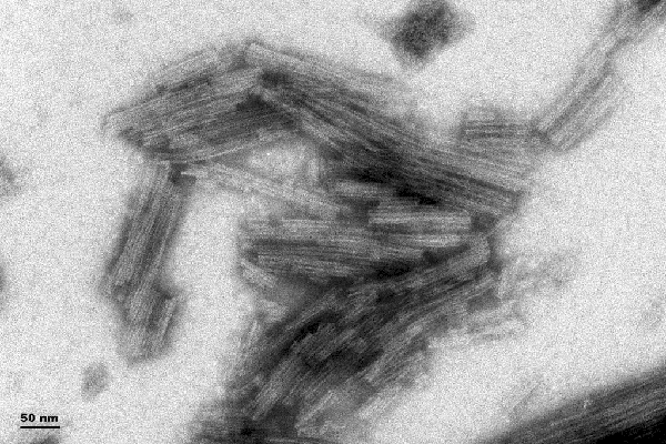
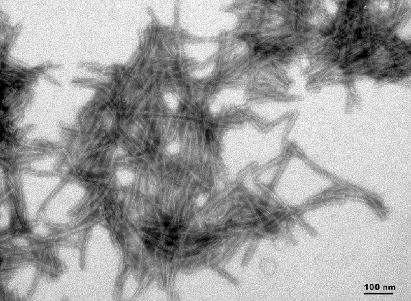
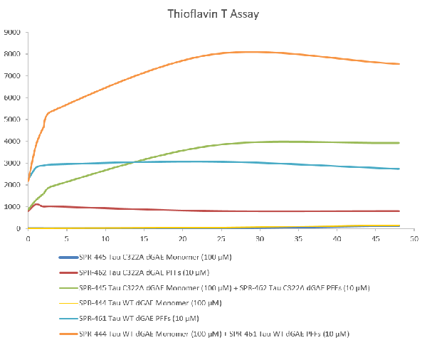
 Application Data
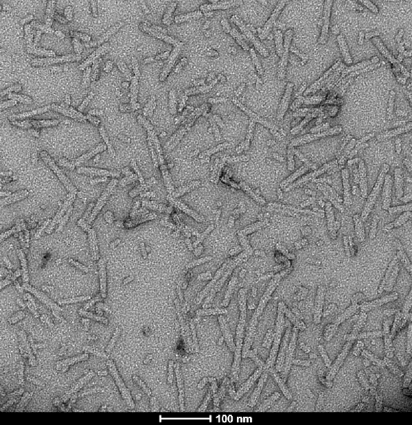
(ThioflavinTisafluorescentdyethatbindstobetasheet-richstructures,suchasthoseintaufibrils.Uponbinding,theemissionspectrumofthedyeexperiencesared-shiftandincreasedfluorescenceintensity.ThioflavinTemissioncurvesshowincreasedfluorescence(correlatedtotauaggregation)overtimewhentruncatedtaufragment(AA297-391)(dGAE)monomeriscombinedwithtruncatedtaufragment(AA297-391)(dGAE)Pre-formedFibrils(Type1).)
Application Data
(ThioflavinTisafluorescentdyethatbindstobetasheet-richstructures,suchasthoseintaufibrils.Uponbinding,theemissionspectrumofthedyeexperiencesared-shiftandincreasedfluorescenceintensity.ThioflavinTemissioncurvesshowincreasedfluorescence(correlatedtotauaggregation)overtimewhentruncatedtaufragment(AA297-391)(dGAE)monomeriscombinedwithtruncatedtaufragment(AA297-391)(dGAE)Pre-formedFibrils(Type1).)
Truncated Tau Fragment (AA297-391)(dGAE), Recombinant Protein (Cat# AAA254091)
 Application Data
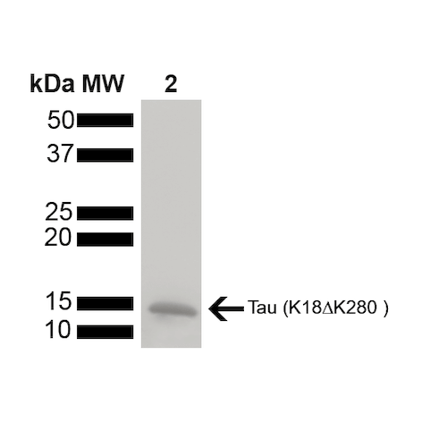
(CoomassiegelstainofHumanK18K280DeletionTauproteinmonomer(AAA254096).Load:2ug.)
Application Data
(CoomassiegelstainofHumanK18K280DeletionTauproteinmonomer(AAA254096).Load:2ug.)
Tau (K18) Delta K280 Mutant Protein Monomers, Recombinant Protein (Cat# AAA254096)
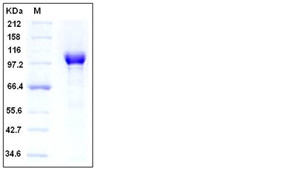
Notch homolog 2 N-terminal-like B (NOTCH2NLB), Recombinant Protein (Cat# AAA244047)
 SDS-PAGE
FGFBP3, Recombinant Protein (Cat# AAA257957)
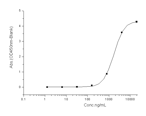
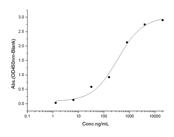 Application Data
(Measured by its binding ability in a functional ELISA. Immobilized Human PARP His at 2 ug/ml (100 ul/well) can bind Human HSP70 His, biotinylated, the EC50 of HSP70 His, biotinylated is 0.2-1.0 ug/mL.)
Application Data
(Measured by its binding ability in a functional ELISA. Immobilized Human PARP His at 2 ug/ml (100 ul/well) can bind Human HSP70 His, biotinylated, the EC50 of HSP70 His, biotinylated is 0.2-1.0 ug/mL.)
PARP, Recombinant Protein (Cat# AAA257960)
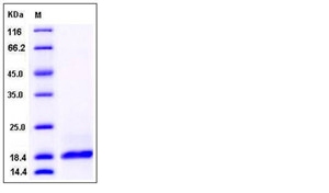 SDS-PAGE
PLA2G2A, Recombinant Protein (Cat# AAA257967)
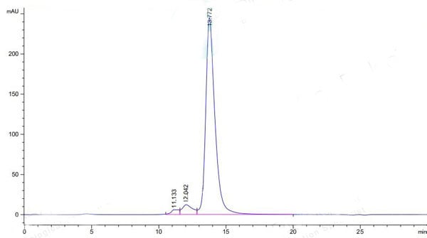
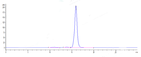
>90% as determined by SEC-HPLC
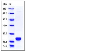 SDS-PAGE
LCN1, Recombinant Protein (Cat# AAA257979)
 Application Data
(Measured in a cell proliferation assay using human umbilical vein endothelial cells (HUVEC). The ED50 for this effect is typically 50-210 ng/mL.)
Application Data
(Measured in a cell proliferation assay using human umbilical vein endothelial cells (HUVEC). The ED50 for this effect is typically 50-210 ng/mL.)
VEGF183, Recombinant Protein (Cat# AAA257397)
 Application Data
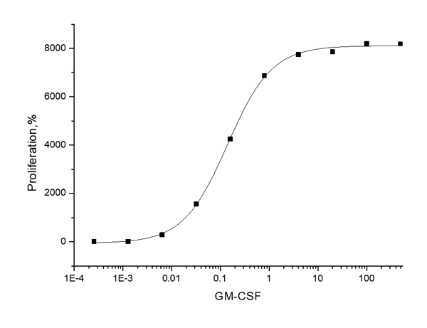
(Measured in a cell proliferation assay using TF-1 human erythroleukemic cells. The ED50 for this effect is typically 0.1-0.6 ng/ml.)
Application Data
(Measured in a cell proliferation assay using TF-1 human erythroleukemic cells. The ED50 for this effect is typically 0.1-0.6 ng/ml.)
GM-CSF, Recombinant Protein (Cat# AAA257398)
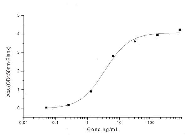
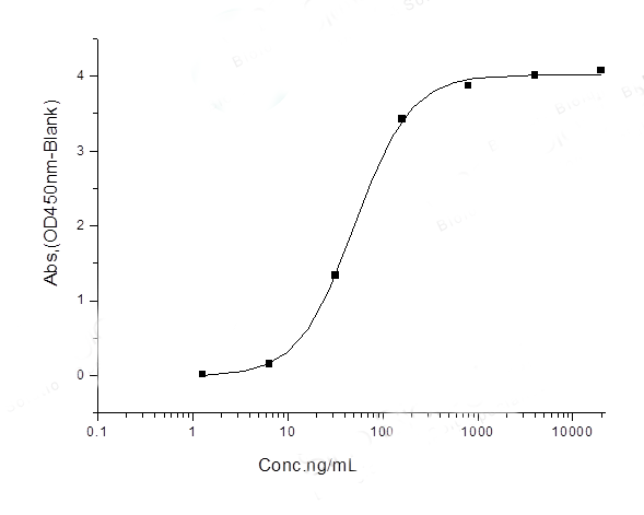
 Application Data
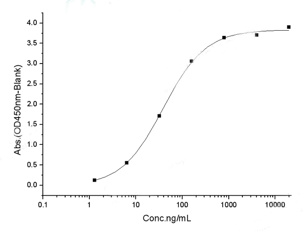
(Measured by its binding ability in a functional ELISA. Immobilized human CD30L-hFc (Cat: 10040-H01H) at 2 ug/ml (100 ul/well) can bind human CD30/TNFRSF8-Fch (Cat: 10777-H03H), the EC50 of human CD30/TNFRSF8-Fch is 15-60 ng/mL.)
Application Data
(Measured by its binding ability in a functional ELISA. Immobilized human CD30L-hFc (Cat: 10040-H01H) at 2 ug/ml (100 ul/well) can bind human CD30/TNFRSF8-Fch (Cat: 10777-H03H), the EC50 of human CD30/TNFRSF8-Fch is 15-60 ng/mL.)
CD30L, Recombinant Protein (Cat# AAA257399)
 Application Data
ACE2, Recombinant Protein (Cat# AAA257402)
 Application Data
(Measured by its ability to inhibit Fas Ligand induced apoptosis of Jurkat human acute T cell leukemia cells. The ED50 for this effect is typically 0.2-1.2 ug/mL in the presence of 200ng/mL recombinant human Fas ligand.)
Application Data
(Measured by its ability to inhibit Fas Ligand induced apoptosis of Jurkat human acute T cell leukemia cells. The ED50 for this effect is typically 0.2-1.2 ug/mL in the presence of 200ng/mL recombinant human Fas ligand.)
Fas/CD95, Recombinant Protein (Cat# AAA257404)
 Application Data
TNF beta, Recombinant Protein (Cat# AAA257408)
 Application Data
(Measured in antiviral assays using WISH human amnion cells infected with vesicular stomatitisvirus (VSV). The ED50 for this effect is 0.1-1 ng/mL.)
Application Data
(Measured in antiviral assays using WISH human amnion cells infected with vesicular stomatitisvirus (VSV). The ED50 for this effect is 0.1-1 ng/mL.)
IFNA7, Recombinant Protein (Cat# AAA257412)
 Application Data
(Measured in antiviral assays using WISH cells infected with vesicular stomatitis virus. The ED50 for this effect is 3.0-15.0pg/mL.)
Application Data
(Measured in antiviral assays using WISH cells infected with vesicular stomatitis virus. The ED50 for this effect is 3.0-15.0pg/mL.)
IFNA7, Recombinant Protein (Cat# AAA257413)
 Application Data
(Measured in antiviral assays using WISH human amnion cells infected with vesicular stomatitisvirus (VSV). The ED50 for this effect is 20-80 pg/mL.)
Application Data
(Measured in antiviral assays using WISH human amnion cells infected with vesicular stomatitisvirus (VSV). The ED50 for this effect is 20-80 pg/mL.)
Interferon alpha 10/IFNA10, Recombinant Protein (Cat# AAA257415)
 Application Data
(Measured in antiviral assays using WISH human amnion cells infected with vesicular stomatitis virus(VSV). The ED50 for this effect is typically 2-8 pg/mL.)
Application Data
(Measured in antiviral assays using WISH human amnion cells infected with vesicular stomatitis virus(VSV). The ED50 for this effect is typically 2-8 pg/mL.)
IFN-omega, Recombinant Protein (Cat# AAA257417)

 FCM/FACS (Flow Cytometry)
(Labeled biotin to PD-1 Protein, Human, Recombinant (Fc Tag) by a certain molar ratio; Using the Octet RED System, the affinity constant (Kd) of PD-1 Protein, Human, Recombinant (Fc Tag), Biotinylated bound to Atezolizumab was 0.3 nM.)
FCM/FACS (Flow Cytometry)
(Labeled biotin to PD-1 Protein, Human, Recombinant (Fc Tag) by a certain molar ratio; Using the Octet RED System, the affinity constant (Kd) of PD-1 Protein, Human, Recombinant (Fc Tag), Biotinylated bound to Atezolizumab was 0.3 nM.)
PD-1, Recombinant Protein (Cat# AAA257419)
 Application Data
(Measured in a cell proliferation assay using TF-1 human erythroleukemic cells. The ED50 for this effect is typically 0.2-1 ng/mL.)
Application Data
(Measured in a cell proliferation assay using TF-1 human erythroleukemic cells. The ED50 for this effect is typically 0.2-1 ng/mL.)
Oncostatin M/OSM, Recombinant Protein (Cat# AAA257422)
 Application Data
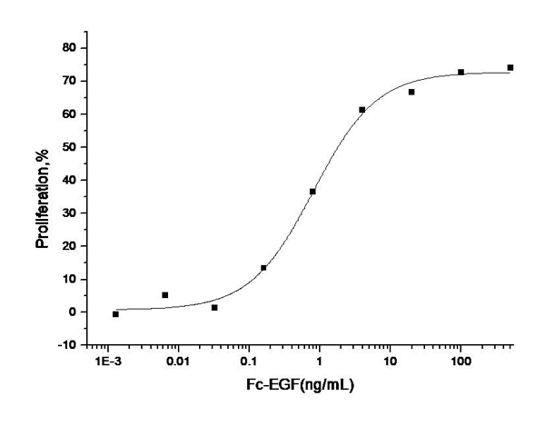
(Measured in a cell proliferation assay using Balb/C 3T3 mouse embryonic fibroblast cells. The ED50 for this effect is typically 0.3-1.5 ng/mL.)
Application Data
(Measured in a cell proliferation assay using Balb/C 3T3 mouse embryonic fibroblast cells. The ED50 for this effect is typically 0.3-1.5 ng/mL.)
EGF, Recombinant Protein (Cat# AAA257429)
 Application Data
(Measured by its binding ability in a functional ELISA. Immobilized human uPA at 5 ug/ml (100 ul/well) can bind mouse PLAUR with a linear ranger of 1.6-40 ng/ml.)
Application Data
(Measured by its binding ability in a functional ELISA. Immobilized human uPA at 5 ug/ml (100 ul/well) can bind mouse PLAUR with a linear ranger of 1.6-40 ng/ml.)
Urokinase/uPA, Recombinant Protein (Cat# AAA257433)
 Application Data
(Measured by its binding ability in a functional ELISA. Immobilized S100A8-His at 10 ug/ml (100 ul/well) can bind S100A9-His (Cat: 11145-H08B), The EC50 of S100A9-His (Cat: 11145-H08B) is 0.35-0.70 ug/mL.)
Application Data
(Measured by its binding ability in a functional ELISA. Immobilized S100A8-His at 10 ug/ml (100 ul/well) can bind S100A9-His (Cat: 11145-H08B), The EC50 of S100A9-His (Cat: 11145-H08B) is 0.35-0.70 ug/mL.)
S100A9, Recombinant Protein (Cat# AAA257436)
 Application Data
(Measured in antiviral assays using HepG2 cells infected with vesicular stomatitis virus. The ED50 for this effect is 1.7-6.8 ug/mL.)
Application Data
(Measured in antiviral assays using HepG2 cells infected with vesicular stomatitis virus. The ED50 for this effect is 1.7-6.8 ug/mL.)
IL28A, Recombinant Protein (Cat# AAA257439)
 Application Data
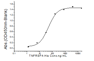
(Measured by its binding ability in a functional ELISA. Immobilized recombinant Human TNFRSF4-His (Cat:10481-H08H) at 10 ug/ml (100 ul/well) can bind human S4-mFc3-TNFSF4 (Cat:13127-H04H)with a linear range of 1.28-20 ug/ml.)
Application Data
(Measured by its binding ability in a functional ELISA. Immobilized recombinant Human TNFRSF4-His (Cat:10481-H08H) at 10 ug/ml (100 ul/well) can bind human S4-mFc3-TNFSF4 (Cat:13127-H04H)with a linear range of 1.28-20 ug/ml.)
OX40L/TNFSF4, Recombinant Protein (Cat# AAA257442)
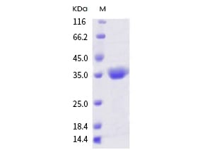
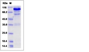
 SDS-PAGE
PRTN3 (203S/A, His), Recombinant Protein (Cat# AAA257443)
 Application Data
(Measured in antiviral assays using WISH human amnion cells infected with vesicular stomatitis virus(VSV). The EC50 for this effect is typically 0.6-4.2pg/mL.)
Application Data
(Measured in antiviral assays using WISH human amnion cells infected with vesicular stomatitis virus(VSV). The EC50 for this effect is typically 0.6-4.2pg/mL.)
Interferon alpha 2/IFNA2, Recombinant Protein (Cat# AAA257444)
 Application Data
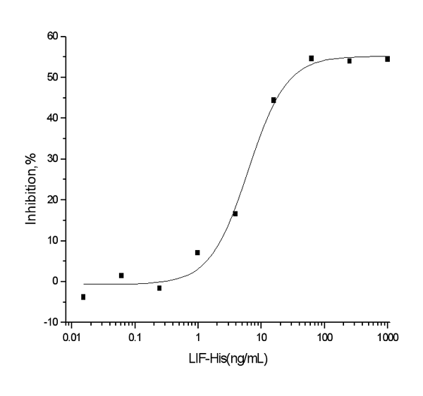
(Measured by its ability to inhibit the proliferation of M1 mouse myeloid leukemia cells. The ED50 for this effect is typically 2-15ng/mL.)
Application Data
(Measured by its ability to inhibit the proliferation of M1 mouse myeloid leukemia cells. The ED50 for this effect is typically 2-15ng/mL.)
LIF, Recombinant Protein (Cat# AAA257446)
 Application Data
COVID 19 Spike Coronavirus, Recombinant Protein (Cat# AAA257457)
 Application Data
(Measured by its binding ability in a functional ELISA. Immobilized human ACE2 protein (Fc tag)(10108-H05H) at 2ug/mL (100uL/well) can bind 2019-nCoV Spike Protein (RBD, His Tag) (40592-V08B), the EC50 of 2019-nCoV Spike Protein (RBD, His Tag) (40592-V08B) is 35-70 ng/mL.)
Application Data
(Measured by its binding ability in a functional ELISA. Immobilized human ACE2 protein (Fc tag)(10108-H05H) at 2ug/mL (100uL/well) can bind 2019-nCoV Spike Protein (RBD, His Tag) (40592-V08B), the EC50 of 2019-nCoV Spike Protein (RBD, His Tag) (40592-V08B) is 35-70 ng/mL.)
COVID 19 Spike Coronavirus, Recombinant Protein (Cat# AAA257459)
 Application Data
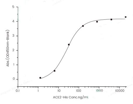
(Measured by its binding ability in a functional ELISA. Immobilized S3-2019-nCov-RBDï(40592-VNAH) at 2 ug/mL (100 uL/well) can bind ACE2 Protein, Human, Recombinant (mFc Tag) (10108-H05H), the EC50 of ACE2 Protein, Human, Recombinant (mFc Tag) (10108-H05H) is 20-80 ng/mL.)
Application Data
(Measured by its binding ability in a functional ELISA. Immobilized S3-2019-nCov-RBDï(40592-VNAH) at 2 ug/mL (100 uL/well) can bind ACE2 Protein, Human, Recombinant (mFc Tag) (10108-H05H), the EC50 of ACE2 Protein, Human, Recombinant (mFc Tag) (10108-H05H) is 20-80 ng/mL.)
COVID 19 Spike Coronavirus, Recombinant Protein (Cat# AAA257462)
 Application Data
(Measured by its ability to neutralize TGF-beta mediated inhibition on Mv-1-Lu cell proliferation. The ED50 for this effect is typically 1-8 ng/mL.)
Application Data
(Measured by its ability to neutralize TGF-beta mediated inhibition on Mv-1-Lu cell proliferation. The ED50 for this effect is typically 1-8 ng/mL.)
HGF, Recombinant Protein (Cat# AAA257466)
 Application Data
(Immobilized mouse Fc-TNFSF11 at 10 ug/ml (100 ul/well) can bind biotinylated human TNFRSF11B-His (Cat: 10271-H08H), The EC50 of biotinylated human TNFRSF11B-His (Cat: 10271-H08H) is 0.07-0.17 ug/ml.)
Application Data
(Immobilized mouse Fc-TNFSF11 at 10 ug/ml (100 ul/well) can bind biotinylated human TNFRSF11B-His (Cat: 10271-H08H), The EC50 of biotinylated human TNFRSF11B-His (Cat: 10271-H08H) is 0.07-0.17 ug/ml.)
RANKL, Recombinant Protein (Cat# AAA257470)
 Application Data
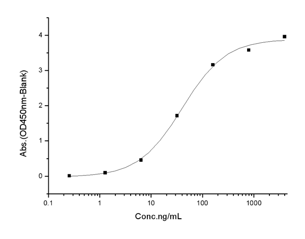
(Measured in a cell proliferation assay using TF-1 human erythroleukemic cells. The ED50 for this effect is 1-8ng/mL.)
Application Data
(Measured in a cell proliferation assay using TF-1 human erythroleukemic cells. The ED50 for this effect is 1-8ng/mL.)
NGF, Recombinant Protein (Cat# AAA257472)
 Application Data
(Measured in a cell proliferation assay using Balb/3T3 mouse embryonic fibroblasts. The ED50 for this effect is typically 0.5-3 ng/mL.)
Application Data
(Measured in a cell proliferation assay using Balb/3T3 mouse embryonic fibroblasts. The ED50 for this effect is typically 0.5-3 ng/mL.)
EGF, Recombinant Protein (Cat# AAA257473)
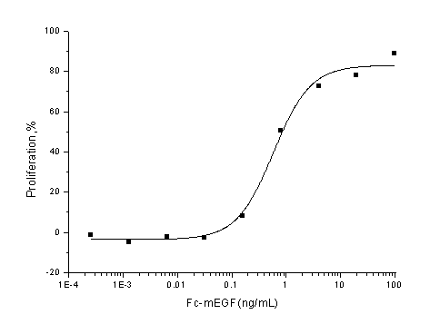
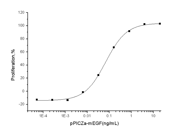 Application Data
(Measured in a cell proliferation assay using Balb/c 3T3 mouse embryonic fibroblast cells. The ED50 for this effect is typically 30-180 pg/mL.)
Application Data
(Measured in a cell proliferation assay using Balb/c 3T3 mouse embryonic fibroblast cells. The ED50 for this effect is typically 30-180 pg/mL.)
EGF, Recombinant Protein (Cat# AAA257474)
 Application Data
(Measured in antiviral assays using L929 cells infected with vesicular stomatitis virusï(VSV). The ED50 for this effect is typically 25-100 pg/mL.)
Application Data
(Measured in antiviral assays using L929 cells infected with vesicular stomatitis virusï(VSV). The ED50 for this effect is typically 25-100 pg/mL.)
Interferon alpha 2/IFNA2, Recombinant Protein (Cat# AAA257478)
 Application Data
(Measured in antiviral assay using L929 cells infected with vesicular stomatitisvirus (VSV). The ED50 for this effect is typically 10-80 pg/mL.)
Application Data
(Measured in antiviral assay using L929 cells infected with vesicular stomatitisvirus (VSV). The ED50 for this effect is typically 10-80 pg/mL.)
IFN-alpha 13, Recombinant Protein (Cat# AAA257483)
 Application Data
(Measured by its binding ability in a functional ELISA. Immobilized mouse PARP1 at 10 ug/mL (100 ul/well) can bind biotinylated human HSP70, The EC50 of biotinylated human HSP70 is 0.021 ug/mL.)
Application Data
(Measured by its binding ability in a functional ELISA. Immobilized mouse PARP1 at 10 ug/mL (100 ul/well) can bind biotinylated human HSP70, The EC50 of biotinylated human HSP70 is 0.021 ug/mL.)
PARP, Recombinant Protein (Cat# AAA257485)
What Are Recombinant Proteins?
A recombinant protein is created by inserting the gene of interest into a host system like E. coli, mammalian cells, or insect cells. This protein expression method results in the large-scale production of protein in a controlled environment with highly precise structures and functionality. Proteins produced via the “Recombinant” methodology have proven themselves crucial in various biological research areas, including immunology, neuroscience, cancer research, drug delivery and development, and more.
Core Recombinant Protein Product Lines:
- Human: Produced from human genes (e.g., growth factors, cytokines).
- Animal: Derived from mouse, rat, or other animals (e.g., mouse antibodies, enzymes).
- Bacterial: Produced in bacteria like E. coli (e.g., enzymes, bacterial toxins).
- Yeast: Produced in yeast cells (e.g., surface proteins, metabolic enzymes).
- Insect:Expressed in insect cells (e.g., viral proteins, glycoproteins).
- Viral: Derived from viral genomes (e.g., spike proteins for vaccine development).
How Does It Work?
- Gene Identification and Cloning: Scientists first find the exact gene that codes for the protein they are interested in producing, and then create a copy of said gene.
- Recombinant DNA Creation: The copied gene is then inserted into a vector (a vehicle-like plasmid) that can be transported safely into your intended expression host cell/system.
- Host Cell Transformation: The vector (with the foreign gene inside) is shuttled into a host cell. This host can be a bacterium (like E. coli), yeast, insect cells, or mammalian cells, depending on the type of protein or protein characteristics needed.
- Protein Expression:The host cell “reads” the copied gene that was shuttled into it, and begins to produce the gene’s coded-for protein using its own cellular machinery. The host can continue to multiply, and with each new cell, more protein is produced.
- Protein Purification:After enough protein has been made, scientists collect the host cell and separate out the target protein. The protein is then carefully purified so it is free of contaminants, ideally active, and also safe for use.
The result is a purified recombinant protein — a lab-made protein that is biologically identical (in most respects) to the natural/native version.
We take this process a step further, offering recombinant proteins that are produced with precise control over sequence modifications, expression levels, and large-scale production. This ensures high-quality, consistent results that will be able to support your research needs and empower your scientific discoveries.
Some of the expression hosts/systems used in recombinant protein generation by AAA Biotech include:
- Escherichia coli (E. coli)
- Human (Homo sapiens)
- Chinese Hamster
- Yeast
- Insect cells (e.g., Baculovirus expression system)
Advantages Of Our Recombinant Proteins:
Recombinant Protein Offerings
| S.No | Protein Expression Systems | Quality and Bioactivity | Formats and Flexibility |
|---|---|---|---|
| 01. | E. coli, HEK293, CHO, yeast, and insect cells | Validated high bioactivity and binding activity | Ready-to-use formats (lyophilized or liquid formulations available) |
| 02. | 675+ recombinant protein products | High protein purity (≥95%) | Available in flexible pack sizes |
| 03. | Custom protein production (His-tag, GST, FLAG, and Fc fusion) | Lot-to-lot consistency through QC protocols | Wide range of targets (Cytokines, growth factors, enzymes, receptors, signaling proteins, etc.) |
Applications Of Our Recombinant Proteins
- Studying protein interactions (protein–protein or protein–DNA).
- Testing biological pathways with functional/activity assays.
- Creating standard curves in ELISA and other tests.
- Making antigens/immunogens for antibody production.
- Drug discovery and screening.
- Finding and confirming biomarkers.
- Researching cell signaling and immunity.
- Developing vaccines.
Read more about the applications of the recombinant proteins here!
Why Buy Recombinant Proteins from AAA Biotech?
1. High Purity & Verified Quality:
Most of our proteins are ≥ 95% pure. We also test the biological activity of our products where it's relevant (it will be indicated on the product page).
2. Multiple Expression Systems:
We offer proteins made in multiple host systems, and this enables researchers to pick the system that gives them the best folding, post-translational modifications, or functionality for their experiment requirements.
3. Custom & Flexible Options:
Need a special tag (His, GST, FLAG, Fc fusion)? Or a custom variant? We can provide it. Our proteins come in formats that are lab-friendly: lyophilized (dry) or “ready-to-use” liquid.
4. Rigorous Quality Control & Documentation:
Every batch is backed by strong QC. This means you can rely on product consistency — two different batches will perform similarly.
5. Wide Variety & Global Availability:
We carry a large and growing catalog of recombinant proteins to cover almost all needs in research (immunology, oncology, vaccine work, etc.)
6. Researcher-Focused Support:
Technical support, clear documentation, and user-friendly product pages help you select the right protein for your work. Plus, our user-friendly packaging and handling, and global shipping are all designed to reduce delays, damage, and hassles.
Order Recombinant Proteins Today!
We are committed to supporting the research community with recombinant proteins that display exceptional performance and reliability. Our proteins are produced using industry-standard methods and are validated to meet the needs of academic, pharmaceutical, and biotechnology laboratories.
We have a track record of delivering proteins that work — large numbers of researchers rely on them in many published studies. Browse our catalog to find the perfect protein for your research applications now!
FAQ
1. What is involved in the purification of recombinant proteins?
The purification process of these recombinant proteins is performed to isolate the specific protein produced by the host cell from unwanted substances. The process generally includes techniques such as “affinity chromatography” to separate the protein from other proteins and impurities. AAA Biotech generally offers proteins ≥ 95% pure, depending on the protein type.
2. What are some examples of popular AAA Biotech recombinant proteins?
Some examples of recombinant proteins include:
Retinoblastoma Protein (Cat # AAA10852) – reportedly used in vaccine research and antibody development. Hepatitis B Surface Antigen (HBsAg) (Cat# AAA13446) – reportedly used in diagnostic tests and vaccine development for Hepatitis B. High-Mobility Group Box 1 (HMGB1) (Cat# AAA10851) – reportedly used in inflammation and immune response research.
3. How are AAA Biotech recombinant proteins validated?
Each batch undergoes stringent quality control checks, including SDS-PAGE analysis, endotoxin testing (for select products), and activity assaying (for select products). Certificates of Analysis are provided with every product (only upon request for some products).
4. Are your proteins suitable for therapeutic development or only research?
AAA Biotech recombinant proteins are strictly for research-use only and are not intended for diagnostic or therapeutic purposes in humans or animals.
5. What types of expression systems do you use for recombinant protein production?
The production labs use a variety of expression platforms, including bacterial (E. coli), yeast, insect (baculovirus), and mammalian (HEK293, CHO) systems. The expression system used depends on the complexity and intended function/use of the protein.