Filters
▼Clonality
▼Type
▼Reactivity
▼Gene Name
▼Isotype
▼Host
▼Application
▼Clone
▼Polyclonal Antibodies
At AAA Biotech also known as AAA Bio or AAABio, we provide a broad range of purified polyclonal antibodies (pAbs) that are able to all be browsed online through our website. Due to their high specificity and strong binding affinity, these antibodies are ideal for wide swathes of research and experimental applications.
Our polyclonal antibodies can easily support your work, whether you use them for Western Blotting, Immunocytochemistry (with or without Immunofluorescence used in conjunction), Immunohistochemistry, Immunoprecipitation, and ELISA tests. We highly encourage you to browse our range of pAbs and choose the one that best suits your experimental model.
Viewing 6750-6800 of 96805 product results
 IHC (Immunohiostchemistry)
(Immunohistochemistry of paraffin-embedded Rat brain tissue using UCH-L1 Polyclonal Antibody at dilution of 1:100.)
IHC (Immunohiostchemistry)
(Immunohistochemistry of paraffin-embedded Rat brain tissue using UCH-L1 Polyclonal Antibody at dilution of 1:100.)
UCH-L1, Polyclonal Antibody (Cat# AAA172160)
 IHC (Immunohiostchemistry)
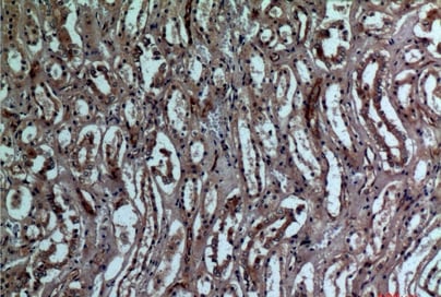
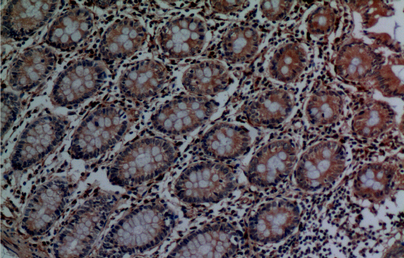
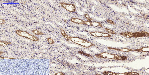
(Immunohistochemistry of paraffin-embedded Human kidney tissue using CD159a/c Polyclonal Antibody at dilution of 1:100.)
IHC (Immunohiostchemistry)
(Immunohistochemistry of paraffin-embedded Human kidney tissue using CD159a/c Polyclonal Antibody at dilution of 1:100.)
CD159a/c, Polyclonal Antibody (Cat# AAA172168)
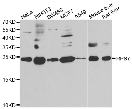
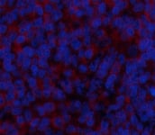
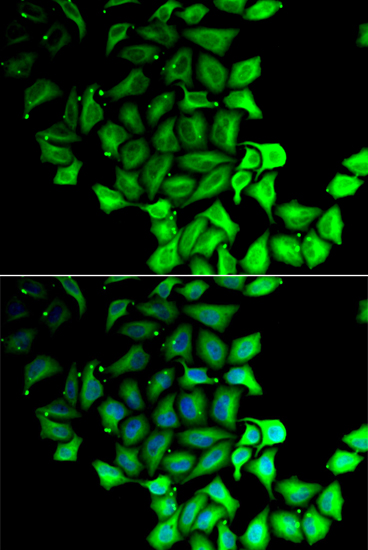 IF (Immunofluorescence)
(Immunofluorescence analysis of MCF-7 cell using RPS7 antibody. Blue: DAPI for nuclear staining.)
IF (Immunofluorescence)
(Immunofluorescence analysis of MCF-7 cell using RPS7 antibody. Blue: DAPI for nuclear staining.)
RPS7 , Polyclonal Antibody (Cat# AAA172170)

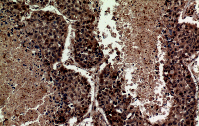
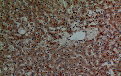
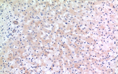 IHC (Immunohiostchemistry)
(Immunohistochemistry of paraffin-embedded Human liver tissue using CD166 Polyclonal Antibody at dilution of 1:100.)
IHC (Immunohiostchemistry)
(Immunohistochemistry of paraffin-embedded Human liver tissue using CD166 Polyclonal Antibody at dilution of 1:100.)
CD166, Polyclonal Antibody (Cat# AAA172177)
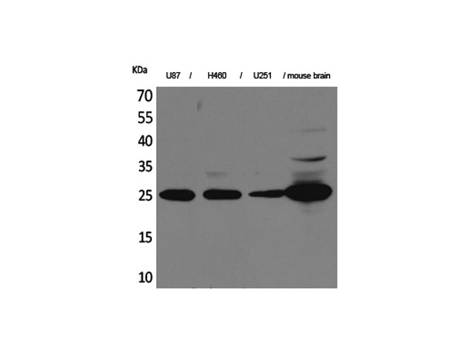
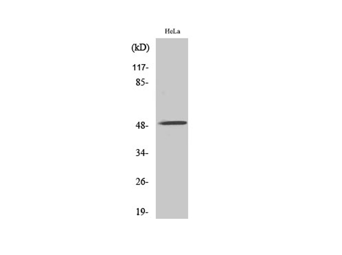
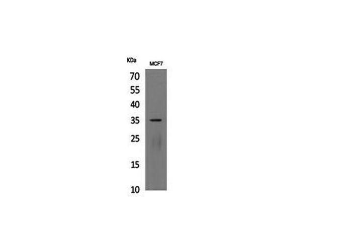
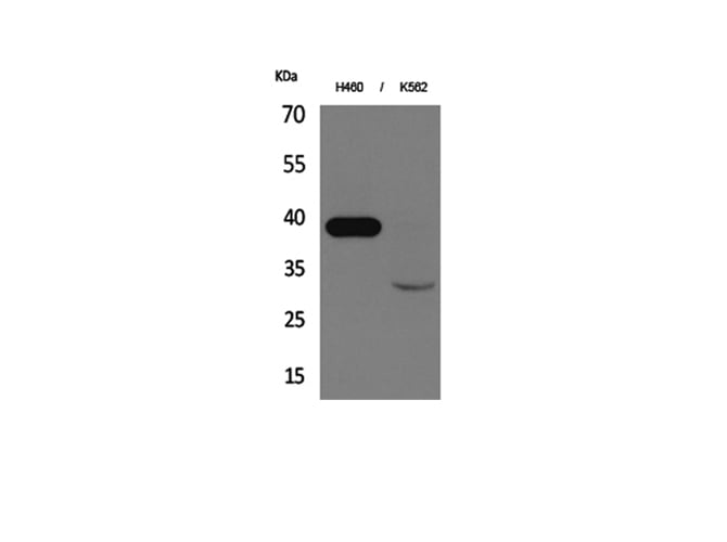
 WB (Western Blot)
(Western Blot analysis of various cells with PAR-4 Polyclonal Antibody.)
WB (Western Blot)
(Western Blot analysis of various cells with PAR-4 Polyclonal Antibody.)
PAR-4, Polyclonal Antibody (Cat# AAA172181)
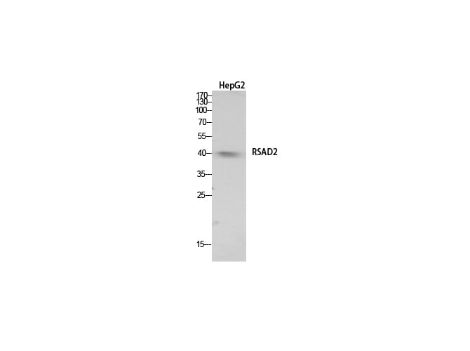
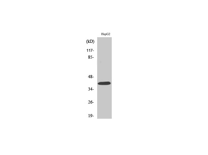
 IHC (Immunohiostchemistry)
(Immunohistochemistry of paraffin-embedded Human prostate cancer tissue using RSAD2 Polyclonal Antibody at dilution of 1:100.)
IHC (Immunohiostchemistry)
(Immunohistochemistry of paraffin-embedded Human prostate cancer tissue using RSAD2 Polyclonal Antibody at dilution of 1:100.)
RSAD2, Polyclonal Antibody (Cat# AAA172185)
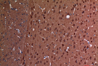
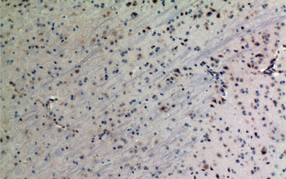
 IHC (Immunohiostchemistry)
(Immunohistochemistry of paraffin-embedded Rat brain tissue using Apelin Polyclonal Antibody at dilution of 1:100.)
IHC (Immunohiostchemistry)
(Immunohistochemistry of paraffin-embedded Rat brain tissue using Apelin Polyclonal Antibody at dilution of 1:100.)
Apelin, Polyclonal Antibody (Cat# AAA172186)
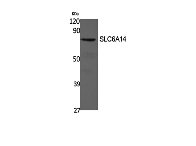
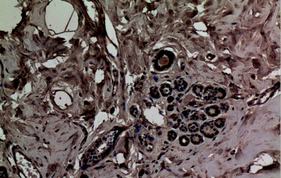 IHC (Immunohiostchemistry)
(Immunohistochemistry of paraffin-embedded Human breast tissue using SLC6A14 Polyclonal Antibody at dilution of 1:100.)
IHC (Immunohiostchemistry)
(Immunohistochemistry of paraffin-embedded Human breast tissue using SLC6A14 Polyclonal Antibody at dilution of 1:100.)
SLC6A14, Polyclonal Antibody (Cat# AAA172187)
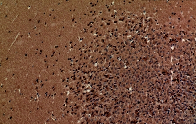
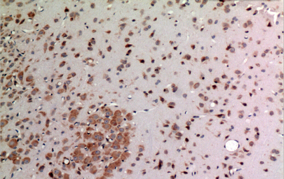
 IHC (Immunohiostchemistry)
(Immunohistochemistry of paraffin-embedded Rat brain tissue using HSP 90 Polyclonal Antibody at dilution of 1:100.)
IHC (Immunohiostchemistry)
(Immunohistochemistry of paraffin-embedded Rat brain tissue using HSP 90 Polyclonal Antibody at dilution of 1:100.)
HSP 90, Polyclonal Antibody (Cat# AAA172190)
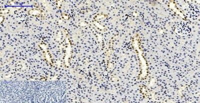
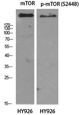
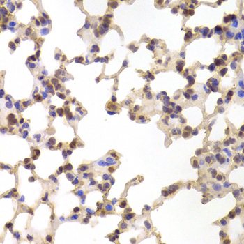
 IF (Immunofluorescence)
(Immunofluorescence analysis of Rat lung tissue with Phospho-mTOR (Ser2448) Polyclonal Antibody at dilution of 1:200)
IF (Immunofluorescence)
(Immunofluorescence analysis of Rat lung tissue with Phospho-mTOR (Ser2448) Polyclonal Antibody at dilution of 1:200)
mTOR, Polyclonal Antibody (Cat# AAA172193)
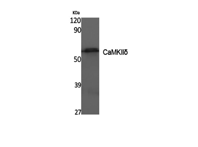
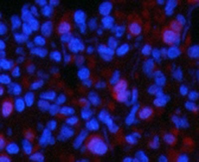
 IHC (Immunohiostchemistry)
(Immunohistochemistry of paraffin-embedded Mouse brain tissue using CaMKII? Polyclonal Antibody at dilution of 1:100.)
IHC (Immunohiostchemistry)
(Immunohistochemistry of paraffin-embedded Mouse brain tissue using CaMKII? Polyclonal Antibody at dilution of 1:100.)
CaMKIIdelta, Polyclonal Antibody (Cat# AAA172194)

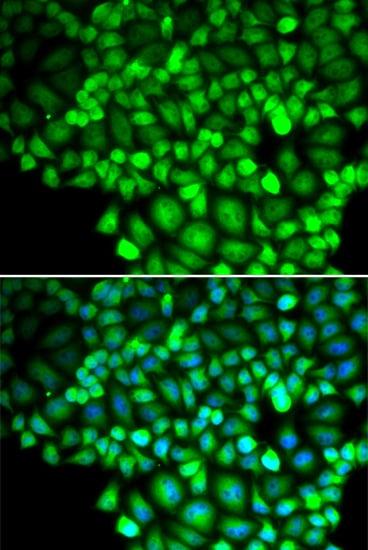 IF (Immunofluorescence)
(Immunofluorescence analysis of HeLa cell using ALOX15B antibody. Blue: DAPI for nuclear staining.)
IF (Immunofluorescence)
(Immunofluorescence analysis of HeLa cell using ALOX15B antibody. Blue: DAPI for nuclear staining.)
ALOX15B, Polyclonal Antibody (Cat# AAA172197)
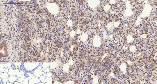
 IHC (Immunohiostchemistry)
(Immunohistochemistry of paraffin-embedded Human pancreas tissue using CD1D Polyclonal Antibody at dilution of 1:100.)
IHC (Immunohiostchemistry)
(Immunohistochemistry of paraffin-embedded Human pancreas tissue using CD1D Polyclonal Antibody at dilution of 1:100.)
CD1D, Polyclonal Antibody (Cat# AAA172205)
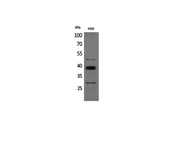
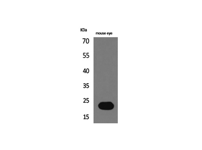
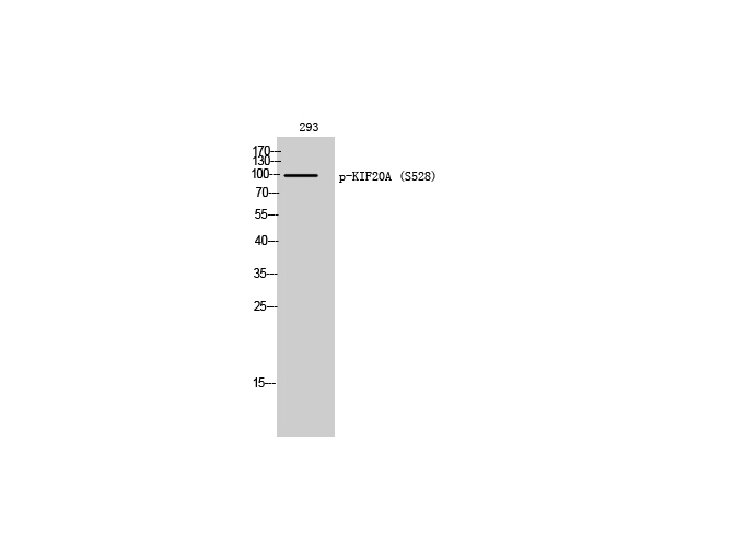 WB (Western Blot)
(Western Blot analysis of 293T cells with Phospho-KIF20A (Ser528) Polyclonal Antibody)
WB (Western Blot)
(Western Blot analysis of 293T cells with Phospho-KIF20A (Ser528) Polyclonal Antibody)
KIF20A, Polyclonal Antibody (Cat# AAA172206)
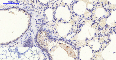
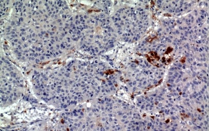 IHC (Immunohistochemisry)
(Immunohistochemistry of paraffin-embedded Human lung tissue using CD163b Polyclonal Antibody at dilution of 1:100.)
IHC (Immunohistochemisry)
(Immunohistochemistry of paraffin-embedded Human lung tissue using CD163b Polyclonal Antibody at dilution of 1:100.)
CD163b, Polyclonal Antibody (Cat# AAA172207)
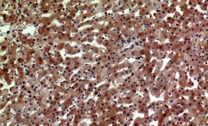
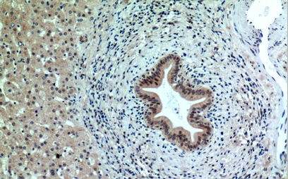 IHC (Immunohiostchemistry)
(Immunohistochemistry of paraffin-embedded Human liver tissue using PPP1R15B Polyclonal Antibody at dilution of 1:100.)
IHC (Immunohiostchemistry)
(Immunohistochemistry of paraffin-embedded Human liver tissue using PPP1R15B Polyclonal Antibody at dilution of 1:100.)
PPP1R15B, Polyclonal Antibody (Cat# AAA172208)
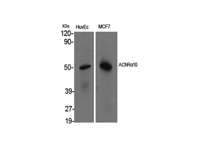
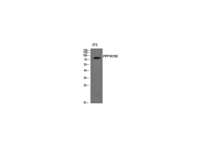
 WB (Western Blot)
(Western Blot analysis of Hela cells with AChR?10 Polyclonal Antibody.)
WB (Western Blot)
(Western Blot analysis of Hela cells with AChR?10 Polyclonal Antibody.)
AChRalpha10, Polyclonal Antibody (Cat# AAA172212)
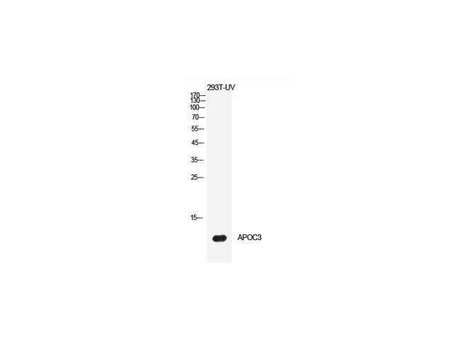
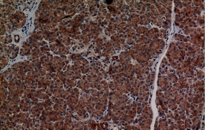
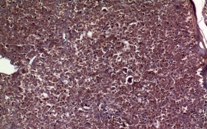 IHC (Immunohiostchemistry)
(Immunohistochemistry of paraffin-embedded Human pancreas tissue using ApoC-III Polyclonal Antibody at dilution of 1:100.)
IHC (Immunohiostchemistry)
(Immunohistochemistry of paraffin-embedded Human pancreas tissue using ApoC-III Polyclonal Antibody at dilution of 1:100.)
ApoC-III, Polyclonal Antibody (Cat# AAA172213)
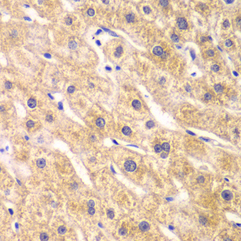
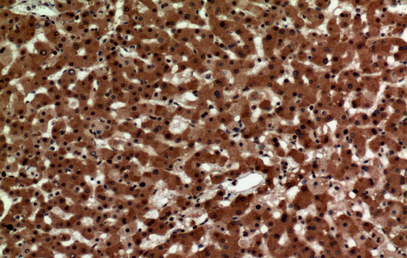
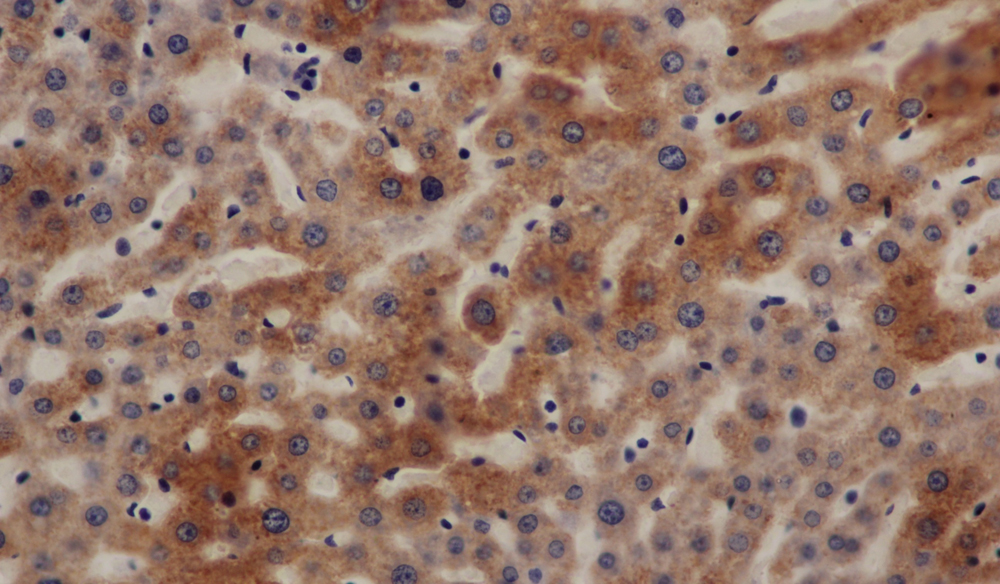 IHC (Immunohiostchemistry)
(Immunohistochemistry of paraffin-embedded Mouse liver using ATP5A antibody at dilution of 1:50)
IHC (Immunohiostchemistry)
(Immunohistochemistry of paraffin-embedded Mouse liver using ATP5A antibody at dilution of 1:50)
ATP5A, Polyclonal Antibody (Cat# AAA172222)
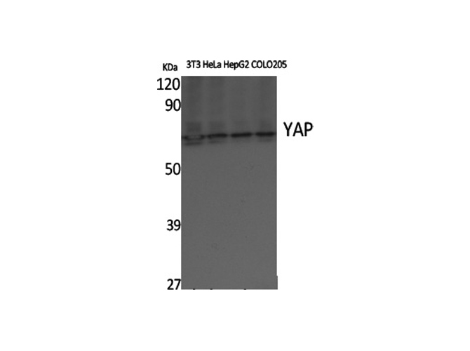
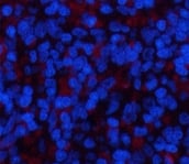 IF (Immunofluorescence)
(Immunofluorescence analysis of Rat spleen tissue using YAP Polyclonal Antibody at dilution of 1:200.)
IF (Immunofluorescence)
(Immunofluorescence analysis of Rat spleen tissue using YAP Polyclonal Antibody at dilution of 1:200.)
YAP, Polyclonal Antibody (Cat# AAA172227)
 IHC (Immunohiostchemistry)
(Immunohistochemistry of paraffin-embedded Mouse brain tissue using Cyclophilin D Polyclonal Antibody at dilution of 1:100.)
IHC (Immunohiostchemistry)
(Immunohistochemistry of paraffin-embedded Mouse brain tissue using Cyclophilin D Polyclonal Antibody at dilution of 1:100.)
Cyclophilin D, Polyclonal Antibody (Cat# AAA172231)
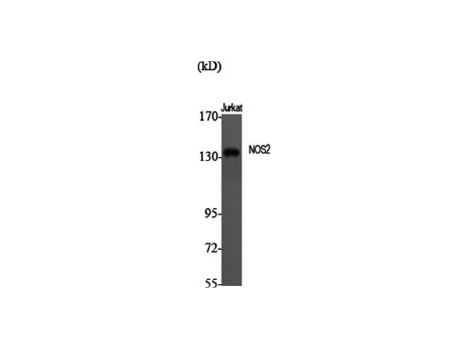
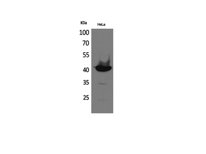
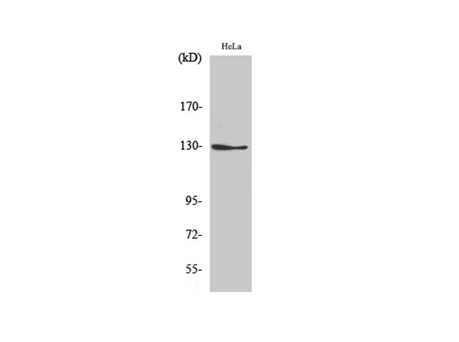 WB (Western Blot)
(Western Blot analysis of Hela cells using NOS2 Polyclonal Antibody at dilution of 1:500.)
WB (Western Blot)
(Western Blot analysis of Hela cells using NOS2 Polyclonal Antibody at dilution of 1:500.)
NOS2, Polyclonal Antibody (Cat# AAA172234)
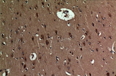 IHC (Immunohistochemisry)
(Immunohistochemistry of paraffin-embedded Human brain tissue using BNIP-3 Polyclonal Antibody at dilution of 1:100.)
IHC (Immunohistochemisry)
(Immunohistochemistry of paraffin-embedded Human brain tissue using BNIP-3 Polyclonal Antibody at dilution of 1:100.)
BNIP-3, Polyclonal Antibody (Cat# AAA172239)
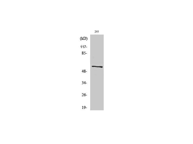
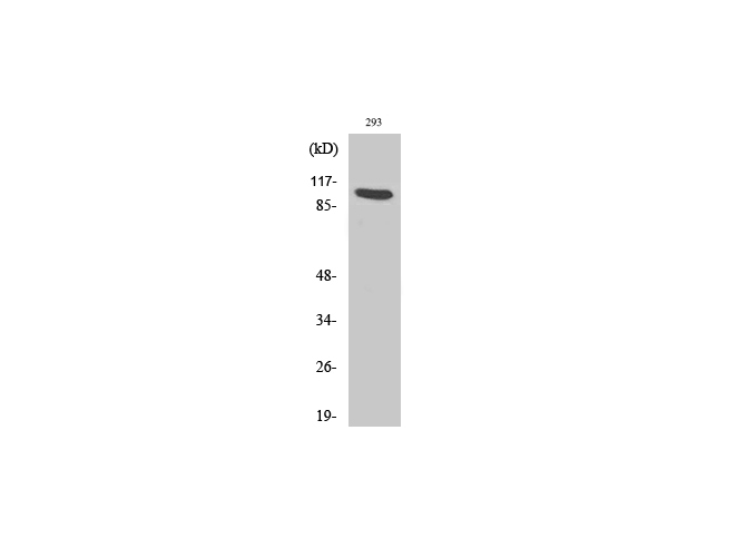 WB (Western Blot)
(Western Blot analysis of 293 cells with AHR/AHRR Polyclonal Antibody)
WB (Western Blot)
(Western Blot analysis of 293 cells with AHR/AHRR Polyclonal Antibody)
Ah Receptor, Polyclonal Antibody (Cat# AAA172240)
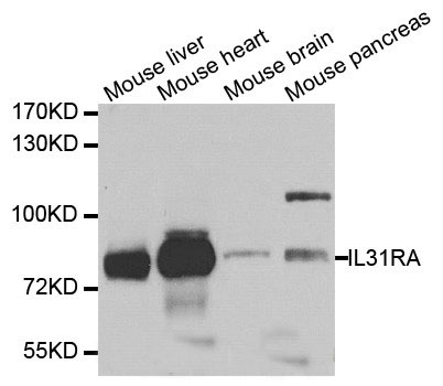 WB (Western Blot)
(Western blot analysis of extracts of various cell lines, using IL31RA antibody.)
WB (Western Blot)
(Western blot analysis of extracts of various cell lines, using IL31RA antibody.)
IL31RA, Polyclonal Antibody (Cat# AAA172325)
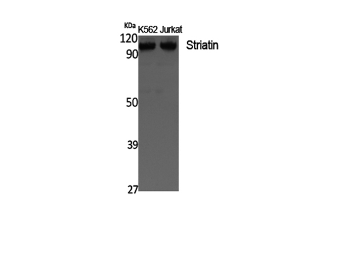
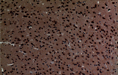 IHC (Immunohiostchemistry)
(Immunohistochemistry of paraffin-embedded Mouse brain tissue using Striatin Polyclonal Antibody at dilution of 1:100.)
IHC (Immunohiostchemistry)
(Immunohistochemistry of paraffin-embedded Mouse brain tissue using Striatin Polyclonal Antibody at dilution of 1:100.)
Striatin, Polyclonal Antibody (Cat# AAA172329)
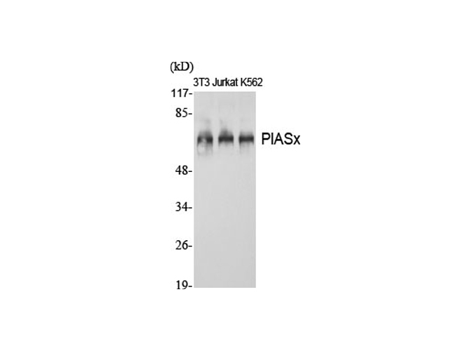
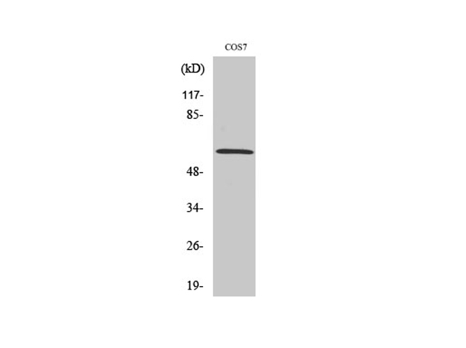 WB (Western Blot)
(Western Blot analysis of COS-7 cells using PIASx Polyclonal Antibody at dilution of 1:1000.)
WB (Western Blot)
(Western Blot analysis of COS-7 cells using PIASx Polyclonal Antibody at dilution of 1:1000.)
PIASx, Polyclonal Antibody (Cat# AAA172331)
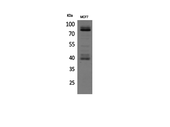
 WB (Western Blot)
(Western Blot analysis of MCF7, 4T1 cells with CD36 Polyclonal Antibody.)
WB (Western Blot)
(Western Blot analysis of MCF7, 4T1 cells with CD36 Polyclonal Antibody.)
CD36, Polyclonal Antibody (Cat# AAA172338)
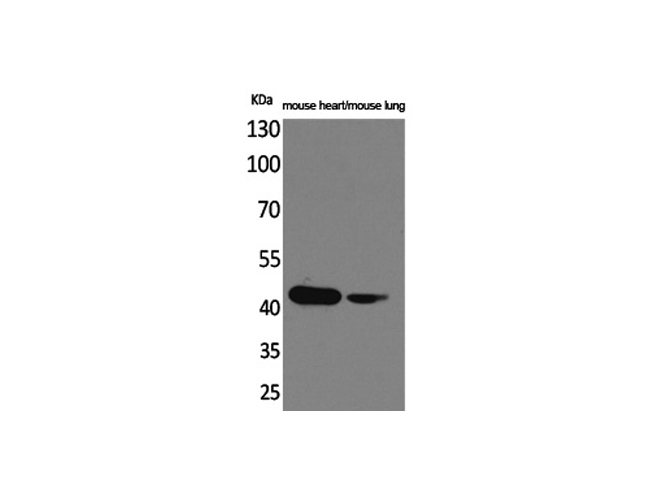
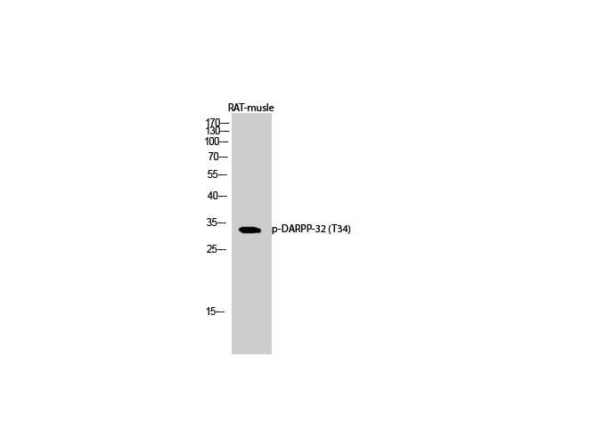 WB (Western Blot)
(Western Blot analysis of Rat musle with Phospho-DARPP-32 (Thr34) Polyclonal Antibody at dilution of 1:1000)
WB (Western Blot)
(Western Blot analysis of Rat musle with Phospho-DARPP-32 (Thr34) Polyclonal Antibody at dilution of 1:1000)
DARPP-32, Polyclonal Antibody (Cat# AAA172345)

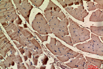
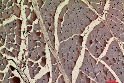
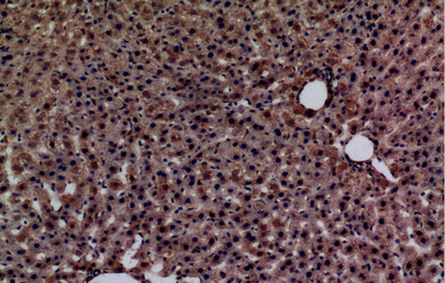 IHC (Immunohiostchemistry)
(Immunohistochemistry of paraffin-embedded Rat liver using Glypican-3 antibody at dilution of 1:100)
IHC (Immunohiostchemistry)
(Immunohistochemistry of paraffin-embedded Rat liver using Glypican-3 antibody at dilution of 1:100)
Glypican-3, Polyclonal Antibody (Cat# AAA172346)
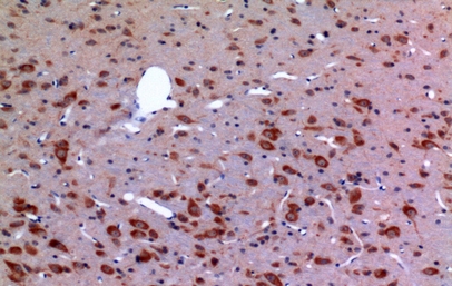
 IHC (Immunohiostchemistry)
(Immunohistochemistry of paraffin-embedded Human brain tissue using IL-1RI Polyclonal Antibody at dilution of 1:100.)
IHC (Immunohiostchemistry)
(Immunohistochemistry of paraffin-embedded Human brain tissue using IL-1RI Polyclonal Antibody at dilution of 1:100.)
IL-1RI, Polyclonal Antibody (Cat# AAA172347)
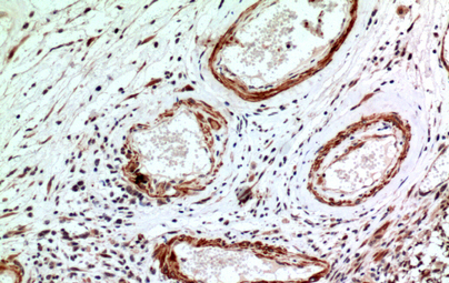
 IHC (Immunohistochemisry)
(Immunohistochemistry of paraffin-embedded Rat muscle tissue using Actin-? cardiac muscle Polyclonal Antibody at dilution of 1:100.)
IHC (Immunohistochemisry)
(Immunohistochemistry of paraffin-embedded Rat muscle tissue using Actin-? cardiac muscle Polyclonal Antibody at dilution of 1:100.)
Actin-alpha cardiac muscle, Polyclonal Antibody (Cat# AAA172352)
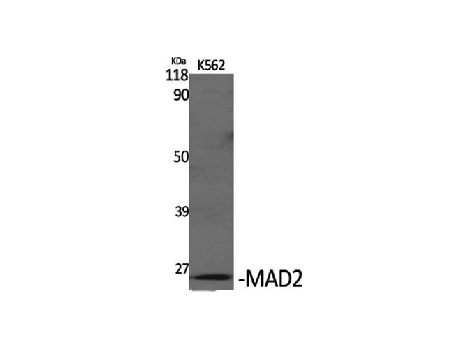
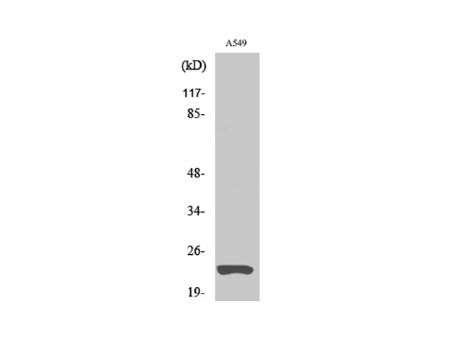 WB (Western Blot)
(Western Blot analysis of A549 cells using MAD2 Polyclonal Antibody at dilution of 1:1000.)
WB (Western Blot)
(Western Blot analysis of A549 cells using MAD2 Polyclonal Antibody at dilution of 1:1000.)
MAD2, Polyclonal Antibody (Cat# AAA172353)
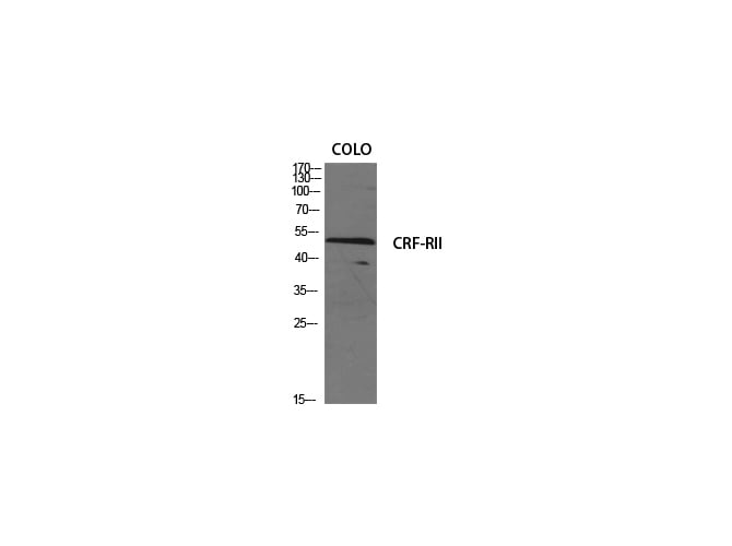 WB (Western Blot)
(Western Blot analysis of various cells using CRF-RII Polyclonal Antibody at dilution of 1:500.)
WB (Western Blot)
(Western Blot analysis of various cells using CRF-RII Polyclonal Antibody at dilution of 1:500.)
CRF-RII, Polyclonal Antibody (Cat# AAA172354)
 IHC (Immunohistochemisry)
(Immunohistochemistry of paraffin-embedded Human lung tissue using IL-12A p35 Polyclonal Antibody at dilution of 1:100.)
IHC (Immunohistochemisry)
(Immunohistochemistry of paraffin-embedded Human lung tissue using IL-12A p35 Polyclonal Antibody at dilution of 1:100.)
IL-12A p35, Polyclonal Antibody (Cat# AAA172360)
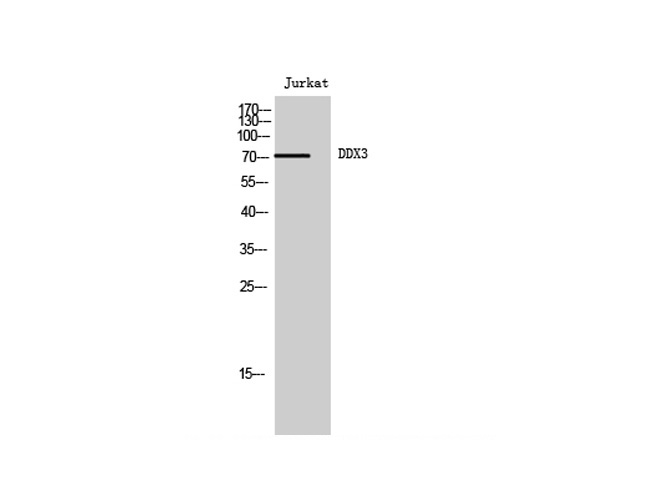
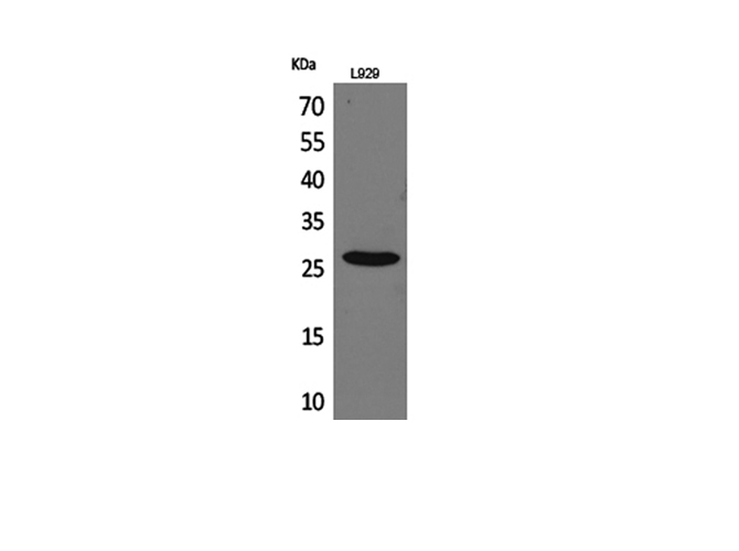
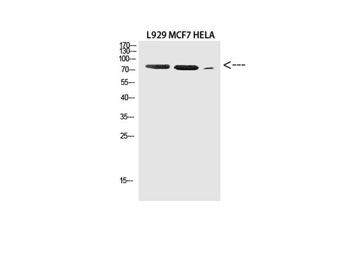 WB (Western Blot)
(Western Blot analysis of L929, MCF-7, Hela cells using DDX3 Polyclonal Antibody at dilution of 1:2000.)
WB (Western Blot)
(Western Blot analysis of L929, MCF-7, Hela cells using DDX3 Polyclonal Antibody at dilution of 1:2000.)
DDX3, Polyclonal Antibody (Cat# AAA172361)
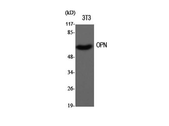 WB (Western Blot)
(Western Blot analysis of 3T3 cells using SPP1 Polyclonal Antibody at dilution of 1:500.)
WB (Western Blot)
(Western Blot analysis of 3T3 cells using SPP1 Polyclonal Antibody at dilution of 1:500.)
OPN, Polyclonal Antibody (Cat# AAA172368)
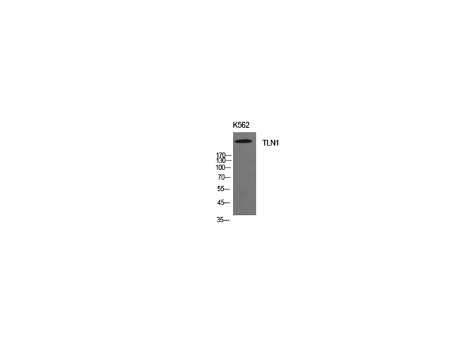
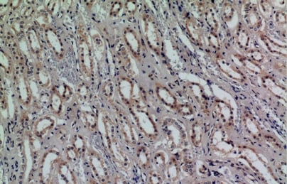
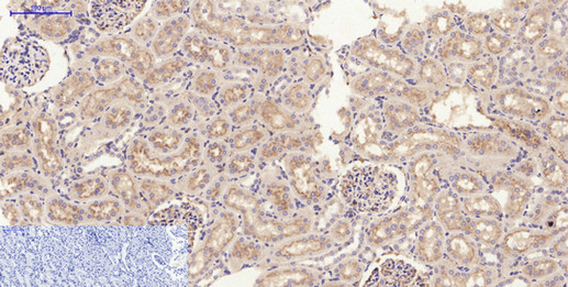
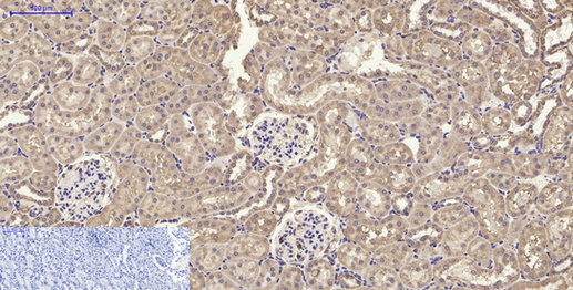
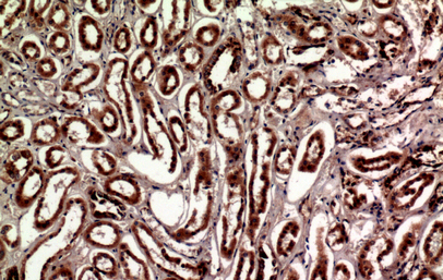 IHC (Immunohiostchemistry)
(Immunohistochemistry of paraffin-embedded Human kidney tissue using Talin-1 Polyclonal Antibody at dilution of 1:100.)
IHC (Immunohiostchemistry)
(Immunohistochemistry of paraffin-embedded Human kidney tissue using Talin-1 Polyclonal Antibody at dilution of 1:100.)
Talin-1, Polyclonal Antibody (Cat# AAA172371)
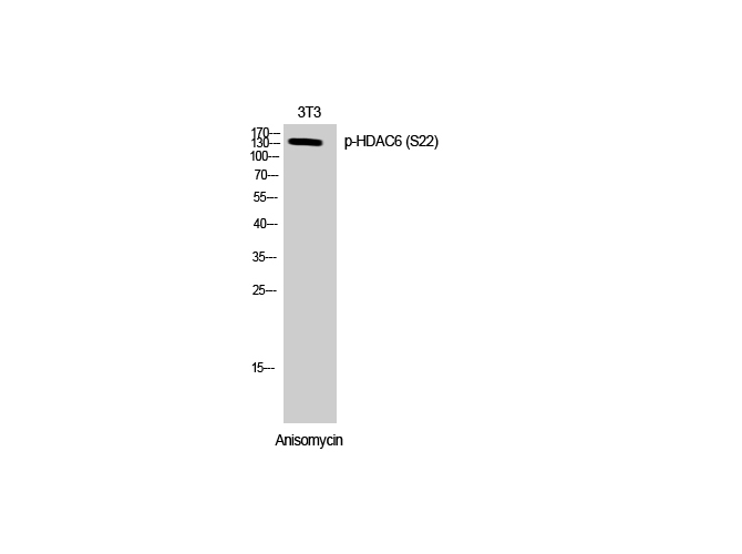 WB (Western Blot)
(Western Blot analysis of 3T3 cells with Phospho-HDAC6 (Ser22) Polyclonal Antibody at dilution of 1:500)
WB (Western Blot)
(Western Blot analysis of 3T3 cells with Phospho-HDAC6 (Ser22) Polyclonal Antibody at dilution of 1:500)
HDAC6, Polyclonal Antibody (Cat# AAA172376)
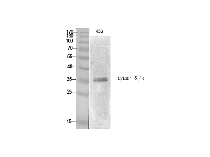
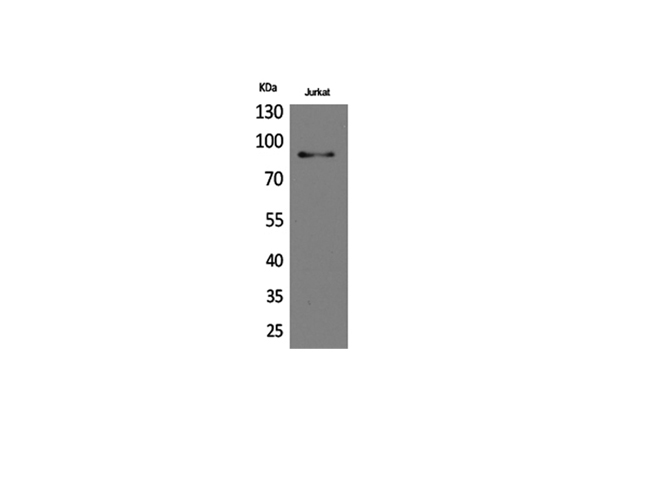
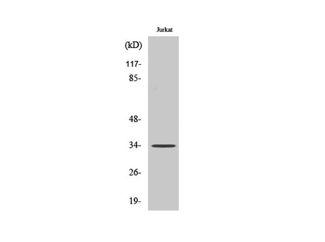 WB (Western Blot)
(Western Blot analysis of Jurkat cells with C/EBP ? Polyclonal Antibody.)
WB (Western Blot)
(Western Blot analysis of Jurkat cells with C/EBP ? Polyclonal Antibody.)
C/EBP epsilon, Polyclonal Antibody (Cat# AAA172378)
 IF (Immunofluorescence)
(Immunofluorescence analysis of Rat spleen tissue using JAK2 Polyclonal Antibody at dilution of 1:200.)
IF (Immunofluorescence)
(Immunofluorescence analysis of Rat spleen tissue using JAK2 Polyclonal Antibody at dilution of 1:200.)
JAK2, Polyclonal Antibody (Cat# AAA172385)
 IHC (Immunohistochemisry)
(Immunohistochemistry of paraffin-embedded Human liver tissue using Cathepsin L Polyclonal Antibody at dilution of 1:100.)
IHC (Immunohistochemisry)
(Immunohistochemistry of paraffin-embedded Human liver tissue using Cathepsin L Polyclonal Antibody at dilution of 1:100.)
Cathepsin L, Polyclonal Antibody (Cat# AAA172389)

 IF (Immunofluorescence)
(Immunofluorescence analysis of Rat spleen tissue with Phospho-Stat1 (Tyr701) Polyclonal Antibody at dilution of 1:200)
IF (Immunofluorescence)
(Immunofluorescence analysis of Rat spleen tissue with Phospho-Stat1 (Tyr701) Polyclonal Antibody at dilution of 1:200)
Stat1, Polyclonal Antibody (Cat# AAA172394)
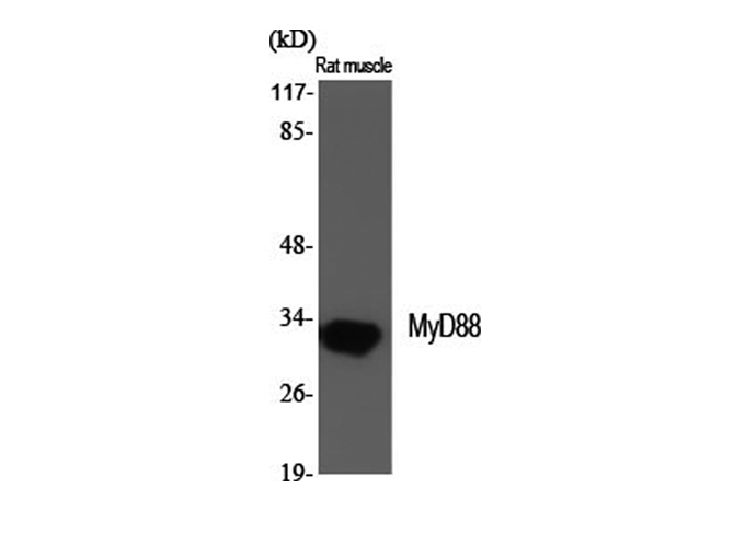
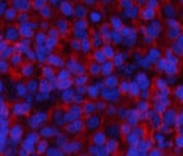
 IF (Immunofluorescence)
(Immunofluorescence analysis of Mouse spleen tissue using MyD88 Polyclonal Antibody at dilution of 1:200.)
IF (Immunofluorescence)
(Immunofluorescence analysis of Mouse spleen tissue using MyD88 Polyclonal Antibody at dilution of 1:200.)
MyD88, Polyclonal Antibody (Cat# AAA172395)
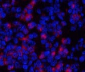
 IF (Immunofluorescence)
(Immunofluorescence analysis of Human liver tissue using JNK1/2/3 Polyclonal Antibody at dilution of 1:200.)
IF (Immunofluorescence)
(Immunofluorescence analysis of Human liver tissue using JNK1/2/3 Polyclonal Antibody at dilution of 1:200.)
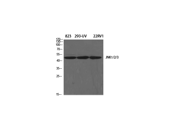
JNK1/2/3, Polyclonal Antibody (Cat# AAA172396)
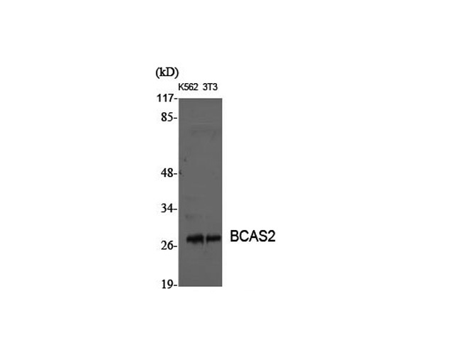
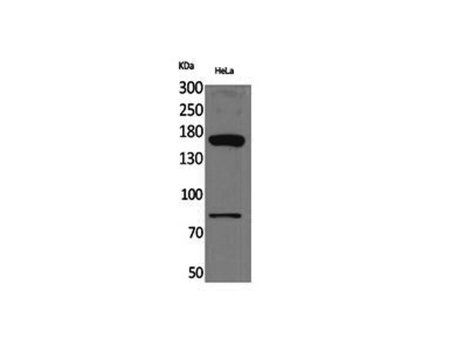
 WB (Western Blot)
(Western Blot analysis of Hela cells using BCAS2 Polyclonal Antibody at dilution of 1:1000.)
WB (Western Blot)
(Western Blot analysis of Hela cells using BCAS2 Polyclonal Antibody at dilution of 1:1000.)
BCAS2, Polyclonal Antibody (Cat# AAA172401)
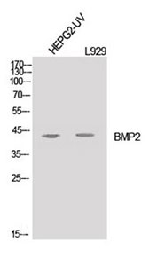
 IF (Immunofluorescence)
(Immunofluorescence analysis of Rat spleen tissue using BMP-2 Polyclonal Antibody at dilution of 1:200.)
IF (Immunofluorescence)
(Immunofluorescence analysis of Rat spleen tissue using BMP-2 Polyclonal Antibody at dilution of 1:200.)
BMP-2, Polyclonal Antibody (Cat# AAA172403)
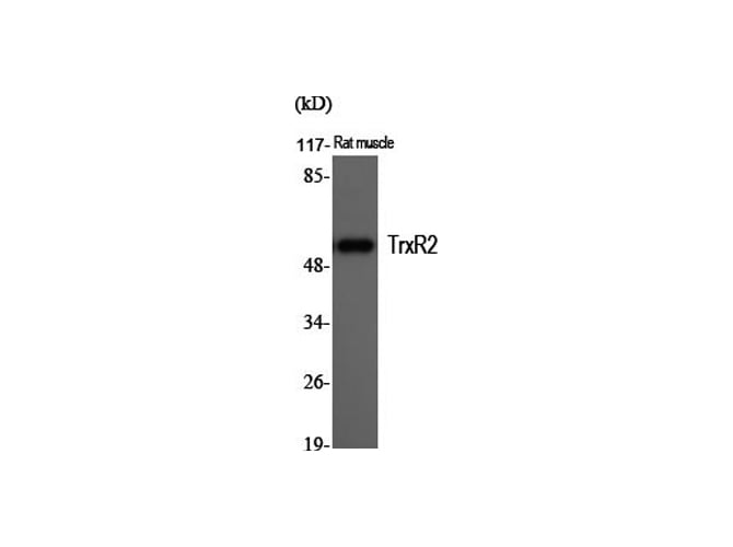
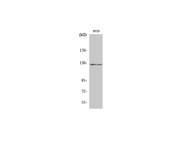
 WB (Western Blot)
(Western Blot analysis of HT29 cells using TrxR2 Polyclonal Antibody at dilution of 1:20000.)
WB (Western Blot)
(Western Blot analysis of HT29 cells using TrxR2 Polyclonal Antibody at dilution of 1:20000.)
TrxR2, Polyclonal Antibody (Cat# AAA172556)
 IHC (Immunohiostchemistry)
(Immunohistochemistry of paraffin-embedded Rat brain tissue using FGF-20 Polyclonal Antibody at dilution of 1:100.)
IHC (Immunohiostchemistry)
(Immunohistochemistry of paraffin-embedded Rat brain tissue using FGF-20 Polyclonal Antibody at dilution of 1:100.)
FGF-20, Polyclonal Antibody (Cat# AAA172557)
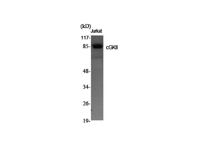
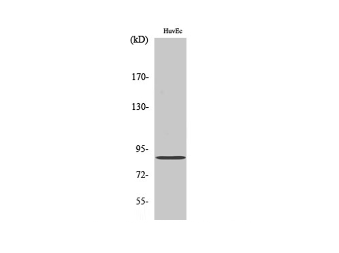 WB (Western Blot)
(Western Blot analysis of HuvEc cells with cGKII Polyclonal Antibody.)
WB (Western Blot)
(Western Blot analysis of HuvEc cells with cGKII Polyclonal Antibody.)
cGKII, Polyclonal Antibody (Cat# AAA172560)
What are Polyclonal Antibodies?
Polyclonal antibodies are antibodies that come from multiple B cell clones of a host animal. The typical hosts used for the majority of polyclonal antibody production are rabbits, goats, sheep, and donkeys. These polyclonal antibodies, once having identified their target, will bind to different epitopes located at different regions or sequences on the same protein/antigen. As a result, they are ideal at locating and binding to the target, even if the target is in very low concentrations (due to many different antibodies being able to bind to the same target molecule, which allows for significant amplification of a downstream signal).
Polyclonal antibodies are typically produced by injecting an antigen into a host animal, which causes the animal’s immune system to attack the foreign antigen by mass generating antibodies against it. After a period of time, serum is collected from the animal and purified using physicochemical fractionation, class-specific affinity purification, and/or antigen-affinity purification.
Key Uses of Polyclonal Antibodies
- Western Blotting: This method is used to find specific proteins in biological samples after separating them by size.
- Immunohistochemistry: IHC helps visualize the location of proteins in tissue sections using various staining techniques.
- ELISA: (Enzyme-Linked Immunosorbent Assay) is typically used to identify specific protein quantities in a sample. ELISAs can be either “Quantitative” or “Qualitative”.
- Flow Cytometry: technique that identifies and measures the specific protein on the surface or inside the cells in a fluid suspension.
- Immunoprecipitation: IP isolates and studies a specific protein from a complex mixture using antibodies.
Why Buy Polyclonal Antibodies from AAA Biotech?
1. Ideal for Various Applications
Our antibodies are generally going to be validated for use in multiple types of assays, including ELISA, Western Blotting, Immunohistochemistry, Immunoprecipitation, amongst others. They are ideal for a wide range of research applications.
2. Rigorous Quality Control
All of the antibodies in our catalog undergo strict quality testing to ensure specificity, sensitivity, and consistent performance. We are confident in the ability of our antibodies to provide you with accurate results.
3. Wide Assortment of Antibodies
Antibodies in are catalog can be found for both common and exotic species, and these antibodies are also available in both conjugated and recombinant forms to suit many diverse experimental needs.
4. Highly Purified
Our antibodies are available in purified forms with over 85% purity, as confirmed by SDS-PAGE. They are also available with tags such as His, Flag, GST, or MBP. We cater to customers worldwide.
FAQ
1. How are polyclonal antibodies produced?
Traditionally, polyclonal antibodies are produced by injecting an antigen into a host animal (such as a rabbit or goat), which then triggers an immune response from the host animal. The animal’s B cells produce antibodies that will recognize different parts of the injected antigen. These antibodies are then collected from the animal’s blood and purified for use.
2. How do polyclonal antibodies differ from monoclonal antibodies?
Polyclonal antibodies are a mix of antibodies that bind to different locations (epitopes) of the same antigen, while monoclonal antibodies are identical and bind to just one specific epitope. This makes polyclonal antibodies more versatile and better at detecting proteins that may be present in low quantities or in altered/modified forms.
3. How should I store polyclonal antibodies?
Polyclonal antibodies should be stored at 4°C for short-term use (up to a few weeks) and at -20°C or -80°C for long-term storage. Avoid repeated freeze-thaw cycles by dividing them into small aliquots. Always check the datasheet for specific storage instructions.