Filters
▼Clonality
▼Type
▼Reactivity
▼Gene Name
▼Isotype
▼Host
▼Application
▼Clone
▼Polyclonal Antibodies
At AAA Biotech also known as AAA Bio or AAABio, we provide a broad range of purified polyclonal antibodies (pAbs) that are able to all be browsed online through our website. Due to their high specificity and strong binding affinity, these antibodies are ideal for wide swathes of research and experimental applications.
Our polyclonal antibodies can easily support your work, whether you use them for Western Blotting, Immunocytochemistry (with or without Immunofluorescence used in conjunction), Immunohistochemistry, Immunoprecipitation, and ELISA tests. We highly encourage you to browse our range of pAbs and choose the one that best suits your experimental model.
Viewing 6700-6750 of 96805 product results
 WB (Western Blot)
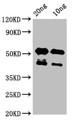
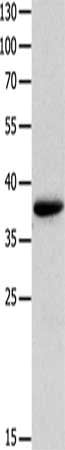
(Western BlotPositive WB detected in Recombinant proteinAll lanes: lats2 at 3ug/mlSecondaryGoat polyclonal to rabbit IgG at 1/50000 dilutionPredicted band size: 43 kDaObserved band size: 43,53 kDa)
WB (Western Blot)
(Western BlotPositive WB detected in Recombinant proteinAll lanes: lats2 at 3ug/mlSecondaryGoat polyclonal to rabbit IgG at 1/50000 dilutionPredicted band size: 43 kDaObserved band size: 43,53 kDa)
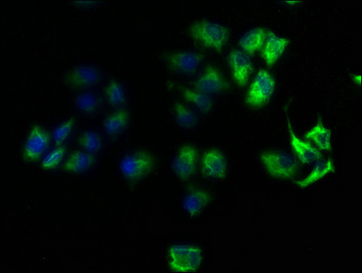
LATS2, Polyclonal Antibody (Cat# AAA235389)
Protein G Purified
ZOT, Polyclonal Antibody (Cat# AAA235396)
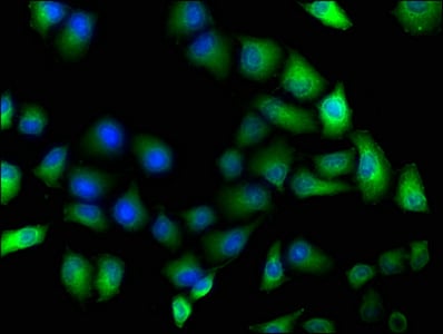
 IF (Immunofluorescence)
(Immunofluorescence staining of Hela cells with CSB-PA614415HA01HU at 1:100, counter-stained with DAPI. The cells were fixed in 4% formaldehyde, permeabilized using 0.2% Triton X-100 and blocked in 10% normal Goat Serum. The cells were then incubated with the antibody overnight at 4 degree C. The secondary antibody was Alexa Fluor 488-congugated AffiniPure Goat Anti-Rabbit IgG(H+L).)
IF (Immunofluorescence)
(Immunofluorescence staining of Hela cells with CSB-PA614415HA01HU at 1:100, counter-stained with DAPI. The cells were fixed in 4% formaldehyde, permeabilized using 0.2% Triton X-100 and blocked in 10% normal Goat Serum. The cells were then incubated with the antibody overnight at 4 degree C. The secondary antibody was Alexa Fluor 488-congugated AffiniPure Goat Anti-Rabbit IgG(H+L).)
ROCK1, Polyclonal Antibody (Cat# AAA235411)
Protein G Purified
 IF (Immunofluorescence)
(Immunofluorescence staining of MCF-7 cells with CSB-PA617999LA01HU at 1:166, counter-stained with DAPI. The cells were fixed in 4% formaldehyde, permeabilized using 0.2% Triton X-100 and blocked in 10% normal Goat Serum. The cells were then incubated with the antibody overnight at 4 degree C. The secondary antibody was Alexa Fluor 488-congugated AffiniPure Goat Anti-Rabbit IgG(H+L).)
IF (Immunofluorescence)
(Immunofluorescence staining of MCF-7 cells with CSB-PA617999LA01HU at 1:166, counter-stained with DAPI. The cells were fixed in 4% formaldehyde, permeabilized using 0.2% Triton X-100 and blocked in 10% normal Goat Serum. The cells were then incubated with the antibody overnight at 4 degree C. The secondary antibody was Alexa Fluor 488-congugated AffiniPure Goat Anti-Rabbit IgG(H+L).)
NCF4, Polyclonal Antibody (Cat# AAA235415)
Protein G Purified
 IF (Immunofluorescence)
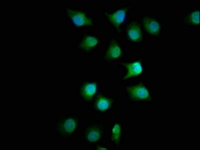
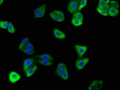
(Immunofluorescence staining of A549 cells with CSB-PA622993LA01HU at 1:33, counter-stained with DAPI. The cells were fixed in 4% formaldehyde, permeabilized using 0.2% Triton X-100 and blocked in 10% normal Goat Serum. The cells were then incubated with the antibody overnight at 4 degree C. The secondary antibody was Alexa Fluor 488-congugated AffiniPure Goat Anti-Rabbit IgG(H+L).)
IF (Immunofluorescence)
(Immunofluorescence staining of A549 cells with CSB-PA622993LA01HU at 1:33, counter-stained with DAPI. The cells were fixed in 4% formaldehyde, permeabilized using 0.2% Triton X-100 and blocked in 10% normal Goat Serum. The cells were then incubated with the antibody overnight at 4 degree C. The secondary antibody was Alexa Fluor 488-congugated AffiniPure Goat Anti-Rabbit IgG(H+L).)
LLGL1, Polyclonal Antibody (Cat# AAA235420)
Protein G Purified
 IF (Immunofluorescence)
(Immunofluorescence staining of A549 cells with CSB-PA715012LA01HU at 1:100, counter-stained with DAPI. The cells were fixed in 4% formaldehyde, permeabilized using 0.2% Triton X-100 and blocked in 10% normal Goat Serum. The cells were then incubated with the antibody overnight at 4 degree C. The secondary antibody was Alexa Fluor 488-congugated AffiniPure Goat Anti-Rabbit IgG(H+L).)
IF (Immunofluorescence)
(Immunofluorescence staining of A549 cells with CSB-PA715012LA01HU at 1:100, counter-stained with DAPI. The cells were fixed in 4% formaldehyde, permeabilized using 0.2% Triton X-100 and blocked in 10% normal Goat Serum. The cells were then incubated with the antibody overnight at 4 degree C. The secondary antibody was Alexa Fluor 488-congugated AffiniPure Goat Anti-Rabbit IgG(H+L).)
FMN1, Polyclonal Antibody (Cat# AAA235425)
Protein G Purified
 IF (Immunofluorescence)
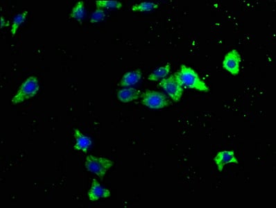
(Immunofluorescence staining of HepG2 cells with CSB-PA719416LA01HU at 1:100, counter-stained with DAPI. The cells were fixed in 4% formaldehyde, permeabilized using 0.2% Triton X-100 and blocked in 10% normal Goat Serum. The cells were then incubated with the antibody overnight at 4 degree C. The secondary antibody was Alexa Fluor 488-congugated AffiniPure Goat Anti-Rabbit IgG(H+L).)
IF (Immunofluorescence)
(Immunofluorescence staining of HepG2 cells with CSB-PA719416LA01HU at 1:100, counter-stained with DAPI. The cells were fixed in 4% formaldehyde, permeabilized using 0.2% Triton X-100 and blocked in 10% normal Goat Serum. The cells were then incubated with the antibody overnight at 4 degree C. The secondary antibody was Alexa Fluor 488-congugated AffiniPure Goat Anti-Rabbit IgG(H+L).)
CPTP, Polyclonal Antibody (Cat# AAA235427)
Protein G Purified
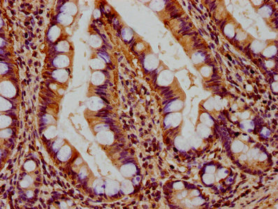
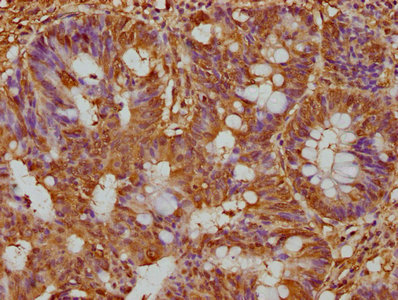
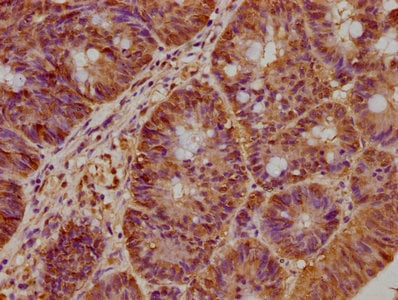 IHC (Immunohistochemisry)
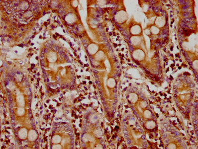
(IHC image of CSB-PA757659LA01HU diluted at 1:300 and staining in paraffin-embedded human colon cancer performed on a Leica BondTM system. After dewaxing and hydration, antigen retrieval was mediated by high pressure in a citrate buffer (pH 6.0). Section was blocked with 10% normal goat serum 30min at RT. Then primary antibody (1% BSA) was incubated at 4 degree C overnight. The primary is detected by a biotinylated secondary antibody and visualized using an HRP conjugated SP system.)
IHC (Immunohistochemisry)
(IHC image of CSB-PA757659LA01HU diluted at 1:300 and staining in paraffin-embedded human colon cancer performed on a Leica BondTM system. After dewaxing and hydration, antigen retrieval was mediated by high pressure in a citrate buffer (pH 6.0). Section was blocked with 10% normal goat serum 30min at RT. Then primary antibody (1% BSA) was incubated at 4 degree C overnight. The primary is detected by a biotinylated secondary antibody and visualized using an HRP conjugated SP system.)
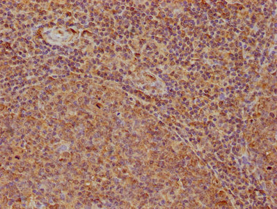
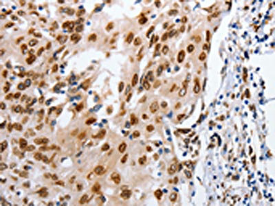
BRAT1, Polyclonal Antibody (Cat# AAA235438)
Protein G Purified
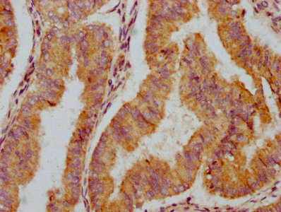
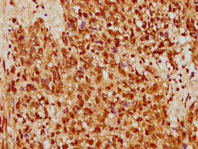 IHC (Immunohiostchemistry)
(IHC image of CSB-PA767212LA01HU diluted at 1:500 and staining in paraffin-embedded human ovarian cancer performed on a Leica BondTM system. After dewaxing and hydration, antigen retrieval was mediated by high pressure in a citrate buffer (pH 6.0). Section was blocked with 10% normal goat serum 30min at RT. Then primary antibody (1% BSA) was incubated at 4 degree C overnight. The primary is detected by a biotinylated secondary antibody and visualized using an HRP conjugated SP system.)
IHC (Immunohiostchemistry)
(IHC image of CSB-PA767212LA01HU diluted at 1:500 and staining in paraffin-embedded human ovarian cancer performed on a Leica BondTM system. After dewaxing and hydration, antigen retrieval was mediated by high pressure in a citrate buffer (pH 6.0). Section was blocked with 10% normal goat serum 30min at RT. Then primary antibody (1% BSA) was incubated at 4 degree C overnight. The primary is detected by a biotinylated secondary antibody and visualized using an HRP conjugated SP system.)
STRA8, Polyclonal Antibody (Cat# AAA235441)
Protein G Purified
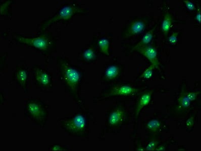
 IF (Immunofluorescence)
(Immunofluorescence staining of U251 cells with CSB-PA769774LA01HU at 1:166, counter-stained with DAPI. The cells were fixed in 4% formaldehyde, permeabilized using 0.2% Triton X-100 and blocked in 10% normal Goat Serum. The cells were then incubated with the antibody overnight at 4 degree C. The secondary antibody was Alexa Fluor 488-congugated AffiniPure Goat Anti-Rabbit IgG(H+L).)
IF (Immunofluorescence)
(Immunofluorescence staining of U251 cells with CSB-PA769774LA01HU at 1:166, counter-stained with DAPI. The cells were fixed in 4% formaldehyde, permeabilized using 0.2% Triton X-100 and blocked in 10% normal Goat Serum. The cells were then incubated with the antibody overnight at 4 degree C. The secondary antibody was Alexa Fluor 488-congugated AffiniPure Goat Anti-Rabbit IgG(H+L).)
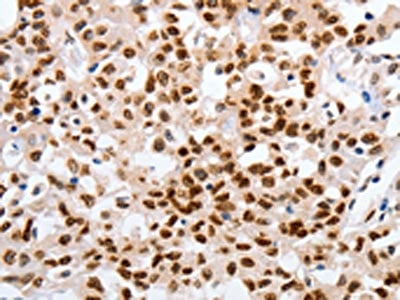
PRPF39, Polyclonal Antibody (Cat# AAA235443)
Protein G Purified
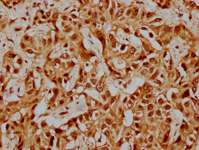
 IHC (Immunohiostchemistry)
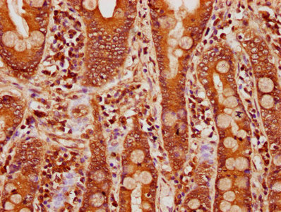
(IHC image of CSB-PA773036LA01HU diluted at 1:100 and staining in paraffin-embedded human cervical cancer performed on a Leica BondTM system. After dewaxing and hydration, antigen retrieval was mediated by high pressure in a citrate buffer (pH 6.0). Section was blocked with 10% normal goat serum 30min at RT. Then primary antibody (1% BSA) was incubated at 4 degree C overnight. The primary is detected by a biotinylated secondary antibody and visualized using an HRP conjugated SP system.)
IHC (Immunohiostchemistry)
(IHC image of CSB-PA773036LA01HU diluted at 1:100 and staining in paraffin-embedded human cervical cancer performed on a Leica BondTM system. After dewaxing and hydration, antigen retrieval was mediated by high pressure in a citrate buffer (pH 6.0). Section was blocked with 10% normal goat serum 30min at RT. Then primary antibody (1% BSA) was incubated at 4 degree C overnight. The primary is detected by a biotinylated secondary antibody and visualized using an HRP conjugated SP system.)
CASZ1, Polyclonal Antibody (Cat# AAA235446)
Protein G Purified
 IF (Immunofluorescence)
(Immunofluorescence staining of MCF-7 cells with CSB-PA774805NA01HU at 1:50, counter-stained with DAPI. The cells were fixed in 4% formaldehyde, permeabilized using 0.2% Triton X-100 and blocked in 10% normal Goat Serum. The cells were then incubated with the antibody overnight at 4 degree C. The secondary antibody was Alexa Fluor 488-congugated AffiniPure Goat Anti-Rabbit IgG(H+L).)
IF (Immunofluorescence)
(Immunofluorescence staining of MCF-7 cells with CSB-PA774805NA01HU at 1:50, counter-stained with DAPI. The cells were fixed in 4% formaldehyde, permeabilized using 0.2% Triton X-100 and blocked in 10% normal Goat Serum. The cells were then incubated with the antibody overnight at 4 degree C. The secondary antibody was Alexa Fluor 488-congugated AffiniPure Goat Anti-Rabbit IgG(H+L).)
RTN4RL2, Polyclonal Antibody (Cat# AAA235447)
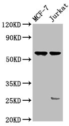
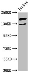
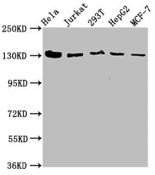 Application Data
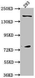
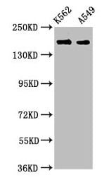
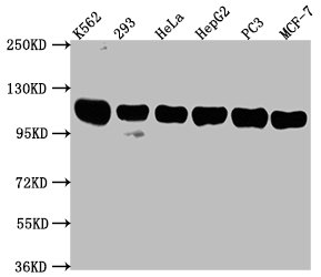
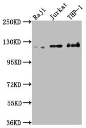
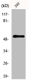
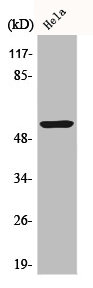
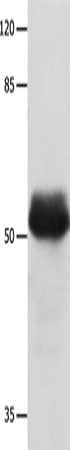
(Western BlotPositive WB detected in: Hela whole cell lysate, Jurkat whole cell lysate, 293T whole cell lysate, HepG2 whole cell lysate, MCF-7 whole cell lysateAll lanes: CAND1 antibody at 4.9ug/mlSecondaryGoat polyclonal to rabbit IgG at 1/50000 dilutionPredicted band size: 137, 118, 46 kDaObserved band size: 137 kDa)
Application Data
(Western BlotPositive WB detected in: Hela whole cell lysate, Jurkat whole cell lysate, 293T whole cell lysate, HepG2 whole cell lysate, MCF-7 whole cell lysateAll lanes: CAND1 antibody at 4.9ug/mlSecondaryGoat polyclonal to rabbit IgG at 1/50000 dilutionPredicted band size: 137, 118, 46 kDaObserved band size: 137 kDa)
CAND1, Polyclonal Antibody (Cat# AAA235448)
Protein G Purified
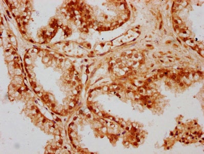
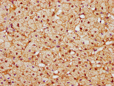
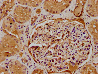
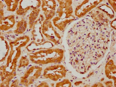
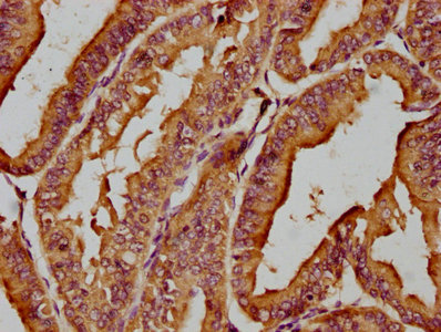
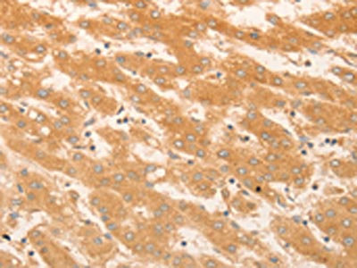
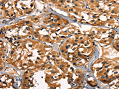
 IHC (Immunohiostchemistry)
(IHC image of CSB-PA810289LA01HU diluted at 1:400 and staining in paraffin-embedded human kidney tissue performed on a Leica BondTM system. After dewaxing and hydration, antigen retrieval was mediated by high pressure in a citrate buffer (pH 6.0). Section was blocked with 10% normal goat serum 30min at RT. Then primary antibody (1% BSA) was incubated at 4 degree C overnight. The primary is detected by a biotinylated secondary antibody and visualized using an HRP conjugated SP system.)
IHC (Immunohiostchemistry)
(IHC image of CSB-PA810289LA01HU diluted at 1:400 and staining in paraffin-embedded human kidney tissue performed on a Leica BondTM system. After dewaxing and hydration, antigen retrieval was mediated by high pressure in a citrate buffer (pH 6.0). Section was blocked with 10% normal goat serum 30min at RT. Then primary antibody (1% BSA) was incubated at 4 degree C overnight. The primary is detected by a biotinylated secondary antibody and visualized using an HRP conjugated SP system.)
RHOT2, Polyclonal Antibody (Cat# AAA235450)
Protein G Purified
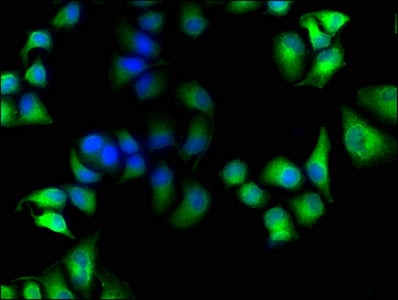
 IF (Immunofluorescence)
(Immunofluorescence staining of U251 cells with CSB-PA818235LA01HU at 1:100, counter-stained with DAPI. The cells were fixed in 4% formaldehyde, permeabilized using 0.2% Triton X-100 and blocked in 10% normal Goat Serum. The cells were then incubated with the antibody overnight at 4 degree C. The secondary antibody was Alexa Fluor 488-congugated AffiniPure Goat Anti-Rabbit IgG(H+L).)
IF (Immunofluorescence)
(Immunofluorescence staining of U251 cells with CSB-PA818235LA01HU at 1:100, counter-stained with DAPI. The cells were fixed in 4% formaldehyde, permeabilized using 0.2% Triton X-100 and blocked in 10% normal Goat Serum. The cells were then incubated with the antibody overnight at 4 degree C. The secondary antibody was Alexa Fluor 488-congugated AffiniPure Goat Anti-Rabbit IgG(H+L).)
TIAM2, Polyclonal Antibody (Cat# AAA235451)
Protein G Purified
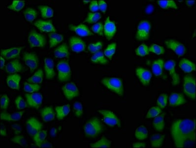
 IF (Immunofluorescence)
(Immunofluorescence staining of HepG2 cells with CSB-PA818735LA01HU at 1:166, counter-stained with DAPI. The cells were fixed in 4% formaldehyde, permeabilized using 0.2% Triton X-100 and blocked in 10% normal Goat Serum. The cells were then incubated with the antibody overnight at 4 degree C. The secondary antibody was Alexa Fluor 488-congugated AffiniPure Goat Anti-Rabbit IgG(H+L).)
IF (Immunofluorescence)
(Immunofluorescence staining of HepG2 cells with CSB-PA818735LA01HU at 1:166, counter-stained with DAPI. The cells were fixed in 4% formaldehyde, permeabilized using 0.2% Triton X-100 and blocked in 10% normal Goat Serum. The cells were then incubated with the antibody overnight at 4 degree C. The secondary antibody was Alexa Fluor 488-congugated AffiniPure Goat Anti-Rabbit IgG(H+L).)
ATP6V0D2, Polyclonal Antibody (Cat# AAA235452)
Protein G Purified
FAR1, Polyclonal Antibody (Cat# AAA235459)
Protein G Purified
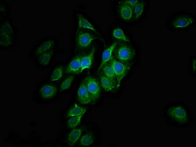
 IF (Immunofluorescence)
(Immunofluorescence staining of HepG2 cells with CSB-PA839346HA01HU at 1:133, counter-stained with DAPI. The cells were fixed in 4% formaldehyde, permeabilized using 0.2% Triton X-100 and blocked in 10% normal Goat Serum. The cells were then incubated with the antibody overnight at 4 degree C. The secondary antibody was Alexa Fluor 488-congugated AffiniPure Goat Anti-Rabbit IgG(H+L).)
IF (Immunofluorescence)
(Immunofluorescence staining of HepG2 cells with CSB-PA839346HA01HU at 1:133, counter-stained with DAPI. The cells were fixed in 4% formaldehyde, permeabilized using 0.2% Triton X-100 and blocked in 10% normal Goat Serum. The cells were then incubated with the antibody overnight at 4 degree C. The secondary antibody was Alexa Fluor 488-congugated AffiniPure Goat Anti-Rabbit IgG(H+L).)
ITCH, Polyclonal Antibody (Cat# AAA235461)
Protein G Purified
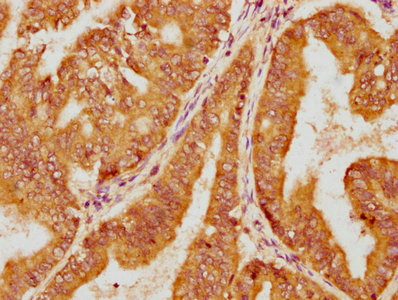
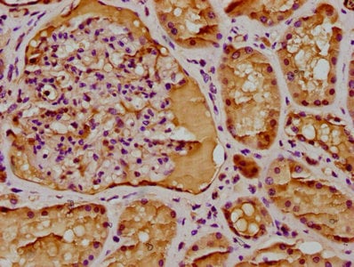
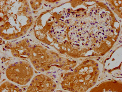
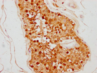
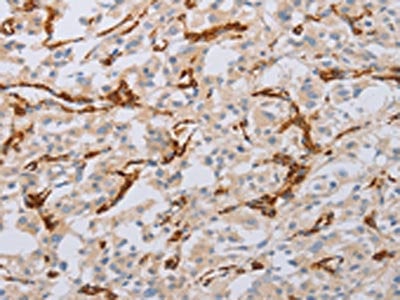
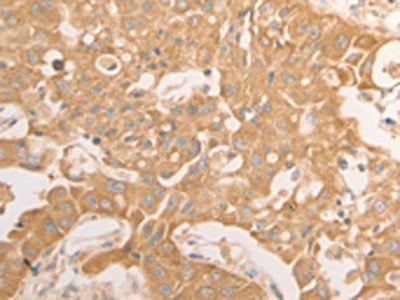
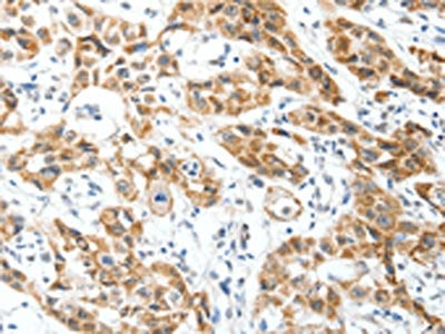
 IHC (Immunohiostchemistry)
(IHC image of CSB-PA839415LA01HU diluted at 1:300 and staining in paraffin-embedded human kidney tissue performed on a Leica BondTM system. After dewaxing and hydration, antigen retrieval was mediated by high pressure in a citrate buffer (pH 6.0). Section was blocked with 10% normal goat serum 30min at RT. Then primary antibody (1% BSA) was incubated at 4 degree C overnight. The primary is detected by a biotinylated secondary antibody and visualized using an HRP conjugated SP system.)
IHC (Immunohiostchemistry)
(IHC image of CSB-PA839415LA01HU diluted at 1:300 and staining in paraffin-embedded human kidney tissue performed on a Leica BondTM system. After dewaxing and hydration, antigen retrieval was mediated by high pressure in a citrate buffer (pH 6.0). Section was blocked with 10% normal goat serum 30min at RT. Then primary antibody (1% BSA) was incubated at 4 degree C overnight. The primary is detected by a biotinylated secondary antibody and visualized using an HRP conjugated SP system.)
FIZ1, Polyclonal Antibody (Cat# AAA235462)
Protein G Purified
ALMS1, Polyclonal Antibody (Cat# AAA235465)
Protein G Purified
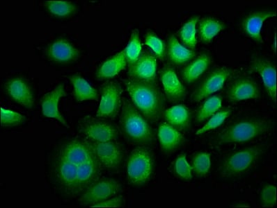
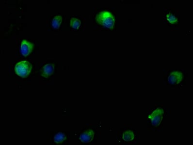
 IF (Immunofluorescence)
(Immunofluorescence staining of Hela cells with CSB-PA847623LA01HU at 1:100, counter-stained with DAPI. The cells were fixed in 4% formaldehyde, permeabilized using 0.2% Triton X-100 and blocked in 10% normal Goat Serum. The cells were then incubated with the antibody overnight at 4 degree C. The secondary antibody was Alexa Fluor 488-congugated AffiniPure Goat Anti-Rabbit IgG(H+L).)
IF (Immunofluorescence)
(Immunofluorescence staining of Hela cells with CSB-PA847623LA01HU at 1:100, counter-stained with DAPI. The cells were fixed in 4% formaldehyde, permeabilized using 0.2% Triton X-100 and blocked in 10% normal Goat Serum. The cells were then incubated with the antibody overnight at 4 degree C. The secondary antibody was Alexa Fluor 488-congugated AffiniPure Goat Anti-Rabbit IgG(H+L).)
PARP8, Polyclonal Antibody (Cat# AAA235467)
Protein G Purified
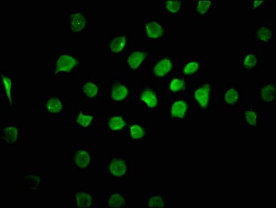
 IF (Immunofluorescence)
(Immunofluorescence staining of Hela cells with CSB-PA851555LA01HU at 1:100, counter-stained with DAPI. The cells were fixed in 4% formaldehyde, permeabilized using 0.2% Triton X-100 and blocked in 10% normal Goat Serum. The cells were then incubated with the antibody overnight at 4 degree C. The secondary antibody was Alexa Fluor 488-congugated AffiniPure Goat Anti-Rabbit IgG(H+L).)
IF (Immunofluorescence)
(Immunofluorescence staining of Hela cells with CSB-PA851555LA01HU at 1:100, counter-stained with DAPI. The cells were fixed in 4% formaldehyde, permeabilized using 0.2% Triton X-100 and blocked in 10% normal Goat Serum. The cells were then incubated with the antibody overnight at 4 degree C. The secondary antibody was Alexa Fluor 488-congugated AffiniPure Goat Anti-Rabbit IgG(H+L).)
MICAL1, Polyclonal Antibody (Cat# AAA235469)
Protein G Purified
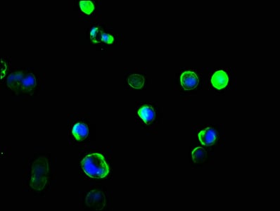
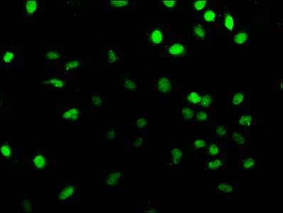
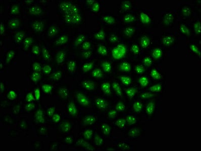
 IF (Immunofluorescence)
(Immunofluorescence staining of Hela cells with AAA235471 at 1:133, counter-stained with DAPI. The cells were fixed in 4% formaldehyde, permeabilized using 0.2% Triton X-100 and blocked in 10% normal Goat Serum. The cells were then incubated with the antibody overnight at 4 degree C. The secondary antibody was Alexa Fluor 488-congugated AffiniPure Goat Anti-Rabbit IgG(H+L).)
IF (Immunofluorescence)
(Immunofluorescence staining of Hela cells with AAA235471 at 1:133, counter-stained with DAPI. The cells were fixed in 4% formaldehyde, permeabilized using 0.2% Triton X-100 and blocked in 10% normal Goat Serum. The cells were then incubated with the antibody overnight at 4 degree C. The secondary antibody was Alexa Fluor 488-congugated AffiniPure Goat Anti-Rabbit IgG(H+L).)
FCRL5, Polyclonal Antibody (Cat# AAA235471)
Protein G Purified
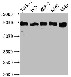
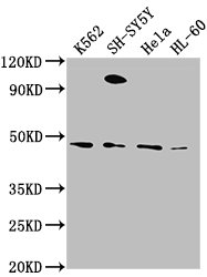
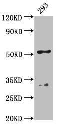
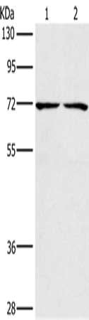
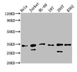 Application Data
(Western BlotPositive WB detected in: Hela whole cell lysate, Jurkat whole cell lysate, HL60 whole cell lysate, U87 whole cell lysate, 293T whole cell lysate, K562 whole cell lysateAll lanes: TSNAX antibody at 2.5ug/mlSecondaryGoat polyclonal to rabbit IgG at 1/50000 dilutionPredicted band size: 34 kDaObserved band size: 34 kDa)
Application Data
(Western BlotPositive WB detected in: Hela whole cell lysate, Jurkat whole cell lysate, HL60 whole cell lysate, U87 whole cell lysate, 293T whole cell lysate, K562 whole cell lysateAll lanes: TSNAX antibody at 2.5ug/mlSecondaryGoat polyclonal to rabbit IgG at 1/50000 dilutionPredicted band size: 34 kDaObserved band size: 34 kDa)
TSNAX, Polyclonal Antibody (Cat# AAA235474)
Protein G Purified
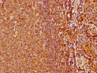
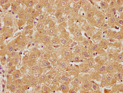
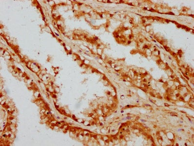
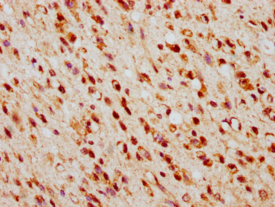
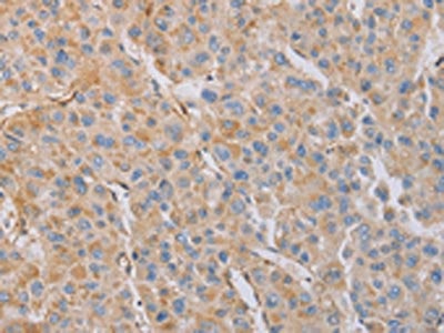
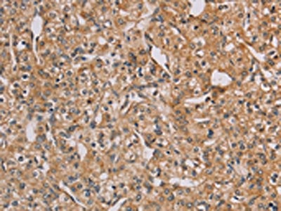
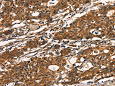
 IHC (Immunohiostchemistry)
(IHC image of CSB-PA858733LA01HU diluted at 1:300 and staining in paraffin-embedded human placenta tissue performed on a Leica BondTM system. After dewaxing and hydration, antigen retrieval was mediated by high pressure in a citrate buffer (pH 6.0). Section was blocked with 10% normal goat serum 30min at RT. Then primary antibody (1% BSA) was incubated at 4 degree C overnight. The primary is detected by a biotinylated secondary antibody and visualized using an HRP conjugated SP system.)
IHC (Immunohiostchemistry)
(IHC image of CSB-PA858733LA01HU diluted at 1:300 and staining in paraffin-embedded human placenta tissue performed on a Leica BondTM system. After dewaxing and hydration, antigen retrieval was mediated by high pressure in a citrate buffer (pH 6.0). Section was blocked with 10% normal goat serum 30min at RT. Then primary antibody (1% BSA) was incubated at 4 degree C overnight. The primary is detected by a biotinylated secondary antibody and visualized using an HRP conjugated SP system.)
VGLL1, Polyclonal Antibody (Cat# AAA235475)
Protein G Purified
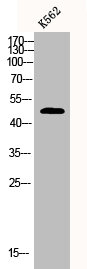
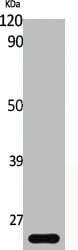
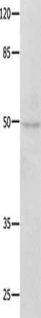
 WB (Western Blot)
(Western Blot analysis of K562 cells using CPA1 Polyclonal Antibody)
WB (Western Blot)
(Western Blot analysis of K562 cells using CPA1 Polyclonal Antibody)
CPA1, Polyclonal Antibody (Cat# AAA236039)
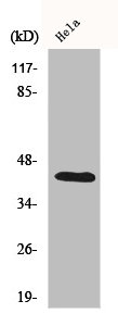
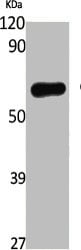
 WB (Western Blot)
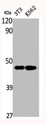
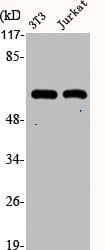
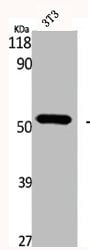
(Western Blot analysis of NIH-3T3 K562 cells using CREB-1 Polyclonal Antibody)
WB (Western Blot)
(Western Blot analysis of NIH-3T3 K562 cells using CREB-1 Polyclonal Antibody)
CREB1, Polyclonal Antibody (Cat# AAA236043)
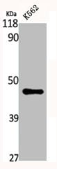
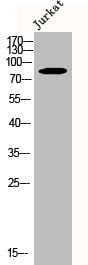
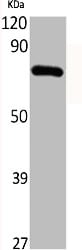
 WB (Western Blot)
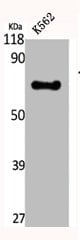
(Western Blot analysis of K562 cells using CREB3L2 Polyclonal Antibody)
WB (Western Blot)
(Western Blot analysis of K562 cells using CREB3L2 Polyclonal Antibody)
CREB3L2, Polyclonal Antibody (Cat# AAA236044)
 WB (Western Blot)
(Western Blot analysis of various cells using Crystallin-alphaB Polyclonal Antibody)
WB (Western Blot)
(Western Blot analysis of various cells using Crystallin-alphaB Polyclonal Antibody)
CRYAB, Polyclonal Antibody (Cat# AAA236046)

 WB (Western Blot)
(Western Blot analysis of various cells using c-Src Polyclonal Antibody)
WB (Western Blot)
(Western Blot analysis of various cells using c-Src Polyclonal Antibody)
SRC, Polyclonal Antibody (Cat# AAA236049)
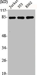 WB (Western Blot)
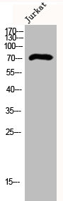
(Western Blot analysis of Jurkat NIH-3T3 K562 cells using CUL-2 Polyclonal Antibody)
WB (Western Blot)
(Western Blot analysis of Jurkat NIH-3T3 K562 cells using CUL-2 Polyclonal Antibody)
CUL2, Polyclonal Antibody (Cat# AAA236055)
 WB (Western Blot)
(Western Blot analysis of various cells using Cyclin B1 Polyclonal Antibody)
WB (Western Blot)
(Western Blot analysis of various cells using Cyclin B1 Polyclonal Antibody)
CCNB1, Polyclonal Antibody (Cat# AAA236059)
 WB (Western Blot)
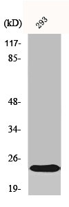
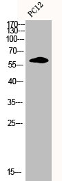
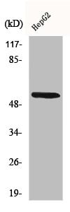
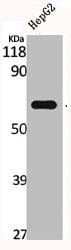
(Western Blot analysis of HepG2 cells using CYP26A1 Polyclonal Antibody)
WB (Western Blot)
(Western Blot analysis of HepG2 cells using CYP26A1 Polyclonal Antibody)
CYP26A1, Polyclonal Antibody (Cat# AAA236066)
 WB (Western Blot)
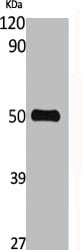
(Western Blot analysis of K562 cells using CYP39A1 Polyclonal Antibody)
WB (Western Blot)
(Western Blot analysis of K562 cells using CYP39A1 Polyclonal Antibody)
CYP39A1, Polyclonal Antibody (Cat# AAA236069)
 WB (Western Blot)
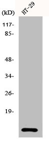
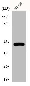
(Western Blot analysis of HT29 cells using Cystatin A Polyclonal Antibody)
WB (Western Blot)
(Western Blot analysis of HT29 cells using Cystatin A Polyclonal Antibody)
CSTA, Polyclonal Antibody (Cat# AAA236074)
 WB (Western Blot)
(Western Blot analysis of NIH-3T3 Jurkat cells using Cytokeratin 10 Polyclonal Antibody)
WB (Western Blot)
(Western Blot analysis of NIH-3T3 Jurkat cells using Cytokeratin 10 Polyclonal Antibody)
KRT10, Polyclonal Antibody (Cat# AAA236077)
 WB (Western Blot)
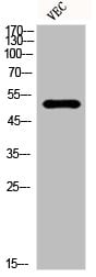
(Western Blot analysis of VEC cells using Cytokeratin 18 Polyclonal Antibody)
WB (Western Blot)
(Western Blot analysis of VEC cells using Cytokeratin 18 Polyclonal Antibody)
KRT18, Polyclonal Antibody (Cat# AAA236080)
 WB (Western Blot)
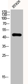
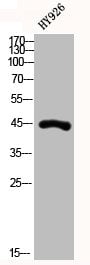
(Western Blot analysis of HY926 cells using Cytokeratin 19 Polyclonal Antibody)
WB (Western Blot)
(Western Blot analysis of HY926 cells using Cytokeratin 19 Polyclonal Antibody)
KRT19, Polyclonal Antibody (Cat# AAA236082)
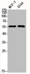 WB (Western Blot)
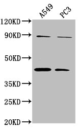
(Western Blot analysis of MCF7 A549 cells using Cytokeratin 8 Polyclonal Antibody)
WB (Western Blot)
(Western Blot analysis of MCF7 A549 cells using Cytokeratin 8 Polyclonal Antibody)
KRT8, Polyclonal Antibody (Cat# AAA236085)
 WB (Western Blot)
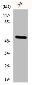
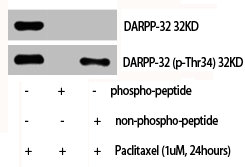
(Western Blot analysis of various cells using DARPP-32 Polyclonal Antibody)
WB (Western Blot)
(Western Blot analysis of various cells using DARPP-32 Polyclonal Antibody)
PPP1R1B, Polyclonal Antibody (Cat# AAA236089)

 WB (Western Blot)
(Western Blot analysis of NIH-3T3 cells using DBP Polyclonal Antibody)
WB (Western Blot)
(Western Blot analysis of NIH-3T3 cells using DBP Polyclonal Antibody)
GC, Polyclonal Antibody (Cat# AAA236091)
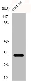
 WB (Western Blot)
(Western Blot analysis of various cells using DCAMKL3 Polyclonal Antibody)
WB (Western Blot)
(Western Blot analysis of various cells using DCAMKL3 Polyclonal Antibody)
DCLK3, Polyclonal Antibody (Cat# AAA236092)
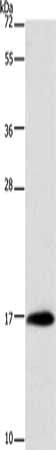
 SDS-PAGE
(Gel: 12%SDS-PAGE, Lysate: 40 ug, Lane: Mouse muscle tissue, Primary antibody: AAA237619(MYOZ1 Antibody) at dilution 1/1000, Secondary antibody: Goat anti rabbit IgG at 1/8000 dilution, Exposure time: 10 seconds)
SDS-PAGE
(Gel: 12%SDS-PAGE, Lysate: 40 ug, Lane: Mouse muscle tissue, Primary antibody: AAA237619(MYOZ1 Antibody) at dilution 1/1000, Secondary antibody: Goat anti rabbit IgG at 1/8000 dilution, Exposure time: 10 seconds)
MYOZ1, Polyclonal Antibody (Cat# AAA237619)
 SDS-PAGE
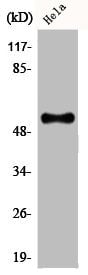
(Gel: 8%SDS-PAGE, Lysate: 40 ug, Lane: Hela cells, Primary antibody: AAA237621(MUTYH Antibody) at dilution 1/500, Secondary antibody: Goat anti rabbit IgG at 1/8000 dilution, Exposure time: 1 minute)
SDS-PAGE
(Gel: 8%SDS-PAGE, Lysate: 40 ug, Lane: Hela cells, Primary antibody: AAA237621(MUTYH Antibody) at dilution 1/500, Secondary antibody: Goat anti rabbit IgG at 1/8000 dilution, Exposure time: 1 minute)
MUTYH, Polyclonal Antibody (Cat# AAA237621)
 SDS-PAGE
(Gel: 10%SDS-PAGE, Lysate: 40 ug, Lane: Mouse stomach tissue, Primary antibody: AAA237625(MYL6 Antibody) at dilution 1/262.5, Secondary antibody: Goat anti rabbit IgG at 1/8000 dilution, Exposure time: 30 seconds)
SDS-PAGE
(Gel: 10%SDS-PAGE, Lysate: 40 ug, Lane: Mouse stomach tissue, Primary antibody: AAA237625(MYL6 Antibody) at dilution 1/262.5, Secondary antibody: Goat anti rabbit IgG at 1/8000 dilution, Exposure time: 30 seconds)
MYL6, Polyclonal Antibody (Cat# AAA237625)
 SDS-PAGE
(Gel: 8%SDS-PAGE, Lysate: 40 ug, Lane 1-2: Lovo cells, 293T cells, Primary antibody: AAA237627(CHRNA4 Antibody) at dilution 1/200, Secondary antibody: Goat anti rabbit IgG at 1/8000 dilution, Exposure time: 5 seconds)
SDS-PAGE
(Gel: 8%SDS-PAGE, Lysate: 40 ug, Lane 1-2: Lovo cells, 293T cells, Primary antibody: AAA237627(CHRNA4 Antibody) at dilution 1/200, Secondary antibody: Goat anti rabbit IgG at 1/8000 dilution, Exposure time: 5 seconds)
CHRNA4, Polyclonal Antibody (Cat# AAA237627)
 SDS-PAGE
(Gel: 10%SDS-PAGE, Lysate: 40 ug, Lane: Jurkat cells, Primary antibody: AAA237630(NAP1L1 Antibody) at dilution 1/650, Secondary antibody: Goat anti rabbit IgG at 1/8000 dilution, Exposure time: 40 seconds)
SDS-PAGE
(Gel: 10%SDS-PAGE, Lysate: 40 ug, Lane: Jurkat cells, Primary antibody: AAA237630(NAP1L1 Antibody) at dilution 1/650, Secondary antibody: Goat anti rabbit IgG at 1/8000 dilution, Exposure time: 40 seconds)
NAP1L1, Polyclonal Antibody (Cat# AAA237630)
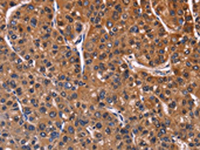
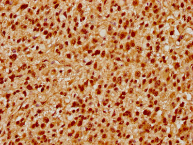
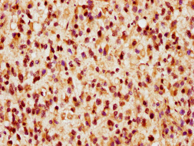
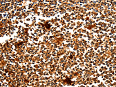
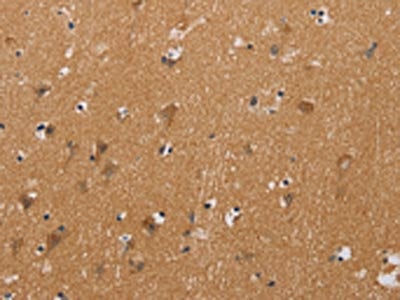
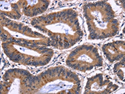 IHC (Immunohiostchemistry)
(The image on the left is immunohistochemistry of paraffin-embedded Human colon cancer tissue using AAA237638(NFATC3 Antibody) at dilution 1/60, on the right is treated with fusion protein. (Original magnification: ×200))
IHC (Immunohiostchemistry)
(The image on the left is immunohistochemistry of paraffin-embedded Human colon cancer tissue using AAA237638(NFATC3 Antibody) at dilution 1/60, on the right is treated with fusion protein. (Original magnification: ×200))
NFATC3, Polyclonal Antibody (Cat# AAA237638)
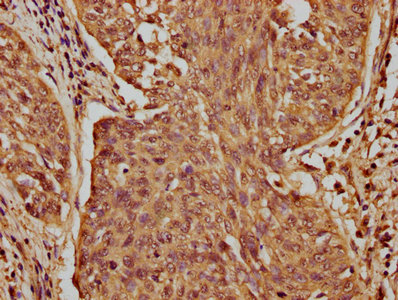
 IHC (Immunohiostchemistry)
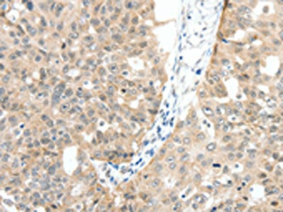
(The image on the left is immunohistochemistry of paraffin-embedded Human lung cancer tissue using AAA237640(MAP3K14 Antibody) at dilution 1/30, on the right is treated with fusion protein. (Original magnification: ×200))
IHC (Immunohiostchemistry)
(The image on the left is immunohistochemistry of paraffin-embedded Human lung cancer tissue using AAA237640(MAP3K14 Antibody) at dilution 1/30, on the right is treated with fusion protein. (Original magnification: ×200))
MAP3K14, Polyclonal Antibody (Cat# AAA237640)
 IHC (Immunohiostchemistry)
(The image on the left is immunohistochemistry of paraffin-embedded Human thyroid cancer tissue using AAA237643(KLRC1 Antibody) at dilution 1/20, on the right is treated with fusion protein. (Original magnification: ×200))
IHC (Immunohiostchemistry)
(The image on the left is immunohistochemistry of paraffin-embedded Human thyroid cancer tissue using AAA237643(KLRC1 Antibody) at dilution 1/20, on the right is treated with fusion protein. (Original magnification: ×200))
KLRC1, Polyclonal Antibody (Cat# AAA237643)
What are Polyclonal Antibodies?
Polyclonal antibodies are antibodies that come from multiple B cell clones of a host animal. The typical hosts used for the majority of polyclonal antibody production are rabbits, goats, sheep, and donkeys. These polyclonal antibodies, once having identified their target, will bind to different epitopes located at different regions or sequences on the same protein/antigen. As a result, they are ideal at locating and binding to the target, even if the target is in very low concentrations (due to many different antibodies being able to bind to the same target molecule, which allows for significant amplification of a downstream signal).
Polyclonal antibodies are typically produced by injecting an antigen into a host animal, which causes the animal’s immune system to attack the foreign antigen by mass generating antibodies against it. After a period of time, serum is collected from the animal and purified using physicochemical fractionation, class-specific affinity purification, and/or antigen-affinity purification.
Key Uses of Polyclonal Antibodies
- Western Blotting: This method is used to find specific proteins in biological samples after separating them by size.
- Immunohistochemistry: IHC helps visualize the location of proteins in tissue sections using various staining techniques.
- ELISA: (Enzyme-Linked Immunosorbent Assay) is typically used to identify specific protein quantities in a sample. ELISAs can be either “Quantitative” or “Qualitative”.
- Flow Cytometry: technique that identifies and measures the specific protein on the surface or inside the cells in a fluid suspension.
- Immunoprecipitation: IP isolates and studies a specific protein from a complex mixture using antibodies.
Why Buy Polyclonal Antibodies from AAA Biotech?
1. Ideal for Various Applications
Our antibodies are generally going to be validated for use in multiple types of assays, including ELISA, Western Blotting, Immunohistochemistry, Immunoprecipitation, amongst others. They are ideal for a wide range of research applications.
2. Rigorous Quality Control
All of the antibodies in our catalog undergo strict quality testing to ensure specificity, sensitivity, and consistent performance. We are confident in the ability of our antibodies to provide you with accurate results.
3. Wide Assortment of Antibodies
Antibodies in are catalog can be found for both common and exotic species, and these antibodies are also available in both conjugated and recombinant forms to suit many diverse experimental needs.
4. Highly Purified
Our antibodies are available in purified forms with over 85% purity, as confirmed by SDS-PAGE. They are also available with tags such as His, Flag, GST, or MBP. We cater to customers worldwide.
FAQ
1. How are polyclonal antibodies produced?
Traditionally, polyclonal antibodies are produced by injecting an antigen into a host animal (such as a rabbit or goat), which then triggers an immune response from the host animal. The animal’s B cells produce antibodies that will recognize different parts of the injected antigen. These antibodies are then collected from the animal’s blood and purified for use.
2. How do polyclonal antibodies differ from monoclonal antibodies?
Polyclonal antibodies are a mix of antibodies that bind to different locations (epitopes) of the same antigen, while monoclonal antibodies are identical and bind to just one specific epitope. This makes polyclonal antibodies more versatile and better at detecting proteins that may be present in low quantities or in altered/modified forms.
3. How should I store polyclonal antibodies?
Polyclonal antibodies should be stored at 4°C for short-term use (up to a few weeks) and at -20°C or -80°C for long-term storage. Avoid repeated freeze-thaw cycles by dividing them into small aliquots. Always check the datasheet for specific storage instructions.