Filters
▼Clonality
▼Type
▼Reactivity
▼Gene Name
▼Isotype
▼Host
▼Application
▼Clone
▼Phospho Antibodies
Phospho-specific antibodies’ typical purpose is to enable researchers to detect changes in proteins. They will exclusively bind to the amino acid sequence on a protein that has been phosphorylated (which is both a physical & chemical change) and do not bind to the same amino acid sequence on said protein if it lacks said phosphorylation. This aids in being able to clearly see and understand the data produced from this particular protein modification.
Viewing 750-800 of 5298 product results

 IHC (Immunohistochemisry)
(Detection of human Phospho XRCC1 (S518/T519/T523) by immunohistochemistry. Sample: FFPE section of human breast carcinoma. Antibody: Affinity purified rabbit anti-Phospho XRCC1 (S518/T519/T523) (Cat. No. AAA213802) used at a dilution of 1:100. Detection: Red-fluorescent Alexa Fluor 555 goat anti-rabbit IgG (Invitrogen) used at a dilution of 1:500.)
IHC (Immunohistochemisry)
(Detection of human Phospho XRCC1 (S518/T519/T523) by immunohistochemistry. Sample: FFPE section of human breast carcinoma. Antibody: Affinity purified rabbit anti-Phospho XRCC1 (S518/T519/T523) (Cat. No. AAA213802) used at a dilution of 1:100. Detection: Red-fluorescent Alexa Fluor 555 goat anti-rabbit IgG (Invitrogen) used at a dilution of 1:500.)
XRCC1, Polyclonal Antibody (Cat# AAA213802)
Phospho-CHK2, Monoclonal Recombinant Antibody (Cat# AAA120239)
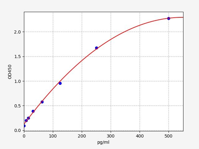
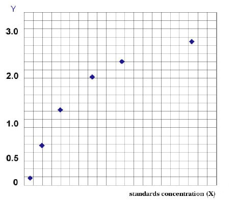
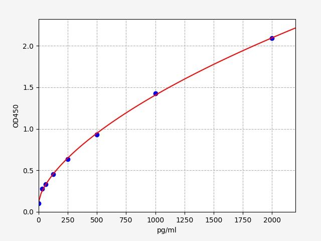 Standard Curve (Sample)
PERK (Phospho Extracellular Signal Regulated Kinase), ELISA Kit (Cat# AAA251370)
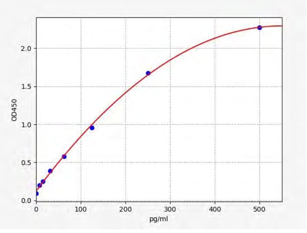 Standard Curve (Sample)
p-tau217 (Phospho Tau 217), ELISA Kit (Cat# AAA250999)
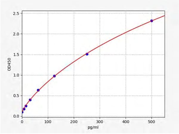 Standard Curve (Sample)
p-tau217 (Phospho Tau 217), ELISA Kit (Cat# AAA251072)
 Standard Curve (Sample)
p-tau217 (Phospho Tau 217), ELISA Kit (Cat# AAA253182)
 Standard Curve (Sample)
p-tau181 (Phospho Tau 181), ELISA Kit (Cat# AAA253448)

 IHC (Immunohiostchemistry)
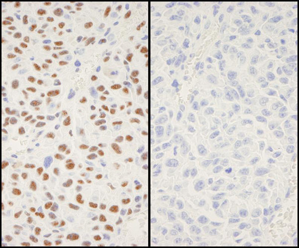
(Immunohistochemical analysis of paraffin-embedded human breast carcinoma tissue using CREB(Phospho-Ser133) Antibody(left) or the same antibody preincubated with blocking peptide(right).)
IHC (Immunohiostchemistry)
(Immunohistochemical analysis of paraffin-embedded human breast carcinoma tissue using CREB(Phospho-Ser133) Antibody(left) or the same antibody preincubated with blocking peptide(right).)
CREB1, Polyclonal Antibody (Cat# AAA243061)
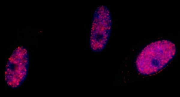
 IF (Immunofluorescence)
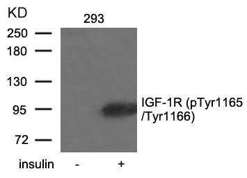
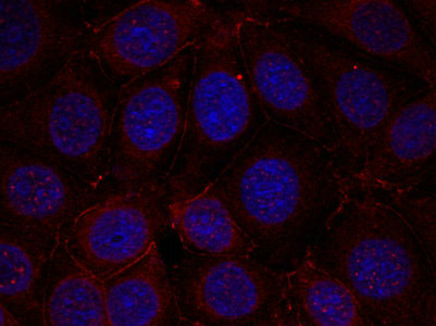
(Immunofluorescence staining of methanol-fixed MCF cells using IGF-1R(Phospho-Tyr1165/Tyr1166) Antibody.)
IF (Immunofluorescence)
(Immunofluorescence staining of methanol-fixed MCF cells using IGF-1R(Phospho-Tyr1165/Tyr1166) Antibody.)
IGF1R, Polyclonal Antibody (Cat# AAA243072)
 IHC (Immunohiostchemistry)
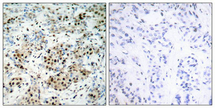
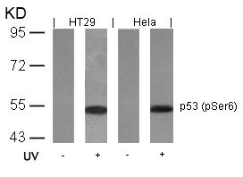
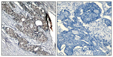
(Immunohistochemical analysis of paraffin-embedded human breast carcinoma tissue using p53(Phospho-Ser6) Antibody(left) or the same antibody preincubated with blocking peptide(right).)
IHC (Immunohiostchemistry)
(Immunohistochemical analysis of paraffin-embedded human breast carcinoma tissue using p53(Phospho-Ser6) Antibody(left) or the same antibody preincubated with blocking peptide(right).)
TP53, Polyclonal Antibody (Cat# AAA243075)
 IHC (Immunohiostchemistry)
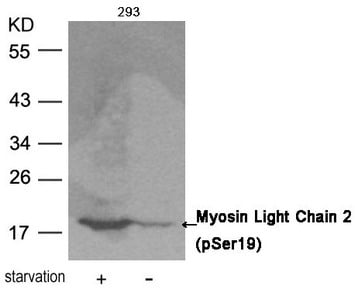
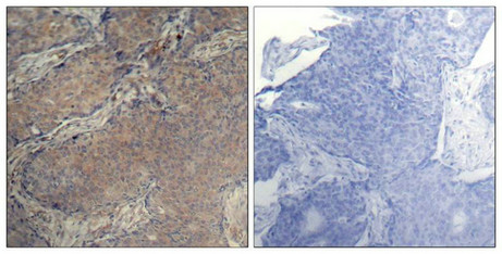
(Immunohistochemical analysis of paraffin-embedded human breast carcinoma tissue using Myosin Light Chain 2 (Phospho-Ser19) Antibody (left) or the same antibody preincubated with blocking peptide (right).)
IHC (Immunohiostchemistry)
(Immunohistochemical analysis of paraffin-embedded human breast carcinoma tissue using Myosin Light Chain 2 (Phospho-Ser19) Antibody (left) or the same antibody preincubated with blocking peptide (right).)
MYL9, Polyclonal Antibody (Cat# AAA243084)
 IF (Immunofluorescence)
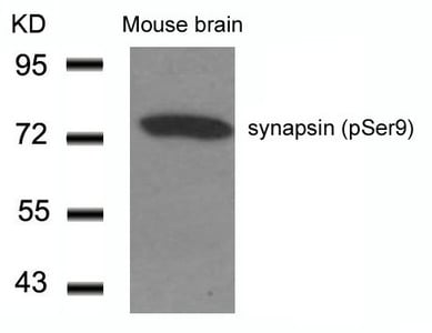
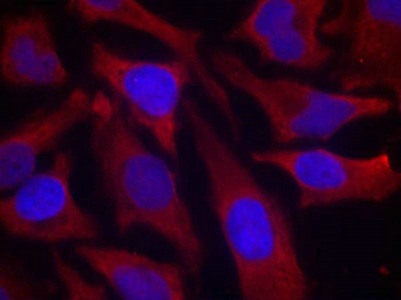
(Immunofluorescence staining of methanol-fixed Hela cells using synapsin(Phospho-Ser9) Antibody.)
IF (Immunofluorescence)
(Immunofluorescence staining of methanol-fixed Hela cells using synapsin(Phospho-Ser9) Antibody.)
SYN1, Polyclonal Antibody (Cat# AAA243134)
 IF (Immunofluorescence)
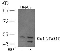
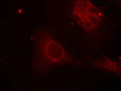
(Immunofluorescence staining of methanol-fixed Hela cells using Shc1(Phospho-Tyr349) Antibody.)
IF (Immunofluorescence)
(Immunofluorescence staining of methanol-fixed Hela cells using Shc1(Phospho-Tyr349) Antibody.)
SHC1, Polyclonal Antibody (Cat# AAA243147)
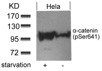
 WB (Western Blot)
(Western blot analysis of extracts from HT29 cells, treated with serum or calf intestinal phosphatase (CIP), using alpha-catenin (Phospho-Ser641) Antibody.)
WB (Western Blot)
(Western blot analysis of extracts from HT29 cells, treated with serum or calf intestinal phosphatase (CIP), using alpha-catenin (Phospho-Ser641) Antibody.)
CTNNA1, Polyclonal Antibody (Cat# AAA243153)
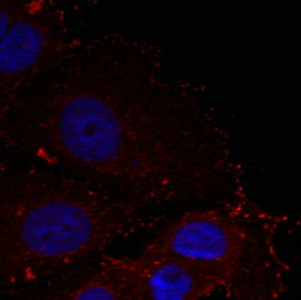 IF (Immunofluorescence)
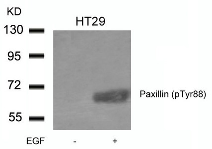
(Immunofluorescence staining of methanol-fixed Hela cells using Paxillin(phospho-Tyr88) Antibody.)
IF (Immunofluorescence)
(Immunofluorescence staining of methanol-fixed Hela cells using Paxillin(phospho-Tyr88) Antibody.)
PXN, Polyclonal Antibody (Cat# AAA243160)
 Standard Curve (Sample)
Phospho-Tyrosine Kinase (p-TrK), ELISA Kit (Cat# AAA209007)
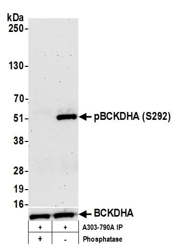
 WB (Western Blot)
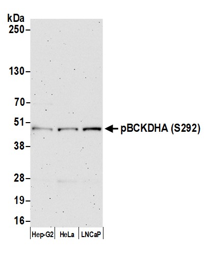
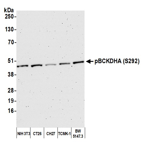
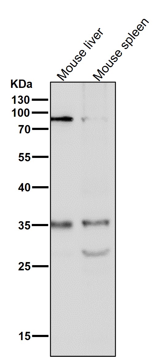
(Detection of mouse pBCKDHA (S292) by western blot. Samples: Whole cell lysate (10 ug) from NIH 3T3, CT26, CH27, TCMK-1, and BW5147.3 cells prepared using NETN lysis buffer. Antibody: Affinity purified rabbit anti-pBCKDHA (S292) antibody (AAA212852 lot 12) used for WB at 0.04 ug/ml. Detection: Chemiluminescence with an exposure time of 3 minutes.)
WB (Western Blot)
(Detection of mouse pBCKDHA (S292) by western blot. Samples: Whole cell lysate (10 ug) from NIH 3T3, CT26, CH27, TCMK-1, and BW5147.3 cells prepared using NETN lysis buffer. Antibody: Affinity purified rabbit anti-pBCKDHA (S292) antibody (AAA212852 lot 12) used for WB at 0.04 ug/ml. Detection: Chemiluminescence with an exposure time of 3 minutes.)
BCKDHA, Polyclonal Antibody (Cat# AAA212852)
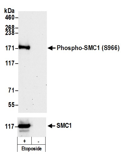 WB (Western Blot)
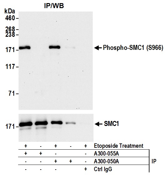
(Detection of Phospho SMC1 (S966) by western blot. Samples: Whole cell lysate (50 ug) from Jurkat cells treated with 100 uM etoposide (+) or mock treated (-). Antibodies: Affinity purified rabbit anti-Phospho SMC1 (S966) antibody AAA210736 (lot AAA210736-7) used at 0.1 ug/ml. Detection: Chemiluminescence with an exposure time of 3 minutes. For detection of total SMC1, rabbit anti-SMC1 antibody was used.)
WB (Western Blot)
(Detection of Phospho SMC1 (S966) by western blot. Samples: Whole cell lysate (50 ug) from Jurkat cells treated with 100 uM etoposide (+) or mock treated (-). Antibodies: Affinity purified rabbit anti-Phospho SMC1 (S966) antibody AAA210736 (lot AAA210736-7) used at 0.1 ug/ml. Detection: Chemiluminescence with an exposure time of 3 minutes. For detection of total SMC1, rabbit anti-SMC1 antibody was used.)
SMC1, Polyclonal Antibody (Cat# AAA210736)
 Standard Curve (Sample)
Phospho-GRK2, ELISA Kit (Cat# AAA210506)
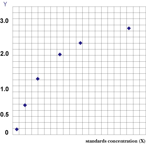 Standard Curve (Sample)
Phospho Endothelial Nitric Oxide Synthase, ELISA Kit (Cat# AAA205097)
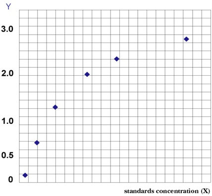 Standard Curve (Sample)
Phospho-Extracellular Signal-Regulated Kinase (PERK), ELISA Kit (Cat# AAA202883)
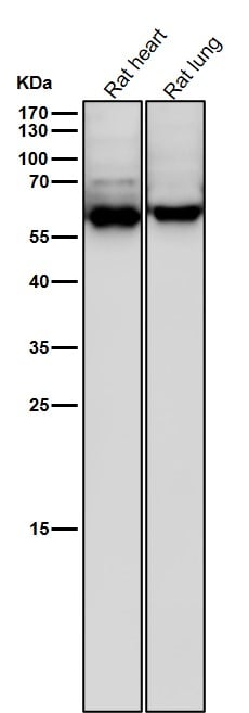
 WB (Western Blot)
(All lanes use the Antibody at 1:6K dilution for 1 hour at room temperature.)
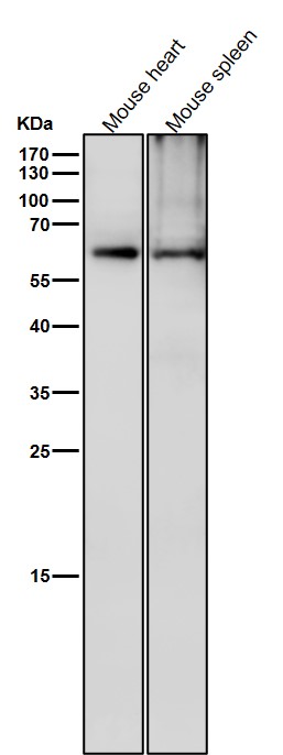
WB (Western Blot)
(All lanes use the Antibody at 1:6K dilution for 1 hour at room temperature.)
CRMP2, Monoclonal Antibody (Cat# AAA128151)
 WB (Western Blot)
(All lanes use the Antibody at 1:1K dilution for 1 hour at room temperature.)
WB (Western Blot)
(All lanes use the Antibody at 1:1K dilution for 1 hour at room temperature.)
beta Catenin, Monoclonal Antibody (Cat# AAA128101)
 WB (Western Blot)
(All lanes use the Antibody at 1:3K dilution for 1 hour at room temperature.)
WB (Western Blot)
(All lanes use the Antibody at 1:3K dilution for 1 hour at room temperature.)
Nucleophosmin, Monoclonal Antibody (Cat# AAA128117)
 WB (Western Blot)
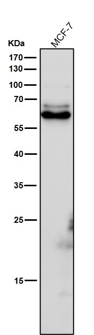
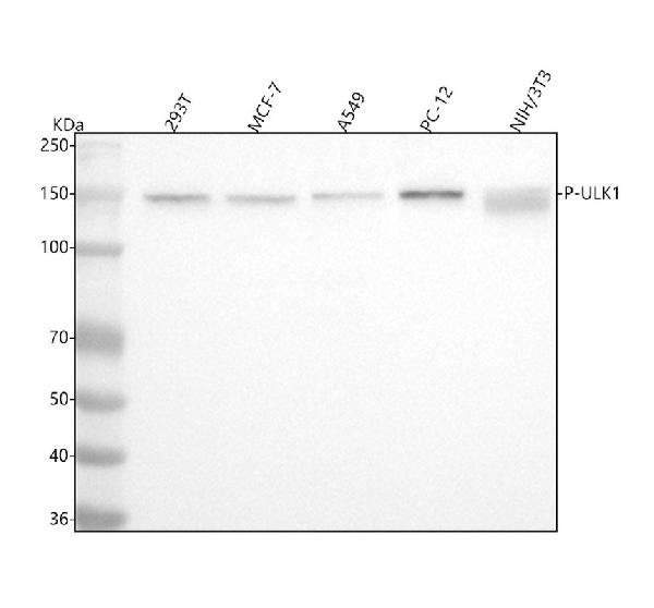
(Figure 1. Western blot analysis of ULK1 using anti-ULK1 antibody (AAA128121).Electrophoresis was performed on a 5-20% SDS-PAGE gel at 70V (Stacking gel)/90V (Resolving gel) for 2-3 hours. The sample well of each lane was loaded with 30 ug of sample under reducing conditions.Lane 1: human 293T whole cell lysates,Lane 2: human MCF-7 whole cell lysates,Lane 3: human A549 whole cell lysates,Lane 4: rat PC-12 whole cell lysates,Lane 5: mouse NIH/3T3 whole cell lysates.After electrophoresis, proteins were transferred to a nitrocellulose membrane at 150 mA for 50-90 minutes. Blocked the membrane with 5% non-fat milk/TBS for 1.5 hour at RT. The membrane was incubated with rabbit anti-ULK1 antigen affinity purified monoclonal antibody (#AAA128121) at 1:500 overnight at 4 degree C, then washed with TBS-0.1%Tween 3 times with 5 minutes each and probed with a goat anti-rabbit IgG-HRP secondary antibody at a dilution of 1:5000 for 1.5 hour at RT. The signal is developed using an Enhanced Chemiluminescent detection (ECL) kit with Tanon 5200 system. A specific band was detected for ULK1 at approximately 150 kDa. The expected band size for ULK1 is at 113 kDa.)
WB (Western Blot)
(Figure 1. Western blot analysis of ULK1 using anti-ULK1 antibody (AAA128121).Electrophoresis was performed on a 5-20% SDS-PAGE gel at 70V (Stacking gel)/90V (Resolving gel) for 2-3 hours. The sample well of each lane was loaded with 30 ug of sample under reducing conditions.Lane 1: human 293T whole cell lysates,Lane 2: human MCF-7 whole cell lysates,Lane 3: human A549 whole cell lysates,Lane 4: rat PC-12 whole cell lysates,Lane 5: mouse NIH/3T3 whole cell lysates.After electrophoresis, proteins were transferred to a nitrocellulose membrane at 150 mA for 50-90 minutes. Blocked the membrane with 5% non-fat milk/TBS for 1.5 hour at RT. The membrane was incubated with rabbit anti-ULK1 antigen affinity purified monoclonal antibody (#AAA128121) at 1:500 overnight at 4 degree C, then washed with TBS-0.1%Tween 3 times with 5 minutes each and probed with a goat anti-rabbit IgG-HRP secondary antibody at a dilution of 1:5000 for 1.5 hour at RT. The signal is developed using an Enhanced Chemiluminescent detection (ECL) kit with Tanon 5200 system. A specific band was detected for ULK1 at approximately 150 kDa. The expected band size for ULK1 is at 113 kDa.)
ULK1, Monoclonal Antibody (Cat# AAA128121)
 WB (Western Blot)
(All lanes use the Antibody at 1:1W dilution for 1 hour at room temperature.)
WB (Western Blot)
(All lanes use the Antibody at 1:1W dilution for 1 hour at room temperature.)
beta Arrestin 1, Monoclonal Antibody (Cat# AAA128142)
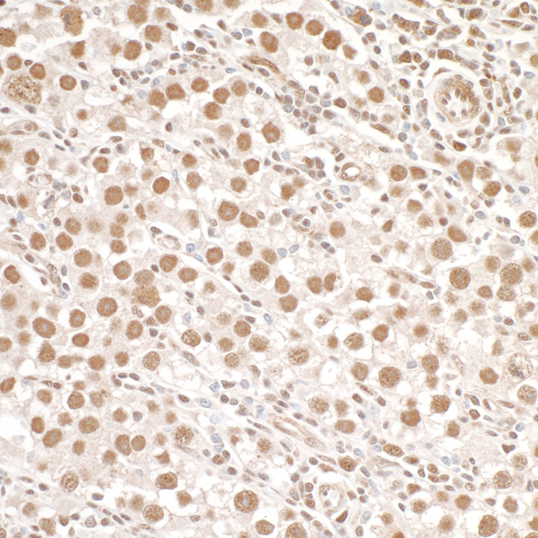
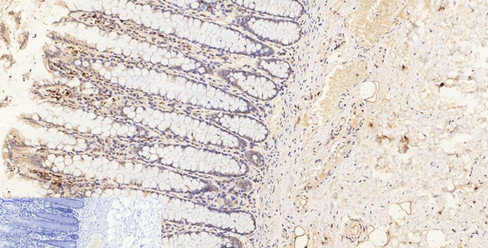
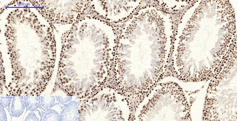
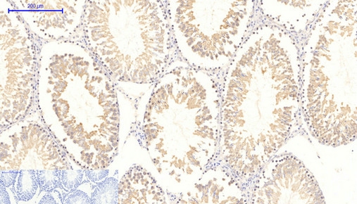
 IHC (Immunohiostchemistry)
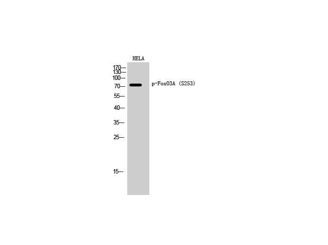
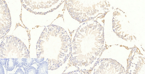
(Immunohistochemistry of paraffin-embedded Rat testis tissue with Phospho-FoxO3A (Ser253) Polyclonal Antibody at dilution of 1:200)
IHC (Immunohiostchemistry)
(Immunohistochemistry of paraffin-embedded Rat testis tissue with Phospho-FoxO3A (Ser253) Polyclonal Antibody at dilution of 1:200)
FoxO3A, Polyclonal Antibody (Cat# AAA171763)
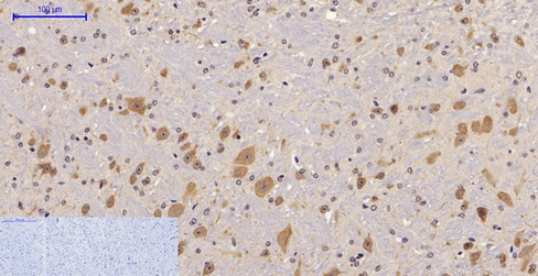 IHC (Immunohistochemisry)
(Immunohistochemistry of paraffin-embedded Mouse brain tissue with Phospho-FAK (Tyr397) Polyclonal Antibody at dilution of 1:200)
IHC (Immunohistochemisry)
(Immunohistochemistry of paraffin-embedded Mouse brain tissue with Phospho-FAK (Tyr397) Polyclonal Antibody at dilution of 1:200)
FAK, Polyclonal Antibody (Cat# AAA171768)
 WB (Western Blot)
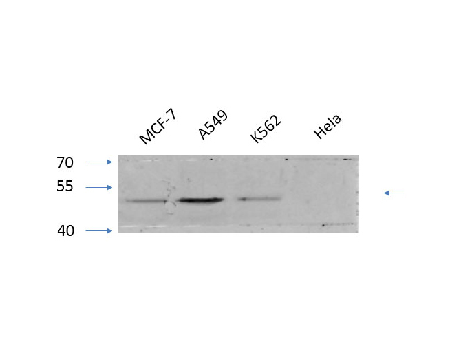
(Western Blot analysis of various cells with Phospho-Cot (Thr290) Polyclonal Antibody)
WB (Western Blot)
(Western Blot analysis of various cells with Phospho-Cot (Thr290) Polyclonal Antibody)
Cot, Polyclonal Antibody (Cat# AAA171783)
 WB (Western Blot)
(Western Blot analysis of various cells with Phospho-HNF4A (Ser313) Polyclonal Antibody at dilution of 1:1000)
WB (Western Blot)
(Western Blot analysis of various cells with Phospho-HNF4A (Ser313) Polyclonal Antibody at dilution of 1:1000)
HNF4-alpha, Polyclonal Antibody (Cat# AAA171845)
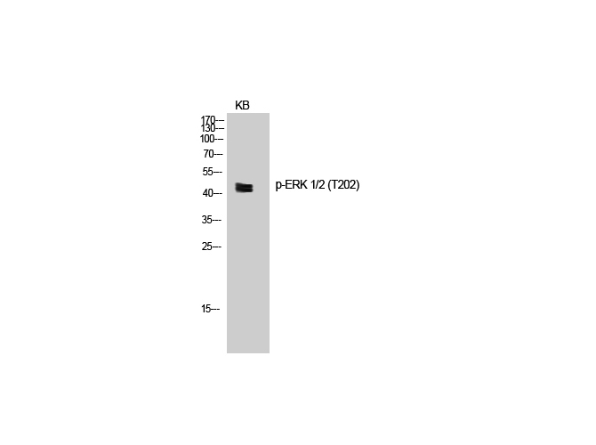
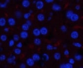
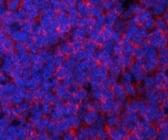 IF (Immunofluorescence)
(Immunofluorescence analysis of Mouse spleen tissue with Phospho-ERK 1/2 (Thr202) Polyclonal Antibody at dilution of 1:200)
IF (Immunofluorescence)
(Immunofluorescence analysis of Mouse spleen tissue with Phospho-ERK 1/2 (Thr202) Polyclonal Antibody at dilution of 1:200)
ERK 1/2, Polyclonal Antibody (Cat# AAA171854)
 WB (Western Blot)
(Western Blot analysis of various cells with Phospho-p21 (Thr145) Polyclonal Antibody)
WB (Western Blot)
(Western Blot analysis of various cells with Phospho-p21 (Thr145) Polyclonal Antibody)
p21, Polyclonal Antibody (Cat# AAA171859)
 IF (Immunofluorescence)
(Immunofluorescence analysis of Human lung tissue with Phospho-JNK1/2/3 (Tyr185) Polyclonal Antibody at dilution of 1:200)
IF (Immunofluorescence)
(Immunofluorescence analysis of Human lung tissue with Phospho-JNK1/2/3 (Tyr185) Polyclonal Antibody at dilution of 1:200)
JNK1/2/3, Polyclonal Antibody (Cat# AAA171865)
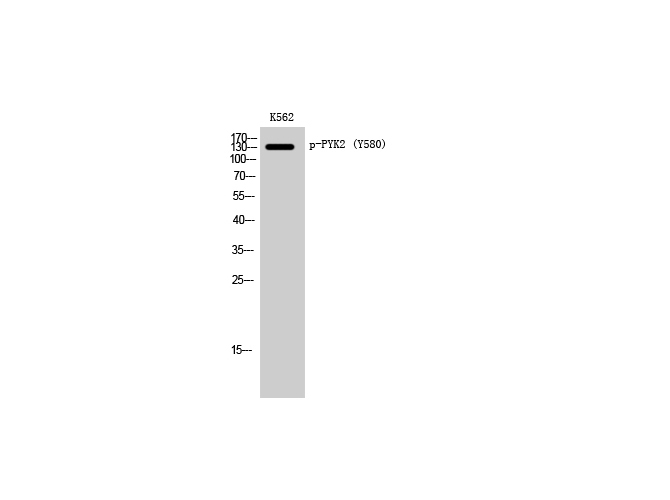 WB (Western Blot)
(Western Blot analysis of K562 cells with Phospho-PYK2 (Tyr580) Polyclonal Antibody)
WB (Western Blot)
(Western Blot analysis of K562 cells with Phospho-PYK2 (Tyr580) Polyclonal Antibody)
PYK2, Polyclonal Antibody (Cat# AAA171871)
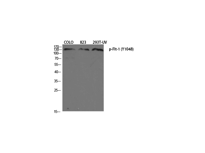 WB (Western Blot)
(Western Blot analysis of various cells with Phospho-Flt-1 (Tyr1048) Polyclonal Antibody at dilution of 1:1000)
WB (Western Blot)
(Western Blot analysis of various cells with Phospho-Flt-1 (Tyr1048) Polyclonal Antibody at dilution of 1:1000)
Flt-1, Polyclonal Antibody (Cat# AAA171910)
 WB (Western Blot)
(Western Blot analysis of various cells with Phospho-FAK (Tyr861) Polyclonal Antibody at dilution of 1:1000)
WB (Western Blot)
(Western Blot analysis of various cells with Phospho-FAK (Tyr861) Polyclonal Antibody at dilution of 1:1000)
FAK, Polyclonal Antibody (Cat# AAA171911)
 IF (Immunofluorescence)
(Immunofluorescence analysis of Mouse liver tissue with Phospho-GSK3?/? (Tyr279/216) Polyclonal Antibody at dilution of 1:200)
IF (Immunofluorescence)
(Immunofluorescence analysis of Mouse liver tissue with Phospho-GSK3?/? (Tyr279/216) Polyclonal Antibody at dilution of 1:200)
GSK3alpha/beta, Polyclonal Antibody (Cat# AAA171664)
 WB (Western Blot)
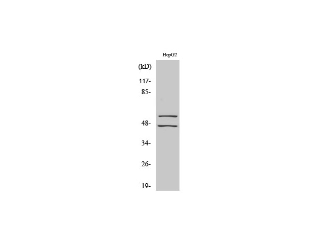
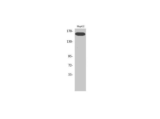
(Western Blot analysis of HepG2 cells with Phospho-FLK1 (Tyr1214) Polyclonal Antibody)
WB (Western Blot)
(Western Blot analysis of HepG2 cells with Phospho-FLK1 (Tyr1214) Polyclonal Antibody)
Flk-1, Polyclonal Antibody (Cat# AAA171925)
 IF (Immunofluorescence)
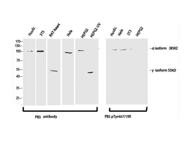
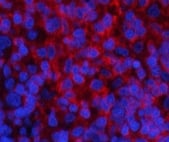
(Immunofluorescence analysis of Rat spleen tissue with Phospho-PI 3-kinase p85/p55 (Tyr467/199) Polyclonal Antibody at dilution of 1:200)
IF (Immunofluorescence)
(Immunofluorescence analysis of Rat spleen tissue with Phospho-PI 3-kinase p85/p55 (Tyr467/199) Polyclonal Antibody at dilution of 1:200)
PI 3-kinase p85/p55, Polyclonal Antibody (Cat# AAA171680)
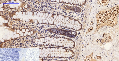
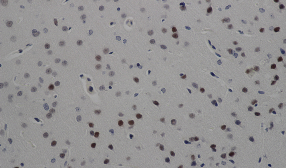 IHC (Immunohiostchemistry)
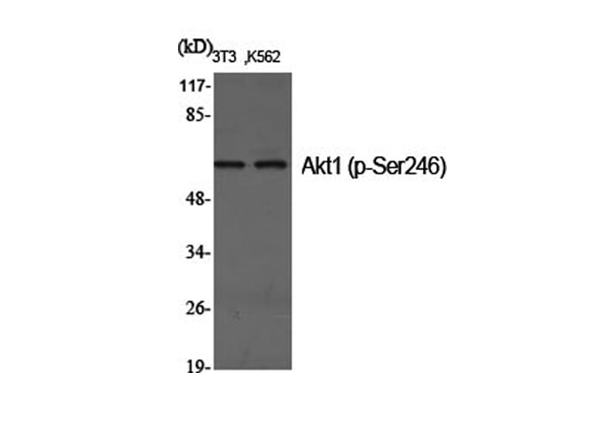
(Immunohistochemistry of paraffin-embedded Rat brain with Phospho-Akt1 (Ser246) Polyclonal Antibody at dilution of 1:200)
IHC (Immunohiostchemistry)
(Immunohistochemistry of paraffin-embedded Rat brain with Phospho-Akt1 (Ser246) Polyclonal Antibody at dilution of 1:200)
Akt1, Polyclonal Antibody (Cat# AAA171710)
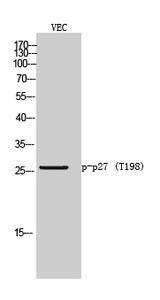 WB (Western Blot)
(Western Blot analysis of VEC cells with Phospho-p27 (Thr198) Polyclonal Antibody)
WB (Western Blot)
(Western Blot analysis of VEC cells with Phospho-p27 (Thr198) Polyclonal Antibody)
p27, Polyclonal Antibody (Cat# AAA171746)
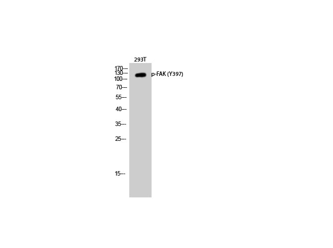
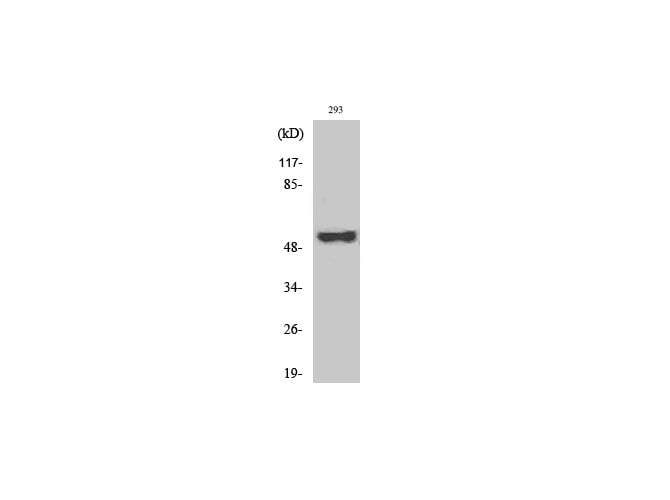
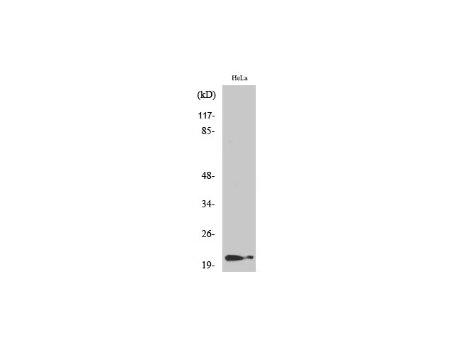
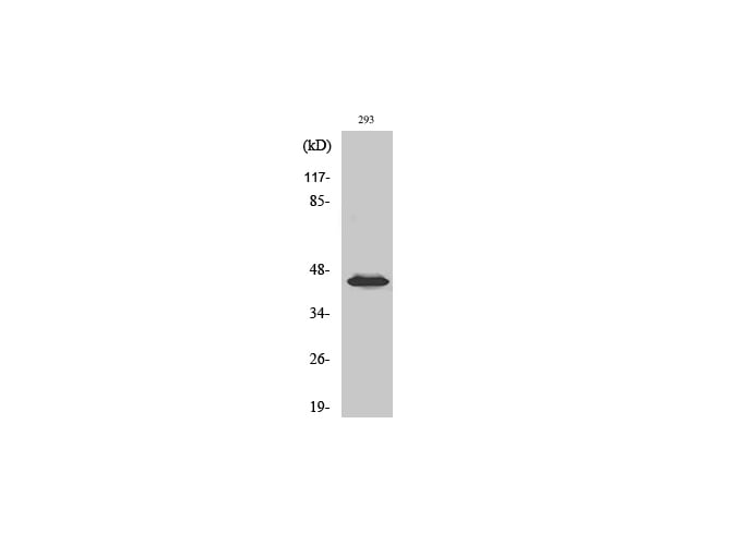
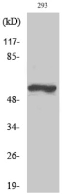 WB (Western Blot)
(Western Blot analysis of 293T cells with Phospho-TH (Ser19) Polyclonal Antibody at dilution of 1:1000)
WB (Western Blot)
(Western Blot analysis of 293T cells with Phospho-TH (Ser19) Polyclonal Antibody at dilution of 1:1000)
TH, Polyclonal Antibody (Cat# AAA171750)
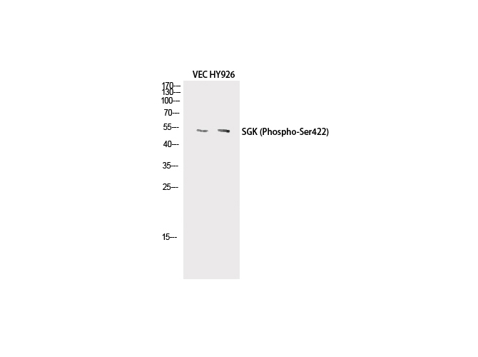 WB (Western Blot)
(Western Blot analysis of VEC, HY926 cells with Phospho-SGK1 (Ser422) Polyclonal Antibody at dilution of 1:500)
WB (Western Blot)
(Western Blot analysis of VEC, HY926 cells with Phospho-SGK1 (Ser422) Polyclonal Antibody at dilution of 1:500)
SGK1, Polyclonal Antibody (Cat# AAA172042)
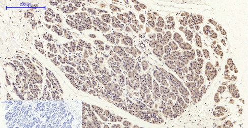
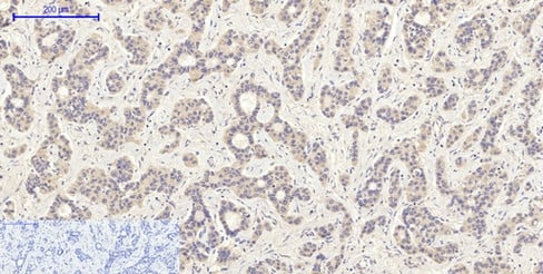
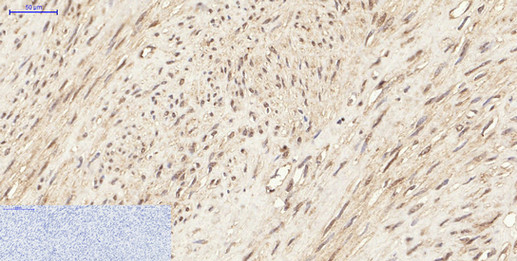
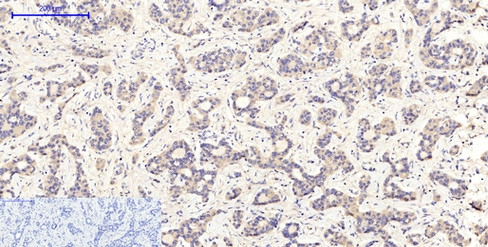 IHC (Immunohiostchemistry)
(Immunohistochemistry of paraffin-embedded Human liver cancer tissue with Phospho-MEK-1/2 (Ser218/222) Polyclonal Antibody at dilution of 1:200)
IHC (Immunohiostchemistry)
(Immunohistochemistry of paraffin-embedded Human liver cancer tissue with Phospho-MEK-1/2 (Ser218/222) Polyclonal Antibody at dilution of 1:200)
MEK-1/2, Polyclonal Antibody (Cat# AAA172055)
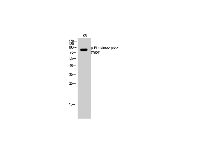
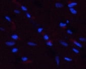 IF (Immunofluorescence)
(Immunofluorescence analysis of Rat heart tissue with Phospho-PI 3-kinase p85? (Tyr607) Polyclonal Antibody at dilution of 1:200)
IF (Immunofluorescence)
(Immunofluorescence analysis of Rat heart tissue with Phospho-PI 3-kinase p85? (Tyr607) Polyclonal Antibody at dilution of 1:200)
PI 3-kinase p85alpha, Polyclonal Antibody (Cat# AAA172246)
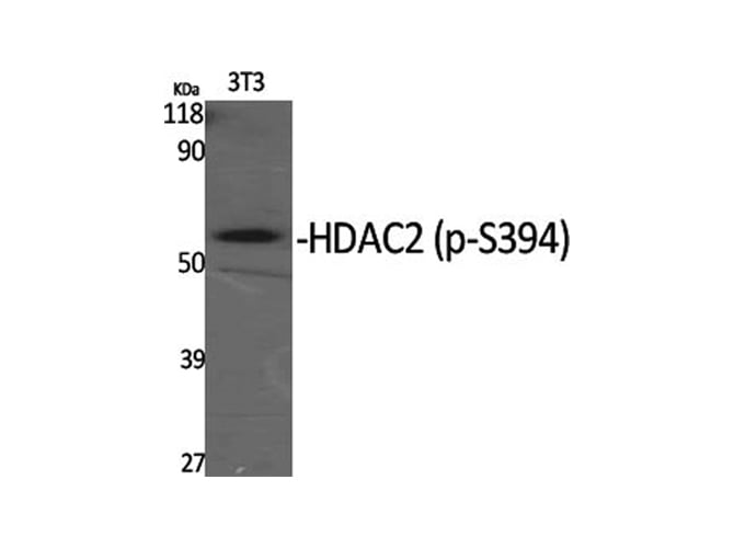 WB (Western Blot)
(Western Blot analysis of various cells with Phospho-HDAC2 (Ser394) Polyclonal Antibody)
WB (Western Blot)
(Western Blot analysis of various cells with Phospho-HDAC2 (Ser394) Polyclonal Antibody)
HDAC2, Polyclonal Antibody (Cat# AAA172255)
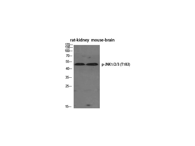
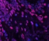
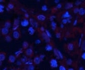 IF (Immunofluorescence)
(Immunofluorescence analysis of Rat testis tissue with Phospho-JNK1/2/3 (Thr183) Polyclonal Antibody at dilution of 1:200)
IF (Immunofluorescence)
(Immunofluorescence analysis of Rat testis tissue with Phospho-JNK1/2/3 (Thr183) Polyclonal Antibody at dilution of 1:200)
JNK1/2/3, Polyclonal Antibody (Cat# AAA172265)
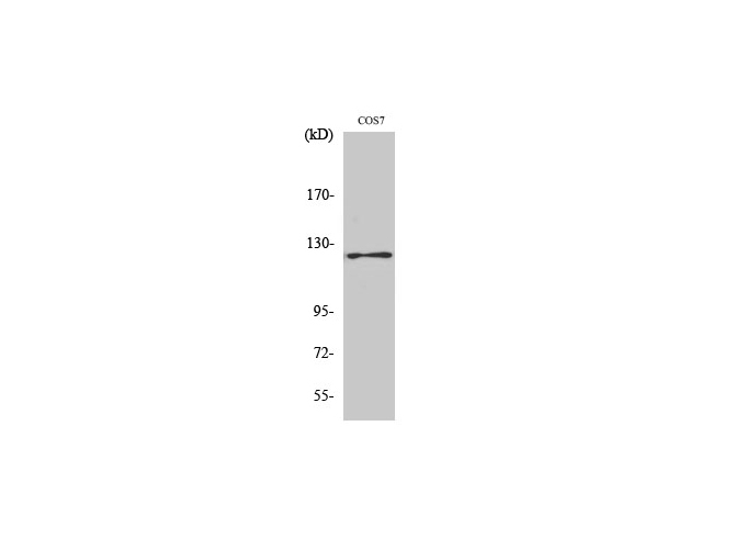
 WB (Western Blot)
(Western Blot analysis of HT-29, COS7 cells with Phospho-SNAI 1 (Ser246) Polyclonal Antibody at dilution of 1:500)
WB (Western Blot)
(Western Blot analysis of HT-29, COS7 cells with Phospho-SNAI 1 (Ser246) Polyclonal Antibody at dilution of 1:500)
SNAI 1, Polyclonal Antibody (Cat# AAA172372)
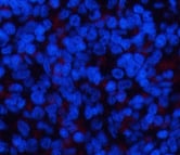 IF (Immunofluorescence)
(Immunofluorescence analysis of Rat spleen tissue with Phospho-ERK 1/2 (Tyr204) Polyclonal Antibody at dilution of 1:200)
IF (Immunofluorescence)
(Immunofluorescence analysis of Rat spleen tissue with Phospho-ERK 1/2 (Tyr204) Polyclonal Antibody at dilution of 1:200)
ERK 1/2, Polyclonal Antibody (Cat# AAA172558)
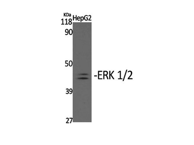
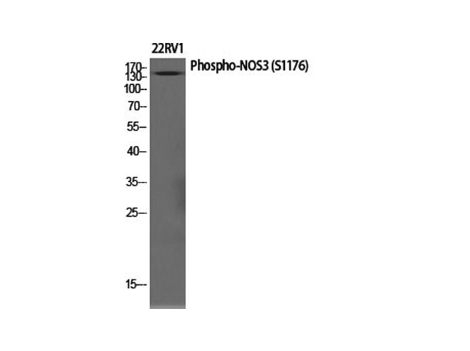 WB (Western Blot)
(Western Blot analysis of various cells with Phospho-NOS3 (Ser1177) Polyclonal Antibody at dilution of 1:1000)
WB (Western Blot)
(Western Blot analysis of various cells with Phospho-NOS3 (Ser1177) Polyclonal Antibody at dilution of 1:1000)
NOS3, Polyclonal Antibody (Cat# AAA172600)
What Are Phospho Antibodies?
Protein phosphorylation is a process where a phosphate group is added to certain amino acid residues of a protein – usually serine (S), threonine (T), or tyrosine (Y) - by enzymes called kinases. This process is integral in controlling cellular signaling, cellular growth, and other biological functions.
Our catalog includes a wide range of phospho-specific antibodies that can accurately detect this important marker. They perform strongly in widely-used laboratory applications such as Western blot, flow cytometry, immunohistochemistry, and immunofluorescence microscopy. We value your trust in us and are committed to providing top-quality products and services. All of our antibodies are guaranteed to work for the applications and species indicated on our website & associated product pages.
What Are The Key Applications of Phospho Antibodies?
1. Western Blotting
One of the first steps a researcher can take in utilizing these phospho-specific antibodies, is to check if the antibody works using a technique referred to as “Western blot”. For those unfamiliar, Western Blot aids in showing whether the protein that the antibody recognizes is appearing at the correct/expected size. These phospho-specific antibodies should also be able to detect changes in the target protein’s phosphorylation (on/off state) when cells are stimulated in certain ways.
2. Staining of Fixed Cells (Immunocytochemistry)
Another routine use of these phospho-specific antibodies, is to test if the antibody is able to demonstrate similar performance when used on fixed cells (intact cells that have been preserved) as it did in the Western blot tests. It is an important aspect in many cases to confirm that the antibody works in actual intact cell samples. Ideally, the method used for cellular fixation should be the same as what is used in pathology labs (like using 10% formalin). To check if the antibody works well in tissue sections (FFPE), researchers will often test it on fixed cells that are processed similar to tissue samples.
3. Specificity Tests Using Peptides
In order to make sure that the antibody is only binding to the right target:
- Laboratory technicians will mix the antibody with phospho-peptides (short segments of the protein containing the phosphate group modification).
- If the antibody signal disappears, it is confirmation that it is binding to the correct phosphorylated location.
- A more robust test is to use both the phosphorylated and non-phosphorylated (dephosphorylated) versions of the protein. The antibody should react only with the phosphorylated one.
- Another method sometimes utilized is to treat the sample with an enzyme, such as alkaline phosphatase, that specifically removes phosphate groups. If the antibody signal disappears after this, it also confirms specificity.
4. Genetic Confirmation
As a final step, scientists can genetically manipulate the nucleotide sequence and alter the target protein by removing the exact site where phosphorylation happens. If the antibody no longer appears to detect the modified protein, it is strong evidence supporting the antibody being specific for that phosphorylated site.
Why Buy Phospho Antibodies Through Us?
- The production laboratory adheres to strict and consistent protocols prior to releasing any of these phospho-specific antibodies:
- Standard methods and proper controls in all tests to ensure high quality.
- These antibodies are tested and validated in different cell types and species.
- High quality control criterion to ensure each batch is consistent, so you will obtain reliable results every time.
FAQ
1. What Are Phospho-Specific Antibodies?
Phospho-specific antibodies are made to detect proteins only when they have a phosphate group linked to a specific amino acid residue. This empowers scientists understand if a protein is "turned on" or active, based on its phosphorylation state.
2. How to Detect Phosphorylated Proteins in a Western Blot?
To find out if a protein is phosphorylated using Western blot:
- Use a phospho-specific antibody that binds only to the phosphorylated form of the protein.
- You can also use a “regular” antibody for the same amino acid sequence of the protein that the phospho-specific antibody is binding to (but in this case, this antibody will not bind if there is a phosphate group present) in order to compare how much of it is phosphorylated versus how much is non-phosphorylated (or “total” protein, if the “normal” antibody’s epitopes are non-phospho-site-specific).
3. How to Choose the Best Antibody?
Here are some simple tips to help you pick the right antibody:
- Know your target
- Match your sample characteristics
- Confirm the intended use is appropriate
- Check “host” and “type”
- Check the “quality” of the presented data/images
- Appraise whether the available validation meets your needs