Filters
▼Clonality
▼Type
▼Reactivity
▼Gene Name
▼Isotype
▼Host
▼Application
▼Clone
▼Monoclonal Antibodies
Get accurate results in your research with our Monoclonal Antibodies, which are specially made to target exactly what you require for your research, and will produce consistent, reliable performance in lab tests.
Viewing 2250-2300 of 27597 product results
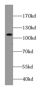 WB (Western Blot)
(human plasma tissue were subjected to SDS PAGE followed by western blot with AAA102716 (C3 Antibody) at dilution of 1:2000)
WB (Western Blot)
(human plasma tissue were subjected to SDS PAGE followed by western blot with AAA102716 (C3 Antibody) at dilution of 1:2000)
C3 / C3b / C3c, Monoclonal Antibody (Cat# AAA102716)
 WB (Western Blot)
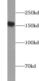
(U-937 cells were subjected to SDS PAGE followed by western blot with AAA102726 (ANPEP Antibody) at dilution of 1:3000)
WB (Western Blot)
(U-937 cells were subjected to SDS PAGE followed by western blot with AAA102726 (ANPEP Antibody) at dilution of 1:3000)
CD13, Monoclonal Antibody (Cat# AAA102726)
Protein A+G purification
 IP (Immunoprecipitation)
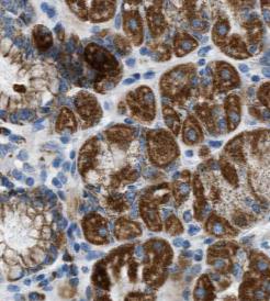
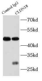
(IP Result of anti-Claudin 18 (IP: AAA102748, 3ug; Detection: AAA102748 1:500) with mouse stomach tissue lysate 3000ug.)
IP (Immunoprecipitation)
(IP Result of anti-Claudin 18 (IP: AAA102748, 3ug; Detection: AAA102748 1:500) with mouse stomach tissue lysate 3000ug.)
Claudin 18, Monoclonal Antibody (Cat# AAA102748)
Protein A+G purification
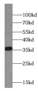 WB (Western Blot)
(pig stomach tissue were subjected to SDS PAGE followed by western blot with AAA102751 (CNN2 Antibody) at dilution of 1:2000)
WB (Western Blot)
(pig stomach tissue were subjected to SDS PAGE followed by western blot with AAA102751 (CNN2 Antibody) at dilution of 1:2000)
CNN2, Monoclonal Antibody (Cat# AAA102751)
Protein A+G purification
 WB (Western Blot)
(fetal human brain tissue were subjected to SDS PAGE followed by western blot with AAA102755 (COTL1 Antibody) at dilution of 1:2000)
WB (Western Blot)
(fetal human brain tissue were subjected to SDS PAGE followed by western blot with AAA102755 (COTL1 Antibody) at dilution of 1:2000)
COTL1, Monoclonal Antibody (Cat# AAA102755)
Protein A+G purification
 WB (Western Blot)
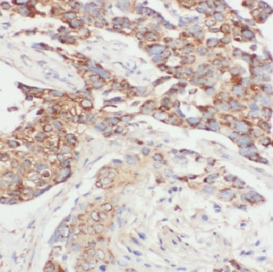
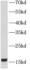
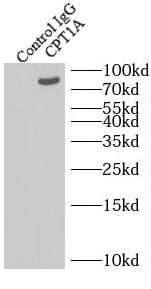
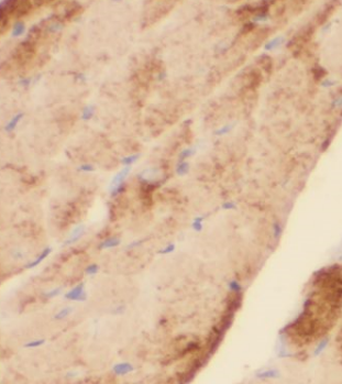
(L02 cells were subjected to SDS PAGE followed by western blot with AAA102757 (CPT1A antibody) at dilution of 1:500)
WB (Western Blot)
(L02 cells were subjected to SDS PAGE followed by western blot with AAA102757 (CPT1A antibody) at dilution of 1:500)
CPT1A, Monoclonal Antibody (Cat# AAA102757)
Protein A+G purification
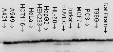
 WB (Western Blot)
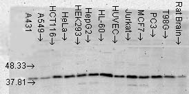
(Western blot of c-SRC in various cell lines with AAA102762 at dilution of 1:4000. AAA102762)
WB (Western Blot)
(Western blot of c-SRC in various cell lines with AAA102762 at dilution of 1:4000. AAA102762)
c-SRC, Monoclonal Antibody (Cat# AAA102762)
Protein A+G purification
 WB (Western Blot)
(Hela cells were subjected to SDS PAGE followed by western blot with AAA102770 (CYPB antibody) at dilution of 1:2000)
WB (Western Blot)
(Hela cells were subjected to SDS PAGE followed by western blot with AAA102770 (CYPB antibody) at dilution of 1:2000)
Cyclophilin B, Monoclonal Antibody (Cat# AAA102770)
Protein A+G purification
 WB (Western Blot)
(HeLa cells were subjected to SDS PAGE followed by western blot with AAA102681 (GLA antibody) at dilution of 1:1000)
WB (Western Blot)
(HeLa cells were subjected to SDS PAGE followed by western blot with AAA102681 (GLA antibody) at dilution of 1:1000)
Alpha galactosidase A, Monoclonal Antibody (Cat# AAA102681)


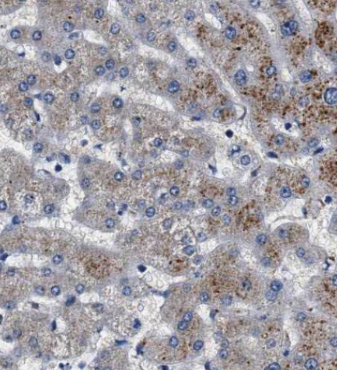
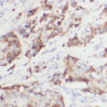
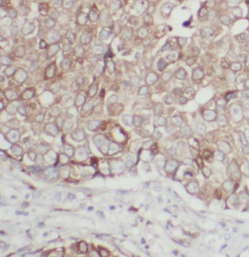
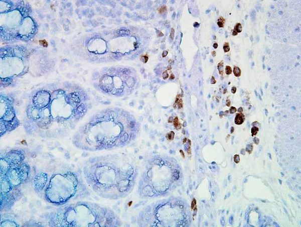
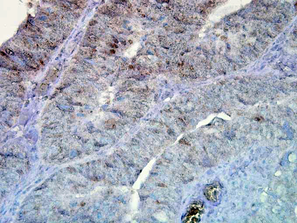
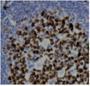
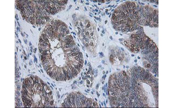
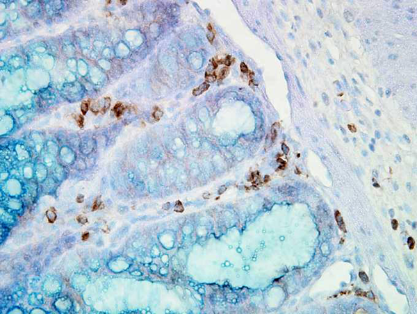 IHC (Immunohistochemisry)
(Immunohistochemistry analysis using Mouse Anti-Hsp90 alpha Monoclonal Antibody, Clone K41009. Tissue: inflamed colon. Species: Mouse. Fixation: Formalin. Primary Antibody: Mouse Anti-Hsp90 alpha Monoclonal Antibody at 1:5000 for 12 hours at 4 degree C. Secondary Antibody: Biotin Goat Anti-Mouse at 1:2000 for 1 hour at RT. Counterstain: Mayer Hematoxylin (purple/blue) nuclear stain at 200 ul for 2 minutes at RT. Localization: Inflammatory cells. Magnification: 40x. Inflammatory cells.)
IHC (Immunohistochemisry)
(Immunohistochemistry analysis using Mouse Anti-Hsp90 alpha Monoclonal Antibody, Clone K41009. Tissue: inflamed colon. Species: Mouse. Fixation: Formalin. Primary Antibody: Mouse Anti-Hsp90 alpha Monoclonal Antibody at 1:5000 for 12 hours at 4 degree C. Secondary Antibody: Biotin Goat Anti-Mouse at 1:2000 for 1 hour at RT. Counterstain: Mayer Hematoxylin (purple/blue) nuclear stain at 200 ul for 2 minutes at RT. Localization: Inflammatory cells. Magnification: 40x. Inflammatory cells.)
HSP90 alpha, Monoclonal Antibody (Cat# AAA103886)

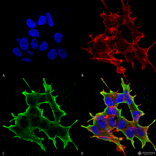
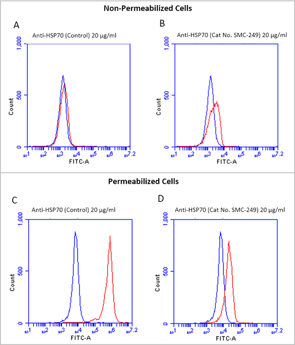 IF (Immunofluorescence)
(Fluorescence-activated cell sorting analysis using Mouse Anti-HSP70 Monoclonal Antibody, Clone 1H11. Tissue: Jurkat E6.1 cells. Species: Human. Fixation: No fixation. Primary Antibody: Mouse Anti-HSP70 Monoclonal Antibody at 20 ug/ml for 40 min at 4 degree C. Counterstain: Propidium Iodide nuclear stain at 2.5 ug/ml for 5 min at RT. Isotype Control: Anti-mouse FITC at 1:32 for 15 min at RT (blue line). Courtesy of: Dr. Elyse Ireland, Institute of Medicine, University of Chester.)
IF (Immunofluorescence)
(Fluorescence-activated cell sorting analysis using Mouse Anti-HSP70 Monoclonal Antibody, Clone 1H11. Tissue: Jurkat E6.1 cells. Species: Human. Fixation: No fixation. Primary Antibody: Mouse Anti-HSP70 Monoclonal Antibody at 20 ug/ml for 40 min at 4 degree C. Counterstain: Propidium Iodide nuclear stain at 2.5 ug/ml for 5 min at RT. Isotype Control: Anti-mouse FITC at 1:32 for 15 min at RT (blue line). Courtesy of: Dr. Elyse Ireland, Institute of Medicine, University of Chester.)
HSP70, Monoclonal Antibody (Cat# AAA103912)
 IF (Immunofluorescence)
(Fluorescence-activated cell sorting analysis using Mouse Anti-HSP70 Monoclonal Antibody, Clone 1H11. Tissue: Jurkat E6.1 cells. Species: Human. Fixation: No fixation. Primary Antibody: Mouse Anti-HSP70 Monoclonal Antibody at 20 ug/ml for 40 min at 4 degree C. Counterstain: Propidium Iodide nuclear stain at 2.5 ug/ml for 5 min at RT. Isotype Control: Anti-mouse FITC at 1:32 for 15 min at RT (blue line). Courtesy of: Dr. Elyse Ireland, Institute of Medicine, University of Chester.)
IF (Immunofluorescence)
(Fluorescence-activated cell sorting analysis using Mouse Anti-HSP70 Monoclonal Antibody, Clone 1H11. Tissue: Jurkat E6.1 cells. Species: Human. Fixation: No fixation. Primary Antibody: Mouse Anti-HSP70 Monoclonal Antibody at 20 ug/ml for 40 min at 4 degree C. Counterstain: Propidium Iodide nuclear stain at 2.5 ug/ml for 5 min at RT. Isotype Control: Anti-mouse FITC at 1:32 for 15 min at RT (blue line). Courtesy of: Dr. Elyse Ireland, Institute of Medicine, University of Chester.)
HSP70, Monoclonal Antibody (Cat# AAA103913)
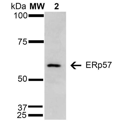 WB (Western Blot)
(Western Blot analysis of Human Cervical Cancer cell line (HeLa) showing detection of 57 kDa Erp57 protein using Mouse Anti-Erp57 Monoclonal Antibody, Clone 4F9 . Lane 1: Molecular Weight Ladder (MW). Lane 2: HeLa cell lysate. Load: 15 ug. Block: 5% Skim Milk in TBST. Primary Antibody: Mouse Anti-Erp57 Monoclonal Antibody at 1:1000 for 2 hours at RT. Secondary Antibody: Goat Anti-Mouse IgG: HRP at 1:1000 for 60 min at RT. Color Development: ECL solution for 5 min in RT. Predicted/Observed Size: 57 kDa.)
WB (Western Blot)
(Western Blot analysis of Human Cervical Cancer cell line (HeLa) showing detection of 57 kDa Erp57 protein using Mouse Anti-Erp57 Monoclonal Antibody, Clone 4F9 . Lane 1: Molecular Weight Ladder (MW). Lane 2: HeLa cell lysate. Load: 15 ug. Block: 5% Skim Milk in TBST. Primary Antibody: Mouse Anti-Erp57 Monoclonal Antibody at 1:1000 for 2 hours at RT. Secondary Antibody: Goat Anti-Mouse IgG: HRP at 1:1000 for 60 min at RT. Color Development: ECL solution for 5 min in RT. Predicted/Observed Size: 57 kDa.)
ERp57, Monoclonal Antibody (Cat# AAA103926)
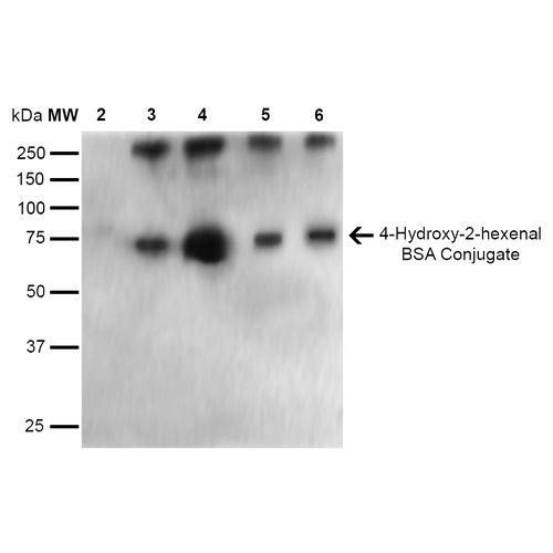
 WB (Western Blot)
(Western Blot analysis of Human Cervical cancer cell line (HeLa) lysate showing detection of Acrolein protein using Mouse Anti-Acrolein Monoclonal Antibody, Clone 10A10. Lane 1: Molecular Weight Ladder (MW). Lane 2: HeLa cell lysate. Lane 3: H2O2 treated HeLa cell lysate. Load: 12 ug. Block: 5% Skim Milk in TBST. Primary Antibody: Mouse Anti-Acrolein Monoclonal Antibody at 1:1000 for 2 hours at RT. Secondary Antibody: Goat Anti-Mouse IgG: HRP at 1:2000 for 60 min at RT. Color Development: ECL solution for 5 min in RT.)
WB (Western Blot)
(Western Blot analysis of Human Cervical cancer cell line (HeLa) lysate showing detection of Acrolein protein using Mouse Anti-Acrolein Monoclonal Antibody, Clone 10A10. Lane 1: Molecular Weight Ladder (MW). Lane 2: HeLa cell lysate. Lane 3: H2O2 treated HeLa cell lysate. Load: 12 ug. Block: 5% Skim Milk in TBST. Primary Antibody: Mouse Anti-Acrolein Monoclonal Antibody at 1:1000 for 2 hours at RT. Secondary Antibody: Goat Anti-Mouse IgG: HRP at 1:2000 for 60 min at RT. Color Development: ECL solution for 5 min in RT.)
Acrolein, Monoclonal Antibody (Cat# AAA104012)

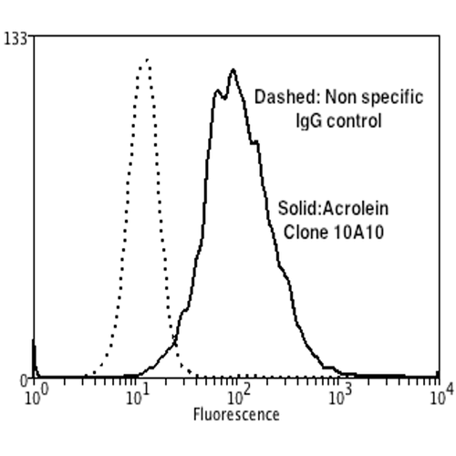
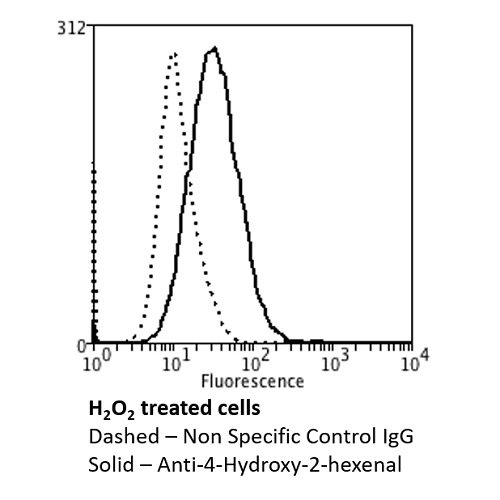
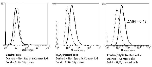
 FCM/FACS (Flow Cytometry)
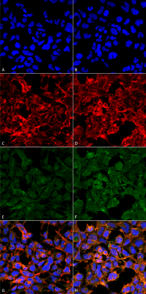
(Flow Cytometry analysis using Mouse Anti-4-hydroxy-2-hexenal Monoclonal Antibody, Clone 6F10. Tissue: Neuroblastoma cells (SH-SY5Y). Species: Human. Fixation: 90% Methanol. Primary Antibody: Mouse Anti-4-hydroxy-2-hexenal Monoclonal Antibody at 1:50 for 30 min on ice. Secondary Antibody: Goat Anti-Mouse: PE at 1:100 for 20 min at RT. Isotype Control: Non Specific IgG. Cells were subject to oxidative stress by treating with 250 uM H2O2 for 24 hours.)
FCM/FACS (Flow Cytometry)
(Flow Cytometry analysis using Mouse Anti-4-hydroxy-2-hexenal Monoclonal Antibody, Clone 6F10. Tissue: Neuroblastoma cells (SH-SY5Y). Species: Human. Fixation: 90% Methanol. Primary Antibody: Mouse Anti-4-hydroxy-2-hexenal Monoclonal Antibody at 1:50 for 30 min on ice. Secondary Antibody: Goat Anti-Mouse: PE at 1:100 for 20 min at RT. Isotype Control: Non Specific IgG. Cells were subject to oxidative stress by treating with 250 uM H2O2 for 24 hours.)
4-Hydroxy-2-hexenal, Monoclonal Antibody (Cat# AAA104033)

 FCM/FACS (Flow Cytometry)
(Flow Cytometry analysis using Mouse Anti-4-hydroxy-2-hexenal Monoclonal Antibody, Clone 6F10. Tissue: Neuroblastoma cells (SH-SY5Y). Species: Human. Fixation: 90% Methanol. Primary Antibody: Mouse Anti-4-hydroxy-2-hexenal Monoclonal Antibody at 1:50 for 30 min on ice. Secondary Antibody: Goat Anti-Mouse: PE at 1:100 for 20 min at RT. Isotype Control: Non Specific IgG. Cells were subject to oxidative stress by treating with 250 uM H2O2 for 24 hours.)
FCM/FACS (Flow Cytometry)
(Flow Cytometry analysis using Mouse Anti-4-hydroxy-2-hexenal Monoclonal Antibody, Clone 6F10. Tissue: Neuroblastoma cells (SH-SY5Y). Species: Human. Fixation: 90% Methanol. Primary Antibody: Mouse Anti-4-hydroxy-2-hexenal Monoclonal Antibody at 1:50 for 30 min on ice. Secondary Antibody: Goat Anti-Mouse: PE at 1:100 for 20 min at RT. Isotype Control: Non Specific IgG. Cells were subject to oxidative stress by treating with 250 uM H2O2 for 24 hours.)
4-Hydroxy-2-hexenal, Monoclonal Antibody (Cat# AAA104035)


 FCM/FACS (Flow Cytometry)
(Flow Cytometry analysis using Mouse Anti-4-hydroxy-2-hexenal Monoclonal Antibody, Clone 6F10. Tissue: Neuroblastoma cells (SH-SY5Y). Species: Human. Fixation: 90% Methanol. Primary Antibody: Mouse Anti-4-hydroxy-2-hexenal Monoclonal Antibody at 1:50 for 30 min on ice. Secondary Antibody: Goat Anti-Mouse: PE at 1:100 for 20 min at RT. Isotype Control: Non Specific IgG. Cells were subject to oxidative stress by treating with 250 uM H2O2 for 24 hours.)
FCM/FACS (Flow Cytometry)
(Flow Cytometry analysis using Mouse Anti-4-hydroxy-2-hexenal Monoclonal Antibody, Clone 6F10. Tissue: Neuroblastoma cells (SH-SY5Y). Species: Human. Fixation: 90% Methanol. Primary Antibody: Mouse Anti-4-hydroxy-2-hexenal Monoclonal Antibody at 1:50 for 30 min on ice. Secondary Antibody: Goat Anti-Mouse: PE at 1:100 for 20 min at RT. Isotype Control: Non Specific IgG. Cells were subject to oxidative stress by treating with 250 uM H2O2 for 24 hours.)
4-Hydroxy-2-hexenal, Monoclonal Antibody (Cat# AAA104036)
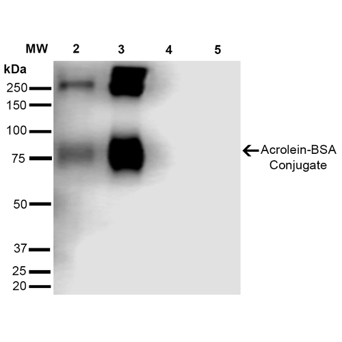
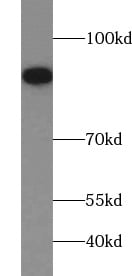
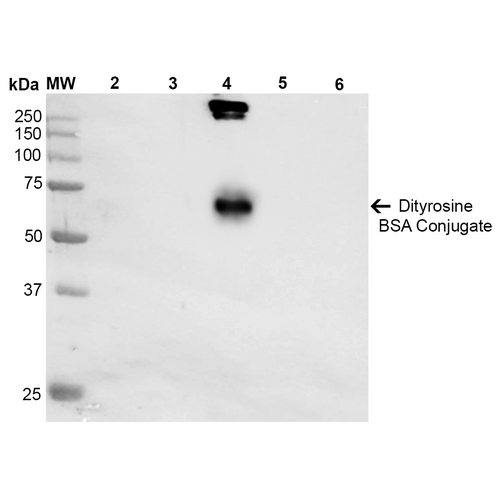
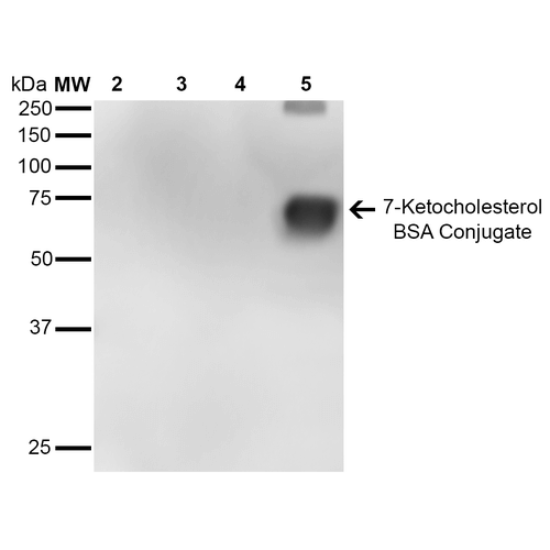 WB (Western Blot)
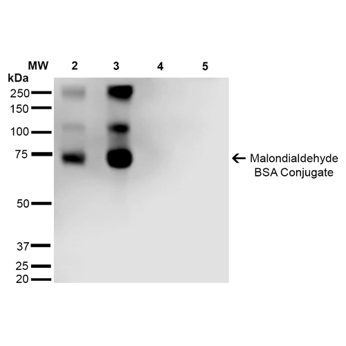
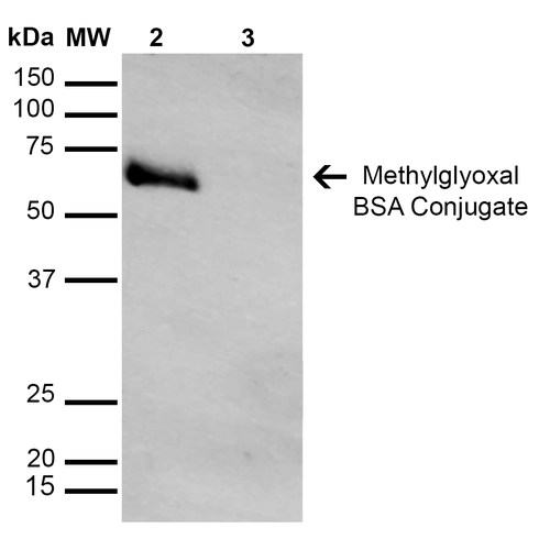
(Western Blot analysis of 7-Ketocholesterol-BSA Conjugate showing detection of 67 kDa 7-Ketocholesterol protein using Mouse Anti-7-Ketocholesterol Monoclonal Antibody, Clone 7E1. Lane 1: Molecular Weight Ladder (MW). Lane 2: BSA (0.5 ug). Lane 3: BSA (2.0 ug). Lane 4: 7-ketocholesterol-BSA (0.5 ug). Lane 5: 7-ketocholesterol-BSA (2.0 ug). Block: 5% Skim Milk in TBST. Primary Antibody: Mouse Anti-7-Ketocholesterol Monoclonal Antibody at 1:1000 for 2 hours at RT. Secondary Antibody: Goat Anti-Mouse IgG: HRP at 1:2000 for 60 min at RT. Color Development: ECL solution for 5 min in RT. Predicted/Observed Size: 67 kDa.)
WB (Western Blot)
(Western Blot analysis of 7-Ketocholesterol-BSA Conjugate showing detection of 67 kDa 7-Ketocholesterol protein using Mouse Anti-7-Ketocholesterol Monoclonal Antibody, Clone 7E1. Lane 1: Molecular Weight Ladder (MW). Lane 2: BSA (0.5 ug). Lane 3: BSA (2.0 ug). Lane 4: 7-ketocholesterol-BSA (0.5 ug). Lane 5: 7-ketocholesterol-BSA (2.0 ug). Block: 5% Skim Milk in TBST. Primary Antibody: Mouse Anti-7-Ketocholesterol Monoclonal Antibody at 1:1000 for 2 hours at RT. Secondary Antibody: Goat Anti-Mouse IgG: HRP at 1:2000 for 60 min at RT. Color Development: ECL solution for 5 min in RT. Predicted/Observed Size: 67 kDa.)
7-Ketocholesterol, Monoclonal Antibody (Cat# AAA104057)
 WB (Western Blot)
(Western Blot analysis of Human Cell line lysates showing detection of GABA A Receptor protein using Mouse Anti-GABA A Receptor Monoclonal Antibody, Clone S95-35. Load: 15 ug. Block: 1.5% BSA for 30 minutes at RT. Primary Antibody: Mouse Anti-GABA A Receptor Monoclonal Antibody at 1:1000 for 2 hours at RT. Secondary Antibody: Sheep Anti-Mouse IgG: HRP for 1 hour at RT.)
WB (Western Blot)
(Western Blot analysis of Human Cell line lysates showing detection of GABA A Receptor protein using Mouse Anti-GABA A Receptor Monoclonal Antibody, Clone S95-35. Load: 15 ug. Block: 1.5% BSA for 30 minutes at RT. Primary Antibody: Mouse Anti-GABA A Receptor Monoclonal Antibody at 1:1000 for 2 hours at RT. Secondary Antibody: Sheep Anti-Mouse IgG: HRP for 1 hour at RT.)
GABA(A) Receptor Alpha1, Monoclonal Antibody (Cat# AAA103355)
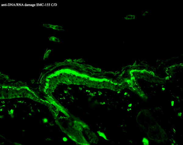
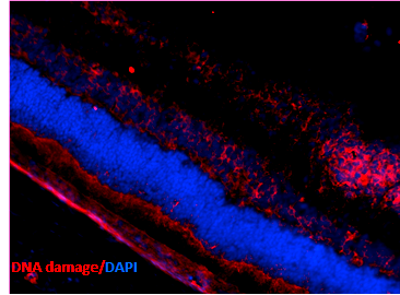
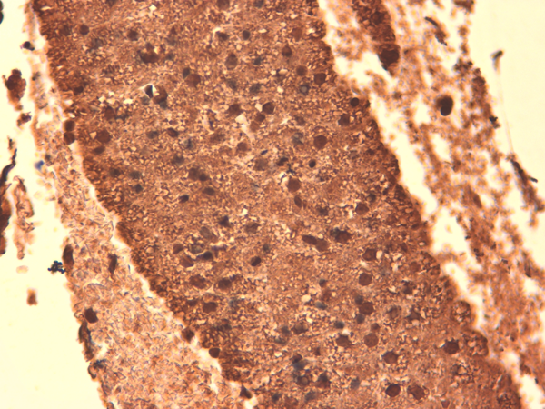
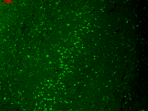
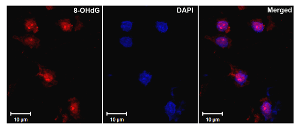 IHC (Immunohistochemistry)
(Immunohistochemistry analysis using Mouse Anti-DNA Damage Monoclonal Antibody, Clone 15A3. Tissue: Ischemic fresh brain tissue. Species: Rat. Primary Antibody: Mouse Anti-DNA Damage Monoclonal Antibody at 1:1000 for 16 hours at RT. Secondary Antibody: Alexa Fluor 546 Goat Anti-mouse (Red) at 1:500 for 1 hour at RT. Localization: Cerebral Cortex. Courtesy of: Dr. Yi Yang, U. New Mexico.)
IHC (Immunohistochemistry)
(Immunohistochemistry analysis using Mouse Anti-DNA Damage Monoclonal Antibody, Clone 15A3. Tissue: Ischemic fresh brain tissue. Species: Rat. Primary Antibody: Mouse Anti-DNA Damage Monoclonal Antibody at 1:1000 for 16 hours at RT. Secondary Antibody: Alexa Fluor 546 Goat Anti-mouse (Red) at 1:500 for 1 hour at RT. Localization: Cerebral Cortex. Courtesy of: Dr. Yi Yang, U. New Mexico.)
DNA/RNA Damage, Monoclonal Antibody (Cat# AAA103271)
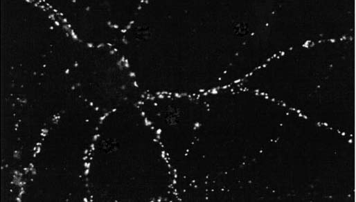
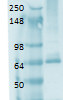
 WB (Western Blot)
(Western Blot analysis of Rat brain membrane lysate showing detection of PSD95 protein using Mouse Anti-PSD95 Monoclonal Antibody, Clone 6G6. Primary Antibody: Mouse Anti-PSD95 Monoclonal Antibody at 1:1000.)
WB (Western Blot)
(Western Blot analysis of Rat brain membrane lysate showing detection of PSD95 protein using Mouse Anti-PSD95 Monoclonal Antibody, Clone 6G6. Primary Antibody: Mouse Anti-PSD95 Monoclonal Antibody at 1:1000.)
PSD95, Monoclonal Antibody (Cat# AAA103323)
 WB (Western Blot)
(Western Blot analysis of Rat tissue lysate showing detection of KDEL Receptor protein using Mouse Anti-KDEL Receptor Monoclonal Antibody, Clone KR-10. Load: 15 ug. Block: 1.5% BSA for 30 minutes at RT. Primary Antibody: Mouse Anti-KDEL Receptor Monoclonal Antibody at 1:1000 for 2 hours at RT. Secondary Antibody: Sheep Anti-Mouse IgG: HRP for 1 hour at RT.)
WB (Western Blot)
(Western Blot analysis of Rat tissue lysate showing detection of KDEL Receptor protein using Mouse Anti-KDEL Receptor Monoclonal Antibody, Clone KR-10. Load: 15 ug. Block: 1.5% BSA for 30 minutes at RT. Primary Antibody: Mouse Anti-KDEL Receptor Monoclonal Antibody at 1:1000 for 2 hours at RT. Secondary Antibody: Sheep Anti-Mouse IgG: HRP for 1 hour at RT.)
KDEL, Monoclonal Antibody (Cat# AAA103092)
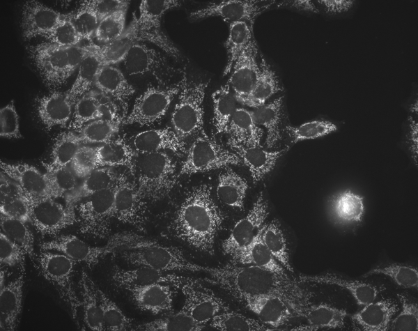
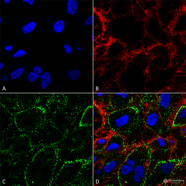
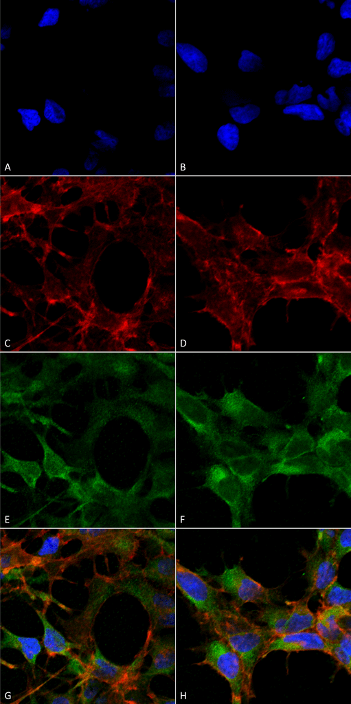
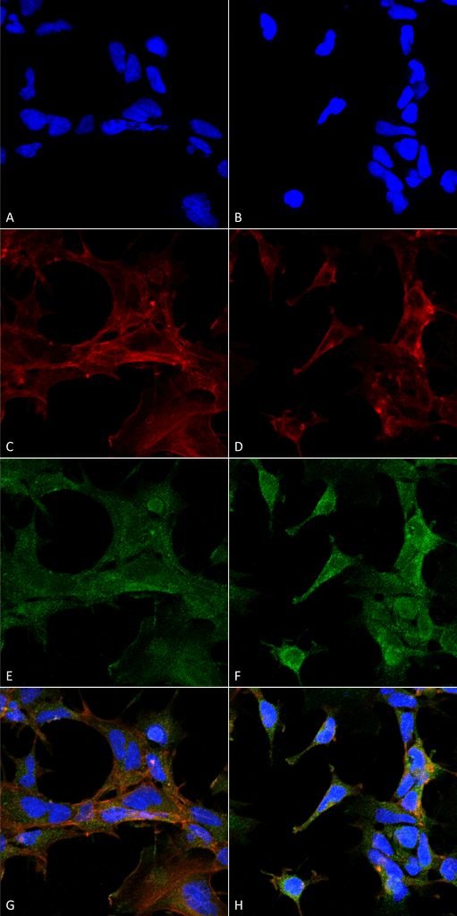
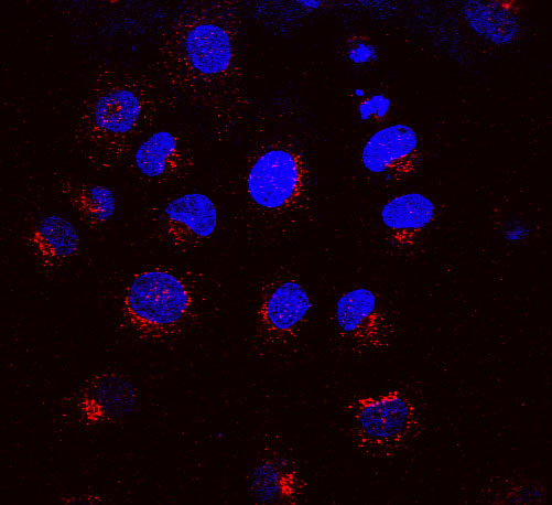
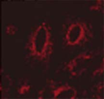
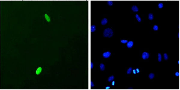
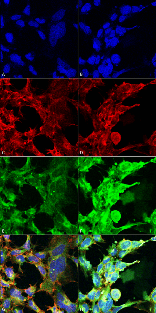
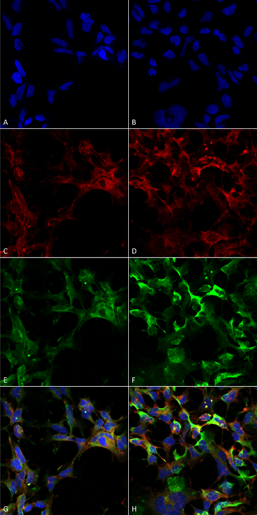
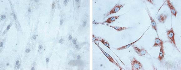 ICC (Immunocytochemistry)
(Immunocytochemistry/Immunofluorescence analysis using Mouse Anti-Hsp60 Monoclonal Antibody, Clone LK1,. Tissue: skin Fibroblasts. Species: Human. Fixation: Cold 100% methanol for 30 minutes at -20 degree C . Primary Antibody: Mouse Anti-Hsp60 Monoclonal Antibody at 1:1000 for 1 hour at RT. Secondary Antibody: DAKO LSAB2 streptavidin-peroxidase system. Counterstain: Mayer Hematoxylin (purple/blue) nuclear stain. Left: control; Right: 24 hours after 7th passage of senescence. Courtesy of: Valentina di Felice, University of Palermo, Italy.)
ICC (Immunocytochemistry)
(Immunocytochemistry/Immunofluorescence analysis using Mouse Anti-Hsp60 Monoclonal Antibody, Clone LK1,. Tissue: skin Fibroblasts. Species: Human. Fixation: Cold 100% methanol for 30 minutes at -20 degree C . Primary Antibody: Mouse Anti-Hsp60 Monoclonal Antibody at 1:1000 for 1 hour at RT. Secondary Antibody: DAKO LSAB2 streptavidin-peroxidase system. Counterstain: Mayer Hematoxylin (purple/blue) nuclear stain. Left: control; Right: 24 hours after 7th passage of senescence. Courtesy of: Valentina di Felice, University of Palermo, Italy.)
HSP60, Monoclonal Antibody (Cat# AAA103103)
 WB (Western Blot)
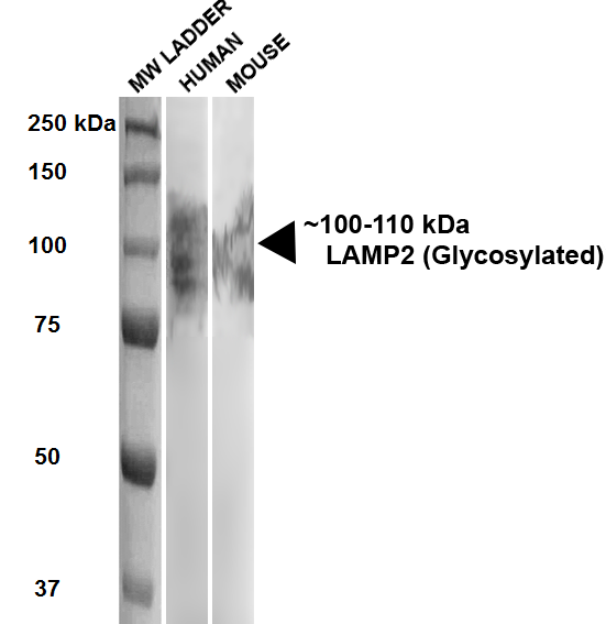
(Western Blot analysis of Human, Mouse HEK293 and 3T3NIH cell lysates showing detection of ~100-110 kDa LAMP2 protein using Rat Anti-LAMP2 Monoclonal Antibody, Clone GL2A7. Lane 1: MW ladder. Lane 2: Human HEK293 lysate (20 ug). Lane 3: Mouse 3T3NIH lysate (10 ug). Block: 5% milk + TBST for 1 hour at RT. Primary Antibody: Rat Anti-LAMP2 Monoclonal Antibody at 1:500 for 1 hour at RT. Secondary Antibody: HRP Goat Anti-Rat at 1:100 for 1 hour at RT. Color Development: TMB solution for 5 min at RT. Predicted/Observed Size: ~100-110 kDa.)
WB (Western Blot)
(Western Blot analysis of Human, Mouse HEK293 and 3T3NIH cell lysates showing detection of ~100-110 kDa LAMP2 protein using Rat Anti-LAMP2 Monoclonal Antibody, Clone GL2A7. Lane 1: MW ladder. Lane 2: Human HEK293 lysate (20 ug). Lane 3: Mouse 3T3NIH lysate (10 ug). Block: 5% milk + TBST for 1 hour at RT. Primary Antibody: Rat Anti-LAMP2 Monoclonal Antibody at 1:500 for 1 hour at RT. Secondary Antibody: HRP Goat Anti-Rat at 1:100 for 1 hour at RT. Color Development: TMB solution for 5 min at RT. Predicted/Observed Size: ~100-110 kDa.)
LAMP2, Monoclonal Antibody (Cat# AAA103116)
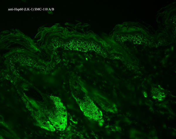
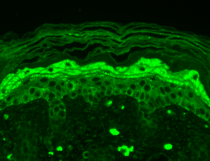
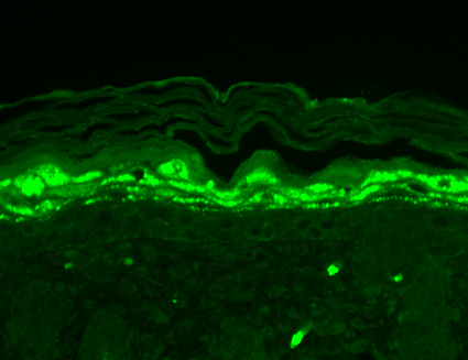
 IHC (Immunohistochemisry)
(Immunohistochemistry analysis using Mouse Anti-Nav1.8 Monoclonal Antibody, Clone S134-12. Tissue: backskin. Species: Mouse. Fixation: Bouin's Fixative and paraffin-embedded. Primary Antibody: Mouse Anti-Nav1.8 Monoclonal Antibody at 1:100 for 1 hour at RT. Secondary Antibody: FITC Goat Anti-Mouse (green) at 1:50 for 1 hour at RT. Localization: Heavy filaggrin-like staining, lower epidermal cells have some staining.)
IHC (Immunohistochemisry)
(Immunohistochemistry analysis using Mouse Anti-Nav1.8 Monoclonal Antibody, Clone S134-12. Tissue: backskin. Species: Mouse. Fixation: Bouin's Fixative and paraffin-embedded. Primary Antibody: Mouse Anti-Nav1.8 Monoclonal Antibody at 1:100 for 1 hour at RT. Secondary Antibody: FITC Goat Anti-Mouse (green) at 1:50 for 1 hour at RT. Localization: Heavy filaggrin-like staining, lower epidermal cells have some staining.)
Nav1.8, Monoclonal Antibody (Cat# AAA103123)
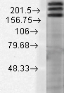
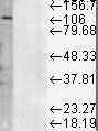
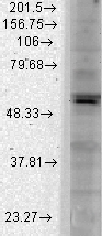
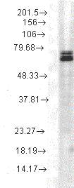
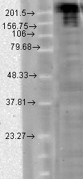
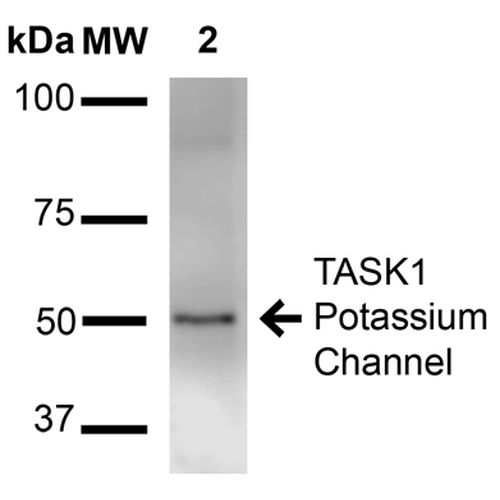 WB (Western Blot)
(Western Blot analysis of Rat Brain Membrane showing detection of ~50 kDa TASK1 Potassium Channel protein using Mouse Anti-TASK1 Potassium Channel Monoclonal Antibody, Clone S374-48 . Lane 1: Molecular Weight Ladder (MW). Lane 2: Rat brain membrane. Load: 15 ug. Block: 2% BSA and 2% Skim Milk in 1X TBST. Primary Antibody: Mouse Anti-TASK1 Potassium Channel Monoclonal Antibody at 1:1000 for 16 hours at 4 degree C. Secondary Antibody: Goat Anti-Mouse IgG: HRP at 1:2000 for 60 min at RT. Color Development: ECL solution for 6 min at RT. Predicted/Observed Size: ~50 kDa.)
WB (Western Blot)
(Western Blot analysis of Rat Brain Membrane showing detection of ~50 kDa TASK1 Potassium Channel protein using Mouse Anti-TASK1 Potassium Channel Monoclonal Antibody, Clone S374-48 . Lane 1: Molecular Weight Ladder (MW). Lane 2: Rat brain membrane. Load: 15 ug. Block: 2% BSA and 2% Skim Milk in 1X TBST. Primary Antibody: Mouse Anti-TASK1 Potassium Channel Monoclonal Antibody at 1:1000 for 16 hours at 4 degree C. Secondary Antibody: Goat Anti-Mouse IgG: HRP at 1:2000 for 60 min at RT. Color Development: ECL solution for 6 min at RT. Predicted/Observed Size: ~50 kDa.)
TASK1 Potassium Channel, Monoclonal Antibody (Cat# AAA103130)
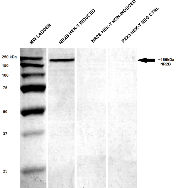
 WB (Western Blot)
(Western Blot analysis of Rat brain membrane lysate showing detection of GluN2B/NR2B protein using Mouse Anti-GluN2B/NR2B Monoclonal Antibody, Clone S59-36. Load: 15 ug. Block: 1.5% BSA for 30 minutes at RT. Primary Antibody: Mouse Anti-GluN2B/NR2B Monoclonal Antibody at 1:1000 for 2 hours at RT. Secondary Antibody: Sheep Anti-Mouse IgG: HRP for 1 hour at RT.)
WB (Western Blot)
(Western Blot analysis of Rat brain membrane lysate showing detection of GluN2B/NR2B protein using Mouse Anti-GluN2B/NR2B Monoclonal Antibody, Clone S59-36. Load: 15 ug. Block: 1.5% BSA for 30 minutes at RT. Primary Antibody: Mouse Anti-GluN2B/NR2B Monoclonal Antibody at 1:1000 for 2 hours at RT. Secondary Antibody: Sheep Anti-Mouse IgG: HRP for 1 hour at RT.)
NR2B, Monoclonal Antibody (Cat# AAA103136)

 WB (Western Blot)
(Western Blot analysis of Human Cell line lysates showing detection of GABA A Receptor protein using Mouse Anti-GABA A Receptor Monoclonal Antibody, Clone S95-35. Load: 15 ug. Block: 1.5% BSA for 30 minutes at RT. Primary Antibody: Mouse Anti-GABA A Receptor Monoclonal Antibody at 1:1000 for 2 hours at RT. Secondary Antibody: Sheep Anti-Mouse IgG: HRP for 1 hour at RT.)
WB (Western Blot)
(Western Blot analysis of Human Cell line lysates showing detection of GABA A Receptor protein using Mouse Anti-GABA A Receptor Monoclonal Antibody, Clone S95-35. Load: 15 ug. Block: 1.5% BSA for 30 minutes at RT. Primary Antibody: Mouse Anti-GABA A Receptor Monoclonal Antibody at 1:1000 for 2 hours at RT. Secondary Antibody: Sheep Anti-Mouse IgG: HRP for 1 hour at RT.)
GABA(A) Receptor Alpha1, Monoclonal Antibody (Cat# AAA103167)
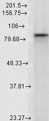
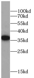
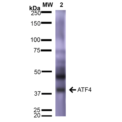 WB (Western Blot)
(Western Blot analysis of Rat Brain showing detection of ~39 kDa (isoform 2) ATF4 protein using Mouse Anti-ATF4 Monoclonal Antibody, Clone S360A-24 . Lane 1: Molecular Weight Ladder (MW). Lane 2: Rat Brain. Load: 15 ug. Block: 5% Skim Milk in 1X TBST. Primary Antibody: Mouse Anti-ATF4 Monoclonal Antibody at 1:1000 for 2 hours at RT. Secondary Antibody: Goat Anti-Mouse IgG: HRP at 1:2000 for 60 min at RT. Color Development: ECL solution for 5 min at RT. Predicted/Observed Size: ~39 kDa (isoform 2).)
WB (Western Blot)
(Western Blot analysis of Rat Brain showing detection of ~39 kDa (isoform 2) ATF4 protein using Mouse Anti-ATF4 Monoclonal Antibody, Clone S360A-24 . Lane 1: Molecular Weight Ladder (MW). Lane 2: Rat Brain. Load: 15 ug. Block: 5% Skim Milk in 1X TBST. Primary Antibody: Mouse Anti-ATF4 Monoclonal Antibody at 1:1000 for 2 hours at RT. Secondary Antibody: Goat Anti-Mouse IgG: HRP at 1:2000 for 60 min at RT. Color Development: ECL solution for 5 min at RT. Predicted/Observed Size: ~39 kDa (isoform 2).)
ATF4, Monoclonal Antibody (Cat# AAA102946)
 WB (Western Blot)
(Western Blot analysis of Rat brain membrane lysate showing detection of SHANK1 protein using Mouse Anti-SHANK1 Monoclonal Antibody, Clone S22-21. Load: 15 ug. Block: 1.5% BSA for 30 minutes at RT. Primary Antibody: Mouse Anti-SHANK1 Monoclonal Antibody at 1:1000 for 2 hours at RT. Secondary Antibody: Sheep Anti-Mouse IgG: HRP for 1 hour at RT.)
WB (Western Blot)
(Western Blot analysis of Rat brain membrane lysate showing detection of SHANK1 protein using Mouse Anti-SHANK1 Monoclonal Antibody, Clone S22-21. Load: 15 ug. Block: 1.5% BSA for 30 minutes at RT. Primary Antibody: Mouse Anti-SHANK1 Monoclonal Antibody at 1:1000 for 2 hours at RT. Secondary Antibody: Sheep Anti-Mouse IgG: HRP for 1 hour at RT.)
Shank1, Monoclonal Antibody (Cat# AAA102982)
 WB (Western Blot)
(Western Blot analysis of Rat liver microsome lysate showing detection of LAMP1 protein using Mouse Anti-LAMP1 Monoclonal Antibody, Clone Ly1C6. Load: 15 ug. Block: 1.5% BSA for 30 minutes at RT. Primary Antibody: Mouse Anti-LAMP1 Monoclonal Antibody at 1:1000 for 2 hours at RT. Secondary Antibody: Sheep Anti-Mouse IgG: HRP for 1 hour at RT.)
WB (Western Blot)
(Western Blot analysis of Rat liver microsome lysate showing detection of LAMP1 protein using Mouse Anti-LAMP1 Monoclonal Antibody, Clone Ly1C6. Load: 15 ug. Block: 1.5% BSA for 30 minutes at RT. Primary Antibody: Mouse Anti-LAMP1 Monoclonal Antibody at 1:1000 for 2 hours at RT. Secondary Antibody: Sheep Anti-Mouse IgG: HRP for 1 hour at RT.)
LAMP1, Monoclonal Antibody (Cat# AAA102984)
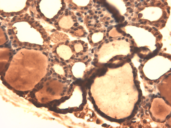

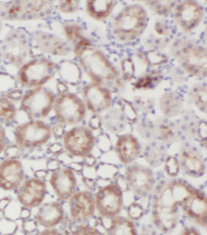
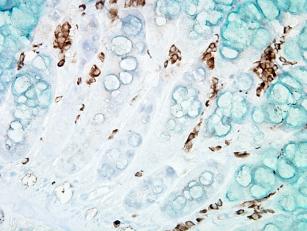
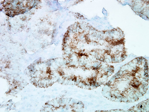
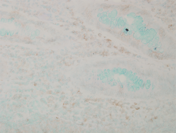
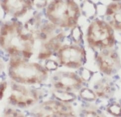
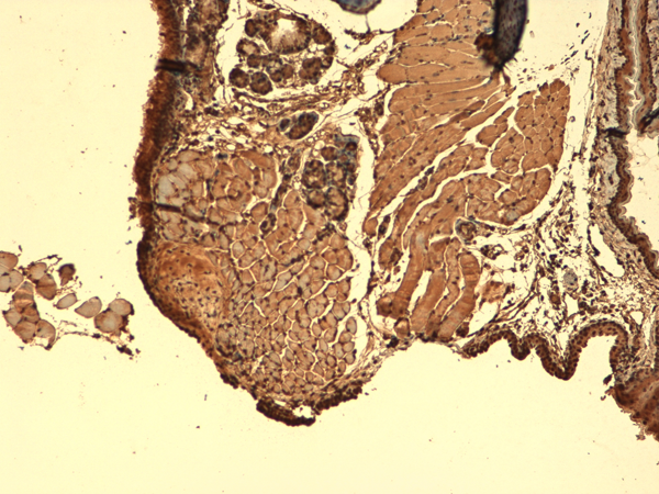 IHC (Immunohistochemistry)
(Immunohistochemistry analysis using Mouse Anti-Sodium Iodide Symporter Monoclonal Antibody, Clone 14F. Tissue: Thyroid. Species: Mouse. Fixation: 10% Formalin Solution for 12-24 hours at RT. Primary Antibody: Mouse Anti-Sodium Iodide Symporter Monoclonal Antibody at 1:1000 for 1 hour at RT. Secondary Antibody: HRP/DAB Detection System: Biotinylated Goat Anti-Mouse, Streptavidin Peroxidase, DAB Chromogen (brown) for 30 minutes at RT. Counterstain: Mayer Hematoxylin (purple/blue) nuclear stain at 250-500 ul for 5 minutes at RT.)
IHC (Immunohistochemistry)
(Immunohistochemistry analysis using Mouse Anti-Sodium Iodide Symporter Monoclonal Antibody, Clone 14F. Tissue: Thyroid. Species: Mouse. Fixation: 10% Formalin Solution for 12-24 hours at RT. Primary Antibody: Mouse Anti-Sodium Iodide Symporter Monoclonal Antibody at 1:1000 for 1 hour at RT. Secondary Antibody: HRP/DAB Detection System: Biotinylated Goat Anti-Mouse, Streptavidin Peroxidase, DAB Chromogen (brown) for 30 minutes at RT. Counterstain: Mayer Hematoxylin (purple/blue) nuclear stain at 250-500 ul for 5 minutes at RT.)
Sodium-Iodide Symporter, Monoclonal Antibody (Cat# AAA102989)
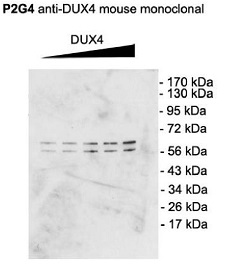 WB (Western Blot)
(Western Blot analysis of Mouse C2C12 cell lysate showing detection of DUX4 protein using Mouse Anti-DUX4 Monoclonal Antibody, Clone P2B1. Primary Antibody: Mouse Anti-DUX4 Monoclonal Antibody at 1:1000. Cells transfected with pCS2+DUX4 which, contains an additional upstream start site.)
WB (Western Blot)
(Western Blot analysis of Mouse C2C12 cell lysate showing detection of DUX4 protein using Mouse Anti-DUX4 Monoclonal Antibody, Clone P2B1. Primary Antibody: Mouse Anti-DUX4 Monoclonal Antibody at 1:1000. Cells transfected with pCS2+DUX4 which, contains an additional upstream start site.)
DUX4, Monoclonal Antibody (Cat# AAA103181)
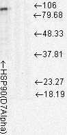 WB (Western Blot)
(Western Blot analysis of Rat cell lysates showing detection of Hsp90 protein using Mouse Anti-Hsp90 Monoclonal Antibody, Clone D7Alpha. Load: 15 ug. Block: 1.5% BSA for 30 minutes at RT. Primary Antibody: Mouse Anti-Hsp90 Monoclonal Antibody at 1:1000 for 2 hours at RT. Secondary Antibody: Sheep Anti-Mouse IgG: HRP for 1 hour at RT.)
WB (Western Blot)
(Western Blot analysis of Rat cell lysates showing detection of Hsp90 protein using Mouse Anti-Hsp90 Monoclonal Antibody, Clone D7Alpha. Load: 15 ug. Block: 1.5% BSA for 30 minutes at RT. Primary Antibody: Mouse Anti-Hsp90 Monoclonal Antibody at 1:1000 for 2 hours at RT. Secondary Antibody: Sheep Anti-Mouse IgG: HRP for 1 hour at RT.)
Hsp90, Monoclonal Antibody (Cat# AAA103245)
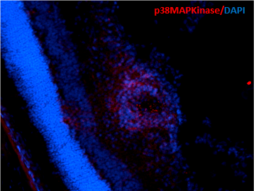
 WB (Western Blot)
(Western Blot analysis of Human Cell lysates showing detection of p38 MAPK protein using Mouse Anti-p38 MAPK Monoclonal Antibody, Clone 9F12. Load: 15 ug. Block: 1.5% BSA for 30 minutes at RT. Primary Antibody: Mouse Anti-p38 MAPK Monoclonal Antibody at 1:1000 for 2 hours at RT. Secondary Antibody: Sheep Anti-Mouse IgG: HRP for 1 hour at RT.)
WB (Western Blot)
(Western Blot analysis of Human Cell lysates showing detection of p38 MAPK protein using Mouse Anti-p38 MAPK Monoclonal Antibody, Clone 9F12. Load: 15 ug. Block: 1.5% BSA for 30 minutes at RT. Primary Antibody: Mouse Anti-p38 MAPK Monoclonal Antibody at 1:1000 for 2 hours at RT. Secondary Antibody: Sheep Anti-Mouse IgG: HRP for 1 hour at RT.)
p38 alpha, Monoclonal Antibody (Cat# AAA102897)
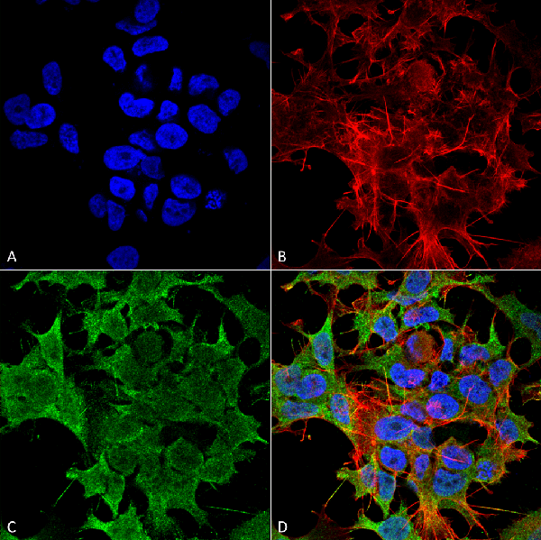
 IHC (Immunohistochemisry)
(Immunohistochemistry analysis using Mouse Anti-Kir2.1 Potassium Channel Monoclonal Antibody, Clone S112B-14. Tissue: hippocampus. Species: Human. Fixation: Bouin's Fixative and paraffin-embedded. Primary Antibody: Mouse Anti-Kir2.1 Potassium Channel Monoclonal Antibody at 1:1000 for 1 hour at RT. Secondary Antibody: FITC Goat Anti-Mouse (green) at 1:50 for 1 hour at RT.)
IHC (Immunohistochemisry)
(Immunohistochemistry analysis using Mouse Anti-Kir2.1 Potassium Channel Monoclonal Antibody, Clone S112B-14. Tissue: hippocampus. Species: Human. Fixation: Bouin's Fixative and paraffin-embedded. Primary Antibody: Mouse Anti-Kir2.1 Potassium Channel Monoclonal Antibody at 1:1000 for 1 hour at RT. Secondary Antibody: FITC Goat Anti-Mouse (green) at 1:50 for 1 hour at RT.)
Kir2.1, Monoclonal Antibody (Cat# AAA102920)
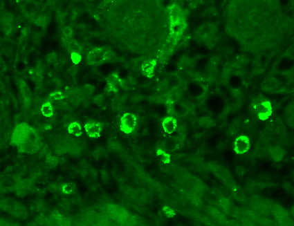
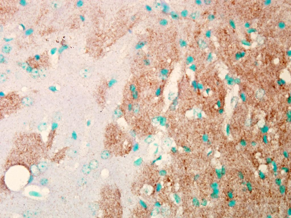
 IHC (Immunohistochemisry)
(Immunohistochemistry analysis using Mouse Anti-HCN2 Monoclonal Antibody, Clone S71-37. Tissue: hippocampus. Species: Human. Fixation: Bouin's Fixative and paraffin-embedded. Primary Antibody: Mouse Anti-HCN2 Monoclonal Antibody at 1:100 for 1 hour at RT. Secondary Antibody: FITC Goat Anti-Mouse (green) at 1:50 for 1 hour at RT.)
IHC (Immunohistochemisry)
(Immunohistochemistry analysis using Mouse Anti-HCN2 Monoclonal Antibody, Clone S71-37. Tissue: hippocampus. Species: Human. Fixation: Bouin's Fixative and paraffin-embedded. Primary Antibody: Mouse Anti-HCN2 Monoclonal Antibody at 1:100 for 1 hour at RT. Secondary Antibody: FITC Goat Anti-Mouse (green) at 1:50 for 1 hour at RT.)
HCN2, Monoclonal Antibody (Cat# AAA103413)
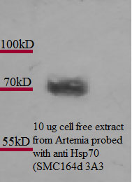
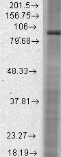
 WB (Western Blot)
(Western Blot analysis of Rat cell lysates showing detection of Hsp70 protein using Mouse Anti-Hsp70 Monoclonal Antibody, Clone 3A3. Load: 15 ug. Block: 1.5% BSA for 30 minutes at RT. Primary Antibody: Mouse Anti-Hsp70 Monoclonal Antibody at 1:1000 for 2 hours at RT. Secondary Antibody: Sheep Anti-Mouse IgG: HRP for 1 hour at RT.)
WB (Western Blot)
(Western Blot analysis of Rat cell lysates showing detection of Hsp70 protein using Mouse Anti-Hsp70 Monoclonal Antibody, Clone 3A3. Load: 15 ug. Block: 1.5% BSA for 30 minutes at RT. Primary Antibody: Mouse Anti-Hsp70 Monoclonal Antibody at 1:1000 for 2 hours at RT. Secondary Antibody: Sheep Anti-Mouse IgG: HRP for 1 hour at RT.)
Hsp70, Monoclonal Antibody (Cat# AAA103441)
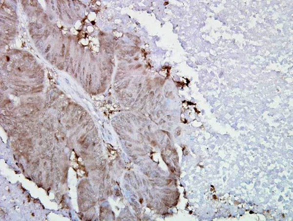
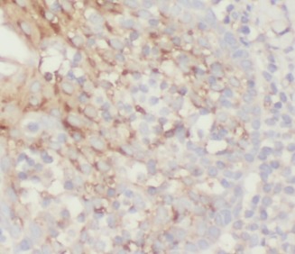
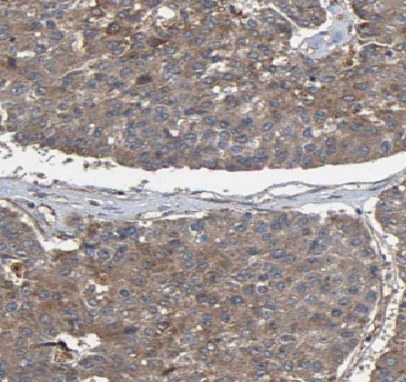
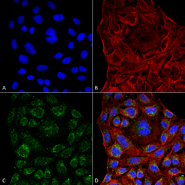
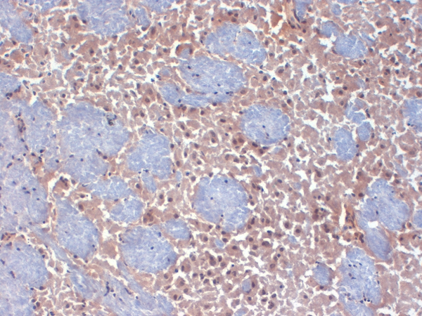
 IHC (Immunohistochemisry)
(Immunohistochemistry analysis using Mouse Anti-Hsp90 alpha Monoclonal Antibody, Clone K41009. Tissue: inflamed colon. Species: Mouse. Fixation: Formalin. Primary Antibody: Mouse Anti-Hsp90 alpha Monoclonal Antibody at 1:5000 for 12 hours at 4 degree C. Secondary Antibody: Biotin Goat Anti-Mouse at 1:2000 for 1 hour at RT. Counterstain: Mayer Hematoxylin (purple/blue) nuclear stain at 200 ul for 2 minutes at RT. Localization: Inflammatory cells. Magnification: 40x. Inflammatory cells.)
IHC (Immunohistochemisry)
(Immunohistochemistry analysis using Mouse Anti-Hsp90 alpha Monoclonal Antibody, Clone K41009. Tissue: inflamed colon. Species: Mouse. Fixation: Formalin. Primary Antibody: Mouse Anti-Hsp90 alpha Monoclonal Antibody at 1:5000 for 12 hours at 4 degree C. Secondary Antibody: Biotin Goat Anti-Mouse at 1:2000 for 1 hour at RT. Counterstain: Mayer Hematoxylin (purple/blue) nuclear stain at 200 ul for 2 minutes at RT. Localization: Inflammatory cells. Magnification: 40x. Inflammatory cells.)
Hsp90 alpha, Monoclonal Antibody (Cat# AAA103448)
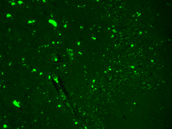
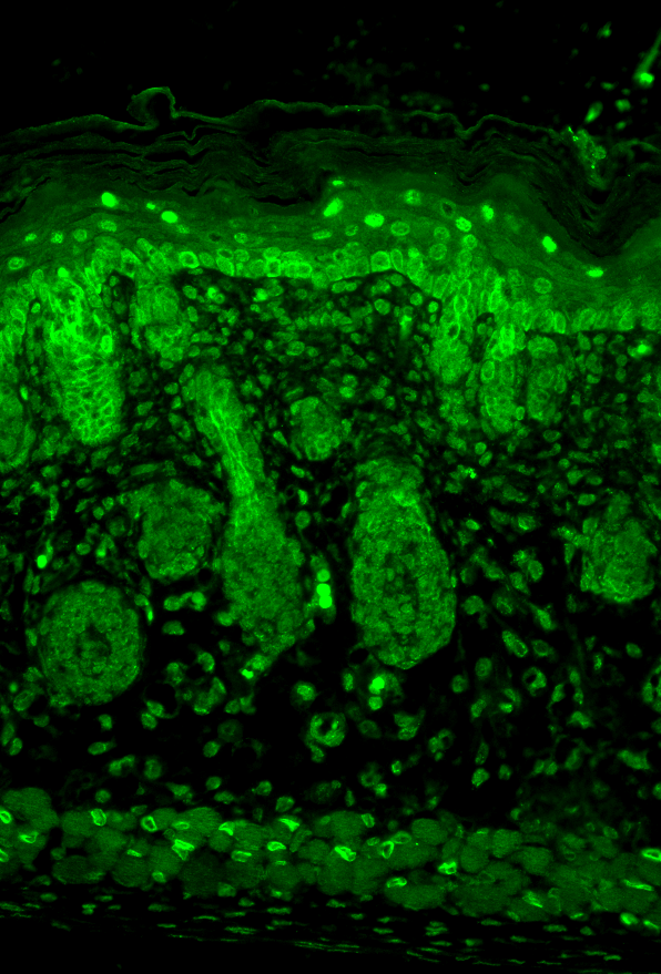
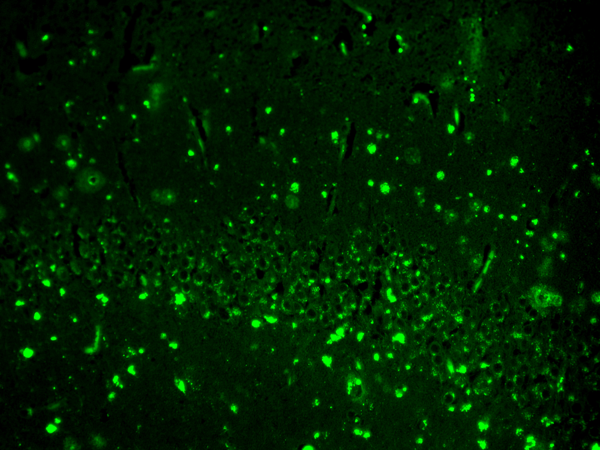
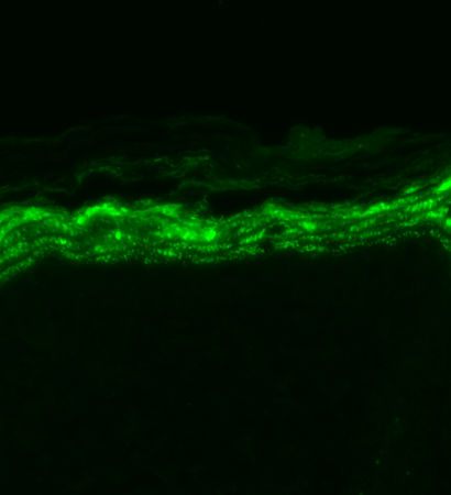 IHC (Immunohistochemistry)
(Immunohistochemistry analysis using Mouse Anti-Nav1.7 Sodium Channel Monoclonal Antibody, Clone S68-6. Tissue: backskin. Species: Mouse. Fixation: Bouin's Fixative and paraffin-embedded. Primary Antibody: Mouse Anti-Nav1.7 Sodium Channel Monoclonal Antibody at 1:100 for 1 hour at RT. Secondary Antibody: FITC Goat Anti-Mouse (green) at 1:50 for 1 hour at RT.)
IHC (Immunohistochemistry)
(Immunohistochemistry analysis using Mouse Anti-Nav1.7 Sodium Channel Monoclonal Antibody, Clone S68-6. Tissue: backskin. Species: Mouse. Fixation: Bouin's Fixative and paraffin-embedded. Primary Antibody: Mouse Anti-Nav1.7 Sodium Channel Monoclonal Antibody at 1:100 for 1 hour at RT. Secondary Antibody: FITC Goat Anti-Mouse (green) at 1:50 for 1 hour at RT.)
Nav1.7, Monoclonal Antibody (Cat# AAA103461)
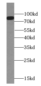
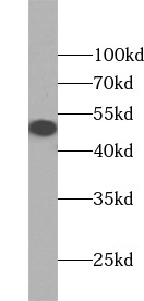
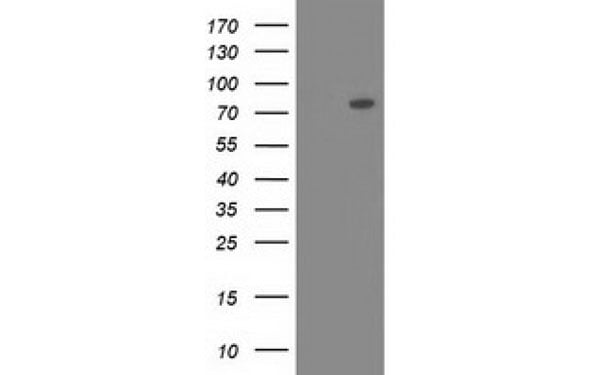
 WB (Western Blot)
(K-562 cells were subjected to SDS PAGE followed by western blot with AAA102795 (FGFR1 Antibody) at dilution of 1:1000)
WB (Western Blot)
(K-562 cells were subjected to SDS PAGE followed by western blot with AAA102795 (FGFR1 Antibody) at dilution of 1:1000)
FGFR1, Monoclonal Antibody (Cat# AAA102795)
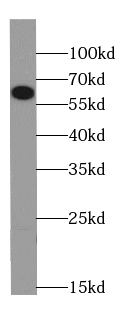
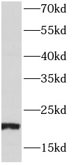
 WB (Western Blot)
(HeLa cells were subjected to SDS PAGE followed by western blot with AAA102804 (FOLR1 Antibody) at dilution of 1:1000)
WB (Western Blot)
(HeLa cells were subjected to SDS PAGE followed by western blot with AAA102804 (FOLR1 Antibody) at dilution of 1:1000)
FOLR1, Monoclonal Antibody (Cat# AAA102804)
Protein A+G purification
 FCM/FACS (Flow Cytometry)
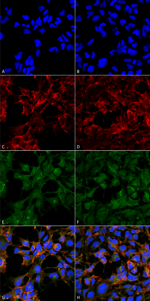
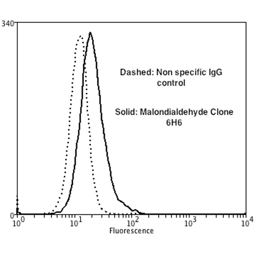
(Flow Cytometry analysis using Mouse Anti-Malondialdehyde Monoclonal Antibody, Clone 6H6. Tissue: Neuroblastoma cells (SH-SY5Y). Species: Human. Fixation: 90% Methanol. Primary Antibody: Mouse Anti-Malondialdehyde Monoclonal Antibody at 1:50 for 30 min on ice. Secondary Antibody: Goat Anti-Mouse: PE at 1:100 for 20 min at RT. Isotype Control: Non Specific IgG. Cells were subject to oxidative stress by treating with 250 uM H2O2 for 24 hours.)
FCM/FACS (Flow Cytometry)
(Flow Cytometry analysis using Mouse Anti-Malondialdehyde Monoclonal Antibody, Clone 6H6. Tissue: Neuroblastoma cells (SH-SY5Y). Species: Human. Fixation: 90% Methanol. Primary Antibody: Mouse Anti-Malondialdehyde Monoclonal Antibody at 1:50 for 30 min on ice. Secondary Antibody: Goat Anti-Mouse: PE at 1:100 for 20 min at RT. Isotype Control: Non Specific IgG. Cells were subject to oxidative stress by treating with 250 uM H2O2 for 24 hours.)
Malondialdehyde, Monoclonal Antibody (Cat# AAA104067)
 WB (Western Blot)
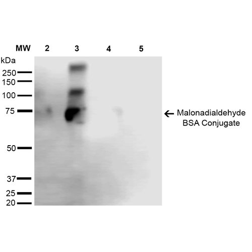
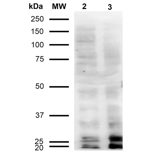
(Western Blot analysis of Human Cervical cancer cell line (HeLa) lysate showing detection of Malondialdehyde protein using Mouse Anti-Malondialdehyde Monoclonal Antibody, Clone 11E3. Lane 1: Molecular Weight Ladder (MW). Lane 2: HeLa cell lysate. Lane 3: H2O2 treated HeLa cell lysate. Load: 12 ug. Block: 5% Skim Milk in TBST. Primary Antibody: Mouse Anti-Malondialdehyde Monoclonal Antibody at 1:1000 for 2 hours at RT. Secondary Antibody: Goat Anti-Mouse IgG: HRP at 1:2000 for 60 min at RT. Color Development: ECL solution for 5 min in RT.)
WB (Western Blot)
(Western Blot analysis of Human Cervical cancer cell line (HeLa) lysate showing detection of Malondialdehyde protein using Mouse Anti-Malondialdehyde Monoclonal Antibody, Clone 11E3. Lane 1: Molecular Weight Ladder (MW). Lane 2: HeLa cell lysate. Lane 3: H2O2 treated HeLa cell lysate. Load: 12 ug. Block: 5% Skim Milk in TBST. Primary Antibody: Mouse Anti-Malondialdehyde Monoclonal Antibody at 1:1000 for 2 hours at RT. Secondary Antibody: Goat Anti-Mouse IgG: HRP at 1:2000 for 60 min at RT. Color Development: ECL solution for 5 min in RT.)
Malondialdehyde, Monoclonal Antibody (Cat# AAA104078)
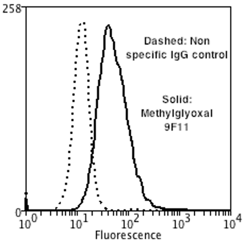 FCM/FACS (Flow Cytometry)
(Flow Cytometry analysis using Mouse Anti-Methylglyoxal Monoclonal Antibody, Clone 9F11. Tissue: Neuroblastoma cells (SH-SY5Y). Species: Human. Fixation: 90% Methanol. Primary Antibody: Mouse Anti-Methylglyoxal Monoclonal Antibody at 1:50 for 30 min on ice. Secondary Antibody: Goat Anti-Mouse: PE at 1:100 for 20 min at RT. Isotype Control: Non Specific IgG. Cells were subject to oxidative stress by treating with 250 uM H2O2 for 24 hours.)
FCM/FACS (Flow Cytometry)
(Flow Cytometry analysis using Mouse Anti-Methylglyoxal Monoclonal Antibody, Clone 9F11. Tissue: Neuroblastoma cells (SH-SY5Y). Species: Human. Fixation: 90% Methanol. Primary Antibody: Mouse Anti-Methylglyoxal Monoclonal Antibody at 1:50 for 30 min on ice. Secondary Antibody: Goat Anti-Mouse: PE at 1:100 for 20 min at RT. Isotype Control: Non Specific IgG. Cells were subject to oxidative stress by treating with 250 uM H2O2 for 24 hours.)
Methylglyoxal, Monoclonal Antibody (Cat# AAA104094)
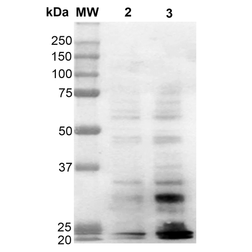 WB (Western Blot)
(Western Blot analysis of Human Cervical cancer cell line (HeLa) lysate showing detection of Dityrosine protein using Mouse Anti-Dityrosine Monoclonal Antibody, Clone 10A6. Lane 1: Molecular Weight Ladder (MW). Lane 2: HeLa cell lysate. Lane 3: H2O2 treated HeLa cell lysate. Load: 12 ug. Block: 5% Skim Milk in TBST. Primary Antibody: Mouse Anti-Dityrosine Monoclonal Antibody at 1:1000 for 2 hours at RT. Secondary Antibody: Goat Anti-Mouse IgG: HRP at 1:2000 for 60 min at RT. Color Development: ECL solution for 5 min in RT.)
WB (Western Blot)
(Western Blot analysis of Human Cervical cancer cell line (HeLa) lysate showing detection of Dityrosine protein using Mouse Anti-Dityrosine Monoclonal Antibody, Clone 10A6. Lane 1: Molecular Weight Ladder (MW). Lane 2: HeLa cell lysate. Lane 3: H2O2 treated HeLa cell lysate. Load: 12 ug. Block: 5% Skim Milk in TBST. Primary Antibody: Mouse Anti-Dityrosine Monoclonal Antibody at 1:1000 for 2 hours at RT. Secondary Antibody: Goat Anti-Mouse IgG: HRP at 1:2000 for 60 min at RT. Color Development: ECL solution for 5 min in RT.)
Dityrosine, Monoclonal Antibody (Cat# AAA104113)
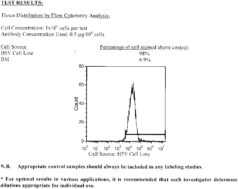 Application Data
CD34, Monoclonal Antibody (Cat# AAA74195)
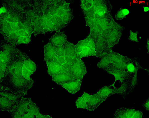
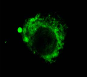
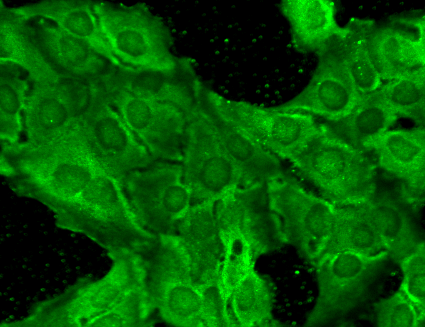
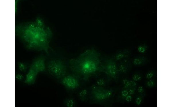
 IF (Immunofluorescence)
(Immunofluorescent staining of COS7 cells transiently transfected with recombinant FGF21 protein using FGF21 antibody)
IF (Immunofluorescence)
(Immunofluorescent staining of COS7 cells transiently transfected with recombinant FGF21 protein using FGF21 antibody)
FGF21, Monoclonal Antibody (Cat# AAA74703)
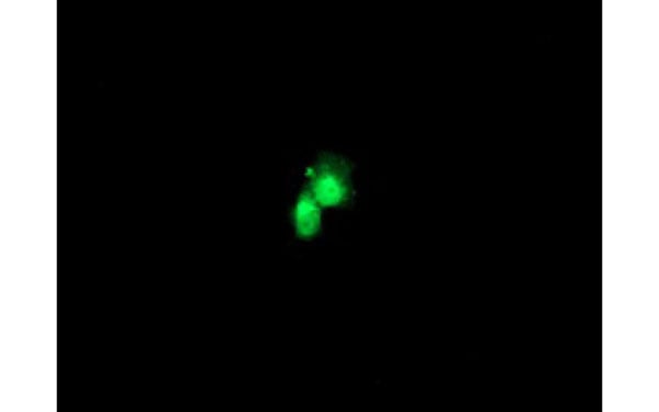
 IF (Immunofluorescence)
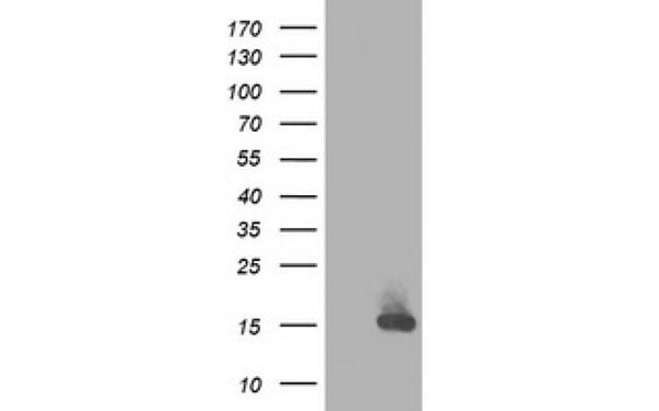
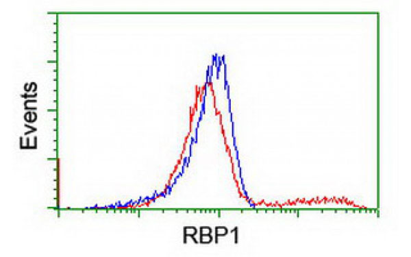
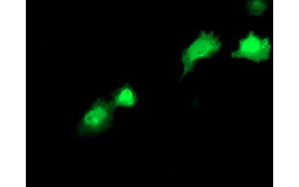
(Immunofluorescent staining of COS7 cells transiently transfected with recombinant RBP1 protein using RBP1 antibody)
IF (Immunofluorescence)
(Immunofluorescent staining of COS7 cells transiently transfected with recombinant RBP1 protein using RBP1 antibody)
RBP1, Monoclonal Antibody (Cat# AAA106642)
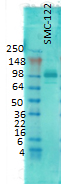
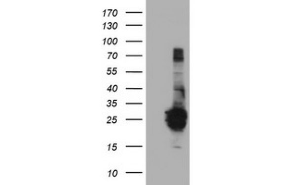
 WB (Western Blot)
(Western Blot analysis of HEK293T cell lysates (5 ug) transfected with either recombinant OSBPL11 protein (Right) or empty vector (Left) detected with OSBPL11 antibody)
WB (Western Blot)
(Western Blot analysis of HEK293T cell lysates (5 ug) transfected with either recombinant OSBPL11 protein (Right) or empty vector (Left) detected with OSBPL11 antibody)
OSBPL11, Monoclonal Antibody (Cat# AAA106683)
What are Monoclonal Antibodies?
Monoclonal antibodies are specialized laboratory-produced proteins developed for binding to specific biological antigens or other molecular targets. Since they come from a single cell (or clone), they are especially consistent and accurate in the data they are involved in producing.
This type of antibody material has been shown to be a powerful tool in finding and subsequently destroying harmful cells in an organism, such as those found in cancers or various autoimmune diseases. This makes them excellent aids in medical testing and research, which is why they are so widely used.
AAA Biotech offers a comprehensive range of high-quality monoclonal antibodies that perform effectively in various laboratory tests, including (amongst others) ELISA, western blotting, immunohistochemistry, and flow cytometry. All of the products in our catalog are thoroughly quality tested to make sure that they are reliable and will consistently perform well in your research.
What Are The Uses of Monoclonal Antibodies
Monoclonal antibodies are used in many lab tests, including (amongst others) ELISA, western blotting, immunohistochemistry, and flow cytometry.
ELISA is a test that helps detect a specific substance/analyte in a sample. It uses antibodies (often monoclonal) bound to a solid surface (such as the well of a microplate) to “capture” the substance/analyte in the sample and immobilize it so that the detection antibody component can then bind to it and produce a signal, which can then be measured.
Western blotting identifies specific proteins in a sample. The sample is first separated on a gel, and then antibodies are applied that will typically bind to the target, which will all be localized to a single band in a lane.
Immunohistochemistry helps locate specific proteins in cells or tissue samples using antibodies.
Flow cytometry looks at and sorts cells. It uses antibodies that are conjugated to reporter molecules called “fluorophores”, which, under special lights, emit light themselves, which can then be measured by a detector instrument.
How Monoclonal Antibodies Are Used as Medicine?
Please note that all of the products listed in AAA Biotech’s also known as AAA Bio or AAABio catalog are strictly for research-use only (RUO).
Monoclonal antibodies can also be used as therapeutic/medical treatments, particularly in the context of cancers. They are designed to find and bind to specific cells or proteins, helping the immune system recognize and attack the cancer. These treatments work in different ways, such as:
- Radioimmunotherapy attaches a small amount of radioactive molecule to the antibody, so it delivers the radiation directly to the cancer cells that the antibody is specifically binding to.
- Antibody-directed enzyme prodrug therapy uses antibodies that are specifically bound to special enzymes. These enzymes activate a harmless drug in the body and turn it into a cancer-killing drug only near the cancer cells—this helps avoid harming healthy cells.
- Immunoliposomes are tiny “bubbles” filled with medicine/drug and coated with antibodies. They carry the drug straight to the cancer cells.
Why Buy Monoclonal Antibodies From Us?
At AAA Biotech, we provide high-performance monoclonal antibodies designed to support a wide range of research needs.
1. Validated for Versatile Applications
The antibodies in our catalog are extensively validated and compatible with multiple techniques, including (but not limited to) ELISA, flow cytometry (FC), immunocytochemistry (ICC), immunofluorescence (IF), immunohistochemistry (IHC), immunoprecipitation (IP), and western blotting (WB).
2. Wide Selection & Specialized Options
We offer antibodies for common and rare species, that are available in various conjugated forms, and also in recombinant formats. Essentially, there is almost anything one might need to meet their experimental model’s requirements.
3. High-Quality Proteins
Our proteins meet high purity standards—90% or more as confirmed by SDS-PAGE. Many are available with tags like His, Flag, GST, or MBP, and we also supply native and biologically active proteins for functional studies.
Frequently Asked Questions
1. Are your monoclonal antibodies validated for specific applications?
Yes, our antibodies are tested and validated for use in methods such as ELISA, western blot, IHC, flow cytometry, and more. Refer to specific product pages or datasheets for individual product information.
2. How do I choose the right monoclonal antibody for my application?
Review the product details directly for application validation, species reactivity, and target information. You may also contact our support team at any time for help.
3. How quickly can I receive my order?
Most orders are processed and shipped within 1–3 business days, depending on product availability and your shipping location.