Filters
Clonality
Type
Reactivity
Gene Name
Isotype
Host
Application
Clone
410 results for " Conjugated Proteins" - showing 300-350


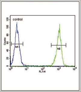
 FCM (Flow Cytometry)
(Figure 7. Flow Cytometry analysis of HELA cells using anti-DNAJC10 antibody (AAA19320).Overlay histogram showing HELA cells stained with AAA19320 (Blue line). The cells were blocked with 10% normal goat serum. And then incubated with rabbit anti-DNAJC10 Antibody (AAA19320, 1μg/1x106 cells) for 30 min at 20 degree C. DyLight®488 conjugated goat anti-rabbit IgG (5-10μg/1x106 cells) was used as secondary antibody for 30 minutes at 20 degree C. Isotype control antibody (Green line) was rabbit IgG (1μg/1x106) used under the same conditions. Unlabelled sample (Red line) was also used as a control.)
FCM (Flow Cytometry)
(Figure 7. Flow Cytometry analysis of HELA cells using anti-DNAJC10 antibody (AAA19320).Overlay histogram showing HELA cells stained with AAA19320 (Blue line). The cells were blocked with 10% normal goat serum. And then incubated with rabbit anti-DNAJC10 Antibody (AAA19320, 1μg/1x106 cells) for 30 min at 20 degree C. DyLight®488 conjugated goat anti-rabbit IgG (5-10μg/1x106 cells) was used as secondary antibody for 30 minutes at 20 degree C. Isotype control antibody (Green line) was rabbit IgG (1μg/1x106) used under the same conditions. Unlabelled sample (Red line) was also used as a control.)
DNAJC10, Polyclonal Antibody (Cat# AAA19320)
Full Name
Anti-DNAJC10 Antibody
Gene Names
DNAJC10; JPDI; MTHr; ERdj5; PDIA19
Reactivity
Human, Mouse, Rat
Applications
WB, IHC-P, ICC, IF, FC/FACS/FCM, EIA
Purity
Immunogen affinity purified.
Pricing


 FCM (Flow Cytometry)
(Figure 6. Flow Cytometry analysis of K562 cells using anti-D2AP antibody (M01756). Overlay histogram showing K562 cells stained with M01756 (Blue line).The cells were blocked with 10% normal goat serum. And then incubated with mouse anti-D2AP Antibody (M01756, 1ug/1x106 cells) for 30 min at 20 degree C. DyLight488 conjugated goat anti-mouse IgG (BA1126, 5-10ug/1x106 cells) was used as secondary antibody for 30 minutes at 20 degree C. Isotype control antibody (Green line) was mouse IgG (1ug/1x106) used under the same conditions. Unlabelled sample (Red line) was also used as a control.)
FCM (Flow Cytometry)
(Figure 6. Flow Cytometry analysis of K562 cells using anti-D2AP antibody (M01756). Overlay histogram showing K562 cells stained with M01756 (Blue line).The cells were blocked with 10% normal goat serum. And then incubated with mouse anti-D2AP Antibody (M01756, 1ug/1x106 cells) for 30 min at 20 degree C. DyLight488 conjugated goat anti-mouse IgG (BA1126, 5-10ug/1x106 cells) was used as secondary antibody for 30 minutes at 20 degree C. Isotype control antibody (Green line) was mouse IgG (1ug/1x106) used under the same conditions. Unlabelled sample (Red line) was also used as a control.)
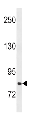
CD2AP, Monoclonal Antibody (Cat# AAA19185)
Full Name
Anti-CD2AP Antibody (monoclonal, 5F8)
Gene Names
CD2AP; CMS
Reactivity
Human, Mouse, Rat
Applications
WB, IHC, FC/FACS
Purity
Immunogen Affinity Purified
Pricing


 FCM (Flow Cytometry)
(Figure 8. Flow Cytometry analysis of A549 cells using anti-Transketolase/TKT antibody (AAA19368).Overlay histogram showing A549 cells stained with AAA19368 (Blue line). The cells were blocked with 10% normal goat serum. And then incubated with mouse anti- Transketolase/TKT Antibody (AAA19368, 1μg/1x106 cells) for 30 min at 20 degree C. DyLight®488 conjugated goat anti-mouse IgG (BA1126, 5-10μg/1x106 cells) was used as secondary antibody for 30 minutes at 20 degree C. Isotype control antibody (Green line) was mouse IgG (1μg/1x106) used under the same conditions. Unlabelled sample (Red line) was also used as a control.)
FCM (Flow Cytometry)
(Figure 8. Flow Cytometry analysis of A549 cells using anti-Transketolase/TKT antibody (AAA19368).Overlay histogram showing A549 cells stained with AAA19368 (Blue line). The cells were blocked with 10% normal goat serum. And then incubated with mouse anti- Transketolase/TKT Antibody (AAA19368, 1μg/1x106 cells) for 30 min at 20 degree C. DyLight®488 conjugated goat anti-mouse IgG (BA1126, 5-10μg/1x106 cells) was used as secondary antibody for 30 minutes at 20 degree C. Isotype control antibody (Green line) was mouse IgG (1μg/1x106) used under the same conditions. Unlabelled sample (Red line) was also used as a control.)
Transketolase/TKT, Monoclonal Antibody (Cat# AAA19368)
Full Name
Anti-Transketolase/TKT Antibody (monoclonal, 2I3)
Gene Names
TKT; TK; TKT1; HEL107
Reactivity
Human, Mouse, Rat
Applications
WB, IHC-P, ICC, IF, FC/FACS/FCM
Purity
Immunogen affinity purified.
Pricing


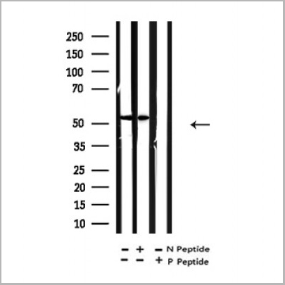
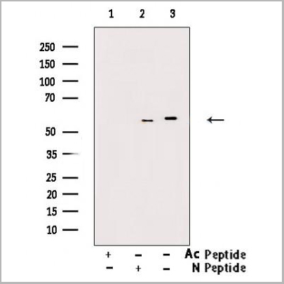
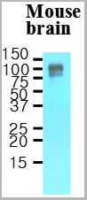
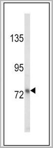
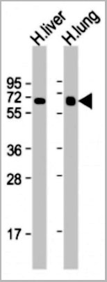
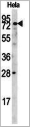
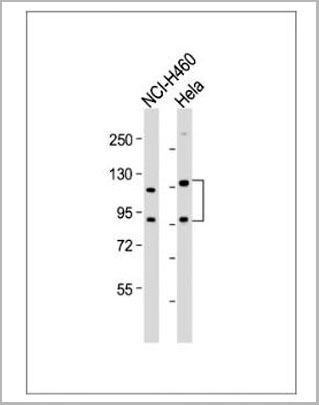
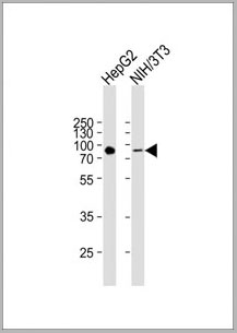
 WB (Western Blot)
(Western blot analysis of MeCP2-S421* in mouse brain, cerebellum tissue and HepG2 cell line lysates (35ug/lane). CP2 (arrow) was detected using the purified Pab.)
WB (Western Blot)
(Western blot analysis of MeCP2-S421* in mouse brain, cerebellum tissue and HepG2 cell line lysates (35ug/lane). CP2 (arrow) was detected using the purified Pab.)
MeCP2, Polyclonal Antibody (Cat# AAA28666)
Full Name
MeCP2 Antibody (S421)
Reactivity
Human, mouse
Applications
WB, EIA
Purity
Peptide Affinity Purified Rabbit Polyclonal Antibody (Pab)
Pricing


 FCM (Flow Cytometry)
(Figure 11. Flow Cytometry analysis of RH35 cells using anti- eRF1/ETF1 antibody (AAA19382).Overlay histogram showing RH35 cells stained with AAA19382 (Blue line). The cells were blocked with 10% normal goat serum. And then incubated with mouse anti-eRF1/ETF1 Antibody (AAA19382, 1μg/1x106 cells) for 30 min at 20 degree C. DyLight®488 conjugated goat anti-mouse IgG (BA1126, 5-10μg/1x106 cells) was used as secondary antibody for 30 minutes at 20 degree C. Isotype control antibody (Green line) was mouse IgG (1μg/1x106) used under the same conditions. Unlabelled sample (Red line) was also used as a control.)
FCM (Flow Cytometry)
(Figure 11. Flow Cytometry analysis of RH35 cells using anti- eRF1/ETF1 antibody (AAA19382).Overlay histogram showing RH35 cells stained with AAA19382 (Blue line). The cells were blocked with 10% normal goat serum. And then incubated with mouse anti-eRF1/ETF1 Antibody (AAA19382, 1μg/1x106 cells) for 30 min at 20 degree C. DyLight®488 conjugated goat anti-mouse IgG (BA1126, 5-10μg/1x106 cells) was used as secondary antibody for 30 minutes at 20 degree C. Isotype control antibody (Green line) was mouse IgG (1μg/1x106) used under the same conditions. Unlabelled sample (Red line) was also used as a control.)
eRF1/ETF1, Monoclonal Antibody (Cat# AAA19382)
Full Name
Anti-eRF1/ETF1 Antibody (monoclonal, 3B6)
Gene Names
ETF1; ERF; RF1; ERF1; TB3-1; D5S1995; SUP45L1
Reactivity
Human, Mouse, Rat
Applications
WB, IHC-P, ICC, IF, FC/FACS/FCM
Purity
Immunogen affinity purified.
Pricing


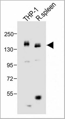
 FCM (Flow Cytometry)
(Figure 8. Flow Cytometry analysis of THP-1 cells using anti-INPPL1 antibody (AAA19364).Overlay histogram showing THP-1 cells stained with AAA19364 (Blue line). The cells were blocked with 10% normal goat serum. And then incubated with mouse anti- INPPL1 Antibody (AAA19364, 1μg/1x106 cells) for 30 min at 20 degree C. DyLight®488 conjugated goat anti-mouse IgG (BA1126, 5-10μg/1x106 cells) was used as secondary antibody for 30 minutes at 20 degree C. Isotype control antibody (Green line) was mouse IgG (1μg/1x106) used under the same conditions. Unlabelled sample (Red line) was also used as a control.)
FCM (Flow Cytometry)
(Figure 8. Flow Cytometry analysis of THP-1 cells using anti-INPPL1 antibody (AAA19364).Overlay histogram showing THP-1 cells stained with AAA19364 (Blue line). The cells were blocked with 10% normal goat serum. And then incubated with mouse anti- INPPL1 Antibody (AAA19364, 1μg/1x106 cells) for 30 min at 20 degree C. DyLight®488 conjugated goat anti-mouse IgG (BA1126, 5-10μg/1x106 cells) was used as secondary antibody for 30 minutes at 20 degree C. Isotype control antibody (Green line) was mouse IgG (1μg/1x106) used under the same conditions. Unlabelled sample (Red line) was also used as a control.)
INPPL1, Monoclonal Antibody (Cat# AAA19364)
Full Name
Anti-INPPL1 Antibody (monoclonal, 8C13)
Gene Names
INPPL1; OPSMD; SHIP2
Reactivity
Human, Mouse, Rat
Applications
WB, IHC-P, ICC, IF, FC/FACS/FCM
Purity
Immunogen affinity purified.
Pricing


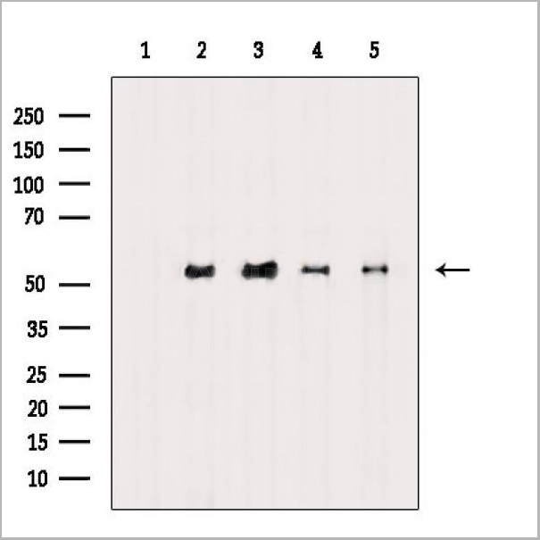
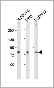
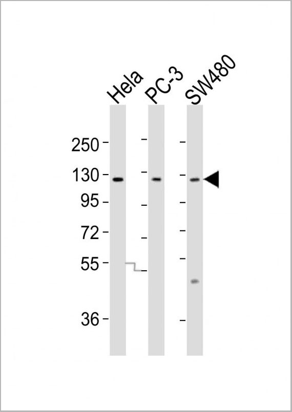
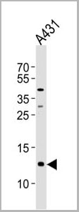
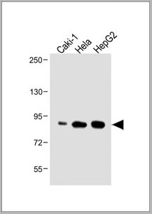
 WB (Western Blot)
(Western blot analysis of CD9 Antibody (Center) in HepG2 cell line lysates (35ug/lane). CD9 (arrow) was detected using the purified Pab.)
WB (Western Blot)
(Western blot analysis of CD9 Antibody (Center) in HepG2 cell line lysates (35ug/lane). CD9 (arrow) was detected using the purified Pab.)
CD9, Polyclonal Antibody (Cat# AAA28656)
Full Name
CD9 Antibody (Center)
Gene Names
CD9; MIC3; MRP-1; BTCC-1; DRAP-27; TSPAN29; TSPAN-29
Reactivity
Human
Applications
WB, EIA, IF, FC/FACS
Purity
Purified Rabbit Polyclonal Antibody (Pab)
Pricing


 FCM (Flow Cytometry)
(Figure 9. Flow Cytometry analysis of U251 cells using anti-RAB1B antibody (AAA19296).Overlay histogram showing U251 cells stained with AAA19296 (Blue line). The cells were blocked with 10% normal goat serum. And then incubated with rabbit anti-RAB1B Antibody (AAA19296, 1μg/1x106 cells) for 30 min at 20 degree C. DyLight®488 conjugated goat anti-rabbit IgG (5-10μg/1x106 cells) was used as secondary antibody for 30 minutes at 20 degree C. Isotype control antibody (Green line) was rabbit IgG (1μg/1x106) used under the same conditions. Unlabelled sample (Red line) was also used as a control.)
FCM (Flow Cytometry)
(Figure 9. Flow Cytometry analysis of U251 cells using anti-RAB1B antibody (AAA19296).Overlay histogram showing U251 cells stained with AAA19296 (Blue line). The cells were blocked with 10% normal goat serum. And then incubated with rabbit anti-RAB1B Antibody (AAA19296, 1μg/1x106 cells) for 30 min at 20 degree C. DyLight®488 conjugated goat anti-rabbit IgG (5-10μg/1x106 cells) was used as secondary antibody for 30 minutes at 20 degree C. Isotype control antibody (Green line) was rabbit IgG (1μg/1x106) used under the same conditions. Unlabelled sample (Red line) was also used as a control.)
RAB1B, Polyclonal Antibody (Cat# AAA19296)
Full Name
Anti-RAB1B Antibody
Reactivity
Human, Mouse, Rat
Applications
WB, IHC-P, ICC, IF, FC/FACS/FCM
Purity
Immunogen affinity purified.
Pricing


 FCM (Flow Cytometry)
(Figure 7. Flow Cytometry analysis of MCF-7 cells using anti-RGS6 antibody (AAA19308).Overlay histogram showing MCF-7 cells stained with AAA19308 (Blue line). The cells were blocked with 10% normal goat serum. And then incubated with rabbit anti-RGS6 Antibody (AAA19308, 1μg/1x106 cells) for 30 min at 20 degree C. DyLight®488 conjugated goat anti-rabbit IgG (5-10μg/1x106 cells) was used as secondary antibody for 30 minutes at 20 degree C. Isotype control antibody (Green line) was rabbit IgG (1μg/1x106) used under the same conditions. Unlabelled sample (Red line) was also used as a control.)
FCM (Flow Cytometry)
(Figure 7. Flow Cytometry analysis of MCF-7 cells using anti-RGS6 antibody (AAA19308).Overlay histogram showing MCF-7 cells stained with AAA19308 (Blue line). The cells were blocked with 10% normal goat serum. And then incubated with rabbit anti-RGS6 Antibody (AAA19308, 1μg/1x106 cells) for 30 min at 20 degree C. DyLight®488 conjugated goat anti-rabbit IgG (5-10μg/1x106 cells) was used as secondary antibody for 30 minutes at 20 degree C. Isotype control antibody (Green line) was rabbit IgG (1μg/1x106) used under the same conditions. Unlabelled sample (Red line) was also used as a control.)
RGS6, Polyclonal Antibody (Cat# AAA19308)
Full Name
Anti-RGS6 Antibody
Gene Names
RGS6; GAP
Reactivity
Human, Mouse, Rat
Applications
WB, IHC-P, FC/FACS/FCM, EIA
Purity
Immunogen affinity purified.
Pricing


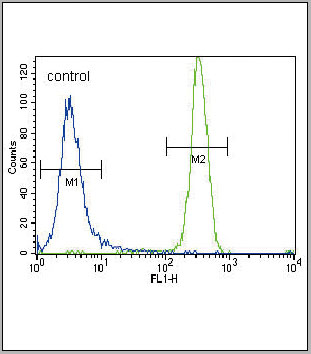
 FCM (Flow Cytometry)
(Figure 6. Flow Cytometry analysis of THP-1 cells using anti-PRMT8 antibody (AAA19321).Overlay histogram showing THP-1 cells stained with AAA19321 (Blue line). The cells were blocked with 10% normal goat serum. And then incubated with rabbit anti-PRMT8 Antibody (AAA19321,1μg/1x106 cells) for 30 min at 20 degree C. DyLight®488 conjugated goat anti-rabbit IgG (5-10μg/1x106 cells) was used as secondary antibody for 30 minutes at 20 degree C. Isotype control antibody (Green line) was rabbit IgG (1μg/1x106) used under the same conditions. Unlabelled sample (Red line) was also used as a control.)
FCM (Flow Cytometry)
(Figure 6. Flow Cytometry analysis of THP-1 cells using anti-PRMT8 antibody (AAA19321).Overlay histogram showing THP-1 cells stained with AAA19321 (Blue line). The cells were blocked with 10% normal goat serum. And then incubated with rabbit anti-PRMT8 Antibody (AAA19321,1μg/1x106 cells) for 30 min at 20 degree C. DyLight®488 conjugated goat anti-rabbit IgG (5-10μg/1x106 cells) was used as secondary antibody for 30 minutes at 20 degree C. Isotype control antibody (Green line) was rabbit IgG (1μg/1x106) used under the same conditions. Unlabelled sample (Red line) was also used as a control.)
PRMT8, Polyclonal Antibody (Cat# AAA19321)
Full Name
Anti-PRMT8 Antibody
Gene Names
PRMT8; HRMT1L3; HRMT1L4
Reactivity
Human, Rat
Applications
WB, IHC-P, FC/FACS/FCM, EIA
Purity
Immunogen affinity purified.
Pricing


 FCM (Flow Cytometry)
(Figure 12. Flow Cytometry analysis of U937 cells using anti-DYNLL1/PIN antibody (AAA19276).Overlay histogram showing U937 cells stained with AAA19276 (Blue line). The cells were blocked with 10% normal goat serum. And then incubated with rabbit anti-DYNLL1/PIN Antibody (AAA19276, 1μg/1x106 cells) for 30 min at 20 degree C. DyLight®488 conjugated goat anti-rabbit IgG (5-10μg/1x106 cells) was used as secondary antibody for 30 minutes at 20 degree C. Isotype control antibody (Green line) was rabbit IgG (1μg/1x106) used under the same conditions. Unlabelled sample (Red line) was also used as a control.)
FCM (Flow Cytometry)
(Figure 12. Flow Cytometry analysis of U937 cells using anti-DYNLL1/PIN antibody (AAA19276).Overlay histogram showing U937 cells stained with AAA19276 (Blue line). The cells were blocked with 10% normal goat serum. And then incubated with rabbit anti-DYNLL1/PIN Antibody (AAA19276, 1μg/1x106 cells) for 30 min at 20 degree C. DyLight®488 conjugated goat anti-rabbit IgG (5-10μg/1x106 cells) was used as secondary antibody for 30 minutes at 20 degree C. Isotype control antibody (Green line) was rabbit IgG (1μg/1x106) used under the same conditions. Unlabelled sample (Red line) was also used as a control.)
DYNLL1/PIN, Polyclonal Antibody (Cat# AAA19276)
Full Name
Anti-DYNLL1/PIN Antibody
Gene Names
DYNLL1; LC8; PIN; DLC1; DLC8; LC8a; DNCL1; hdlc1; DNCLC1
Reactivity
Human, Mouse, Rat
Applications
WB, IHC-P, ICC, IF, FC/FACS/FCM, EIA
Purity
Immunogen affinity purified.
Pricing


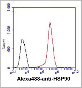
 FCM (Flow Cytometry)
(Figure 7. Flow Cytometry analysis of A549 cells using anti-Hsp90 alpha antibody (AAA19359).Overlay histogram showing A549 cells stained with AAA19359 (Blue line). The cells were blocked with 10% normal goat serum. And then incubated with mouse anti-Hsp90 alpha Antibody (AAA19359, 1μg/1x106 cells) for 30 min at 20 degree C. DyLight®488 conjugated goat anti-mouse IgG (BA1126, 5-10μg/1x106 cells) was used as secondary antibody for 30 minutes at 20 degree C. Isotype control antibody (Green line) was mouse IgG (1μg/1x106) used under the same conditions. Unlabelled sample (Red line) was also used as a control.)
FCM (Flow Cytometry)
(Figure 7. Flow Cytometry analysis of A549 cells using anti-Hsp90 alpha antibody (AAA19359).Overlay histogram showing A549 cells stained with AAA19359 (Blue line). The cells were blocked with 10% normal goat serum. And then incubated with mouse anti-Hsp90 alpha Antibody (AAA19359, 1μg/1x106 cells) for 30 min at 20 degree C. DyLight®488 conjugated goat anti-mouse IgG (BA1126, 5-10μg/1x106 cells) was used as secondary antibody for 30 minutes at 20 degree C. Isotype control antibody (Green line) was mouse IgG (1μg/1x106) used under the same conditions. Unlabelled sample (Red line) was also used as a control.)
Hsp90 alpha, Monoclonal Antibody (Cat# AAA19359)
Full Name
Anti-Hsp90 alpha Antibody (monoclonal, 6B5)
Gene Names
HSP90AA1; EL52; HSPN; LAP2; HSP86; HSPC1; HSPCA; Hsp89; Hsp90; HSP89A; HSP90A; HSP90N; HSPCAL1; HSPCAL4
Reactivity
Human, Mouse, Rat, Monkey
Applications
WB, IHC-P, ICC, IF, FC/FACS/FCM
Purity
Immunogen affinity purified.
Pricing


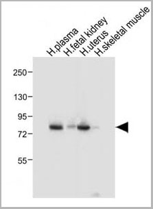
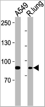
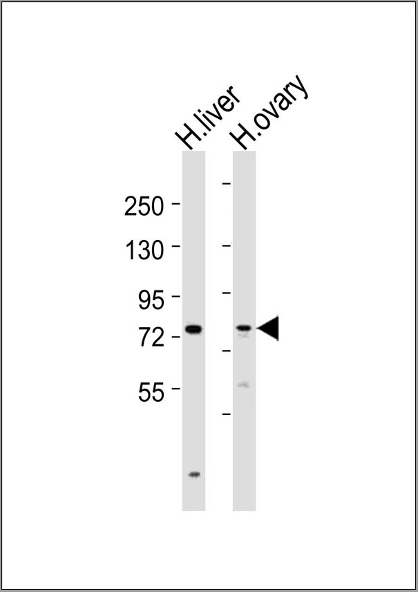
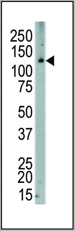
 WB (Western Blot)
(SLC16A11 Antibody (N-term) western blot analysis in mouse brain,stomach tissue lysates (35ug/lane).This demonstrates the SLC16A11 antibody detected the SLC16A11 protein (arrow).)
WB (Western Blot)
(SLC16A11 Antibody (N-term) western blot analysis in mouse brain,stomach tissue lysates (35ug/lane).This demonstrates the SLC16A11 antibody detected the SLC16A11 protein (arrow).)
SLC16A11, Polyclonal Antibody (Cat# AAA28655)
Full Name
SLC16A11 Antibody (N-term)
Gene Names
SLC16A11; MCT11
Reactivity
Human, mouse
Applications
WB, EIA, FC/FACS, IHC
Purity
Peptide Affinity Purified Rabbit Polyclonal Antibody (Pab)
Pricing


 FCM (Flow Cytometry)
(Figure 7. Flow Cytometry analysis of U20S cells using anti- ADA antibody (AAA19184). Overlay histogram showing U20S cells stained with AAA19184 (Blue line).The cells were blocked with 10% normal goat serum. And then incubated with mouse anti- ADA Antibody (AAA19184, 1ug/1x106 cells) for 30 min at 20 degree C. DyLight488 conjugated goat anti-mouse IgG (BA1126, 5-10ug/1x106 cells) was used as secondary antibody for 30 minutes at 20 degree C. Isotype control antibody (Green line) was mouse IgG (1ug/1x106) used under the same conditions. Unlabelled sample (Red line) was also used as a control.)
FCM (Flow Cytometry)
(Figure 7. Flow Cytometry analysis of U20S cells using anti- ADA antibody (AAA19184). Overlay histogram showing U20S cells stained with AAA19184 (Blue line).The cells were blocked with 10% normal goat serum. And then incubated with mouse anti- ADA Antibody (AAA19184, 1ug/1x106 cells) for 30 min at 20 degree C. DyLight488 conjugated goat anti-mouse IgG (BA1126, 5-10ug/1x106 cells) was used as secondary antibody for 30 minutes at 20 degree C. Isotype control antibody (Green line) was mouse IgG (1ug/1x106) used under the same conditions. Unlabelled sample (Red line) was also used as a control.)
ADA, Monoclonal Antibody (Cat# AAA19184)
Full Name
Anti-ADA Antibody (monoclonal, 6D4)
Reactivity
Human
Applications
WB, IHC, FC/FACS
Purity
Immunogen Affinity Purified
Pricing


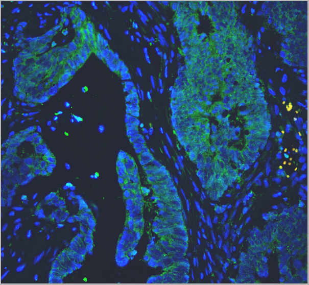
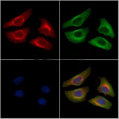
 IF (Immunofluorescence)
(Figure 11. IF analysis of Tubulin alpha using anti- Tubulin alpha antibody (AAA19381).Tubulin alpha was detected in immunocytochemical section of CACO-2 cells. Enzyme antigen retrieval was performed using IHC enzyme antigen retrieval reagent for 15 mins. The cells were blocked with 10% goat serum. And then incubated with 5μg/mL mouse anti- Tubulin alpha Antibody (AAA19381) overnight at 4 degree C. DyLight®594 Conjugated Goat Anti-Mouse IgG (BA1141) was used as secondary antibody at 1:100 dilution and incubated for 30 minutes at 37 degree C. The section was counterstained with DAPI. Visualize using a fluorescence microscope and filter sets appropriate for the label used.)
IF (Immunofluorescence)
(Figure 11. IF analysis of Tubulin alpha using anti- Tubulin alpha antibody (AAA19381).Tubulin alpha was detected in immunocytochemical section of CACO-2 cells. Enzyme antigen retrieval was performed using IHC enzyme antigen retrieval reagent for 15 mins. The cells were blocked with 10% goat serum. And then incubated with 5μg/mL mouse anti- Tubulin alpha Antibody (AAA19381) overnight at 4 degree C. DyLight®594 Conjugated Goat Anti-Mouse IgG (BA1141) was used as secondary antibody at 1:100 dilution and incubated for 30 minutes at 37 degree C. The section was counterstained with DAPI. Visualize using a fluorescence microscope and filter sets appropriate for the label used.)
Tubulin alpha, Monoclonal Antibody (Cat# AAA19381)
Full Name
Anti-Tubulin alpha Antibody (monoclonal, 7B12)
Gene Names
TUBA1A; LIS3; TUBA3; B-ALPHA-1
Reactivity
Human, Mouse, Rat
Applications
WB, IHC-P, ICC, IF, FC/FACS/FCM
Purity
Immunogen affinity purified.
Pricing


 FCM (Flow Cytometry)
(Figure 8. Flow Cytometry analysis of K562 cells using anti-PDIA6 antibody (AAA19282).Overlay histogram showing K562 cells stained with AAA19282 (Blue line). The cells were blocked with 10% normal goat serum. And then incubated with rabbit anti-PDIA6 Antibody (AAA19282, 1μg/1x106 cells) for 30 min at 20 degree C. DyLight®488 conjugated goat anti-rabbit IgG (5-10μg/1x106 cells) was used as secondary antibody for 30 minutes at 20 degree C. Isotype control antibody (Green line) was rabbit IgG (1μg/1x106) used under the same conditions. Unlabelled sample (Red line) was also used as a control.)
FCM (Flow Cytometry)
(Figure 8. Flow Cytometry analysis of K562 cells using anti-PDIA6 antibody (AAA19282).Overlay histogram showing K562 cells stained with AAA19282 (Blue line). The cells were blocked with 10% normal goat serum. And then incubated with rabbit anti-PDIA6 Antibody (AAA19282, 1μg/1x106 cells) for 30 min at 20 degree C. DyLight®488 conjugated goat anti-rabbit IgG (5-10μg/1x106 cells) was used as secondary antibody for 30 minutes at 20 degree C. Isotype control antibody (Green line) was rabbit IgG (1μg/1x106) used under the same conditions. Unlabelled sample (Red line) was also used as a control.)
PDIA6, Polyclonal Antibody (Cat# AAA19282)
Full Name
Anti-PDIA6 Antibody
Gene Names
PDIA6; P5; ERP5; TXNDC7
Reactivity
Human, Mouse, Rat
Applications
WB, IHC-P, ICC, IF, FC/FACS/FCM, EIA
Purity
Immunogen affinity purified.
Pricing


 FCM (Flow Cytometry)
(Figure 9. Flow Cytometry analysis of K562 cells using anti-MCM6 antibody (AAA19372).Overlay histogram showing K562 cells stained with AAA19372 (Blue line). The cells were blocked with 10% normal goat serum. And then incubated with mouse anti- MCM6 Antibody (AAA19372, 1μg/1x106 cells) for 30 min at 20 degree C. DyLight®488 conjugated goat anti-mouse IgG (BA1126, 5-10μg/1x106 cells) was used as secondary antibody for 30 minutes at 20 degree C. Isotype control antibody (Green line) was mouse IgG (1μg/1x106) used under the same conditions. Unlabelled sample (Red line) was also used as a control.)
FCM (Flow Cytometry)
(Figure 9. Flow Cytometry analysis of K562 cells using anti-MCM6 antibody (AAA19372).Overlay histogram showing K562 cells stained with AAA19372 (Blue line). The cells were blocked with 10% normal goat serum. And then incubated with mouse anti- MCM6 Antibody (AAA19372, 1μg/1x106 cells) for 30 min at 20 degree C. DyLight®488 conjugated goat anti-mouse IgG (BA1126, 5-10μg/1x106 cells) was used as secondary antibody for 30 minutes at 20 degree C. Isotype control antibody (Green line) was mouse IgG (1μg/1x106) used under the same conditions. Unlabelled sample (Red line) was also used as a control.)
MCM6, Monoclonal Antibody (Cat# AAA19372)
Full Name
Anti-MCM6 Antibody (monoclonal, 10I9)
Gene Names
MCM6; Mis5; P105MCM; MCG40308
Reactivity
Human
Applications
WB, IHC-P, ICC, IF, FC/FACS/FCM
Purity
Immunogen affinity purified.
Pricing
Transferrin, Native Protein (Cat# AAA14353)
Full Name
Transferrin protein (HRP)
Gene Names
TF; TFQTL1; PRO1557; PRO2086
Purity
Transferrin protein (HRP) was purified by selective precipitation and tandem chromatography followed by dialysis.
Pricing
 WB (Western Blot)
(Fig1: Western blot analysis of Annexin A1 on different lysates. Proteins were transferred to a PVDF membrane and blocked with 5% BSA in PBS for 1 hour at room temperature. The primary antibody was used at a 1:500 dilution in 5% BSA at room temperature for 2 hours. Goat Anti-Rabbit IgG - HRP Secondary Antibody at 1:5,000 dilution was used for 1 hour at room temperature.Positive control:Lane 1: A431 cell lysateLane 2: Rat uterus tissue lysate)
WB (Western Blot)
(Fig1: Western blot analysis of Annexin A1 on different lysates. Proteins were transferred to a PVDF membrane and blocked with 5% BSA in PBS for 1 hour at room temperature. The primary antibody was used at a 1:500 dilution in 5% BSA at room temperature for 2 hours. Goat Anti-Rabbit IgG - HRP Secondary Antibody at 1:5,000 dilution was used for 1 hour at room temperature.Positive control:Lane 1: A431 cell lysateLane 2: Rat uterus tissue lysate)
Annexin A1 Autoantibody (Anti-ANXA1), ELISA Kit (Cat# AAA10593)
Full Name
Human Annexin A1 Autoantibody (Anti-ANXA1) ELISA Kit
Reactivity
Human, Mouse, Rat
Applications
Western Blot, Immunocytochemistry, Immunohistochemistry
Pricing
C9 Tag, Polyclonal Antibody (Cat# AAA13688)
Full Name
Anti-C9 Tag Polyclonal Antibody, rabbit IgG
Applications
Western Blot
Purity
Protein A immunoaffinity chromatography
Pricing
M13 + fd + F1 Filamentous Phages, Monoclonal Antibody (Cat# AAA14300)
Full Name
M13 + fd + F1 Filamentous Phages antibody (FITC)
Reactivity
B62-FE2 binds to an epitope on pVIII (phage coat protein) covering the N-Terminal region of g8p AEGDDPAKAAFDSLQASAT
Purity
Protein A affinity chromatography
Pricing
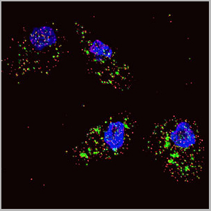
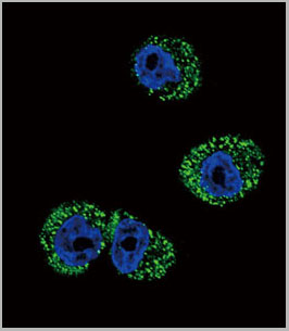
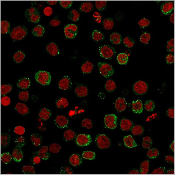
 IF (Immunofluorescence)
(Figure 3 and 4: ICC/IF analysis of MIF in Balb/3T3 cells. The cell was stained with AAA11724 (1;100). The secondary antibody (green) was used Alexa Fluor 488. DAPI was stained the cell nucelus (blue).Figure 5 and 6: ICC/IF analysis of MIF in HeLa cells line. The cell was stained with AAA11724 (1:100). The secondary antibody (green) was used Alexa Fluor 488. DAPI was stained the cell nucleus (blue))
IF (Immunofluorescence)
(Figure 3 and 4: ICC/IF analysis of MIF in Balb/3T3 cells. The cell was stained with AAA11724 (1;100). The secondary antibody (green) was used Alexa Fluor 488. DAPI was stained the cell nucelus (blue).Figure 5 and 6: ICC/IF analysis of MIF in HeLa cells line. The cell was stained with AAA11724 (1:100). The secondary antibody (green) was used Alexa Fluor 488. DAPI was stained the cell nucleus (blue))
MIF, Monoclonal Antibody (Cat# AAA11724)
Full Name
MIF antibody
Gene Names
MIF; GIF; GLIF; MMIF
Reactivity
Human
Applications
Western Blot, Immunocytochemistry, Immunofluorescence
Purity
By protein-G affinity chromatography
Pricing
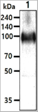
 WB (Western Blot)
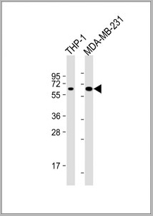
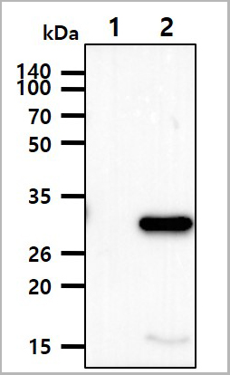
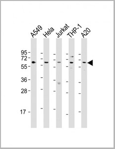
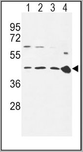
(All lanes : Anti-MB21D1 Antibody at 1:4000 dilutionLane 1: THP-1 whole cell lysateLane 2: MDA-MB-231 whole cell lysateLysates/proteins at 20 ug per lane. SecondaryGoat Anti-mouse IgG, (H+L), Peroxidase conjugated at 1/10000 dilution. Predicted band size : 59kDaBlocking/Dilution buffer: 5% NFDM/TBST.)
WB (Western Blot)
(All lanes : Anti-MB21D1 Antibody at 1:4000 dilutionLane 1: THP-1 whole cell lysateLane 2: MDA-MB-231 whole cell lysateLysates/proteins at 20 ug per lane. SecondaryGoat Anti-mouse IgG, (H+L), Peroxidase conjugated at 1/10000 dilution. Predicted band size : 59kDaBlocking/Dilution buffer: 5% NFDM/TBST.)
MB21D1, Monoclonal Antibody (Cat# AAA28800)
Full Name
MB21D1 Antibody
Gene Names
MB21D1; cGAS; h-cGAS; C6orf150
Reactivity
Human
Applications
Western Blot
Pricing
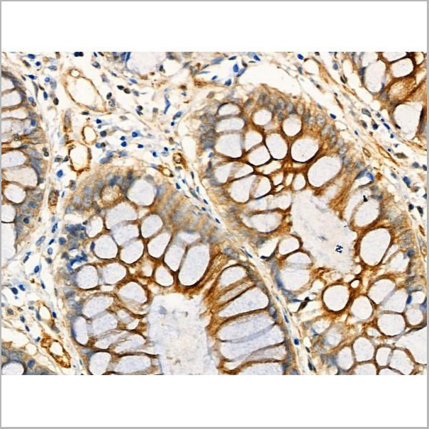
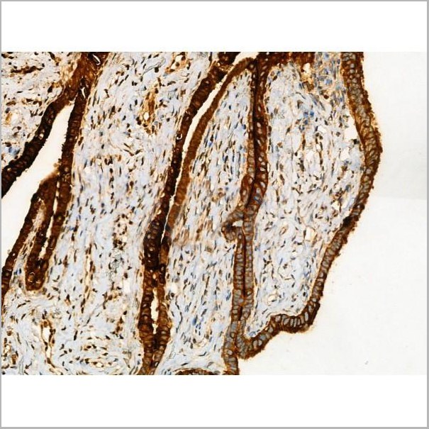
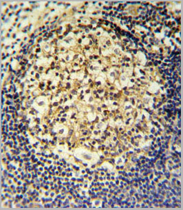
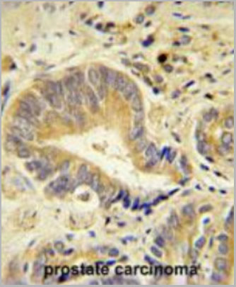
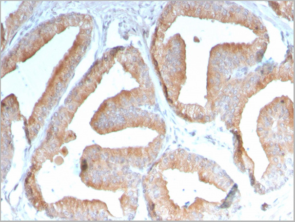
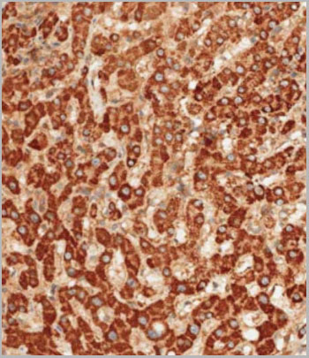
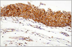 IHC (Immunohistochemistry)
(Paraffin embedded sections of human urothelium were incubated with anti-human Hsp90 (1:100)for 2 hours at room temperature. Antigen retrieval was performed in 0.1M sodium citrate buffer and detectedusing Diaminobenzidine (DAB))
IHC (Immunohistochemistry)
(Paraffin embedded sections of human urothelium were incubated with anti-human Hsp90 (1:100)for 2 hours at room temperature. Antigen retrieval was performed in 0.1M sodium citrate buffer and detectedusing Diaminobenzidine (DAB))
Hsp90, Monoclonal Antibody (Cat# AAA11726)
Full Name
Hsp90 antibody
Gene Names
HSP90AA1; EL52; HSPN; LAP2; HSP86; HSPC1; HSPCA; Hsp89; Hsp90; LAP-2; HSP89A; HSP90A; HSP90N; Hsp103; HSPCAL1; HSPCAL4; HEL-S-65p
Reactivity
Human,
Applications
Western Blot, Immunohistochemistry
Purity
By protein-G affinity chromatography
Pricing
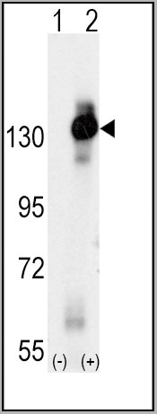
 WB (Western Blot)
(The cell lysates(10ug) were resolved by SDS-PAGE, transferred to PVDF membrane and probed with anti-human NMNAT-1 antibody (1:2000). Proteins were visualized using a goat anti-mouse secondary antibody conjugated to HRP and an ECL detection system.Lane 1.: 293T cell lysateLane 2.: NMNAT-1 Transfected 293T cell lysate)
WB (Western Blot)
(The cell lysates(10ug) were resolved by SDS-PAGE, transferred to PVDF membrane and probed with anti-human NMNAT-1 antibody (1:2000). Proteins were visualized using a goat anti-mouse secondary antibody conjugated to HRP and an ECL detection system.Lane 1.: 293T cell lysateLane 2.: NMNAT-1 Transfected 293T cell lysate)
NMNAT-1, Monoclonal Antibody (Cat# AAA11734)
Full Name
NMNAT-1
Gene Names
NMNAT1; LCA9; NMNAT; PNAT1
Reactivity
Human
Applications
Western Blot
Purity
Protein-A affinity chromatography
Pricing
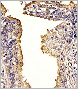
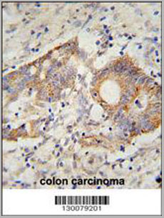
 IF (Immunofluorescence)
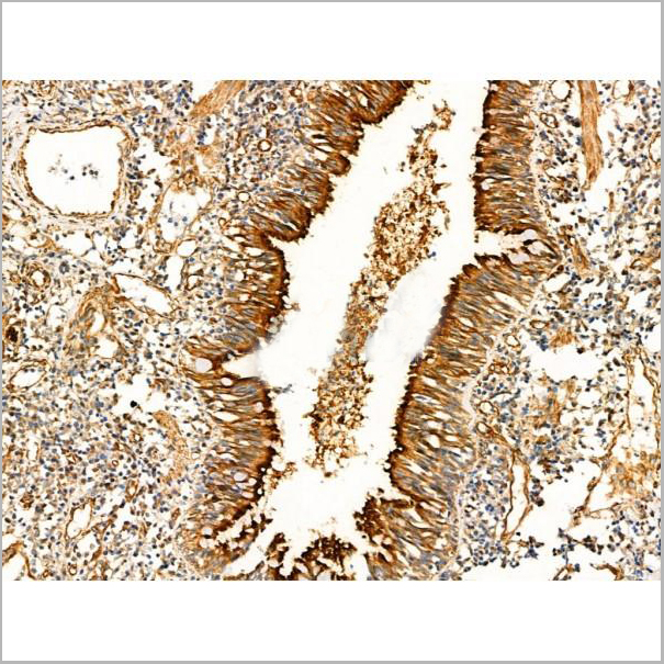
(Figure 3. IF analysis of PC1/3/PCSK1 using anti-PC1/3/PCSK1 antibody PC1/3/PCSK1 was detected in paraffin-embedded section of human colon cancer tissues. Heat mediated antigen retrieval was performed in citrate buffer (pH6, epitope retrieval solution ) for 20 mins. The tissue section was blocked with 10% goat serum. The tissue section was then incubated with 1ug/mL rabbit anti-PC1/3/PCSK1 Antibody overnight at 4 degree C. Biotin conjugated goat anti-rabbit IgG was used as secondary antibody and incubated for 30 minutes at 37 degree C. The tissue section was developed using DyLight 488 Conjugated Avidin. The section was counterstained with DAPI. Visualize using a fluorescence microscope and filter sets appropriate for the label used.)
IF (Immunofluorescence)
(Figure 3. IF analysis of PC1/3/PCSK1 using anti-PC1/3/PCSK1 antibody PC1/3/PCSK1 was detected in paraffin-embedded section of human colon cancer tissues. Heat mediated antigen retrieval was performed in citrate buffer (pH6, epitope retrieval solution ) for 20 mins. The tissue section was blocked with 10% goat serum. The tissue section was then incubated with 1ug/mL rabbit anti-PC1/3/PCSK1 Antibody overnight at 4 degree C. Biotin conjugated goat anti-rabbit IgG was used as secondary antibody and incubated for 30 minutes at 37 degree C. The tissue section was developed using DyLight 488 Conjugated Avidin. The section was counterstained with DAPI. Visualize using a fluorescence microscope and filter sets appropriate for the label used.)
PC1/3, Polyclonal Antibody (Cat# AAA11585)
Full Name
Anti-PC1/3 antibody
Gene Names
PCSK1; PC1; PC3; NEC1; SPC3; BMIQ12
Reactivity
Reacts with: Human, rat; Predicted to work with: Mouse
Applications
Western Blot, Immunohistochemistry, Immunofluorescence
Purity
Immunogen affinity purified.
Pricing
 Application Data
(At 25 degree C. Samples were then incubated with primary Ab(At 37 degree C. An AlexaFluor594 conjugated goat anti-rabbit IgG(H+L) Ab(Red) and an AlexaFluor488 conjugated goat anti-mouse IgG(H+L) Ab(Green) were used as the secondary antibody.The nuclear counter stain is DAPI(blue).)
Application Data
(At 25 degree C. Samples were then incubated with primary Ab(At 37 degree C. An AlexaFluor594 conjugated goat anti-rabbit IgG(H+L) Ab(Red) and an AlexaFluor488 conjugated goat anti-mouse IgG(H+L) Ab(Green) were used as the secondary antibody.The nuclear counter stain is DAPI(blue).)
alpha Tubulin, Polyclonal Antibody (Cat# AAA31344)
Full Name
Acetyl-alpha Tubulin (Lys40) Antibody
Gene Names
TUBA1B; K-ALPHA-1
Reactivity
Human, Mouse, Rat
Applications
Western Blot, Immunohistochemistry, Immunofluorescence, Immunocytochemistry
Purity
The antiserum was purified by peptide affinity chromatography using SulfoLink Coupling Resin (Thermo Fisher Scientific).
Pricing
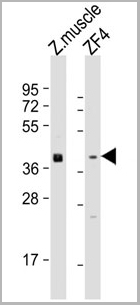 WB (Western Blot)
(All lanes: Anti-Zebrafish wnt8a Antibody (Center) at 1:1000-1:2000 dilutionLane 1: zebrafish muscle lysateLane 2: ZF4 whole cell lysateLysates/proteins at 20 ug per lane.Secondary Goat Anti-Rabbit IgG, (H+L), Peroxidase conjugated at 1/10000 dilution.Predicted band size : 40 kDaBlocking/Dilution buffer: 5% NFDM/TBST.)
WB (Western Blot)
(All lanes: Anti-Zebrafish wnt8a Antibody (Center) at 1:1000-1:2000 dilutionLane 1: zebrafish muscle lysateLane 2: ZF4 whole cell lysateLysates/proteins at 20 ug per lane.Secondary Goat Anti-Rabbit IgG, (H+L), Peroxidase conjugated at 1/10000 dilution.Predicted band size : 40 kDaBlocking/Dilution buffer: 5% NFDM/TBST.)
wnt8a, Polyclonal Antibody (Cat# AAA28796)
Full Name
Zebrafish wnt8a Antibody (Center)
Gene Names
wnt8a; wnt8; wnt-8; wnt8.1; wu:fa20e02; wu:fe05d07
Reactivity
Zebrafish
Applications
Western Blot
Purity
This antibody is purified through a protein A column, followed by peptide affinity purification.
Pricing
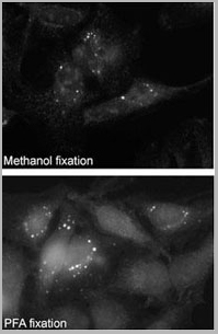 Application Data
(Immunofluorescence staining of Autophagy SQSTM1 (p62) Antibody (C-term) (AAA28682) on Methanol-fixed and PFA fixed HeLa cells. Data courtesy of Dr. Eeva-Liisa Eskelinen, University of Helsinki,Finland.)
Application Data
(Immunofluorescence staining of Autophagy SQSTM1 (p62) Antibody (C-term) (AAA28682) on Methanol-fixed and PFA fixed HeLa cells. Data courtesy of Dr. Eeva-Liisa Eskelinen, University of Helsinki,Finland.)
SQSTM1 (p62), Polyclonal Antibody (Cat# AAA28682)
Full Name
SQSTM1 (p62) Antibody (C-term)
Gene Names
SQSTM1; p60; p62; A170; OSIL; PDB3; ZIP3; p62B; FTDALS3
Reactivity
Human,Mouse,Rat
Predicted: Rat
Predicted: Rat
Applications
Western Blot, Immunohistochemistry, Immunofluorescence
Purity
Purified Rabbit Polyclonal Antibody (Pab)
Pricing
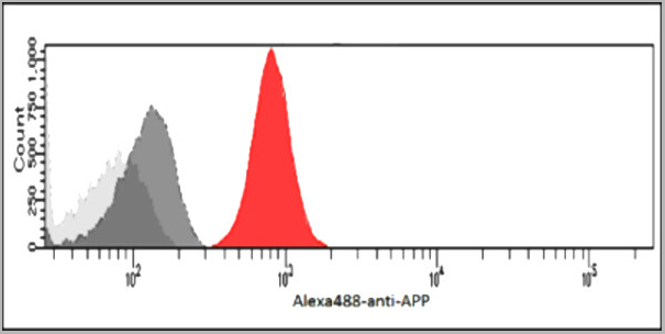
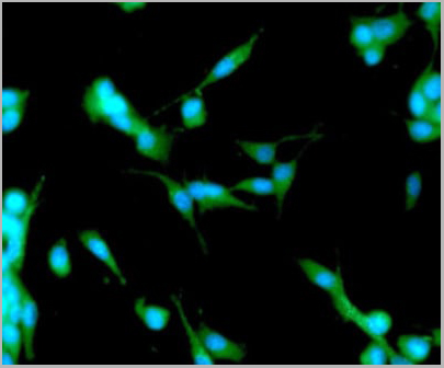 IF (Immunofluorescence)
(ICC/IF analysis of APP/Protease Nexin II in U87MG cells. The cell was stained with AAA11729 (1:100). The secondary antibody (green) was used Alexa Fluor 488. DAPI was stained the cell nucleus (blue).)
IF (Immunofluorescence)
(ICC/IF analysis of APP/Protease Nexin II in U87MG cells. The cell was stained with AAA11729 (1:100). The secondary antibody (green) was used Alexa Fluor 488. DAPI was stained the cell nucleus (blue).)
APP, Monoclonal Antibody (Cat# AAA11729)
Full Name
APP antibody
Gene Names
APP; AAA; AD1; PN2; ABPP; APPI; CVAP; ABETA; PN-II; preA4; CTFgamma
Reactivity
Human
Applications
Western Blot, Flow Cytometry, Immunocytochemistry, Immunofluorescence
Purity
By protein-G affinity chromatography
Pricing
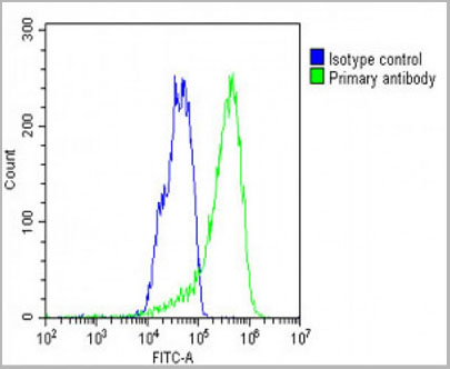
 FCM (Flow Cytometry)
(CCR7 Antibody (N-term) (AAA28701) flow cytometric analysis of 293 cells (right histogram) compared to a negative controlcell (left histogram). FITC-conjugated goat-anti-rabbit secondary antibodies were used for the analysis.)
FCM (Flow Cytometry)
(CCR7 Antibody (N-term) (AAA28701) flow cytometric analysis of 293 cells (right histogram) compared to a negative controlcell (left histogram). FITC-conjugated goat-anti-rabbit secondary antibodies were used for the analysis.)
CCR7, Polyclonal Antibody (Cat# AAA28701)
Full Name
CCR7 Antibody (N-term)
Gene Names
CCR7; BLR2; EBI1; CCR-7; CD197; CDw197; CMKBR7; CC-CKR-7
Reactivity
Human, mouse
Applications
Western Blot, Immunohistochemistry, Immunofluorescence, Flow Cytometry
Purity
Peptide Affinity Purified Rabbit Polyclonal Antibody (Pab)
Pricing

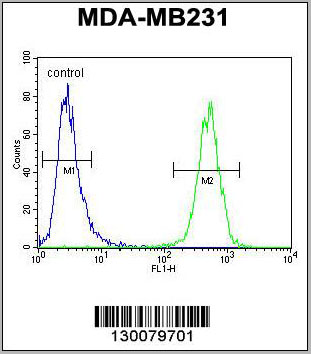
 FCM (Flow Cytometry)
(LTF Antibody flow cytometric analysis of MDA-MB231 cells (right histogram) compared to a negative control cell (lefthistogram).FITC-conjugated goat-anti-rabbit secondary antibodies were used for theanalysis.)
FCM (Flow Cytometry)
(LTF Antibody flow cytometric analysis of MDA-MB231 cells (right histogram) compared to a negative control cell (lefthistogram).FITC-conjugated goat-anti-rabbit secondary antibodies were used for theanalysis.)
LTF, Polyclonal Antibody (Cat# AAA28680)
Full Name
LTF Antibody
Gene Names
LTF; LF; HLF2; GIG12; HEL110
Reactivity
Human, mouse
Applications
Western Blot, Flow Cytometry
Purity
Peptide Affinity Purified Rabbit Polyclonal Antibody (Pab)
Pricing

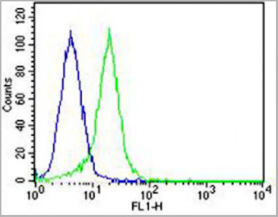 FCM (Flow Cytometry)
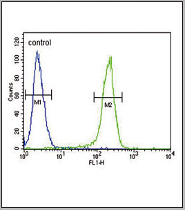
(Overlay histogram showing Hela cells stained with AAA28788 (green line). The cells were fixed with 2% paraformaldehyde (10 min) and then permeabilized with 90% methanol for 10 min. The cells were then icubated in 2% bovine serum albumin to block non-specific protein-protein interactions followed by the antibody (AAA28788, 1:25 dilution) for 60 min at 37ºC. The secondary antibody used was Goat-Anti-Rabbit IgG, DyLight® 488 Conjugated Highly Cross-Adsorbed at 1/400 dilution for 40 min at 37ºC. Isotype control antibody (blue line) was Rabbit IgG (1ug/1x10^6 cells) used under the same conditions. Acquisition of >10, 000 events was performed.)
FCM (Flow Cytometry)
(Overlay histogram showing Hela cells stained with AAA28788 (green line). The cells were fixed with 2% paraformaldehyde (10 min) and then permeabilized with 90% methanol for 10 min. The cells were then icubated in 2% bovine serum albumin to block non-specific protein-protein interactions followed by the antibody (AAA28788, 1:25 dilution) for 60 min at 37ºC. The secondary antibody used was Goat-Anti-Rabbit IgG, DyLight® 488 Conjugated Highly Cross-Adsorbed at 1/400 dilution for 40 min at 37ºC. Isotype control antibody (blue line) was Rabbit IgG (1ug/1x10^6 cells) used under the same conditions. Acquisition of >10, 000 events was performed.)
IDUA, Polyclonal Antibody (Cat# AAA28788)
Full Name
IDUA Antibody (Center)
Gene Names
IDUA; IDA; MPS1
Reactivity
Human
Applications
Western Blot, Immunohistochemistry, Flow Cytometry
Purity
This antibody is purified through a protein A column, followed by peptide affinity purification.
Pricing
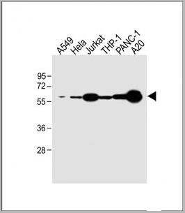
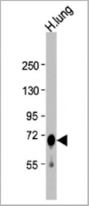
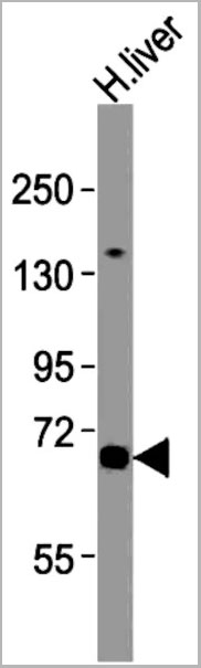
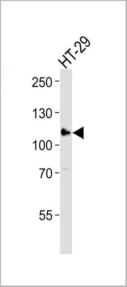 WB (Western Blot)
(Western blot analysis of lysate from HT-29 cell line, using ERN2 Antibody (N-term). AAA28749 was diluted at 1:1000. A goat anti-rabbit IgG H&L(HRP) at 1:10000 dilution was used as the secondary antibody. Lysate at 35ug.)
WB (Western Blot)
(Western blot analysis of lysate from HT-29 cell line, using ERN2 Antibody (N-term). AAA28749 was diluted at 1:1000. A goat anti-rabbit IgG H&L(HRP) at 1:10000 dilution was used as the secondary antibody. Lysate at 35ug.)
ERN2, Polyclonal Antibody (Cat# AAA28749)
Full Name
ERN2 Antibody (N-term)
Gene Names
ERN2; IRE1b; IRE2p; hIRE2p; IRE1-BETA
Reactivity
Human
Applications
Western Blot
Purity
Purified Rabbit Polyclonal Antibody (Pab)
Pricing
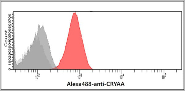 FCM (Flow Cytometry)
(Flow cytometry analysis of Crystallin alpha A in Balb/3T3 cells. The cell was stained with AAA11723 at 2-5ug for 1x10^6cells (red). A Goat anti mouse IgG (Alexa fluor 488) was used as the secondary antibody. Mouse monoclonal IgG was used as the isotype control (dark gray), cells without incubation with primary and secondary antibody was used as the negative control (light gray).)
FCM (Flow Cytometry)
(Flow cytometry analysis of Crystallin alpha A in Balb/3T3 cells. The cell was stained with AAA11723 at 2-5ug for 1x10^6cells (red). A Goat anti mouse IgG (Alexa fluor 488) was used as the secondary antibody. Mouse monoclonal IgG was used as the isotype control (dark gray), cells without incubation with primary and secondary antibody was used as the negative control (light gray).)
A crystallin A, Monoclonal Antibody (Cat# AAA11723)
Full Name
A crystallin A antibody
Gene Names
CRYAA; CRYA1; HSPB4; CTRCT9
Reactivity
Human
Applications
Western Blot, Flow Cytometry
Purity
By protein-G affinity chromatography
Pricing
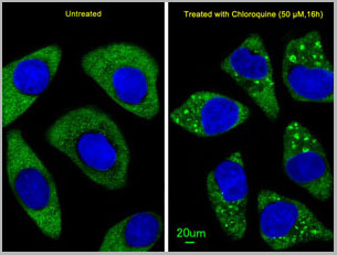
 WB (Western Blot)
(Immunofluorescent analysis of U251 cells, using LC3 Antibody (APG8B)(N-term). U251 cells(right) were treated with Chloroquine (50 uM,16h). AAA28778 was diluted at 1:25 dilution. Alexa Fluor 488-conjugated goat anti-rabbit lgG at 1:400 dilution was used as the secondary antibody (green).DAPI was used to stain the cell nuclear (blue).)
WB (Western Blot)
(Immunofluorescent analysis of U251 cells, using LC3 Antibody (APG8B)(N-term). U251 cells(right) were treated with Chloroquine (50 uM,16h). AAA28778 was diluted at 1:25 dilution. Alexa Fluor 488-conjugated goat anti-rabbit lgG at 1:400 dilution was used as the secondary antibody (green).DAPI was used to stain the cell nuclear (blue).)
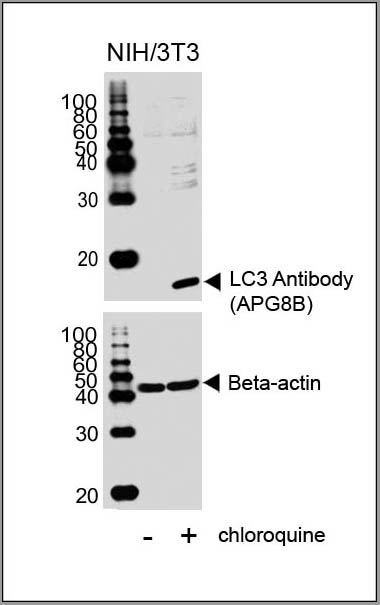
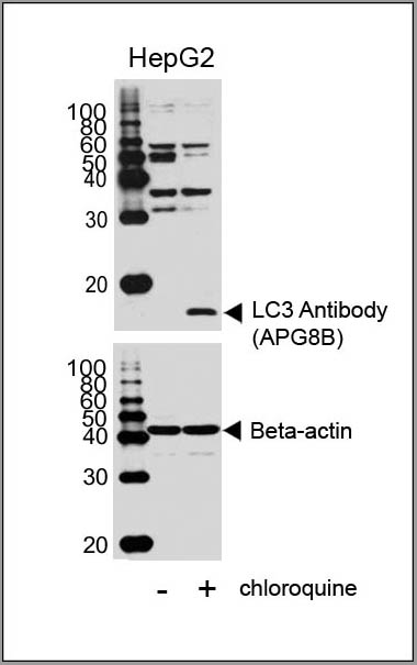
LC3, Polyclonal Antibody (Cat# AAA28778)
Full Name
LC3 Antibody (APG8B) (N-term)
Gene Names
MAP1LC3B; LC3B; ATG8F; MAP1LC3B-a; MAP1A/1BLC3
Reactivity
Human, rat (Predicted Reactivity: Bovine)
Applications
Immunofluorescence, Western Blot, Immunohistochemistry
Purity
Purified Rabbit Polyclonal Antibody (Pab)
Pricing
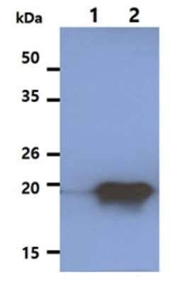
 WB (Western Blot)
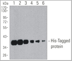
(Western blot analysis of his-tagged fusion protein.Each lane was loaded with 5ng of his-tagged fusion protein.Lane 1-6: 1: 0.5k, 1: 1k, 1: 2k, 1: 5k, 1: 10k, 1: 20k)
WB (Western Blot)
(Western blot analysis of his-tagged fusion protein.Each lane was loaded with 5ng of his-tagged fusion protein.Lane 1-6: 1: 0.5k, 1: 1k, 1: 2k, 1: 5k, 1: 10k, 1: 20k)
His-Tag, Monoclonal Antibody (Cat# AAA13799)
Full Name
His-Tag mAb (HRP Conjugated)
Reactivity
All
Applications
Western Blot
Pricing
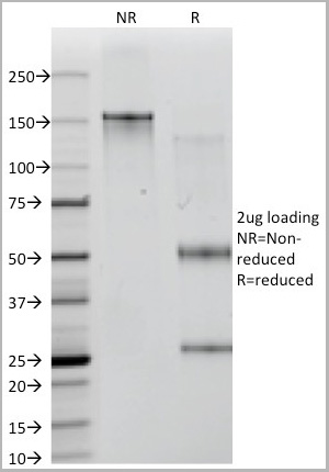
 SDS-PAGE
(The antibody wsas purified by protein G affinity achromatography from cell culture supernatants and verified by SDS-Page (Fig.2).Fig.2: SDS-PAGE analysis of purified YD-4E9 monoclonal antibody.Lane 1: molecular weight markerLane 2: 2ug of purified YD-4E9 antibody. Proteins were separated by SDSPAGE and stained with RAPID StainTM Reagent.)
SDS-PAGE
(The antibody wsas purified by protein G affinity achromatography from cell culture supernatants and verified by SDS-Page (Fig.2).Fig.2: SDS-PAGE analysis of purified YD-4E9 monoclonal antibody.Lane 1: molecular weight markerLane 2: 2ug of purified YD-4E9 antibody. Proteins were separated by SDSPAGE and stained with RAPID StainTM Reagent.)
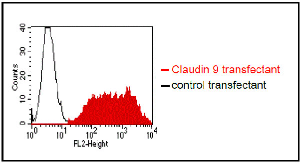
CLAUDIN9, Monoclonal Antibody (Cat# AAA14098)
Full Name
anti-human Claudin 9 monoclonal antibody
Reactivity
Human
Applications
Flow Cytometry, Competitive ELISA
Purity
Protein G
Pricing
 FCM (Flow Cytometry)
(BDKRB1 Antibody (Center) flow cytometric analysis of HepG2 cells (bottom histogram) compared to a negative control cell (top histogram).FITC-conjugated goat-anti-rabbit secondary antibodies were used for the analysis.)
FCM (Flow Cytometry)
(BDKRB1 Antibody (Center) flow cytometric analysis of HepG2 cells (bottom histogram) compared to a negative control cell (top histogram).FITC-conjugated goat-anti-rabbit secondary antibodies were used for the analysis.)
BDK_1, Polyclonal Antibody (Cat# AAA28760)
Full Name
BDK_1 Antibody (Center)
Gene Names
BDKRB1; B1R; BKR1; B1BKR; BKB1R; BDKRB2; BRADYB1
Reactivity
Human
Applications
Western Blot, Flow Cytometry
Purity
This antibody is purified through a protein A column, followed by peptide affinity purification.
Pricing
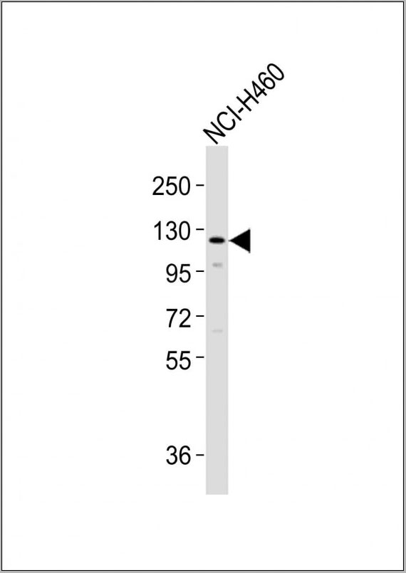
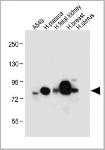
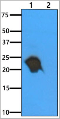
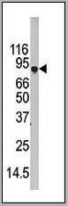
 WB (Western Blot)
(SLCO1B3 Antibody (C-term) western blot analysis in NCI-H460 cell line lysates (35ug/lane).This demonstrates the SLCO1B3 antibody detected the SLCO1B3 protein (arrow).)
WB (Western Blot)
(SLCO1B3 Antibody (C-term) western blot analysis in NCI-H460 cell line lysates (35ug/lane).This demonstrates the SLCO1B3 antibody detected the SLCO1B3 protein (arrow).)
SLCO1B3, Polyclonal Antibody (Cat# AAA28785)
Full Name
SLCO1B3 Antibody (C-term)
Gene Names
SLCO1B3; LST3; HBLRR; LST-2; OATP8; OATP-8; OATP1B3; SLC21A8; LST-3TM13
Reactivity
Human
Applications
Western Blot
Purity
Peptide Affinity Purified Rabbit Polyclonal Antibody (Pab)
Pricing
 WB (Western Blot)
(Western blot analysis of lysate from A431 cell line, using SPRR2A Antibody (C-term). AAA28741 was diluted at 1:1000 at each lane. A goat anti-rabbit IgG H&L(HRP) at 1:5000 dilution was used as the secondary antibody. Lysate at 35ug per lane.)
WB (Western Blot)
(Western blot analysis of lysate from A431 cell line, using SPRR2A Antibody (C-term). AAA28741 was diluted at 1:1000 at each lane. A goat anti-rabbit IgG H&L(HRP) at 1:5000 dilution was used as the secondary antibody. Lysate at 35ug per lane.)
SPRR2A, Polyclonal Antibody (Cat# AAA28741)
Full Name
SPRR2A Antibody (C-term)
Reactivity
Human
Applications
Western Blot
Purity
This antibody is purified through a protein A column, followed by peptide affinity purification.
Pricing
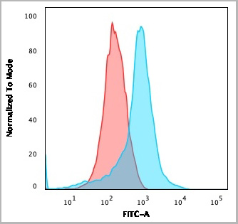
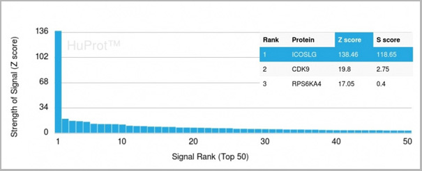 Application Data
(Analysis of Protein Array containing more than 19,000 full-length human proteins using ICOS-L Mouse Monoclonal Antibody (ICOSL/3111). Z- and S- Score: The Z-score represents the strength of a signal that a monoclonal antibody (MAb) (in combination with a fluorescently-tagged anti-IgG secondary antibody) produces when binding to a particular protein on the HuProtTM array. Z-scores are described in units of standard deviations (SD’s) above the mean value of all signals generated on that array. If targets on HuProtTM are arranged in descending order of the Z-score, the S-score is the difference (also in units of SD’s) between the Z-score. S-score therefore represents the relative target specificity of a MAb to its intended target. A MAb is considered to specific to its intended target, if the MAb has an S-score of at least 2.5. For example, if a MAb binds to protein X with a Z-score of 43 and to protein Y with a Z-score of 14, then the S-score for the binding of that MAb to protein X is equal to 29.)
Application Data
(Analysis of Protein Array containing more than 19,000 full-length human proteins using ICOS-L Mouse Monoclonal Antibody (ICOSL/3111). Z- and S- Score: The Z-score represents the strength of a signal that a monoclonal antibody (MAb) (in combination with a fluorescently-tagged anti-IgG secondary antibody) produces when binding to a particular protein on the HuProtTM array. Z-scores are described in units of standard deviations (SD’s) above the mean value of all signals generated on that array. If targets on HuProtTM are arranged in descending order of the Z-score, the S-score is the difference (also in units of SD’s) between the Z-score. S-score therefore represents the relative target specificity of a MAb to its intended target. A MAb is considered to specific to its intended target, if the MAb has an S-score of at least 2.5. For example, if a MAb binds to protein X with a Z-score of 43 and to protein Y with a Z-score of 14, then the S-score for the binding of that MAb to protein X is equal to 29.)
ICOS-L/ICOS Ligand/B7RP-1 (Immuno-Oncology Target), Monoclonal Antibody (Cat# AAA23916)
Full Name
ICOS-L/ICOS Ligand/B7RP-1 (Immuno-Oncology Target)
Gene Names
ICOSLG; B7h; B7H2; GL50; B7-H2; B7RP1; CD275; ICOSL; LICOS; B7RP-1; ICOS-L
Reactivity
Human
Applications
Flow Cytometry, Immunofluorescence, Immunohistochemistry
Purity
Purified Ab with BSA and Azide at 200ug/ml
Pricing
 FCM (Flow Cytometry)
(WB - MCSF Receptor (CSF1R) Antibody (C-term) AAA28757 detail IHC-P - MCSF Receptor (CSF1R) Antibody (C-term) AAA28757 detail IHC-P - MCSF Receptor (CSF1R) Antibody (C-term) AAA28757 detail FC - MCSF Receptor (CSF1R) Antibody (C-term) AAA28757 detail Overlay histogram showing HepG2 cells stained with AAA28757(green line). The cells were fixed with 2% paraformaldehyde (10 min) and then permeabilized with 90% methanol for 10 min. The cells were then icubated in 2% bovine serum albumin to block non-specific protein-protein interactions followed by the antibody (AAA28757, 1:25 dilution) for 60 min at 37ºC. The secondary antibody used was Goat-Anti-Rabbit IgG, DyLight® 488 Conjugated Highly Cross-Adsorbed(1583138) at 1/200 dilution for 40 min at 37ºC. Isotype control antibody (blue line) was rabbit IgG1 (1ug/1x10^6 cells) used under the same conditions. Acquisition of >10, 000 events was performed.)
FCM (Flow Cytometry)
(WB - MCSF Receptor (CSF1R) Antibody (C-term) AAA28757 detail IHC-P - MCSF Receptor (CSF1R) Antibody (C-term) AAA28757 detail IHC-P - MCSF Receptor (CSF1R) Antibody (C-term) AAA28757 detail FC - MCSF Receptor (CSF1R) Antibody (C-term) AAA28757 detail Overlay histogram showing HepG2 cells stained with AAA28757(green line). The cells were fixed with 2% paraformaldehyde (10 min) and then permeabilized with 90% methanol for 10 min. The cells were then icubated in 2% bovine serum albumin to block non-specific protein-protein interactions followed by the antibody (AAA28757, 1:25 dilution) for 60 min at 37ºC. The secondary antibody used was Goat-Anti-Rabbit IgG, DyLight® 488 Conjugated Highly Cross-Adsorbed(1583138) at 1/200 dilution for 40 min at 37ºC. Isotype control antibody (blue line) was rabbit IgG1 (1ug/1x10^6 cells) used under the same conditions. Acquisition of >10, 000 events was performed.)
MCSF Receptor (CSF1R), Polyclonal Antibody (Cat# AAA28757)
Full Name
MCSF Receptor (CSF1R) Antibody (C-term)
Gene Names
CSF1R; FMS; CSFR; FIM2; HDLS; C-FMS; CD115; CSF-1R; M-CSF-R
Reactivity
Human
Applications
Western Blot, Flow Cytometry, Immunohistochemistry
Purity
Purified Rabbit Polyclonal Antibody (Pab)
Pricing
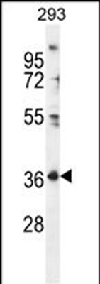
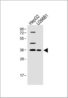 WB (Western Blot)
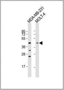
(All lanes: FOLR4 Antibody (N-term) at 1:500 dilution Lane 1: HepG2 whole cell lysateLane 2: U266B1 whole cell lysate Lysates/proteins at 20 ug per lane.Secondary Goat Anti-Rabbit IgG, (H+L), Peroxidase conjugated at 1/10000 dilution.Predicted band size: 36 kDaBlocking/Dilution buffer: 5% NFDM/TBST.)
WB (Western Blot)
(All lanes: FOLR4 Antibody (N-term) at 1:500 dilution Lane 1: HepG2 whole cell lysateLane 2: U266B1 whole cell lysate Lysates/proteins at 20 ug per lane.Secondary Goat Anti-Rabbit IgG, (H+L), Peroxidase conjugated at 1/10000 dilution.Predicted band size: 36 kDaBlocking/Dilution buffer: 5% NFDM/TBST.)
FOLR4, Polyclonal Antibody (Cat# AAA28763)
Full Name
FOLR4 Antibody (N-term)
Gene Names
IZUMO1R; JUNO; FOLR4; Folbp3
Reactivity
Human
Applications
Western Blot
Pricing
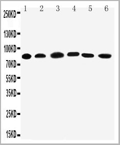
 WB (Western Blot)
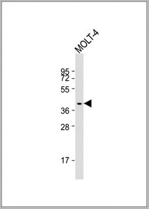
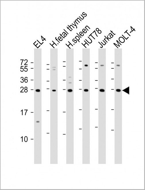
(All lanes : Anti-TIGIT Antibody at 1:2000 dilutionLane 1: EL4 whole cell lysateLane 2: human fetal thymus lysateLane 3: human spleen lysateLane 4: HUT78 whole cell lysateLane 5: Jurkat whole cell lysateLane 6: MOLT-4 whole cell lysateLysates/proteins at 20 ug per lane. SecondaryGoat Anti-Rabbit IgG, (H+L), Peroxidase conjugated at 1/10000 dilution. Predicted band size : 26 kDaBlocking/Dilution buffer: 5% NFDM/TBST.)
WB (Western Blot)
(All lanes : Anti-TIGIT Antibody at 1:2000 dilutionLane 1: EL4 whole cell lysateLane 2: human fetal thymus lysateLane 3: human spleen lysateLane 4: HUT78 whole cell lysateLane 5: Jurkat whole cell lysateLane 6: MOLT-4 whole cell lysateLysates/proteins at 20 ug per lane. SecondaryGoat Anti-Rabbit IgG, (H+L), Peroxidase conjugated at 1/10000 dilution. Predicted band size : 26 kDaBlocking/Dilution buffer: 5% NFDM/TBST.)
TIGIT, Polyclonal Antibody (Cat# AAA28803)
Full Name
TIGIT Antibody
Gene Names
TIGIT; VSIG9; VSTM3; WUCAM
Reactivity
Human, Mouse
Applications
Western Blot
Purity
This antibody is purified through a protein A column, followed by peptide affinity purification.
Pricing
 Application Data
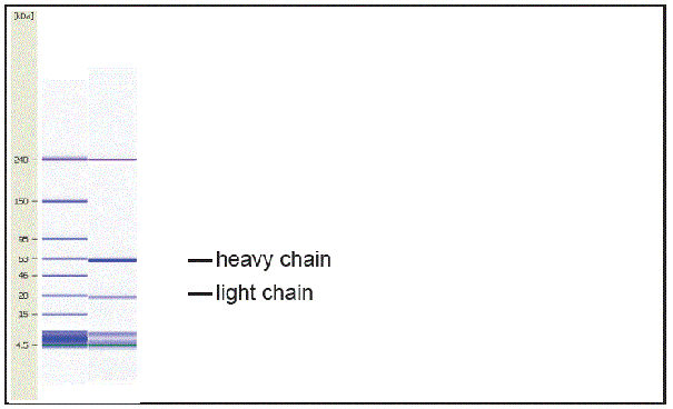
(Fig.2: CGE analysis of purified EQ-8D11-C1 monoclonal antibody. Lane 1: molecular weight marker, Lane 2: 2ug of purified EQ-8D11-C1 antibody. Proteins were separated by CGE (capillary gel electrophoresis, Agilent 2100 Bioanalyzer). Internal control bands (240 kDa / 7 kDa / 4,5 kDa).The antibody was purified by protein G affinity chromatography from cell culture supernatants and verified by CGE (Fig.2).)
Application Data
(Fig.2: CGE analysis of purified EQ-8D11-C1 monoclonal antibody. Lane 1: molecular weight marker, Lane 2: 2ug of purified EQ-8D11-C1 antibody. Proteins were separated by CGE (capillary gel electrophoresis, Agilent 2100 Bioanalyzer). Internal control bands (240 kDa / 7 kDa / 4,5 kDa).The antibody was purified by protein G affinity chromatography from cell culture supernatants and verified by CGE (Fig.2).)
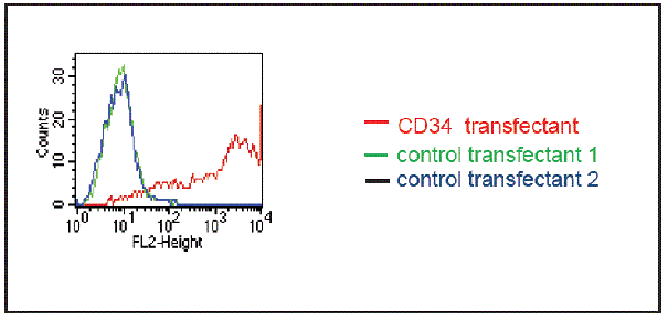
Ovine CD34, Monoclonal Antibody (Cat# AAA14097)
Full Name
anti-ovine CD34 monoclonal antibody
Applications
Flow Cytometry, Competitive ELISA
Purity
Protein G purified
Pricing

 Application Data
(At 25 degree C. Samples were then incubated with primary Ab(At 37 degree C. An AlexaFluor594 conjugated goat anti-rabbit IgG(H+L) Ab(Red) and an AlexaFluor488 conjugated goat anti-mouse IgG(H+L) Ab(Green) were used as the secondary antibody.The nuclear counter stain is DAPI (blue).)
Application Data
(At 25 degree C. Samples were then incubated with primary Ab(At 37 degree C. An AlexaFluor594 conjugated goat anti-rabbit IgG(H+L) Ab(Red) and an AlexaFluor488 conjugated goat anti-mouse IgG(H+L) Ab(Green) were used as the secondary antibody.The nuclear counter stain is DAPI (blue).)
IRF7, Polyclonal Antibody (Cat# AAA31330)
Full Name
Phospho-IRF7 (Ser471/Ser472) Antibody
Gene Names
IRF7; IRF7A; IRF7B; IRF7C; IRF7H; IRF-7H
Reactivity
Human, Mouse, Rat
Applications
Western Blot, Immunohistochemistry, Immunofluorescence, Immunocytochemistry, Peptide ELISA
Purity
The antibody is from purified rabbit serum by affinity purification via sequential chromatography on phospho-peptide and non-phospho-peptide affinity columns.
Pricing


 Application Data
(Published customer image: Mouse anti V5 tag antibody, clone SV5-Pk1 used for the detection of V5 tagged WEEV_nsP3 protein by western blotting and immunofluorescenceImage caption: WEEV nsP3 interaction with host IKKbeta. A) U87MGs were transfected in a 6-well plate with 5 ug of pUC19 and WEEV_nsP3_HA for 24 hours. Cell lysates were resolved using SDS-PAGE and subsequently immunoblotted with V5 antibody and beta-actin served as a loading control. B) U87MGs were transfected with WEEV_nsP3_V5; cells were fixed after 24 hours and stained with antibodies against the endogenous IKKbeta and the V5 tag. Cells were incubated with appropriate secondary Alexa Fluor antibodies and the nuclei stained with DAPI. Co-localization of IKKbeta with WEEV_nsP3_V5 (yellow) was observed as shown by the arrows. B) Panels E -H serve as an example of transfected cells in a given field of view that show co-localization of IKKbeta and WEEV_nsP3_V5 24 hours post transfection. Panels I-L represent magnified images of other cells showing co-localization of IKKbeta and WEEV_nsP3_V5. Panel M is a magnified image of panel L. The co-localization was confirmed by Z-stack analysis. Co-localization was calculated to be approximately in 61% of cells (163 cells were counted of which 44% demonstrated expression of nsP3. Of those cells that expressed nsP3, 61% showed co-localization of both proteins). Images were taken using Nikon Eclipse TE2000-U at 60x magnification and are representative of 2 independent experiments.From: Amaya M, Voss K, Sampey G, Senina S, de la Fuente C, et al. (2014) The Role of IKKbeta in Venezuelan Equine Encephalitis Virus Infection. PLoS ONE 9(2): e86745.)
Application Data
(Published customer image: Mouse anti V5 tag antibody, clone SV5-Pk1 used for the detection of V5 tagged WEEV_nsP3 protein by western blotting and immunofluorescenceImage caption: WEEV nsP3 interaction with host IKKbeta. A) U87MGs were transfected in a 6-well plate with 5 ug of pUC19 and WEEV_nsP3_HA for 24 hours. Cell lysates were resolved using SDS-PAGE and subsequently immunoblotted with V5 antibody and beta-actin served as a loading control. B) U87MGs were transfected with WEEV_nsP3_V5; cells were fixed after 24 hours and stained with antibodies against the endogenous IKKbeta and the V5 tag. Cells were incubated with appropriate secondary Alexa Fluor antibodies and the nuclei stained with DAPI. Co-localization of IKKbeta with WEEV_nsP3_V5 (yellow) was observed as shown by the arrows. B) Panels E -H serve as an example of transfected cells in a given field of view that show co-localization of IKKbeta and WEEV_nsP3_V5 24 hours post transfection. Panels I-L represent magnified images of other cells showing co-localization of IKKbeta and WEEV_nsP3_V5. Panel M is a magnified image of panel L. The co-localization was confirmed by Z-stack analysis. Co-localization was calculated to be approximately in 61% of cells (163 cells were counted of which 44% demonstrated expression of nsP3. Of those cells that expressed nsP3, 61% showed co-localization of both proteins). Images were taken using Nikon Eclipse TE2000-U at 60x magnification and are representative of 2 independent experiments.From: Amaya M, Voss K, Sampey G, Senina S, de la Fuente C, et al. (2014) The Role of IKKbeta in Venezuelan Equine Encephalitis Virus Infection. PLoS ONE 9(2): e86745.)
V5-TAG, Monoclonal Antibody (Cat# AAA12081)
Full Name
MOUSE ANTI V5-TAG:HRP
Applications
Western Blot
Pricing
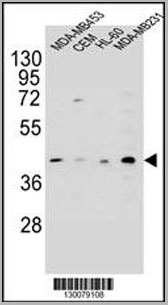 WB (Western Blot)
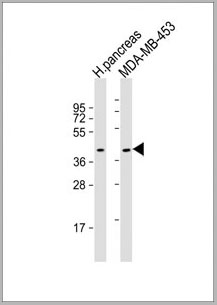
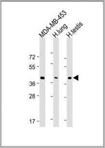
(HHLA2 Antibody (N-term) western blot analysis in MDA-MB453,CEM,HL-60,MDA-MB231 cell line lysates (35ug/lane).This demonstrates the HHLA2 antibody detected the HHLA2 protein (arrow).)
WB (Western Blot)
(HHLA2 Antibody (N-term) western blot analysis in MDA-MB453,CEM,HL-60,MDA-MB231 cell line lysates (35ug/lane).This demonstrates the HHLA2 antibody detected the HHLA2 protein (arrow).)
HHLA2, Polyclonal Antibody (Cat# AAA28722)
Full Name
HHLA2 Antibody (N-term)
Gene Names
HHLA2; B7H7; B7-H7
Reactivity
Human
Applications
Western Blot, Immunohistochemistry, Flow Cytometry
Purity
Peptide Affinity Purified Rabbit Polyclonal Antibody (Pab)
Pricing
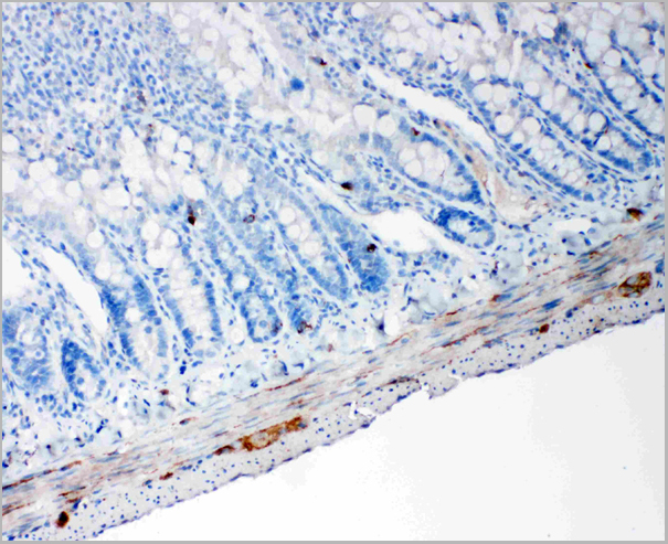
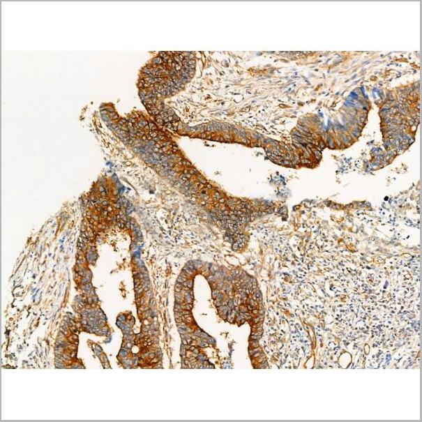
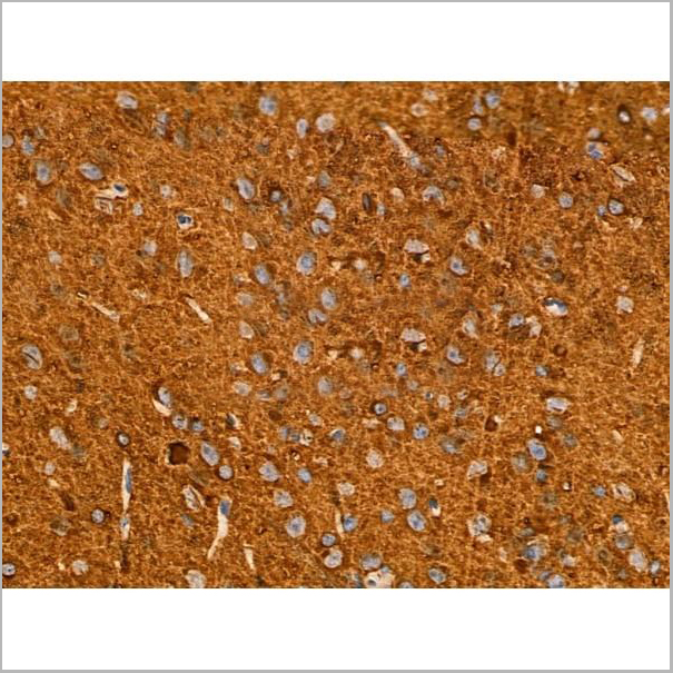
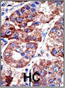
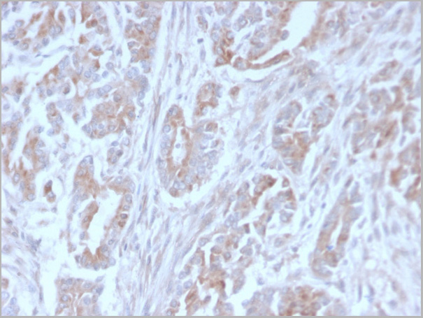
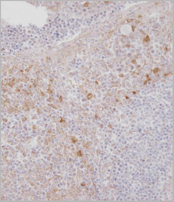
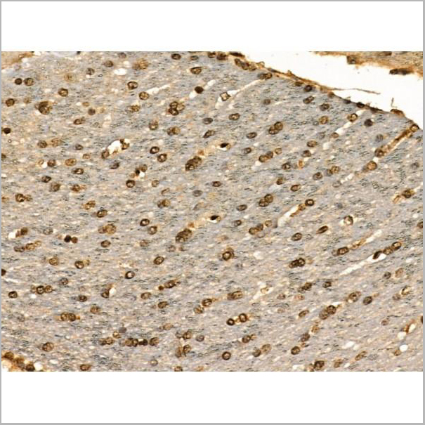
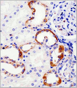
 IHC (Immunohistchemistry)
(Immunohistochemical analysis of paraffin-embedded human liver tissue using AAA28677 performed on the Leica® BOND RXm. Tissue was fixed with formaldehyde at room temperature; antigen retrieval was by heat mediation with a EDTA buffer (pH9. 0). Samples were incubated with primary antibody(1:1000) for 1 hours at room temperature. A undiluted biotinylated CRF Anti-Polyvalent HRP Polymer antibody was used as the secondary antibody.)
IHC (Immunohistchemistry)
(Immunohistochemical analysis of paraffin-embedded human liver tissue using AAA28677 performed on the Leica® BOND RXm. Tissue was fixed with formaldehyde at room temperature; antigen retrieval was by heat mediation with a EDTA buffer (pH9. 0). Samples were incubated with primary antibody(1:1000) for 1 hours at room temperature. A undiluted biotinylated CRF Anti-Polyvalent HRP Polymer antibody was used as the secondary antibody.)
ATG7, Polyclonal Antibody (Cat# AAA28677)
Full Name
ATG7 Antibody (C-term)
Gene Names
ATG7; GSA7; APG7L; APG7-LIKE
Reactivity
Human, Mouse
Predicted reactivity: Chicken, Rat
Predicted reactivity: Chicken, Rat
Applications
Western Blot, Immunohistochemistry, Immunofluorescence
Purity
Purified Rabbit Polyclonal Antibody (Pab)
Pricing