Filters
Clonality
Type
Reactivity
Gene Name
Isotype
Host
Application
Clone
410 results for " Conjugated Proteins" - showing 150-200


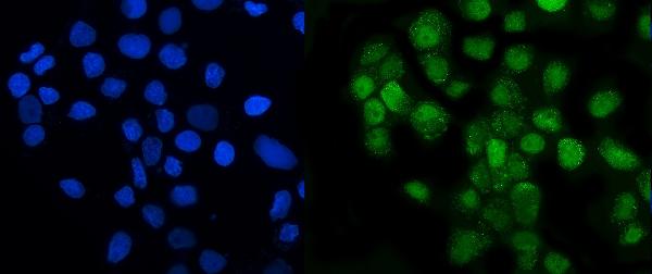
 IF (Immunofluorescence)
(ICC/IF analysis of FABP7 in PC3 cells. The cell was stained with AAA11731 (1:100). The secondary antibody (green) was used Alexa Fluor 488. DAPI was stained the cell nucleus (blue).)
IF (Immunofluorescence)
(ICC/IF analysis of FABP7 in PC3 cells. The cell was stained with AAA11731 (1:100). The secondary antibody (green) was used Alexa Fluor 488. DAPI was stained the cell nucleus (blue).)
FABP7, Monoclonal Antibody (Cat# AAA11731)
Full Name
FABP7 antibody
Gene Names
FABP7; MRG; BLBP; FABPB; B-FABP
Reactivity
Human, Mouse
Applications
Western Blot, Immunocytochemistry, Immunofluorescence
Purity
By protein-G affinity chromatography
Pricing


 IHC (Immunohistchemistry)
(Immunohistochemistry Analysis: Representative lot data. (Fig. 1 and 2) Paraffin-embedded mouse and human brain tissue was prepared using heat-induced epitope retrieval in citrate buffer, pH 6.0. Immunostaining was performed using a 1:100 dilution. Reactivity was detected using the IHC-Select Detection Kit. Staining pattern appears as cytoplasmic. (Fig. 3 and 4) Paraffin-embedded mouse and mouse olfactory lobe and cerebellum brain tissue was prepared using heat-induced epitope retrieval in citrate buffer, pH 6.0. Immunostaining was performed using a Chicken IgY Antibody 1:100 dilution of Cat. No. AB15894, anti-Tbr2. Reactivity was detected using the IHC-Select Detection Kit. Immunoreactivity seen here is mostly nuclear.)
IHC (Immunohistchemistry)
(Immunohistochemistry Analysis: Representative lot data. (Fig. 1 and 2) Paraffin-embedded mouse and human brain tissue was prepared using heat-induced epitope retrieval in citrate buffer, pH 6.0. Immunostaining was performed using a 1:100 dilution. Reactivity was detected using the IHC-Select Detection Kit. Staining pattern appears as cytoplasmic. (Fig. 3 and 4) Paraffin-embedded mouse and mouse olfactory lobe and cerebellum brain tissue was prepared using heat-induced epitope retrieval in citrate buffer, pH 6.0. Immunostaining was performed using a Chicken IgY Antibody 1:100 dilution of Cat. No. AB15894, anti-Tbr2. Reactivity was detected using the IHC-Select Detection Kit. Immunoreactivity seen here is mostly nuclear.)
EOMES, Polyclonal Antibody (Cat# AAA26884)
Full Name
EOMES (Eomesodermin Homolog, T-box Brain Protein 2, TBR2, T-brain-2, TBR-2)
Gene Names
Eomes; Tbr2; TBR-2; C77258
Reactivity
Mouse, Human, Rat
Applications
Immunohistochemistry, Western Blot
Purity
Purified by affinity chromatography.
Pricing
 FCM (Flow Cytometry)
(Figure 2. Flow Cytometry analysis of A549 cells using anti-WEE1 antibody (AAA19240).Overlay histogram showing A549 cells stained with AAA19240 (Blue line). The cells were blocked with 10% normal goat serum. And then incubated with rabbit anti-WEE1 Antibody (AAA19240, 1μg/1x106 cells) for 30 min at 20 degree C. DyLight®488 conjugated goat anti-rabbit IgG (5-10μg/1x106 cells) was used as secondary antibody for 30 minutes at 20 degree C. Isotype control antibody (Green line) was rabbit IgG (1μg/1x106) used under the same conditions. Unlabelled sample (Red line) was also used as a control.)
FCM (Flow Cytometry)
(Figure 2. Flow Cytometry analysis of A549 cells using anti-WEE1 antibody (AAA19240).Overlay histogram showing A549 cells stained with AAA19240 (Blue line). The cells were blocked with 10% normal goat serum. And then incubated with rabbit anti-WEE1 Antibody (AAA19240, 1μg/1x106 cells) for 30 min at 20 degree C. DyLight®488 conjugated goat anti-rabbit IgG (5-10μg/1x106 cells) was used as secondary antibody for 30 minutes at 20 degree C. Isotype control antibody (Green line) was rabbit IgG (1μg/1x106) used under the same conditions. Unlabelled sample (Red line) was also used as a control.)
WEE1, Polyclonal Antibody (Cat# AAA19240)
Full Name
Anti-WEE1 Antibody
Gene Names
WEE1; WEE1A; WEE1hu
Reactivity
Human, Mouse
Applications
Western Blot, Flow Cytometry, Direct ELISA
Purity
Immunogen affinity purified.
Pricing


 Application Data
(Staining of KG1 lymphocytes with Mouse anti Human CD59:FITC)
Application Data
(Staining of KG1 lymphocytes with Mouse anti Human CD59:FITC)
CD59, Monoclonal Antibody (Cat# AAA12068)
Full Name
MOUSE ANTI HUMAN CD59:FITC
Gene Names
CD59; 1F5; EJ16; EJ30; EL32; G344; MIN1; MIN2; MIN3; MIRL; HRF20; MACIF; MEM43; MIC11; MSK21; 16.3A5; HRF-20; MAC-IP; p18-20
Applications
Flow Cytometry
Pricing


 FCM (Flow Cytometry)
(Figure 10. Flow Cytometry analysis of K562 cells using anti-RCC1 antibody (AAA19263).Overlay histogram showing K562 cells stained with AAA19263 (Blue line). The cells were blocked with 10% normal goat serum. And then incubated with rabbit anti-RCC1 Antibody (AAA19263,1μg/1x106 cells) for 30 min at 20 degree C. DyLight®488 conjugated goat anti-rabbit IgG (5-10μg/1x106 cells) was used as secondary antibody for 30 minutes at 20 degree C. Isotype control antibody (Green line) was rabbit IgG (1μg/1x106) used under the same conditions. Unlabelled sample (Red line) was also used as a control.)
FCM (Flow Cytometry)
(Figure 10. Flow Cytometry analysis of K562 cells using anti-RCC1 antibody (AAA19263).Overlay histogram showing K562 cells stained with AAA19263 (Blue line). The cells were blocked with 10% normal goat serum. And then incubated with rabbit anti-RCC1 Antibody (AAA19263,1μg/1x106 cells) for 30 min at 20 degree C. DyLight®488 conjugated goat anti-rabbit IgG (5-10μg/1x106 cells) was used as secondary antibody for 30 minutes at 20 degree C. Isotype control antibody (Green line) was rabbit IgG (1μg/1x106) used under the same conditions. Unlabelled sample (Red line) was also used as a control.)
RCC1, Polyclonal Antibody (Cat# AAA19263)
Full Name
Anti-RCC1 Antibody
Gene Names
RCC1; CHC1; RCC1-I; SNHG3-RCC1
Reactivity
Human, Mouse, Rat
Applications
Western Blot, Immunohistochemistry, Immunohistochemistry, Immunocytochemistry, Immunofluorescence, Flow Cytometry, Direct ELISA
Purity
Immunogen affinity purified.
Pricing


 FCM (Flow Cytometry)
(Figure 8. Flow Cytometry analysis of SiHa cells using anti- ASS1 antibody (AAA19369).Overlay histogram showing SiHa cells stained with AAA19369 (Blue line). The cells were blocked with 10% normal goat serum. And then incubated with mouse anti-ASS1 Antibody (AAA19369, 1μg/1x106 cells) for 30 min at 20 degree C. DyLight®488 conjugated goat anti-mouse IgG (BA1126, 5-10μg/1x106 cells) was used as secondary antibody for 30 minutes at 20 degree C. Isotype control antibody (Green line) was mouse IgG (1μg/1x106) used under the same conditions. Unlabelled sample (Red line) was also used as a control.)
FCM (Flow Cytometry)
(Figure 8. Flow Cytometry analysis of SiHa cells using anti- ASS1 antibody (AAA19369).Overlay histogram showing SiHa cells stained with AAA19369 (Blue line). The cells were blocked with 10% normal goat serum. And then incubated with mouse anti-ASS1 Antibody (AAA19369, 1μg/1x106 cells) for 30 min at 20 degree C. DyLight®488 conjugated goat anti-mouse IgG (BA1126, 5-10μg/1x106 cells) was used as secondary antibody for 30 minutes at 20 degree C. Isotype control antibody (Green line) was mouse IgG (1μg/1x106) used under the same conditions. Unlabelled sample (Red line) was also used as a control.)
ASS1, Monoclonal Antibody (Cat# AAA19369)
Full Name
Anti-ASS1 Antibody (monoclonal, 5I5)
Gene Names
ASS1; ASS; CTLN1
Reactivity
Human, Mouse, Rat, Monkey
Applications
WB, IHC-P, ICC, IF, FC/FACS/FCM
Purity
Immunogen affinity purified.
Pricing


 IHC (Immunohistchemistry)
(Immunohistochemistry Analysis: Representative lot data. (Fig. 1 and 2) Paraffin-embedded mouse and human brain tissue was prepared using heat-induced epitope retrieval in citrate buffer, pH 6.0. Immunostaining was performed using a 1:100 dilution. Reactivity was detected using the IHC-Select Detection Kit. Staining pattern appears as cytoplasmic. (Fig. 3 and 4) Paraffin-embedded mouse and mouse olfactory lobe and cerebellum brain tissue was prepared using heat-induced epitope retrieval in citrate buffer, pH 6.0. Immunostaining was performed using a Chicken IgY Antibody 1:100 dilution of Cat. No. AB15894, anti-Tbr2. Reactivity was detected using the IHC-Select Detection Kit. Immunoreactivity seen here is mostly nuclear.)
IHC (Immunohistchemistry)
(Immunohistochemistry Analysis: Representative lot data. (Fig. 1 and 2) Paraffin-embedded mouse and human brain tissue was prepared using heat-induced epitope retrieval in citrate buffer, pH 6.0. Immunostaining was performed using a 1:100 dilution. Reactivity was detected using the IHC-Select Detection Kit. Staining pattern appears as cytoplasmic. (Fig. 3 and 4) Paraffin-embedded mouse and mouse olfactory lobe and cerebellum brain tissue was prepared using heat-induced epitope retrieval in citrate buffer, pH 6.0. Immunostaining was performed using a Chicken IgY Antibody 1:100 dilution of Cat. No. AB15894, anti-Tbr2. Reactivity was detected using the IHC-Select Detection Kit. Immunoreactivity seen here is mostly nuclear.)
EOMES, Polyclonal Antibody (Cat# AAA26891)
Full Name
EOMES (Eomesodermin Homolog, T-box Brain Protein 2, TBR2, T-brain-2, TBR-2)
Gene Names
Eomes; Tbr2; TBR-2; C77258
Reactivity
Mouse, Human, Rat
Applications
Immunohistochemistry, Western Blot
Purity
Purified by affinity chromatography.
Pricing


 IHC (Immunohistchemistry)
(Immunohistochemistry Analysis: Representative lot data. (Fig. 1 and 2) Paraffin-embedded mouse and human brain tissue was prepared using heat-induced epitope retrieval in citrate buffer, pH 6.0. Immunostaining was performed using a 1:100 dilution. Reactivity was detected using the IHC-Select Detection Kit. Staining pattern appears as cytoplasmic. (Fig. 3 and 4) Paraffin-embedded mouse and mouse olfactory lobe and cerebellum brain tissue was prepared using heat-induced epitope retrieval in citrate buffer, pH 6.0. Immunostaining was performed using a Chicken IgY Antibody 1:100 dilution of Cat. No. AB15894, anti-Tbr2. Reactivity was detected using the IHC-Select Detection Kit. Immunoreactivity seen here is mostly nuclear.)
IHC (Immunohistchemistry)
(Immunohistochemistry Analysis: Representative lot data. (Fig. 1 and 2) Paraffin-embedded mouse and human brain tissue was prepared using heat-induced epitope retrieval in citrate buffer, pH 6.0. Immunostaining was performed using a 1:100 dilution. Reactivity was detected using the IHC-Select Detection Kit. Staining pattern appears as cytoplasmic. (Fig. 3 and 4) Paraffin-embedded mouse and mouse olfactory lobe and cerebellum brain tissue was prepared using heat-induced epitope retrieval in citrate buffer, pH 6.0. Immunostaining was performed using a Chicken IgY Antibody 1:100 dilution of Cat. No. AB15894, anti-Tbr2. Reactivity was detected using the IHC-Select Detection Kit. Immunoreactivity seen here is mostly nuclear.)
EOMES, Polyclonal Antibody (Cat# AAA26892)
Full Name
EOMES (Eomesodermin Homolog, T-box Brain Protein 2, TBR2, T-brain-2, TBR-2)
Gene Names
Eomes; Tbr2; TBR-2; C77258
Reactivity
Mouse, Human, Rat
Applications
Immunohistochemistry, Western Blot
Purity
Purified by affinity chromatography.
Pricing


 FCM (Flow Cytometry)
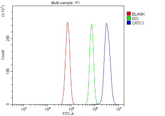
(Figure 8. Flow Cytometry analysis of RH35 cells using anti-TORC1/CRTC1 antibody (AAA19251).Overlay histogram showing RH35 cells stained with AAA19251 (Blue line). The cells were blocked with 10% normal goat serum. And then incubated with rabbit anti-TORC1/CRTC1 Antibody (AAA19251, 1μg/1x106 cells) for 30 min at 20 degree C. DyLight®488 conjugated goat anti-rabbit IgG (5-10μg/1x106 cells) was used as secondary antibody for 30 minutes at 20 degree C. Isotype control antibody (Green line) was rabbit IgG (1μg/1x106) used under the same conditions. Unlabelled sample (Red line) was also used as a control.)
FCM (Flow Cytometry)
(Figure 8. Flow Cytometry analysis of RH35 cells using anti-TORC1/CRTC1 antibody (AAA19251).Overlay histogram showing RH35 cells stained with AAA19251 (Blue line). The cells were blocked with 10% normal goat serum. And then incubated with rabbit anti-TORC1/CRTC1 Antibody (AAA19251, 1μg/1x106 cells) for 30 min at 20 degree C. DyLight®488 conjugated goat anti-rabbit IgG (5-10μg/1x106 cells) was used as secondary antibody for 30 minutes at 20 degree C. Isotype control antibody (Green line) was rabbit IgG (1μg/1x106) used under the same conditions. Unlabelled sample (Red line) was also used as a control.)
TORC1/CRTC1, Polyclonal Antibody (Cat# AAA19251)
Full Name
Anti-TORC1/CRTC1 Antibody
Gene Names
CRTC1; MECT1; TORC1; TORC-1; WAMTP1
Reactivity
Human, Mouse, Rat
Applications
Western Blot, Immunohistochemistry, Immunocytochemistry, Immunofluorescence, Flow Cytometry
Purity
Immunogen affinity purified.
Pricing


 FCM (Flow Cytometry)
(Figure 6. Flow Cytometry analysis of HL-60 cells using anti-MCM6 antibody (AAA19264).Overlay histogram showing HL-60 cells stained with AAA19264 (Blue line). The cells were blocked with 10% normal goat serum. And then incubated with rabbit anti-MCM6 Antibody (AAA19264,1μg/1x106 cells) for 30 min at 20 degree C. DyLight®488 conjugated goat anti-rabbit IgG (5-10μg/1x106 cells) was used as secondary antibody for 30 minutes at 20 degree C. Isotype control antibody (Green line) was rabbit IgG (1μg/1x106) used under the same conditions. Unlabelled sample (Red line) was also used as a control.)
FCM (Flow Cytometry)
(Figure 6. Flow Cytometry analysis of HL-60 cells using anti-MCM6 antibody (AAA19264).Overlay histogram showing HL-60 cells stained with AAA19264 (Blue line). The cells were blocked with 10% normal goat serum. And then incubated with rabbit anti-MCM6 Antibody (AAA19264,1μg/1x106 cells) for 30 min at 20 degree C. DyLight®488 conjugated goat anti-rabbit IgG (5-10μg/1x106 cells) was used as secondary antibody for 30 minutes at 20 degree C. Isotype control antibody (Green line) was rabbit IgG (1μg/1x106) used under the same conditions. Unlabelled sample (Red line) was also used as a control.)
MCM6, Polyclonal Antibody (Cat# AAA19264)
Full Name
Anti-MCM6 Antibody
Gene Names
MCM6; Mis5; P105MCM; MCG40308
Reactivity
Human, Mouse, Rat, Monkey
Applications
Western Blot, Immunohistochemistry, Immunocytochemistry, Immunofluorescence, Flow Cytometry, Direct ELISA
Purity
Immunogen affinity purified.
Pricing
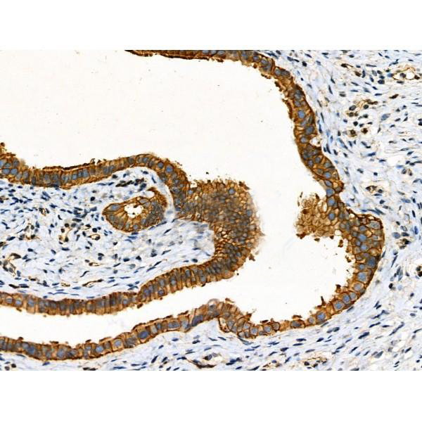


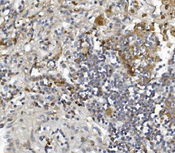
 IHC (Immunohistochemistry)
(At 1/100 staining Human gastric cancer by IHC-P. The sample was formaldehyde fixed and a heat mediated antigen retrieval step in citrate buffer was performed. The sample was then blocked and incubated with the primary antibody at 4 degree C overnight. An HRP conjugated anti-Rabbit antibody was used as the secondary antibody.)
IHC (Immunohistochemistry)
(At 1/100 staining Human gastric cancer by IHC-P. The sample was formaldehyde fixed and a heat mediated antigen retrieval step in citrate buffer was performed. The sample was then blocked and incubated with the primary antibody at 4 degree C overnight. An HRP conjugated anti-Rabbit antibody was used as the secondary antibody.)
alpha 1 Catenin, Polyclonal Antibody (Cat# AAA31379)
Full Name
alpha 1 Catenin Antibody
Gene Names
CTNNA1; CAP102
Reactivity
Human, Mouse, Rat
Applications
Western Blot, Immunohistochemistry, Peptide ELISA
Purity
The antiserum was purified by peptide affinity chromatography using SulfoLink Coupling Resin
Pricing


 FCM (Flow Cytometry)
(Figure 7. Flow Cytometry analysis of Hela cells using anti-ATG9A antibody (AAA19280).Overlay histogram showing Hela cells stained with AAA19280 (Blue line). The cells were blocked with 10% normal goat serum. And then incubated with rabbit anti-ATG9A Antibody (AAA19280, 1μg/1x106 cells) for 30 min at 20 degree C. DyLight®488 conjugated goat anti-rabbit IgG (5-10μg/1x106 cells) was used as secondary antibody for 30 minutes at 20 degree C. Isotype control antibody (Green line) was rabbit IgG (1μg/1x106) used under the same conditions. Unlabelled sample (Red line) was also used as a control.)
FCM (Flow Cytometry)
(Figure 7. Flow Cytometry analysis of Hela cells using anti-ATG9A antibody (AAA19280).Overlay histogram showing Hela cells stained with AAA19280 (Blue line). The cells were blocked with 10% normal goat serum. And then incubated with rabbit anti-ATG9A Antibody (AAA19280, 1μg/1x106 cells) for 30 min at 20 degree C. DyLight®488 conjugated goat anti-rabbit IgG (5-10μg/1x106 cells) was used as secondary antibody for 30 minutes at 20 degree C. Isotype control antibody (Green line) was rabbit IgG (1μg/1x106) used under the same conditions. Unlabelled sample (Red line) was also used as a control.)
ATG9A, Polyclonal Antibody (Cat# AAA19280)
Full Name
Anti-ATG9A Antibody
Gene Names
ATG9A; mATG9; APG9L1; MGD3208
Reactivity
Human, Mouse, Rat
Applications
WB, IHC-P, FC/FACS/FCM, EIA
Purity
Immunogen affinity purified.
Pricing


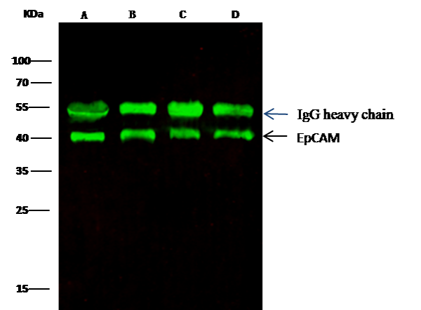 IP (Immunoprecipitation)
(EpCAM was immunoprecipitated using:Lane A:0.5 mg MCF-7 Whole Cell LysateLane B:0.5 mg A431 Whole Cell LysateLane C:0.5 mg HepG2 Whole Cell LysateLane D:0.5 mg Caco-2 Whole Cell Lysate0.5 uL anti-EpCAM rabbit monoclonal antibody and 15 ul of 50 % Protein G agarose.Primary antibody:Anti-EpCAM rabbit monoclonal antibody,at 1:5000 dilution Secondary antibody:Dylight 800-labeled antibody to rabbit IgG (H+L), at 1:5000 dilution Developed using the odssey technique.Performed under reducing conditions.Predicted band size: 40 kDaObserved band size: 40 kDa)
IP (Immunoprecipitation)
(EpCAM was immunoprecipitated using:Lane A:0.5 mg MCF-7 Whole Cell LysateLane B:0.5 mg A431 Whole Cell LysateLane C:0.5 mg HepG2 Whole Cell LysateLane D:0.5 mg Caco-2 Whole Cell Lysate0.5 uL anti-EpCAM rabbit monoclonal antibody and 15 ul of 50 % Protein G agarose.Primary antibody:Anti-EpCAM rabbit monoclonal antibody,at 1:5000 dilution Secondary antibody:Dylight 800-labeled antibody to rabbit IgG (H+L), at 1:5000 dilution Developed using the odssey technique.Performed under reducing conditions.Predicted band size: 40 kDaObserved band size: 40 kDa)
EpCAM, Monoclonal Antibody (Cat# AAA27736)
Full Name
Recombinant Anti-EpCAM Antibody, Rabbit Monoclonal
Gene Names
EPCAM; ESA; KSA; M4S1; MK-1; DIAR5; EGP-2; EGP40; KS1/4; MIC18; TROP1; EGP314; HNPCC8; TACSTD1
Reactivity
Human
Applications
Western Blot, Immunohistochemistry, Flow Cytometry, Immunocytochemistry, Immunofluorescence, Immunoprecipitation
Purity
Protein A
Pricing


 FCM (Flow Cytometry)
(Figure 6. Flow Cytometry analysis of A549 cells using anti-MCM5 antibody (AAA19377).Overlay histogram showing A549 cells stained with AAA19377 (Blue line). The cells were blocked with 10% normal goat serum. And then incubated with mouse anti- MCM5 Antibody (AAA19377, 1μg/1x106 cells) for 30 min at 20 degree C. DyLight®488 conjugated goat anti-mouse IgG (BA1126, 5-10μg/1x106 cells) was used as secondary antibody for 30 minutes at 20 degree C. Isotype control antibody (Green line) was mouse IgG (1μg/1x106) used under the same conditions. Unlabelled sample (Red line) was also used as a control.)
FCM (Flow Cytometry)
(Figure 6. Flow Cytometry analysis of A549 cells using anti-MCM5 antibody (AAA19377).Overlay histogram showing A549 cells stained with AAA19377 (Blue line). The cells were blocked with 10% normal goat serum. And then incubated with mouse anti- MCM5 Antibody (AAA19377, 1μg/1x106 cells) for 30 min at 20 degree C. DyLight®488 conjugated goat anti-mouse IgG (BA1126, 5-10μg/1x106 cells) was used as secondary antibody for 30 minutes at 20 degree C. Isotype control antibody (Green line) was mouse IgG (1μg/1x106) used under the same conditions. Unlabelled sample (Red line) was also used as a control.)
MCM5, Monoclonal Antibody (Cat# AAA19377)
Full Name
Anti-MCM5 Antibody (monoclonal, 4G10)
Gene Names
MCM5; CDC46; P1-CDC46
Reactivity
Human, Mouse, Rat
Applications
WB, IHC-P, ICC, IF, FC/FACS/FCM
Purity
Immunogen affinity purified.
Pricing


 IHC (Immunohistchemistry)
(Immunohistochemistry Analysis: Representative lot data. (Fig. 1 and 2) Paraffin-embedded mouse and human brain tissue was prepared using heat-induced epitope retrieval in citrate buffer, pH 6.0. Immunostaining was performed using a 1:100 dilution. Reactivity was detected using the IHC-Select Detection Kit. Staining pattern appears as cytoplasmic. (Fig. 3 and 4) Paraffin-embedded mouse and mouse olfactory lobe and cerebellum brain tissue was prepared using heat-induced epitope retrieval in citrate buffer, pH 6.0. Immunostaining was performed using a Chicken IgY Antibody 1:100 dilution of Cat. No. AB15894, anti-Tbr2. Reactivity was detected using the IHC-Select Detection Kit. Immunoreactivity seen here is mostly nuclear.)
IHC (Immunohistchemistry)
(Immunohistochemistry Analysis: Representative lot data. (Fig. 1 and 2) Paraffin-embedded mouse and human brain tissue was prepared using heat-induced epitope retrieval in citrate buffer, pH 6.0. Immunostaining was performed using a 1:100 dilution. Reactivity was detected using the IHC-Select Detection Kit. Staining pattern appears as cytoplasmic. (Fig. 3 and 4) Paraffin-embedded mouse and mouse olfactory lobe and cerebellum brain tissue was prepared using heat-induced epitope retrieval in citrate buffer, pH 6.0. Immunostaining was performed using a Chicken IgY Antibody 1:100 dilution of Cat. No. AB15894, anti-Tbr2. Reactivity was detected using the IHC-Select Detection Kit. Immunoreactivity seen here is mostly nuclear.)
EOMES, Polyclonal Antibody (Cat# AAA26886)
Full Name
EOMES (Eomesodermin Homolog, T-box Brain Protein 2, TBR2, T-brain-2, TBR-2)
Gene Names
Eomes; Tbr2; TBR-2; C77258
Reactivity
Mouse, Human, Rat
Applications
Immunohistochemistry, Western Blot
Purity
Purified by affinity chromatography.
Pricing


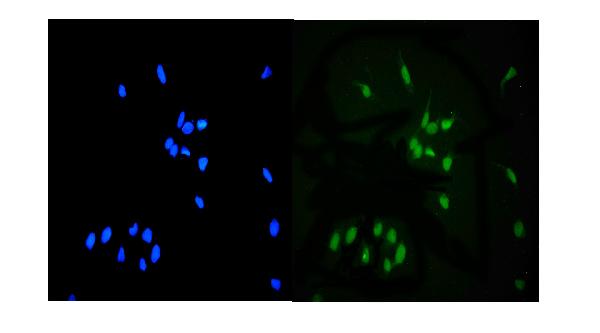
 IF (Immunofluorescence)
(Immunofluorescent analysis of 4% paraformaldehyde-fixed, 0.1% Triton X-100 permeabilized MCF-7 (human breast cancer cell line) cells labeling Pdx1 with at 1:25 dilution, followed by DyLight 488-conjugated IgG goat anti-rabbit secondary antibody at 1:200 dilution (green). Immunofluorescence image showing cytoplasm staining on MCF-7 cell line. Cytoplasmic actin is detected with DyLight 554 Phalloidin (PD18466410) at 1:100 dilution (red). The nuclear counter stain is DAPI (blue).)
IF (Immunofluorescence)
(Immunofluorescent analysis of 4% paraformaldehyde-fixed, 0.1% Triton X-100 permeabilized MCF-7 (human breast cancer cell line) cells labeling Pdx1 with at 1:25 dilution, followed by DyLight 488-conjugated IgG goat anti-rabbit secondary antibody at 1:200 dilution (green). Immunofluorescence image showing cytoplasm staining on MCF-7 cell line. Cytoplasmic actin is detected with DyLight 554 Phalloidin (PD18466410) at 1:100 dilution (red). The nuclear counter stain is DAPI (blue).)
OPN-a/b, Polyclonal Antibody (Cat# AAA26865)
Full Name
OPN-a/b, NT (SPP1, BNSP, OPN, Osteopontin, Bone sialoprotein 1, Nephropontin, Secreted phosphoprotein 1, Urinary stone protein, Uropontin) (PE)
Gene Names
SPP1; OPN; BNSP; BSPI; ETA-1
Reactivity
Human
Applications
WB, IHC, IF
Purity
Purified by Protein A and Peptide Affinity Chromatography.
Pricing


 FCM (Flow Cytometry)
(Figure 8. Flow Cytometry analysis of U20S cells using anti-NFIA antibody (AAA11691).Overlay histogram showing U20S cells stained with AAA11691 (Blue line).The cells were blocked with 10% normal goat serum. And then incubated with rabbit anti-NFIA Antibody (AAA11691,1ug/1x10^6 cells) for 30 min at 20 degree C. DyLight®488 conjugated goat anti-rabbit IgG (5-10ug/1x10^6 cells) was used as secondary antibody for 30 minutes at 20 degree C. Isotype control antibody (Green line) was rabbit IgG (1ug/1x106) used under the same conditions. Unlabelled sample (Red line) was also used as a control.)
FCM (Flow Cytometry)
(Figure 8. Flow Cytometry analysis of U20S cells using anti-NFIA antibody (AAA11691).Overlay histogram showing U20S cells stained with AAA11691 (Blue line).The cells were blocked with 10% normal goat serum. And then incubated with rabbit anti-NFIA Antibody (AAA11691,1ug/1x10^6 cells) for 30 min at 20 degree C. DyLight®488 conjugated goat anti-rabbit IgG (5-10ug/1x10^6 cells) was used as secondary antibody for 30 minutes at 20 degree C. Isotype control antibody (Green line) was rabbit IgG (1ug/1x106) used under the same conditions. Unlabelled sample (Red line) was also used as a control.)
NFIA, Polyclonal Antibody (Cat# AAA11691)
Full Name
Anti-NFIA Antibody
Gene Names
NFIA; CTF; NF1-A; NFI-A; NFI-L; NF-I/A
Reactivity
Human, Mouse, Rat
Applications
Western Blot, Immunohistochemistry
Purity
Immunogen affinity purified.
Pricing


 IHC (Immunohistchemistry)
(Immunohistochemistry Analysis: Representative lot data. (Fig. 1 and 2) Paraffin-embedded mouse and human brain tissue was prepared using heat-induced epitope retrieval in citrate buffer, pH 6.0. Immunostaining was performed using a 1:100 dilution of AAA14728. Reactivity was detected using the IHC-Select Detection Kit. Staining pattern appears as cytoplasmic. (Fig. 3 and 4) Paraffin-embedded mouse and mouse olfactory lobe and cerebellum brain tissue was prepared using heat-induced epitope retrieval in citrate buffer, pH 6.0. Immunostaining was performed using a Chicken IgY Antibody 1:100 dilution of Cat. No. AB15894, anti-Tbr2. Reactivity was detected using the IHC-Select Detection Kit . Immunoreactivity seen here is mostly nuclear.)
IHC (Immunohistchemistry)
(Immunohistochemistry Analysis: Representative lot data. (Fig. 1 and 2) Paraffin-embedded mouse and human brain tissue was prepared using heat-induced epitope retrieval in citrate buffer, pH 6.0. Immunostaining was performed using a 1:100 dilution of AAA14728. Reactivity was detected using the IHC-Select Detection Kit. Staining pattern appears as cytoplasmic. (Fig. 3 and 4) Paraffin-embedded mouse and mouse olfactory lobe and cerebellum brain tissue was prepared using heat-induced epitope retrieval in citrate buffer, pH 6.0. Immunostaining was performed using a Chicken IgY Antibody 1:100 dilution of Cat. No. AB15894, anti-Tbr2. Reactivity was detected using the IHC-Select Detection Kit . Immunoreactivity seen here is mostly nuclear.)
EOMES, Polyclonal Antibody (Cat# AAA14728)
Full Name
EOMES, NT (Eomesodermin Homolog, T-box Brain Protein 2, TBR2, T-brain-2, TBR-2)
Gene Names
EOMES; TBR2
Reactivity
Human, Mouse, Rat
Applications
Western Blot, Immunohistochemistry
Purity
Affinity Purified
Purified by affinity chromatography.
Purified by affinity chromatography.
Pricing


 FCM (Flow Cytometry)
(Figure 6. Flow Cytometry analysis of U20S cells using anti-PPID antibody (AAA19159).Overlay histogram showing U20S cells stained with AAA19159 (Blue line).The cells were blocked with 10% normal goat serum. And then incubated with rabbit anti-PPID Antibody (AAA19159,1ug/1x10^6 cells) for 30 min at 20 degree C. DyLight®488 conjugated goat anti-rabbit IgG (5-10ug/1x10^6 cells) was used as secondary antibody for 30 minutes at 20 degree C. Isotype control antibody (Green line) was rabbit IgG (1ug/1x106) used under the same conditions. Unlabelled sample (Red line) was also used as a control.)
FCM (Flow Cytometry)
(Figure 6. Flow Cytometry analysis of U20S cells using anti-PPID antibody (AAA19159).Overlay histogram showing U20S cells stained with AAA19159 (Blue line).The cells were blocked with 10% normal goat serum. And then incubated with rabbit anti-PPID Antibody (AAA19159,1ug/1x10^6 cells) for 30 min at 20 degree C. DyLight®488 conjugated goat anti-rabbit IgG (5-10ug/1x10^6 cells) was used as secondary antibody for 30 minutes at 20 degree C. Isotype control antibody (Green line) was rabbit IgG (1ug/1x106) used under the same conditions. Unlabelled sample (Red line) was also used as a control.)
PPID/Cyclophilin 40, Polyclonal Antibody (Cat# AAA19159)
Full Name
Anti-PPID/Cyclophilin 40 Picoband antibody
Gene Names
PPID; CYPD; CYP-40
Reactivity
Human, Mouse, Rat
No cross reactivity with other proteins.
No cross reactivity with other proteins.
Applications
EIA, FC/FACS, IHC, ICC, WB
Pricing


 FCM (Flow Cytometry)
(Figure 8. Flow Cytometry analysis of HepG2 cells using anti-MERTK antibody (AAA19219).Overlay histogram showing HepG2 cells stained with AAA19219 (Blue line). The cells were blocked with 10% normal goat serum. And then incubated with rabbit anti-MERTK Antibody (AAA19219, 1ug/1x106 cells) for 30 min at 20 degree C. DyLight488 conjugated goat anti-rabbit IgG (5-10ug/1x106 cells) was used as secondary antibody for 30 minutes at 20 degree C. Isotype control antibody (Green line) was rabbit IgG (1ug/1x106) used under the same conditions. Unlabelled sample (Red line) was also used as a control.)
FCM (Flow Cytometry)
(Figure 8. Flow Cytometry analysis of HepG2 cells using anti-MERTK antibody (AAA19219).Overlay histogram showing HepG2 cells stained with AAA19219 (Blue line). The cells were blocked with 10% normal goat serum. And then incubated with rabbit anti-MERTK Antibody (AAA19219, 1ug/1x106 cells) for 30 min at 20 degree C. DyLight488 conjugated goat anti-rabbit IgG (5-10ug/1x106 cells) was used as secondary antibody for 30 minutes at 20 degree C. Isotype control antibody (Green line) was rabbit IgG (1ug/1x106) used under the same conditions. Unlabelled sample (Red line) was also used as a control.)
MERTK, Polyclonal Antibody (Cat# AAA19219)
Full Name
Anti-MERTK Antibody
Gene Names
MERTK; MER; RP38; c-mer
Reactivity
Human, Mouse, Rat
Applications
Western Blot, Immunohistochemistry, Flow Cytometry, Direct ELISA
Purity
Immunogen affinity purified.
Pricing
 Application Data
HIV-1 p24, Monoclonal Antibody (Cat# AAA13685)
Full Name
Anti-HIV-1 p24
Reactivity
Reacts with P24(HIC-1) proteins
Applications
Western Blot
Purity
Ion exchange column
Pricing


 IHC (Immunohistochemistry)
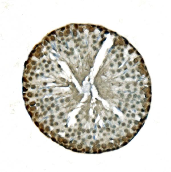
(Figure 7. IHC analysis of GALNS using anti-GALNS antibody (AAA19164).GALNS was detected in paraffin-embedded section of rat small intestine tissue. Heat mediated antigen retrieval was performed in citrate buffer (pH6, epitope retrieval solution) for 20 mins. The tissue section was blocked with 10% goat serum. The tissue section was then incubated with 1ug/ml rabbit anti-GALNS Antibody (AAA19164) overnight at 4 degree C. Biotinylated goat anti-rabbit IgG was used as secondary antibody and incubated for 30 minutes at 37 degree C. The tissue section was developed using Strepavidin-Biotin-Complex (SABC) with DAB as the chromogen.)
IHC (Immunohistochemistry)
(Figure 7. IHC analysis of GALNS using anti-GALNS antibody (AAA19164).GALNS was detected in paraffin-embedded section of rat small intestine tissue. Heat mediated antigen retrieval was performed in citrate buffer (pH6, epitope retrieval solution) for 20 mins. The tissue section was blocked with 10% goat serum. The tissue section was then incubated with 1ug/ml rabbit anti-GALNS Antibody (AAA19164) overnight at 4 degree C. Biotinylated goat anti-rabbit IgG was used as secondary antibody and incubated for 30 minutes at 37 degree C. The tissue section was developed using Strepavidin-Biotin-Complex (SABC) with DAB as the chromogen.)
GALNS, Antibody (Cat# AAA19164)
Full Name
Anti-GALNS Picoband antibody
Gene Names
GALNS; GAS; MPS4A; GalN6S; GALNAC6S
Reactivity
Reacts with: Human, Mouse, Rat
Applications
WB, EIA
Purity
Immunogen affinity purified
Pricing


 FCM (Flow Cytometry)
(Figure 7. Flow Cytometry analysis of HEPA1-6 cells using anti-Rad9b antibody (AAA19340).Overlay histogram showing HEPA1-6 cells stained with AAA19340 (Blue line). The cells were blocked with 10% normal goat serum. And then incubated with rabbit anti-Rad9b Antibody (AAA19340, 1μg/1x106 cells) for 30 min at 20 degree C. DyLight®488 conjugated goat anti-rabbit IgG (5-10μg/1x106 cells) was used as secondary antibody for 30 minutes at 20 degree C. Isotype control antibody (Green line) was rabbit IgG (1μg/1x106) used under the same conditions. Unlabelled sample (Red line) was also used as a control.)
FCM (Flow Cytometry)
(Figure 7. Flow Cytometry analysis of HEPA1-6 cells using anti-Rad9b antibody (AAA19340).Overlay histogram showing HEPA1-6 cells stained with AAA19340 (Blue line). The cells were blocked with 10% normal goat serum. And then incubated with rabbit anti-Rad9b Antibody (AAA19340, 1μg/1x106 cells) for 30 min at 20 degree C. DyLight®488 conjugated goat anti-rabbit IgG (5-10μg/1x106 cells) was used as secondary antibody for 30 minutes at 20 degree C. Isotype control antibody (Green line) was rabbit IgG (1μg/1x106) used under the same conditions. Unlabelled sample (Red line) was also used as a control.)
Rad9b, Polyclonal Antibody (Cat# AAA19340)
Full Name
Anti-Rad9b Antibody
Gene Names
Rad9b; BC021784; A630082N15Rik
Reactivity
Mouse, Rat
Applications
WB, IHC-P, FC/FACS/FCM, EIA
Purity
Immunogen affinity purified.
Pricing


 IF (Immunofluorescence)
(Figure 6. IF analysis of UNG using anti-UNG antibody (AAA19245).UNG was detected in immunocytochemical section of MCF-7 cells. Enzyme antigen retrieval was performed using IHC enzyme antigen retrieval reagent for 15 mins. The cells were blocked with 10% goat serum. And then incubated with 5μg/mL rabbit anti- UNG Antibody (AAA19245) overnight at 4 degree C. DyLight®594 Conjugated Goat Anti-Rabbit IgG (BA1142) was used as secondary antibody at 1:100 dilution and incubated for 30 minutes at 37 degree C. The section was counterstained with DAPI. Visualize using a fluorescence microscope and filter sets appropriate for the label used.)
IF (Immunofluorescence)
(Figure 6. IF analysis of UNG using anti-UNG antibody (AAA19245).UNG was detected in immunocytochemical section of MCF-7 cells. Enzyme antigen retrieval was performed using IHC enzyme antigen retrieval reagent for 15 mins. The cells were blocked with 10% goat serum. And then incubated with 5μg/mL rabbit anti- UNG Antibody (AAA19245) overnight at 4 degree C. DyLight®594 Conjugated Goat Anti-Rabbit IgG (BA1142) was used as secondary antibody at 1:100 dilution and incubated for 30 minutes at 37 degree C. The section was counterstained with DAPI. Visualize using a fluorescence microscope and filter sets appropriate for the label used.)
UNG, Polyclonal Antibody (Cat# AAA19245)
Full Name
Anti-UNG Antibody
Gene Names
UNG; DGU; UDG; UNG1; UNG2; HIGM4; HIGM5; UNG15
Reactivity
Human, Mouse, Rat, Monkey
Applications
Western Blot, Immunohistochemistry, Immunocytochemistry, Immunofluorescence, Flow Cytometry, Direct ELISA
Purity
Immunogen affinity purified.
Pricing


 FCM (Flow Cytometry)
(Figure 8. Flow Cytometry analysis of U87 cells using anti-Transketolase/TKT antibody (AAA19254).Overlay histogram showing U87 cells stained with AAA19254 (Blue line). The cells were blocked with 10% normal goat serum. And then incubated with rabbit anti-Transketolase/TKT Antibody (AAA19254,1μg/1x106 cells) for 30 min at 20 degree C. DyLight®488 conjugated goat anti-rabbit IgG (5-10μg/1x106 cells) was used as secondary antibody for 30 minutes at 20 degree C. Isotype control antibody (Green line) was rabbit IgG (1μg/1x106) used under the same conditions. Unlabelled sample (Red line) was also used as a control.)
FCM (Flow Cytometry)
(Figure 8. Flow Cytometry analysis of U87 cells using anti-Transketolase/TKT antibody (AAA19254).Overlay histogram showing U87 cells stained with AAA19254 (Blue line). The cells were blocked with 10% normal goat serum. And then incubated with rabbit anti-Transketolase/TKT Antibody (AAA19254,1μg/1x106 cells) for 30 min at 20 degree C. DyLight®488 conjugated goat anti-rabbit IgG (5-10μg/1x106 cells) was used as secondary antibody for 30 minutes at 20 degree C. Isotype control antibody (Green line) was rabbit IgG (1μg/1x106) used under the same conditions. Unlabelled sample (Red line) was also used as a control.)
Transketolase/TKT, Polyclonal Antibody (Cat# AAA19254)
Full Name
Anti-Transketolase/TKT Antibody
Gene Names
TKT; TK; TKT1; HEL107
Reactivity
Human, Mouse, Rat
Applications
Western Blot, Immunohistochemistry, Immunocytochemistry, Immunofluorescence, Flow Cytometry, Direct ELISA
Purity
Immunogen affinity purified.
Pricing


 IHC (Immunohistchemistry)
(Figure 6. IHC analysis of Integrin beta 4/ITGB4 using anti-Integrin beta 4/ITGB4 antibody (AAA19269).Integrin beta 4/ITGB4 was detected in paraffin-embedded section of rat brain tissue. Heat mediated antigen retrieval was performed in EDTA buffer (pH8. 0, epitope retrieval solution). The tissue section was blocked with 10% goat serum. The tissue section was then incubated with 1μg/ml rabbit anti-Integrin beta 4/ITGB4 Antibody (AAA19269) overnight at 4 degree C. Biotinylated goat anti-rabbit IgG was used as secondary antibody and incubated for 30 minutes at 37 degree C. The tissue section was developed using Strepavidin-Biotin-Complex (SABC) (Catalog # with DAB as the chromogen.)
IHC (Immunohistchemistry)
(Figure 6. IHC analysis of Integrin beta 4/ITGB4 using anti-Integrin beta 4/ITGB4 antibody (AAA19269).Integrin beta 4/ITGB4 was detected in paraffin-embedded section of rat brain tissue. Heat mediated antigen retrieval was performed in EDTA buffer (pH8. 0, epitope retrieval solution). The tissue section was blocked with 10% goat serum. The tissue section was then incubated with 1μg/ml rabbit anti-Integrin beta 4/ITGB4 Antibody (AAA19269) overnight at 4 degree C. Biotinylated goat anti-rabbit IgG was used as secondary antibody and incubated for 30 minutes at 37 degree C. The tissue section was developed using Strepavidin-Biotin-Complex (SABC) (Catalog # with DAB as the chromogen.)
mGluR1/GRM1, Polyclonal Antibody (Cat# AAA19269)
Full Name
Anti-mGluR1/GRM1 Antibody
Gene Names
GRM1; MGLU1; GPRC1A; MGLUR1; SCAR13; PPP1R85
Reactivity
Human, Mouse, Rat
Applications
WB, IHC-P, FC/FACS/FCM, EIA
Purity
Immunogen affinity purified.
Pricing


 FCM (Flow Cytometry)
(Figure 8. Flow Cytometry analysis of HL-60 cells using anti-REA/PHB2 antibody (AAA19273).Overlay histogram showing HL-60 cells stained with AAA19273 (Blue line). The cells were blocked with 10% normal goat serum. And then incubated with rabbit anti-REA/PHB2 Antibody (AAA19273,1μg/1x106 cells) for 30 min at 20 degree C. DyLight®488 conjugated goat anti-rabbit IgG (5-10μg/1x106 cells) was used as secondary antibody for 30 minutes at 20 degree C. Isotype control antibody (Green line) was rabbit IgG (1μg/1x106) used under the same conditions. Unlabelled sample (Red line) was also used as a control.)
FCM (Flow Cytometry)
(Figure 8. Flow Cytometry analysis of HL-60 cells using anti-REA/PHB2 antibody (AAA19273).Overlay histogram showing HL-60 cells stained with AAA19273 (Blue line). The cells were blocked with 10% normal goat serum. And then incubated with rabbit anti-REA/PHB2 Antibody (AAA19273,1μg/1x106 cells) for 30 min at 20 degree C. DyLight®488 conjugated goat anti-rabbit IgG (5-10μg/1x106 cells) was used as secondary antibody for 30 minutes at 20 degree C. Isotype control antibody (Green line) was rabbit IgG (1μg/1x106) used under the same conditions. Unlabelled sample (Red line) was also used as a control.)
REA/PHB2, Polyclonal Antibody (Cat# AAA19273)
Full Name
Anti-REA/PHB2 Antibody
Gene Names
PHB2; BAP; REA; p22; Bap37; BCAP37; PNAS-141
Reactivity
Human, Mouse, Rat
Applications
WB, IHC-P, ICC, IF, FC/FACS/FCM, EIA
Purity
Immunogen affinity purified.
Pricing


 FCM (Flow Cytometry)
(Figure 9. Flow Cytometry analysis of MCF-7 cells using anti-Claudin 3/CLDN3 antibody (AAA19294).Overlay histogram showing MCF-7 cells stained with AAA19294 (Blue line). The cells were blocked with 10% normal goat serum. And then incubated with rabbit anti-Claudin 3/CLDN3 Antibody (AAA19294, 1μg/1x106 cells) for 30 min at 20 degree C. DyLight®488 conjugated goat anti-rabbit IgG (5-10μg/1x106 cells) was used as secondary antibody for 30 minutes at 20 degree C. Isotype control antibody (Green line) was rabbit IgG (1μg/1x106) used under the same conditions. Unlabelled sample (Red line) was also used as a control.)
FCM (Flow Cytometry)
(Figure 9. Flow Cytometry analysis of MCF-7 cells using anti-Claudin 3/CLDN3 antibody (AAA19294).Overlay histogram showing MCF-7 cells stained with AAA19294 (Blue line). The cells were blocked with 10% normal goat serum. And then incubated with rabbit anti-Claudin 3/CLDN3 Antibody (AAA19294, 1μg/1x106 cells) for 30 min at 20 degree C. DyLight®488 conjugated goat anti-rabbit IgG (5-10μg/1x106 cells) was used as secondary antibody for 30 minutes at 20 degree C. Isotype control antibody (Green line) was rabbit IgG (1μg/1x106) used under the same conditions. Unlabelled sample (Red line) was also used as a control.)
Claudin 3/CLDN3, Polyclonal Antibody (Cat# AAA19294)
Full Name
Anti-Claudin 3/CLDN3 Antibody
Gene Names
CLDN3; RVP1; HRVP1; C7orf1; CPE-R2; CPETR2
Reactivity
Human
Applications
WB, IHC-P, ICC, IF, FC/FACS/FCM, EIA
Purity
Immunogen affinity purified.
Pricing


 FCM (Flow Cytometry)
(AGXT Antibody (Center) flow cytometric analysis of HepG2 cells (right histogram) compared to a negative control cell (left histogram).FITC-conjugated goat-anti-rabbit secondary antibodies were used for the analysis.)
FCM (Flow Cytometry)
(AGXT Antibody (Center) flow cytometric analysis of HepG2 cells (right histogram) compared to a negative control cell (left histogram).FITC-conjugated goat-anti-rabbit secondary antibodies were used for the analysis.)
AGXT, Polyclonal Antibody (Cat# AAA28732)
Full Name
AGXT Antibody (Center)
Gene Names
AGXT; AGT; PH1; SPT; AGT1; SPAT; TLH6; AGXT1
Reactivity
Human
Applications
Western Blot, Immunohistochemistry, Flow Cytometry
Purity
Peptide Affinity Purified Rabbit Polyclonal Antibody (Pab)
Pricing


 IHC (Immunohistochemistry)
(At 1/100 staining Human kidney cancer and adjacent normal tissues by IHC-P. The sample was formaldehyde fixed and a heat mediated antigen retrieval step in citrate buffer was performed. The sample was then blocked and incubated with the primary antibody at 4 degree C overnight. An HRP conjugated anti-Rabbit antibody was used as the secondary antibody.)
IHC (Immunohistochemistry)
(At 1/100 staining Human kidney cancer and adjacent normal tissues by IHC-P. The sample was formaldehyde fixed and a heat mediated antigen retrieval step in citrate buffer was performed. The sample was then blocked and incubated with the primary antibody at 4 degree C overnight. An HRP conjugated anti-Rabbit antibody was used as the secondary antibody.)
Smad5, Polyclonal Antibody (Cat# AAA31331)
Full Name
Phospho-Smad5 (Ser463/Ser465) Antibody
Gene Names
SMAD5; DWFC; JV5-1; MADH5
Reactivity
Human, Mouse, Rat
Applications
Western Blot, Immunohistochemistry, Peptide ELISA
Purity
The antibody is from purified rabbit serum by affinity purification via sequential chromatography on phospho-peptide and non-phospho-peptide affinity columns.
Pricing


 FCM (Flow Cytometry)
(Figure 6. Flow Cytometry analysis of MCF-7 cells using anti- PCK2 antibody (AAA19384).Overlay histogram showing MCF-7 cells stained with AAA19384 (Blue line). The cells were blocked with 10% normal goat serum. And then incubated with mouse anti-PCK2 Antibody (AAA19384, 1μg/1x106 cells) for 30 min at 20 degree C. DyLight®488 conjugated goat anti-mouse IgG (BA1126, 5-10μg/1x106 cells) was used as secondary antibody for 30 minutes at 20 degree C. Isotype control antibody (Green line) was mouse IgG (1μg/1x106) used under the same conditions. Unlabelled sample (Red line) was also used as a control.)
FCM (Flow Cytometry)
(Figure 6. Flow Cytometry analysis of MCF-7 cells using anti- PCK2 antibody (AAA19384).Overlay histogram showing MCF-7 cells stained with AAA19384 (Blue line). The cells were blocked with 10% normal goat serum. And then incubated with mouse anti-PCK2 Antibody (AAA19384, 1μg/1x106 cells) for 30 min at 20 degree C. DyLight®488 conjugated goat anti-mouse IgG (BA1126, 5-10μg/1x106 cells) was used as secondary antibody for 30 minutes at 20 degree C. Isotype control antibody (Green line) was mouse IgG (1μg/1x106) used under the same conditions. Unlabelled sample (Red line) was also used as a control.)
PCK2, Monoclonal Antibody (Cat# AAA19384)
Full Name
Anti-PCK2 Antibody (monoclonal, 3F7)
Gene Names
PCK2; PEPCK; PEPCK2; PEPCK-M
Reactivity
Human, Mouse, Rat, Monkey
Applications
WB, IHC-P, ICC, IF, FC/FACS/FCM
Purity
Immunogen affinity purified.
Pricing


 FCM (Flow Cytometry)
(Figure 9. Flow Cytometry analysis of HL-60 cells using anti-NDUFB10 antibody (AAA19339).Overlay histogram showing HL-60 cells stained with AAA19339 (Blue line). The cells were blocked with 10% normal goat serum. And then incubated with rabbit anti-NDUFB10 Antibody (AAA19339, 1μg/1x106 cells) for 30 min at 20 degree C. DyLight®488 conjugated goat anti-rabbit IgG (5-10μg/1x106 cells) was used as secondary antibody for 30 minutes at 20 degree C. Isotype control antibody (Green line) was rabbit IgG (1μg/1x106) used under the same conditions. Unlabelled sample (Red line) was also used as a control.)
FCM (Flow Cytometry)
(Figure 9. Flow Cytometry analysis of HL-60 cells using anti-NDUFB10 antibody (AAA19339).Overlay histogram showing HL-60 cells stained with AAA19339 (Blue line). The cells were blocked with 10% normal goat serum. And then incubated with rabbit anti-NDUFB10 Antibody (AAA19339, 1μg/1x106 cells) for 30 min at 20 degree C. DyLight®488 conjugated goat anti-rabbit IgG (5-10μg/1x106 cells) was used as secondary antibody for 30 minutes at 20 degree C. Isotype control antibody (Green line) was rabbit IgG (1μg/1x106) used under the same conditions. Unlabelled sample (Red line) was also used as a control.)
NDUFB10, Polyclonal Antibody (Cat# AAA19339)
Full Name
Anti-NDUFB10 Antibody
Gene Names
NDUFB10; PDSW
Reactivity
Human, Mouse, Rat
Applications
WB, IHC-P, ICC, IF, FC/FACS/FCM, EIA
Purity
Immunogen affinity purified.
Pricing


 IHC (Immunohistchemistry)
(Figure 5. IHC analysis of GNG4 using anti-GNG4 antibody (AAA19341).GNG4 was detected in paraffin-embedded section of human placenta tissue. Heat mediated antigen retrieval was performed in EDTA buffer (pH8. 0, epitope retrieval solution). The tissue section was blocked with 10% goat serum. The tissue section was then incubated with 2μg/ml rabbit anti-GNG4 Antibody (AAA19341) overnight at 4 degree C. Biotinylated goat anti-rabbit IgG was used as secondary antibody and incubated for 30 minutes at 37 degree C. The tissue section was developed using Strepavidin-Biotin-Complex (SABC) (Catalog # with DAB as the chromogen.)
IHC (Immunohistchemistry)
(Figure 5. IHC analysis of GNG4 using anti-GNG4 antibody (AAA19341).GNG4 was detected in paraffin-embedded section of human placenta tissue. Heat mediated antigen retrieval was performed in EDTA buffer (pH8. 0, epitope retrieval solution). The tissue section was blocked with 10% goat serum. The tissue section was then incubated with 2μg/ml rabbit anti-GNG4 Antibody (AAA19341) overnight at 4 degree C. Biotinylated goat anti-rabbit IgG was used as secondary antibody and incubated for 30 minutes at 37 degree C. The tissue section was developed using Strepavidin-Biotin-Complex (SABC) (Catalog # with DAB as the chromogen.)
GNG4, Polyclonal Antibody (Cat# AAA19341)
Full Name
Anti-GNG4 Antibody
Reactivity
Human, Mouse, Rat
Applications
WB, IHC-P, FC/FACS/FCM, EIA
Purity
Immunogen affinity purified.
Pricing


 FCM (Flow Cytometry)
(Figure 9. Flow Cytometry analysis of MCF-7 cells using anti-CHRNA5 antibody (AAA11685).Overlay histogram showing MCF-7 cells stained with AAA11685 (Blue line).The cells were blocked with 10% normal goat serum. And then incubated with rabbit anti-CHRNA5 Antibody (AAA11685,1ug/1x10^6 cells) for 30 min at 20 degree C. DyLight®488 conjugated goat anti-rabbit IgG (5-10ug/1x10^6 cells) was used as secondary antibody for 30 minutes at 20 degree C. Isotype control antibody (Green line) was rabbit IgG (1ug/1x106) used under the same conditions. Unlabelled sample (Red line) was also used as a control.)
FCM (Flow Cytometry)
(Figure 9. Flow Cytometry analysis of MCF-7 cells using anti-CHRNA5 antibody (AAA11685).Overlay histogram showing MCF-7 cells stained with AAA11685 (Blue line).The cells were blocked with 10% normal goat serum. And then incubated with rabbit anti-CHRNA5 Antibody (AAA11685,1ug/1x10^6 cells) for 30 min at 20 degree C. DyLight®488 conjugated goat anti-rabbit IgG (5-10ug/1x10^6 cells) was used as secondary antibody for 30 minutes at 20 degree C. Isotype control antibody (Green line) was rabbit IgG (1ug/1x106) used under the same conditions. Unlabelled sample (Red line) was also used as a control.)
CHRNA5, Polyclonal Antibody (Cat# AAA11685)
Full Name
Anti-CHRNA5 Antibody
Gene Names
CHRNA5; LNCR2
Reactivity
Human, Mouse, Rat
Applications
Western Blot, Immunohistochemistry
Purity
Immunogen affinity purified.
Pricing


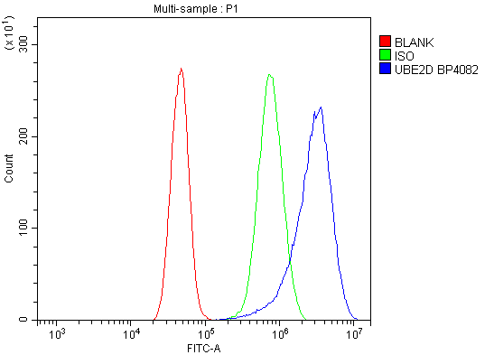 FCM (Flow Cytometry)
(Figure 7. Flow Cytometry analysis of A431 cells using anti-UBE2D1/2/3/4 antibody (AAA19298).Overlay histogram showing A431 cells stained with AAA19298 (Blue line). The cells were blocked with 10% normal goat serum. And then incubated with rabbit anti-UBE2D1/2/3/4 Antibody (AAA19298, 1μg/1x106 cells) for 30 min at 20 degree C. DyLight®488 conjugated goat anti-rabbit IgG (5-10μg/1x106 cells) was used as secondary antibody for 30 minutes at 20 degree C. Isotype control antibody (Green line) was rabbit IgG (1μg/1x106) used under the same conditions. Unlabelled sample (Red line) was also used as a control.)
FCM (Flow Cytometry)
(Figure 7. Flow Cytometry analysis of A431 cells using anti-UBE2D1/2/3/4 antibody (AAA19298).Overlay histogram showing A431 cells stained with AAA19298 (Blue line). The cells were blocked with 10% normal goat serum. And then incubated with rabbit anti-UBE2D1/2/3/4 Antibody (AAA19298, 1μg/1x106 cells) for 30 min at 20 degree C. DyLight®488 conjugated goat anti-rabbit IgG (5-10μg/1x106 cells) was used as secondary antibody for 30 minutes at 20 degree C. Isotype control antibody (Green line) was rabbit IgG (1μg/1x106) used under the same conditions. Unlabelled sample (Red line) was also used as a control.)
UBE2D1/2/3/4, Polyclonal Antibody (Cat# AAA19298)
Full Name
Anti-UBE2D1/2/3/4 Antibody
Gene Names
UBE2D1; SFT; UBCH5; UBC4/5; UBCH5A; E2(17)KB1
Reactivity
Human, Mouse, Rat
Applications
WB, IHC-P, ICC, IF, FC/FACS/FCM, EIA
Purity
Immunogen affinity purified.
Pricing


 FCM (Flow Cytometry)
(Figure 9. Flow Cytometry analysis of THP-1 cells using anti-Ku70 antibody (AAA19362).Overlay histogram showing THP-1 cells stained with AAA19362 (Blue line). The cells were blocked with 10% normal goat serum. And then incubated with mouse anti- Ku70 Antibody (AAA19362, 1μg/1x106 cells) for 30 min at 20 degree C. DyLight®488 conjugated goat anti-mouse IgG (BA1126, 5-10μg/1x106 cells) was used as secondary antibody for 30 minutes at 20 degree C. Isotype control antibody (Green line) was mouse IgG (1μg/1x106) used under the same conditions. Unlabelled sample (Red line) was also used as a control.)
FCM (Flow Cytometry)
(Figure 9. Flow Cytometry analysis of THP-1 cells using anti-Ku70 antibody (AAA19362).Overlay histogram showing THP-1 cells stained with AAA19362 (Blue line). The cells were blocked with 10% normal goat serum. And then incubated with mouse anti- Ku70 Antibody (AAA19362, 1μg/1x106 cells) for 30 min at 20 degree C. DyLight®488 conjugated goat anti-mouse IgG (BA1126, 5-10μg/1x106 cells) was used as secondary antibody for 30 minutes at 20 degree C. Isotype control antibody (Green line) was mouse IgG (1μg/1x106) used under the same conditions. Unlabelled sample (Red line) was also used as a control.)
Ku70, Monoclonal Antibody (Cat# AAA19362)
Full Name
Anti-Ku70 Antibody (monoclonal, 3D7)
Gene Names
XRCC6; ML8; KU70; TLAA; CTC75; CTCBF; G22P1
Reactivity
Human
Applications
WB, IHC-P, ICC, IF, FC/FACS/FCM
Purity
Immunogen affinity purified.
Pricing


 FCM (Flow Cytometry)
(Figure 7. Flow Cytometry analysis of A549 cells using anti-AGO3 antibody (AAA19290).Overlay histogram showing A549 cells stained with AAA19290 (Blue line). The cells were blocked with 10% normal goat serum. And then incubated with rabbit anti-AGO3 Antibody (AAA19290, 1μg/1x106 cells) for 30 min at 20 degree C. DyLight®488 conjugated goat anti-rabbit IgG (5-10μg/1x106 cells) was used as secondary antibody for 30 minutes at 20 degree C. Isotype control antibody (Green line) was rabbit IgG (1μg/1x106) used under the same conditions. Unlabelled sample (Red line) was also used as a control.)
FCM (Flow Cytometry)
(Figure 7. Flow Cytometry analysis of A549 cells using anti-AGO3 antibody (AAA19290).Overlay histogram showing A549 cells stained with AAA19290 (Blue line). The cells were blocked with 10% normal goat serum. And then incubated with rabbit anti-AGO3 Antibody (AAA19290, 1μg/1x106 cells) for 30 min at 20 degree C. DyLight®488 conjugated goat anti-rabbit IgG (5-10μg/1x106 cells) was used as secondary antibody for 30 minutes at 20 degree C. Isotype control antibody (Green line) was rabbit IgG (1μg/1x106) used under the same conditions. Unlabelled sample (Red line) was also used as a control.)
AGO3, Polyclonal Antibody (Cat# AAA19290)
Full Name
Anti-AGO3 Antibody
Gene Names
AGO3; EIF2C3
Reactivity
Human
Applications
WB, IHC-P, ICC, IF, FC/FACS/FCM, EIA
Purity
Immunogen affinity purified.
Pricing


 FCM (Flow Cytometry)
(Figure 10. Flow Cytometry analysis of K562 cells using anti-HNRNPH3 antibody (AAA19338).Overlay histogram showing K562 cells stained with AAA19338 (Blue line). The cells were blocked with 10% normal goat serum. And then incubated with rabbit anti-HNRNPH3 Antibody (AAA19338, 1μg/1x106 cells) for 30 min at 20 degree C. DyLight®488 conjugated goat anti-rabbit IgG (5-10μg/1x106 cells) was used as secondary antibody for 30 minutes at 20 degree C. Isotype control antibody (Green line) was rabbit IgG (1μg/1x106) used under the same conditions. Unlabelled sample (Red line) was also used as a control.)
FCM (Flow Cytometry)
(Figure 10. Flow Cytometry analysis of K562 cells using anti-HNRNPH3 antibody (AAA19338).Overlay histogram showing K562 cells stained with AAA19338 (Blue line). The cells were blocked with 10% normal goat serum. And then incubated with rabbit anti-HNRNPH3 Antibody (AAA19338, 1μg/1x106 cells) for 30 min at 20 degree C. DyLight®488 conjugated goat anti-rabbit IgG (5-10μg/1x106 cells) was used as secondary antibody for 30 minutes at 20 degree C. Isotype control antibody (Green line) was rabbit IgG (1μg/1x106) used under the same conditions. Unlabelled sample (Red line) was also used as a control.)
HNRNPH3, Polyclonal Antibody (Cat# AAA19338)
Full Name
Anti-HNRNPH3 Antibody
Gene Names
HNRNPH3; 2H9; HNRPH3
Reactivity
Human, Mouse, Rat
Applications
WB, IHC-P, ICC, IF, FC/FACS/FCM, EIA
Purity
Immunogen affinity purified.
Pricing


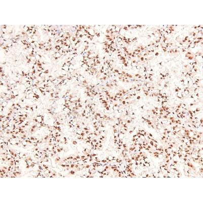 IHC (Immunohistchemistry)
(At 1/200 staining Human lung cancer tissue sections by IHC-P. The tissue was formaldehyde fixed and a heat mediated antigen retrieval step in citrate buffer was performed. The tissue was then blocked and incubated with the antibody for 1.5 hours at 22 degree C. An HRP conjugated goat anti-rabbit antibody was used as the secondary antibody.)
IHC (Immunohistchemistry)
(At 1/200 staining Human lung cancer tissue sections by IHC-P. The tissue was formaldehyde fixed and a heat mediated antigen retrieval step in citrate buffer was performed. The tissue was then blocked and incubated with the antibody for 1.5 hours at 22 degree C. An HRP conjugated goat anti-rabbit antibody was used as the secondary antibody.)
CUTL1, Polyclonal Antibody (Cat# AAA31424)
Full Name
Phospho-CUTL1 (Ser1237) Antibody
Gene Names
CUX1; CDP; CUX; p75; CASP; CDP1; COY1; Clox; p100; p110; p200; CUTL1; GOLIM6; CDP/Cut; Cux/CDP; Nbla10317
Reactivity
Human, Mouse, Rat
Predicted Reactivity: Pig (100%), Sheep (100%), Dog (100%), Xenopus (100%)
Predicted Reactivity: Pig (100%), Sheep (100%), Dog (100%), Xenopus (100%)
Applications
Western Blot, Immunohistochemistry, Peptide ELISA
Purity
The antibody is from purified rabbit serum by affinity purification via sequential chromatography on phospho-peptide and non-phospho-peptide affinity columns.
Pricing

 WB (Western Blot)
(WB of AAA24032 with 1:500 antibody dilution in DiluObuffer . Apparent MW is 78kDa.)
WB (Western Blot)
(WB of AAA24032 with 1:500 antibody dilution in DiluObuffer . Apparent MW is 78kDa.)
SPNS2, Polyclonal Antibody (Cat# AAA24032)
Full Name
SPNS2 Antibody
Reactivity
D. melanogaster, Monkey, Mouse, Rat
Applications
EIA, IHC, IP, WB
Pricing


 FCM (Flow Cytometry)
(Figure 6. Flow Cytometry analysis of Raji cells using anti-CD38 antibody (AAA19217).Overlay histogram showing Raji cells stained with AAA19217 (Blue line). The cells were blocked with 10% normal goat serum. And then incubated with rabbit anti-CD38 Antibody (AAA19217, 1μg/1x106 cells) for 30 min at 20 degree C. DyLight®488 conjugated goat anti-rabbit IgG (5-10μg/1x106 cells) was used as secondary antibody for 30 minutes at 20 degree C. Isotype control antibody (Green line) was rabbit IgG (1μg/1x106) used under the same conditions. Unlabelled sample (Red line) was also used as a control.)
FCM (Flow Cytometry)
(Figure 6. Flow Cytometry analysis of Raji cells using anti-CD38 antibody (AAA19217).Overlay histogram showing Raji cells stained with AAA19217 (Blue line). The cells were blocked with 10% normal goat serum. And then incubated with rabbit anti-CD38 Antibody (AAA19217, 1μg/1x106 cells) for 30 min at 20 degree C. DyLight®488 conjugated goat anti-rabbit IgG (5-10μg/1x106 cells) was used as secondary antibody for 30 minutes at 20 degree C. Isotype control antibody (Green line) was rabbit IgG (1μg/1x106) used under the same conditions. Unlabelled sample (Red line) was also used as a control.)
CD38, Polyclonal Antibody (Cat# AAA19217)
Full Name
Anti-CD38 Antibody
Gene Names
CD38; T10; ADPRC 1
Reactivity
Human
Applications
Western Blot, Immunohistochemistry, Immunocytochemistry, Immunofluorescence, Flow Cytometry
Purity
Immunogen affinity purified.
Pricing


 FCM (Flow Cytometry)
(Figure 6. Flow Cytometry analysis of RAW264. 7 cells using anti-TRIM25/EFP antibody (AAA19272).Overlay histogram showing RAW264. 7 cells stained with AAA19272 (Blue line). The cells were blocked with 10% normal goat serum. And then incubated with rabbit anti-TRIM25/EFP Antibody (AAA19272, 1μg/1x106 cells) for 30 min at 20 degree C. DyLight®488 conjugated goat anti-rabbit IgG (5-10μg/1x106 cells) was used as secondary antibody for 30 minutes at 20 degree C. Isotype control antibody (Green line) was rabbit IgG (1μg/1x106) used under the same conditions. Unlabelled sample (Red line) was also used as a control.)
FCM (Flow Cytometry)
(Figure 6. Flow Cytometry analysis of RAW264. 7 cells using anti-TRIM25/EFP antibody (AAA19272).Overlay histogram showing RAW264. 7 cells stained with AAA19272 (Blue line). The cells were blocked with 10% normal goat serum. And then incubated with rabbit anti-TRIM25/EFP Antibody (AAA19272, 1μg/1x106 cells) for 30 min at 20 degree C. DyLight®488 conjugated goat anti-rabbit IgG (5-10μg/1x106 cells) was used as secondary antibody for 30 minutes at 20 degree C. Isotype control antibody (Green line) was rabbit IgG (1μg/1x106) used under the same conditions. Unlabelled sample (Red line) was also used as a control.)
TRIM25/EFP, Polyclonal Antibody (Cat# AAA19272)
Full Name
Anti-TRIM25/EFP Antibody
Gene Names
Trim25; EFP; Zfp147; AA960166; AL022677
Reactivity
Mouse, Rat
Applications
WB, IHC-P, ICC, IF, FC/FACS/FCM, EIA
Purity
Immunogen affinity purified.
Pricing


 Application Data
(Staining of KG1 lymphocytes with Mouse anti Human CD59:FITC)
Application Data
(Staining of KG1 lymphocytes with Mouse anti Human CD59:FITC)
CD59, Monoclonal Antibody (Cat# AAA11857)
Full Name
MOUSE ANTI HUMAN CD59:FITC
Gene Names
CD59; 1F5; EJ16; EJ30; EL32; G344; MIN1; MIN2; MIN3; MIRL; HRF20; MACIF; MEM43; MIC11; MSK21; 16.3A5; HRF-20; MAC-IP; p18-20
Applications
Flow Cytometry
Pricing


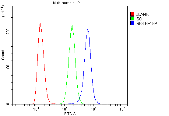 FCM (Flow Cytometry)
(Figure 6. Flow Cytometry analysis of K562 cells using anti-IRF3 antibody (AAA19216).Overlay histogram showing K562 cells stained with AAA19216 (Blue line). The cells were blocked with 10% normal goat serum. And then incubated with rabbit anti-IRF3 Antibody (AAA19216, 1μg/1x106 cells) for 30 min at 20 degree C. DyLight®488 conjugated goat anti-rabbit IgG (5-10μg/1x106 cells) was used as secondary antibody for 30 minutes at 20 degree C. Isotype control antibody (Green line) was rabbit IgG (1μg/1x106) used under the same conditions. Unlabelled sample (Red line) was also used as a control.)
FCM (Flow Cytometry)
(Figure 6. Flow Cytometry analysis of K562 cells using anti-IRF3 antibody (AAA19216).Overlay histogram showing K562 cells stained with AAA19216 (Blue line). The cells were blocked with 10% normal goat serum. And then incubated with rabbit anti-IRF3 Antibody (AAA19216, 1μg/1x106 cells) for 30 min at 20 degree C. DyLight®488 conjugated goat anti-rabbit IgG (5-10μg/1x106 cells) was used as secondary antibody for 30 minutes at 20 degree C. Isotype control antibody (Green line) was rabbit IgG (1μg/1x106) used under the same conditions. Unlabelled sample (Red line) was also used as a control.)
IRF3, Polyclonal Antibody (Cat# AAA19216)
Full Name
Anti-IRF3 Antibody
Reactivity
Human
Applications
Western Blot, Immunohistochemistry, Flow Cytometry, Direct ELISA
Purity
Immunogen affinity purified.
Pricing


 FCM (Flow Cytometry)
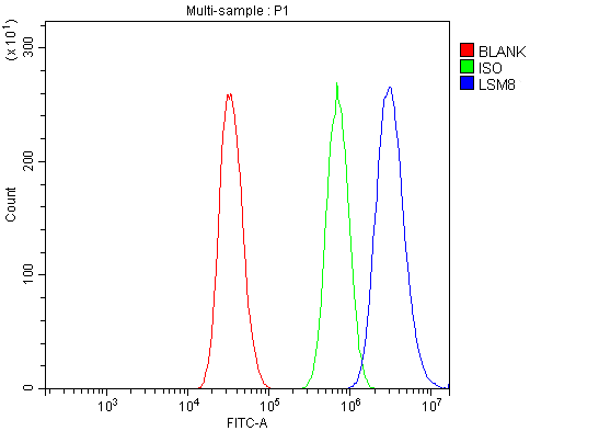
(Figure 13. Flow Cytometry analysis of A431 cells using anti-LSM8 antibody (AAA19337).Overlay histogram showing A431 cells stained with AAA19337 (Blue line). The cells were blocked with 10% normal goat serum. And then incubated with rabbit anti-LSM8 Antibody (AAA19337, 1μg/1x106 cells) for 30 min at 20 degree C. DyLight®488 conjugated goat anti-rabbit IgG (5-10μg/1x106 cells) was used as secondary antibody for 30 minutes at 20 degree C. Isotype control antibody (Green line) was rabbit IgG (1μg/1x106) used under the same conditions. Unlabelled sample (Red line) was also used as a control.)
FCM (Flow Cytometry)
(Figure 13. Flow Cytometry analysis of A431 cells using anti-LSM8 antibody (AAA19337).Overlay histogram showing A431 cells stained with AAA19337 (Blue line). The cells were blocked with 10% normal goat serum. And then incubated with rabbit anti-LSM8 Antibody (AAA19337, 1μg/1x106 cells) for 30 min at 20 degree C. DyLight®488 conjugated goat anti-rabbit IgG (5-10μg/1x106 cells) was used as secondary antibody for 30 minutes at 20 degree C. Isotype control antibody (Green line) was rabbit IgG (1μg/1x106) used under the same conditions. Unlabelled sample (Red line) was also used as a control.)
LSM8, Polyclonal Antibody (Cat# AAA19337)
Full Name
Anti-LSM8 Antibody
Reactivity
Human, Mouse, Rat
Applications
WB, IHC-P, ICC, IF, FC/FACS/FCM, EIA
Purity
Immunogen affinity purified.
Pricing


 FCM (Flow Cytometry)
(Figure 7. Flow Cytometry analysis of SiHa cells using anti-Aconitase 1/ACO1 antibody (AAA19267).Overlay histogram showing SiHa cells stained with AAA19267 (Blue line). The cells were blocked with 10% normal goat serum. And then incubated with rabbit anti-Aconitase 1/ACO1 Antibody (AAA19267,1μg/1x106 cells) for 30 min at 20 degree C. DyLight®488 conjugated goat anti-rabbit IgG (5-10μg/1x106 cells) was used as secondary antibody for 30 minutes at 20 degree C. Isotype control antibody (Green line) was rabbit IgG (1μg/1x106) used under the same conditions. Unlabelled sample (Red line) was also used as a control.)
FCM (Flow Cytometry)
(Figure 7. Flow Cytometry analysis of SiHa cells using anti-Aconitase 1/ACO1 antibody (AAA19267).Overlay histogram showing SiHa cells stained with AAA19267 (Blue line). The cells were blocked with 10% normal goat serum. And then incubated with rabbit anti-Aconitase 1/ACO1 Antibody (AAA19267,1μg/1x106 cells) for 30 min at 20 degree C. DyLight®488 conjugated goat anti-rabbit IgG (5-10μg/1x106 cells) was used as secondary antibody for 30 minutes at 20 degree C. Isotype control antibody (Green line) was rabbit IgG (1μg/1x106) used under the same conditions. Unlabelled sample (Red line) was also used as a control.)
Aconitase 1/ACO1, Polyclonal Antibody (Cat# AAA19267)
Full Name
Anti-Aconitase 1/ACO1 Antibody
Gene Names
ACO1; IRP1; ACONS; IREB1; IREBP; IREBP1
Reactivity
Human, Mouse, Monkey, Rat
Applications
WB, IHC-P, ICC, IF, FC/FACS/FCM, EIA
Purity
Immunogen affinity purified.
Pricing


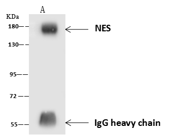 IP (Immunoprecipitation)
(NES was immunoprecipitated using: Lane A:0.5 mg U251MG Whole Cell Lysate 4 uL anti-NES mouse monoclonal antibody and 60 ug of Immunomagnetic beads Protein A/G. Primary antibody: Anti-NES mouse monoclonal antibody,at 1:100 dilution Secondary antibody: Rabbit Anti-Mouse IgG F(ab)2/HRP at 1/10000 dilution Developed using the ECL technique. Performed under reducing conditions. Predicted band size: 177 kDa Observed band size :177 kDa)
IP (Immunoprecipitation)
(NES was immunoprecipitated using: Lane A:0.5 mg U251MG Whole Cell Lysate 4 uL anti-NES mouse monoclonal antibody and 60 ug of Immunomagnetic beads Protein A/G. Primary antibody: Anti-NES mouse monoclonal antibody,at 1:100 dilution Secondary antibody: Rabbit Anti-Mouse IgG F(ab)2/HRP at 1/10000 dilution Developed using the ECL technique. Performed under reducing conditions. Predicted band size: 177 kDa Observed band size :177 kDa)
Nestin, Monoclonal Antibody (Cat# AAA27730)
Full Name
Anti-Nestin Antibody, Mouse Monoclonal
Gene Names
NES; Nbla00170
Reactivity
Human
Applications
Western Blot, Immunohistochemistry, Flow Cytometry, Immunoprecipitation
Purity
Protein A
Pricing


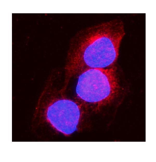
 IF (Immunofluorescence)
(Immunofluorescent analysis of 4% paraformaldehyde-fixed, 0.1% Triton X-100 permeabilized MCF-7 (human breast cancer cell line) cells labeling Pdx1 with at 1:25 dilution, followed by DyLight 488-conjugated IgG goat anti-rabbit secondary antibody at 1:200 dilution (green). Immunofluorescence image showing cytoplasm staining on MCF-7 cell line. Cytoplasmic actin is detected with DyLight 554 Phalloidin (PD18466410) at 1:100 dilution (red). The nuclear counter stain is DAPI (blue).)
IF (Immunofluorescence)
(Immunofluorescent analysis of 4% paraformaldehyde-fixed, 0.1% Triton X-100 permeabilized MCF-7 (human breast cancer cell line) cells labeling Pdx1 with at 1:25 dilution, followed by DyLight 488-conjugated IgG goat anti-rabbit secondary antibody at 1:200 dilution (green). Immunofluorescence image showing cytoplasm staining on MCF-7 cell line. Cytoplasmic actin is detected with DyLight 554 Phalloidin (PD18466410) at 1:100 dilution (red). The nuclear counter stain is DAPI (blue).)
OPN-a/b, Polyclonal Antibody (Cat# AAA26857)
Full Name
OPN-a/b, NT (SPP1, BNSP, OPN, Osteopontin, Bone sialoprotein 1, Nephropontin, Secreted phosphoprotein 1, Urinary stone protein, Uropontin) (APC)
Gene Names
SPP1; OPN; BNSP; BSPI; ETA-1
Reactivity
Human
Applications
WB, IHC, IF
Purity
Purified by Protein A and Peptide Affinity Chromatography.
Pricing


 FCM (Flow Cytometry)
(Figure 6. Flow Cytometry analysis of PC-3 cells using anti-MitoNEET/CISD1 antibody (AAA19293).Overlay histogram showing PC-3 cells stained with AAA19293 (Blue line). The cells were blocked with 10% normal goat serum. And then incubated with rabbit anti-MitoNEET/CISD1 Antibody (AAA19293,1μg/1x106 cells) for 30 min at 20 degree C. DyLight®488 conjugated goat anti-rabbit IgG (5-10μg/1x106 cells) was used as secondary antibody for 30 minutes at 20 degree C. Isotype control antibody (Green line) was rabbit IgG (1μg/1x106) used under the same conditions. Unlabelled sample (Red line) was also used as a control.)
FCM (Flow Cytometry)
(Figure 6. Flow Cytometry analysis of PC-3 cells using anti-MitoNEET/CISD1 antibody (AAA19293).Overlay histogram showing PC-3 cells stained with AAA19293 (Blue line). The cells were blocked with 10% normal goat serum. And then incubated with rabbit anti-MitoNEET/CISD1 Antibody (AAA19293,1μg/1x106 cells) for 30 min at 20 degree C. DyLight®488 conjugated goat anti-rabbit IgG (5-10μg/1x106 cells) was used as secondary antibody for 30 minutes at 20 degree C. Isotype control antibody (Green line) was rabbit IgG (1μg/1x106) used under the same conditions. Unlabelled sample (Red line) was also used as a control.)
MitoNEET/CISD1, Polyclonal Antibody (Cat# AAA19293)
Full Name
Anti-MitoNEET/CISD1 Antibody
Gene Names
CISD1; ZCD1; MDS029; C10orf70; mitoNEET
Reactivity
Human, Mouse, Rat, Monkey
Applications
WB, IHC-P, FC/FACS/FCM, EIA
Purity
Immunogen affinity purified.
Pricing


 IF (Immunofluorescence)
(Figure 6. IF analysis of GRSF1 using anti- GRSF1 antibody (AAA19315).GRSF1 was detected in immunocytochemical section of MCF-7 cells. Enzyme antigen retrieval was performed using IHC enzyme antigen retrieval reagent for 15 mins. The cells were blocked with 10% goat serum. And then incubated with 5μg/mL rabbit anti- GRSF1 Antibody (AAA19315) overnight at 4 degree C. DyLight®488 Conjugated Goat Anti-Rabbit IgG was used as secondary antibody at 1:100 dilution and incubated for 30 minutes at 37 degree C. The section was counterstained with DAPI. Visualize using a fluorescence microscope and filter sets appropriate for the label used.)
IF (Immunofluorescence)
(Figure 6. IF analysis of GRSF1 using anti- GRSF1 antibody (AAA19315).GRSF1 was detected in immunocytochemical section of MCF-7 cells. Enzyme antigen retrieval was performed using IHC enzyme antigen retrieval reagent for 15 mins. The cells were blocked with 10% goat serum. And then incubated with 5μg/mL rabbit anti- GRSF1 Antibody (AAA19315) overnight at 4 degree C. DyLight®488 Conjugated Goat Anti-Rabbit IgG was used as secondary antibody at 1:100 dilution and incubated for 30 minutes at 37 degree C. The section was counterstained with DAPI. Visualize using a fluorescence microscope and filter sets appropriate for the label used.)
GRSF1, Polyclonal Antibody (Cat# AAA19315)
Full Name
Anti-GRSF1 Antibody
Reactivity
Human
Applications
WB, IHC-P, ICC, IF, FC/FACS/FCM, EIA
Purity
Immunogen affinity purified.
Pricing